26
Microscopy Münchenwiler, March 25, 2010 Hans L Rieder Picture drawings: Koch R. Mittheil Kaiserl Gesundheitsamt 1884;2:1‐88

Microscopy
Münchenwiler, March 25, 2010
Hans L Rieder
Picture drawings: Koch R. Mittheil Kaiserl Gesundheitsamt 1884;2:1‐88

Sensitivity of Direct Sputum Smear Examinationin Identifying Pulmonary Tuberculosis and Transmitters
Frac
tion
of c
ases
/ in
fect
ed
0.0
0.2
0.4
0.6
0.8
1.0
Smear-negCulture-pos
Smear-posCulture-pos
Fraction due tosmear-neg cases
Fraction due tosmear-pos cases
Calculated from data from:Grzybowski S, et al. Bull Int Union Tuberc Lung Dis 1975;50:90-106
Cases ofpulmonary
tuberculosis
Infectedcontacts< 15 yr

Rieder H L, Van Deun A, Kam K M, Kim S J, Chonde T M, Trébucq A, Urbanczik R.Priorities for tuberculosis bacteriology services in low‐income countries. Second edition.
Paris: International Union Against Tuberculosis and Lung Disease, 2007
Schematic Presentation of Relative Frequency of Patients,Number of bacilli, and Available Diagnostic Methods
106102 103 104 105100 101
Poor microscopy (35%)
Excellent microscopy (65%)
Nucleic acid amplification techniques (80%)Culture (85%)
Radiography and clinical (95%)
10-1
Rel
ativ
e fre
quen
cy
Mycobacteria per mL sputum

Where things may not be optimum and adversely impact on sensitivity of the sputum smear microscopy result
Step Problem area
Sputum production Patient instruction on what constitutes a good sputum sample
Sputum processing Non‐homogeneity of sputum, smear preparation
Staining Quality of fuchsin
Examination Time spent on examination, actual number of fields properly examined

Improving yield of sputum smear microscopy by simplesputum-submission instructions, Pakistan, 2005
Diff
eren
ce in
stru
cted
-usu
al (%
)
-15
-10
-5
0
5
10
MenWomen
Spot Earlymorning
PositiveSaliva
Spot Earlymorning
Sameer Khan M, et al. Lancet 2007;369:1955-60
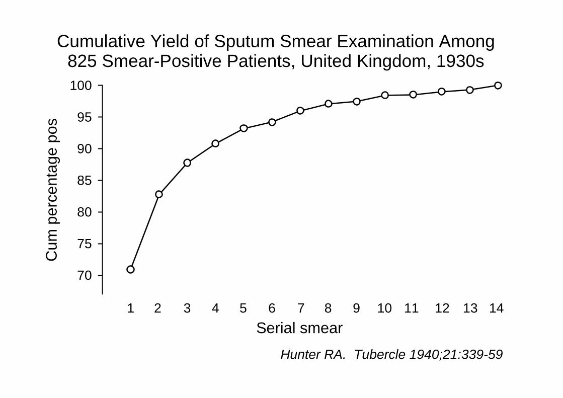
Cumulative Yield of Sputum Smear Examination Among825 Smear-Positive Patients, United Kingdom, 1930s
Cum
per
cent
age
pos
70
75
80
85
90
95
100
Hunter RA. Tubercle 1940;21:339-59
1Serial smear
2 3 4 5 6 7 8 9 10 11 12 13 14

Number of Examinations Required for SuccessiveSerial Smears to Identify one Additional Case, by Country
Number of smears required
3 10 30 100 300 1000
FirstSecond
Third
First
First
Second
SecondThird
Third
FirstSecond
Third
Mol
dova
Mon
golia
Uga
nda
Zim
babw
eSeria
l sm
ear /
cou
ntry
Mabaera B, et al. Int J Tuberc Lung Dis 2006;10:1030-5Katamba A, et al. Int J Tuberc Lung Dis 2007;11:659-64
Arbitrary cutoff (differsfrom country decision)125 slides = 1week

10mm
20mm
0.02mm2
1 Oil immersion field
1 Length = 100 OIF
1 mL Sputum
Wireloop = 0.01 mL
To see:1 AFB in 100 fields
requires:smear surface of 200 mm2
containing 100 AFB
which requires:1 mL sputumcontaining 10,000 AFB
Toman K. Tuberculosis case‐finding and chemotherapy.World Health Organization, Geneva, 1979

Distribution of Graded Results among SputumSmear Microscopy Positive Cases, by Country
Perc
enta
ge
0
20
40
60
80
100
Scanty positive
1+ positive
2+ positive
3+ positive
Moldova Mongolia Uganda Zimbabwe
Unpublished data, Union Tuberculosis Laboratory Collaborative Study
1,097 1,715 6,735 2,758

Distribution of AFB Smear Results Among Culture-PositiveSpecimens, by Relative Centrifugal Force, 15 Minutes
Per c
ent p
ositi
ve(lo
g sc
ale)
15
20
30
40
50
Ratnam S, et al. J Clin Microbiol 1986;23:582-5
2,000 g3,000 g3,900 g
RCF
Negative 1-2 per300 OIF
>2 per300 OIF
Cost of centrifuge: 3,800 Euros

AFB Non AFB
carbol‐fuchsin (or auramine)
decolorize
Principle of staining
counterstain
Slide courtesy: Van Deun A, unpublished lecture notes,Union International Tuberculosis Course, Arusha, November 2009

Principles in two types of microscopy
StainingBright‐field microscopy
Flurorescence microscopy
Primary stain(stains everything) Fuchsin Auramine
Decolorant(destains everything except mycobacteria)
Hydrochloric acidor
Sulfuric acidHydrochloric acid
Counter‐stain(stains everything except color‐saturated mycobacteria)
Methylene blue
Potassium permanganate
orInk

The “Ziehl‐Neelsen” staining technique: an experimental path to optimization, ready and all set since 1882
Contributor Contribution
Robert Koch Primary stain: methylene blue, alkaline potassium hydrate as mordant, vesuvium as both decolorant and counterstain
Paul Ehrlich Fuchsin as primary stain, alkaline alinine as mordant, nitric acid as decolorant, and proposal of a blue counterstain
Franz Ziehl Replace mordant with phenol
Friedrich Neelsen Combine the best of all: primary and counterstain from Ehrlich, mordant from Ziehl, and replacing decolorant with sulphuric acid
Bishop P J, Neumann G. The history of the Ziehl‐Neelsen stain. Tubercle 1970;51:196‐206

Stained with above recipe (HLR, Med Pol, ZH, 1978), but addedpotassium permanganate for background quenching
Magnification: 1,000x, oil immersion(Granulation disappears in photo due to long exposure time)

Picture courtesy:Kim SJ. Unpublished lecture notes, Union Hanoi course, 4 September 2008
Appearance of AFB in bright-field and fluorescence microscopy
Ziehl-Neelsen Fluorescence

Variation in fuchsin content in 10 samples collectedduring routine field visits, China, 2005-2006
Per c
ent o
f sta
ted
cont
ent
0
20
40
60
80
100
120
140
Zhao YL, et al. Int J Tuberc Lung Dis 2009;13:126-9

Fuchsin Absorbance by Wave Length and Manufacturer
Wave length (nm)
540 545 550 555 560
Abso
rban
ce
0.3
0.4
0.5
0.6
Avonchem
Ludeco
Merck NF
Data courtesy: Van Deun A, unpublished experiments, 2008
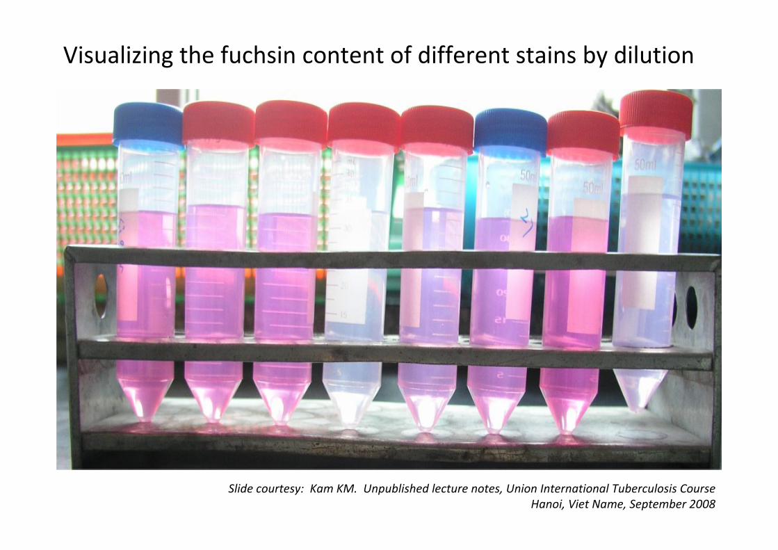
Visualizing the fuchsin content of different stains by dilution
Slide courtesy: Kam KM. Unpublished lecture notes, Union International Tuberculosis CourseHanoi, Viet Name, September 2008
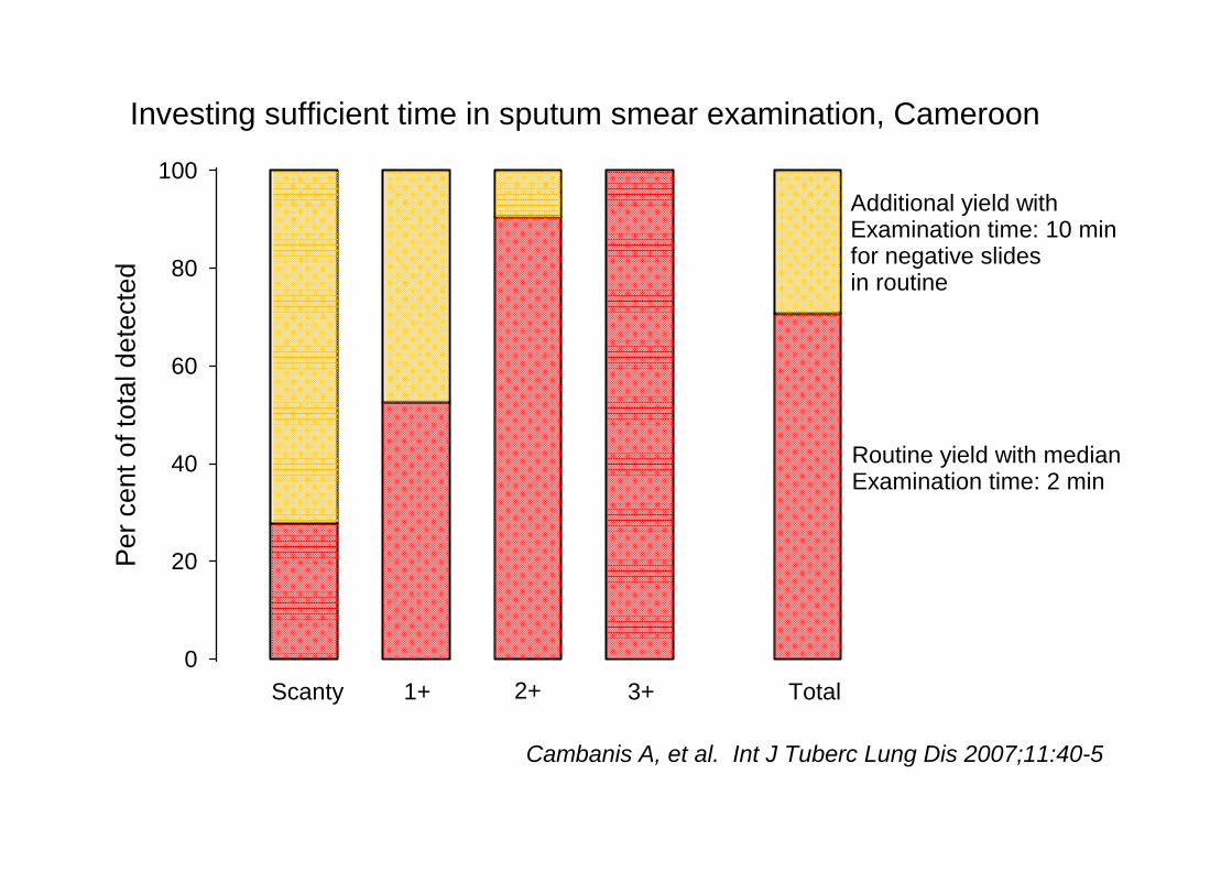
Investing sufficient time in sputum smear examination, CameroonPe
r cen
t of t
otal
det
ecte
d
0
20
40
60
80
100
Routine yield with medianExamination time: 2 min
Additional yield withExamination time: 10 minfor negative slidesin routine
Scanty 1+ 2+ 3+ Total
Cambanis A, et al. Int J Tuberc Lung Dis 2007;11:40-5

Optical system
Slide
Light source
Optical system
Slide
Light source
Optical system
Slide
Light source
Bright fieldmicroscopy
Fluorescencemicroscopy
LED fluorescencemicroscopy
LED fluorescencemicroscopy
Epi‐fluorescence

Principle of classical epi‐fluorescence(Example: Nikon system)
Principle of “add‐on” LED modulein transmission mode
(Example: Fraen system)
Added to an existing bright‐fieldmicroscope without interference
with the existing white‐light function

Readings using LEDReadings using mercury vapor lamp
Negative Scanty 1+ 2+ 3+ Total
Negative 396 6 2 0 0 404
Scanty 1 7 4 0 0 12
1+ 1 1 23 2 0 27
2+ 0 0 2 15 4 21
3+ 0 0 0 4 24 28
Total 398 14 31 21 28 492
Classical vapor lamp versus light emitting diode fluorescence microscopy
Hung NV, et al. Lancet Infect Dis 2007;7:238‐9

Yield from bright field versus fluorescence (LED) microscopyin two districts, Tanzania, 2007-2008
Per c
ent p
ositi
ve
0
5
10
15
20
Jan FebMar May Jun Jul Aug Sep Oct Nov DecApr
FluorescenceBright field
Microscopy:
Study month
Per c
ent p
ositi
ve
0
5
10
15
20
Jan FebMar May Jun Jul Aug Sep Oct Nov DecApr
Temeke
Mwananyamala
Van Deun A, et al. Int J Tuberc Lung Dis 2008;12:1009-14

Some differences between microscopy techniques
Bright‐fieldFluorescence
(classic)Fluorescence
(LED)
Stain Fuchsin Auramine Auramine
Time for proper examination 5 to 10 min 2 min 2 min
Microscope cost (€) 1,000 8,000 1,000
Module cost (€) ‐‐ ‐‐ 1,000
Lamp life (hr) 10,000 200 100,000
Maintenance cost Low Very high Low
Acceptability High Often very low High

Tuberculosis Among Primary Health Care Attendeeswith Prolonged Cough, Harare, Zimbabwe, 2003 (?)
Per c
ent
0
20
40
60
80
100
TB -
TB +
TB -
TB +
HIV negative HIV positive
90 27 454207
sm - / cul +
sm - / cul -
Munyati SS, et al. Clin Infect Dis 2005;40:1818-27
smear pos

Main issue with fluorescence microscopy: acceptability, not test operating characteristics!
o Fluorescence microscopy not accepted in Zurich until 1980 (Policy change introduced by Pr Dr A von Graevenitz, technique by Dr M Salfinger )!
o Fluorescence microscopy break‐through acceptance in the US only after 1962 (Truant, et al), “selling” that combining auramine plus rhodamine was the solution to earlier non‐acceptance – everybody forgot that quirky piece of history – there is “evidence” and “evidence”!
o “Classical” fluorescence microscopy not accepted in Dar es Salaam in the 1990s
o LED fluorescence microscopy in Dar es Salaam 2007‐2008: microscopists refuse to return to Ziehl‐Neelsen


