Model studies in bio-organic processes : sodium transport across biological membranes : an experimental study : quantum chemical calculations on the stereochemistry of coenzyme B12 dependent carbon- skeleton rearrangements Merkelbach, I.I. DOI: 10.6100/IR179116 Published: 01/01/1985 Document Version Publisher’s PDF, also known as Version of Record (includes final page, issue and volume numbers) Please check the document version of this publication: • A submitted manuscript is the author's version of the article upon submission and before peer-review. There can be important differences between the submitted version and the official published version of record. People interested in the research are advised to contact the author for the final version of the publication, or visit the DOI to the publisher's website. • The final author version and the galley proof are versions of the publication after peer review. • The final published version features the final layout of the paper including the volume, issue and page numbers. Link to publication Citation for published version (APA): Merkelbach, I. I. (1985). Model studies in bio-organic processes : sodium transport across biological membranes : an experimental study : quantum chemical calculations on the stereochemistry of coenzyme B12 dependent carbon-skeleton rearrangements Eindhoven: Technische Hogeschool Eindhoven DOI: 10.6100/IR179116 General rights Copyright and moral rights for the publications made accessible in the public portal are retained by the authors and/or other copyright owners and it is a condition of accessing publications that users recognise and abide by the legal requirements associated with these rights. • Users may download and print one copy of any publication from the public portal for the purpose of private study or research. • You may not further distribute the material or use it for any profit-making activity or commercial gain • You may freely distribute the URL identifying the publication in the public portal ? Take down policy If you believe that this document breaches copyright please contact us providing details, and we will remove access to the work immediately and investigate your claim. Download date: 25. Jun. 2018
Transcript
Model studies in bio-organic processes : sodiumtransport across biological membranes : an experimentalstudy : quantum chemical calculations on thestereochemistry of coenzyme B12 dependent carbon-skeleton rearrangementsMerkelbach, I.I.
DOI:10.6100/IR179116
Published: 01/01/1985
Document VersionPublisher’s PDF, also known as Version of Record (includes final page, issue and volume numbers)
Please check the document version of this publication:
• A submitted manuscript is the author's version of the article upon submission and before peer-review. There can be important differencesbetween the submitted version and the official published version of record. People interested in the research are advised to contact theauthor for the final version of the publication, or visit the DOI to the publisher's website.• The final author version and the galley proof are versions of the publication after peer review.• The final published version features the final layout of the paper including the volume, issue and page numbers.
Link to publication
Citation for published version (APA):Merkelbach, I. I. (1985). Model studies in bio-organic processes : sodium transport across biological membranes: an experimental study : quantum chemical calculations on the stereochemistry of coenzyme B12 dependentcarbon-skeleton rearrangements Eindhoven: Technische Hogeschool Eindhoven DOI: 10.6100/IR179116
General rightsCopyright and moral rights for the publications made accessible in the public portal are retained by the authors and/or other copyright ownersand it is a condition of accessing publications that users recognise and abide by the legal requirements associated with these rights.
• Users may download and print one copy of any publication from the public portal for the purpose of private study or research. • You may not further distribute the material or use it for any profit-making activity or commercial gain • You may freely distribute the URL identifying the publication in the public portal ?
Take down policyIf you believe that this document breaches copyright please contact us providing details, and we will remove access to the work immediatelyand investigate your claim.
III.l Structure and function of coenzyme B12• III.2. Mechanism of action of the carbon-skeleton
rearrangements. III.3. The stereochemistry of the carbon-skeleton
rearrangements as 'test' for the carbanionic
machanism. Scope of the second part of this
thesis. III.4. The nature of the hydrogen transferred
temporarily to coenzyme B12 during the carbon-skeleton rearrangements.
References and notes.
IV. Quantum chemical calculations.
IV. 1.
IV. 2.
IV. 3.
IV. 4.
IV. 5.
Introduction.
The choice of the calculational method. Results. Discussion.
Conclusion.
References and notes 1
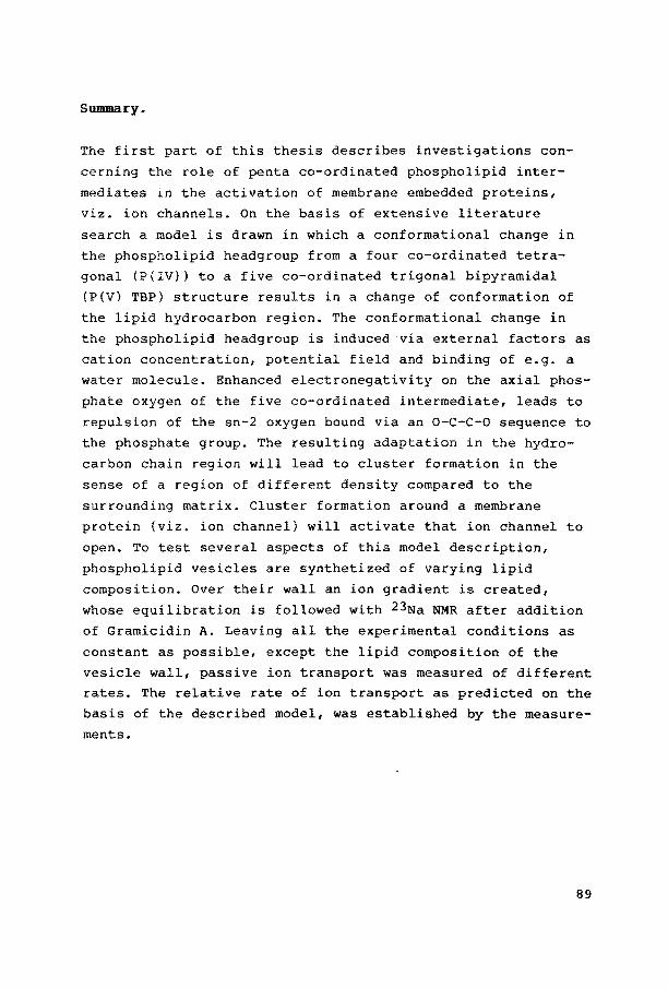
SUIIIlllary
Samenvatting Curriculum Vitae Dankwoord
59
62
65
67
69
73
74
76
85
87
88
89.
91
93
94
Voor het slagen van het kwaad is niets anders
nodig dan dat de goede mensen niets doen.
Maarten Luther King.
Sodium ion transport across biologica! membranes.
An experimental study.
I. Introduction.
I.l. Membrane properties.
Biologica! membranes play a crucial role in almost all
cellular phenomena, yet our understanding of the molecular
organization of membranes still can be called far from
exhaustive. While the composition of membranes varies with
their source, they generally contain approximately 40% of
their dry weight as lipid and 60% as protein1. Usually
carbohydrate is present to the extent of 1-10% of the total
dry weight. In addition to these components, membranes
contain some 20% of their total weight as water, which is
tightly bound and essential to the maintenance of their
I.l. The fluid mosaic mode of Singerand Niaolson. Integral proteins
(crossing the lipid bilayer), pheripheral proteins (bound to the exterior
the bilayer) and proteins embedded in the matrix are bound to
a functional complex or dissolved individually in the membrane bilayer.
9
structure. These components are organized according to the
fluid mosaic model of Singer and Nicolson2 (see Figure I.1.). The lipids span a discontinuous bilayer, with their
hydrophobic tails pointing towards the interior of the membranes and their hydrophilic head groups in contact with
the water phase outside the membrane. In this bilayer integral proteins are embedded, occasionally crossing the
total lipid bilayer matrix, while pheripheral proteins are
bound exterior to the bilayer. Dependent on e.g. temperature
and water content3, the more or less extended hydracarbon ebains of the lipide tilt away from the perpendicular to the
plane of the membrane, thus changing the ratio of the cross
sectional area~ of head group and chain region4. In this way a modification in density of the membrane can be reached, comparable to the melting phenomena of classica! chemica! compounds, e.g. from a fluid liquid-crystalline to a solid gel-like phase. This melting can, even for pure lipids, not be described as a thermodynamic first-order phase transition, since the transition is certainly not discontinuous, as shown, for example, by measurements of volume changes5. One can imagine a gel-like phase in which an appreciable lateral
diffusion of the lipid molecules exists, while on increasing temperature this lateral diffusion will be accelerated
throughout a phase-transition region to the fluid liquid
crystalline phase. The width of this phase-transition region will be increased when mixing different lipids with each other, with proteins or with other membrane constituents5. In
natura! membranes local gel-like domains6 exist over a large
temperature range in liquid-crystalline matrices and viceversa. These domains, often called clusters, develop in a continuing process of ordering and successive relaxation to a
disordered state. They consist of mainly lipide, mainly proteins or a mixture of both, but always can be described as a region of different density compared to the surrounding matrix. Diffusion over longer distances sametimes will be
opposed in biologica! membranes by a cytoskeletal system locallzing essential proteins in a well-defined region and by
10
a number of multivalent ligands that can induce aggregation
of proteins into clusters, patches or caps? in an alternative
way as mentioned above. Here proteins reside in a defined
region near to each other, but are not necessarily located in
one and the same cluster in the sense of same degree of order
or same density. When the word cluster is used throughout this thesis, a small region of one aggregational state in a
matrix of another will be ment, the first description of
clusters given above.
I. 2. Membrane function. Scope of the first part of this
thesis.
There is a lot of controversy concerning the role of lipids
in a membrane. Some authors state that lipids are the
insulating constituents of the membrane, separating different
cellular compartments8. They form the structural support of
membrane proteins, thus maintaining a constant spatial
relationship between them. In this view ion transport across
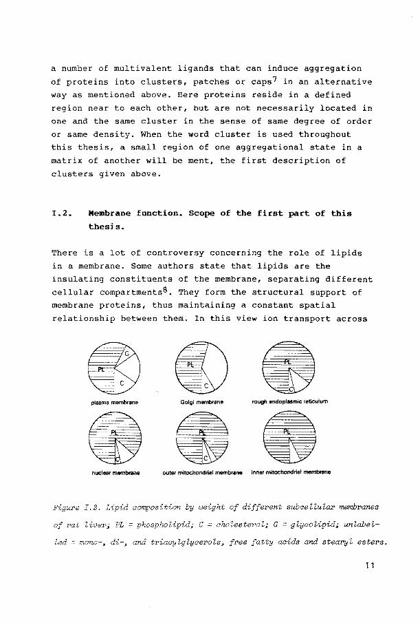
Figure I.3. Phospholipid aomposition by weight of different subaelluLar
membranes of rat Ziver; PC = phosphatidyZahoZine; PE = phosphatidyZethanoZ
amine; PI = phosphatidylinositoZ; PS = phosphatidylserine; S sphingo-
myeZin.
membranes is a property of integral proteins, that are influenced by external factors like metal-ions, protons, potential field etc. If one conaiders however, that lipid
composition of animal membranes vary both with their tissue souree and intracellular location9 (see Figure I.2 and I.3), the question is raised whether the lipids could play a role
themselves. So it has been suggested that the level of free fatty acids, that varies with the functional state of the
membrane, may be involved in changes in membrane permeability10. Until now, little attention has been paid to the
varying amounts of phospholipids in the membranes, in
particular to the·transition of the four co-ordinated to the
five co-ordinated state of the phosphate group, bound on its crucial position between the polar headgroup and hydrophobic
hydracarbon region, in relation to the phenomenon of ion
transport. Recent studies on a number of model compounds for biologica! reactives11,12 suggest a general principle in
passing biologica! information from ionic polar regions to
hydrocarbon zones of natura! molecules via a five co-ordinated (P(V)) trigonal bipyramidal (TBP} intermediate (see
12
Cbapter !.3. and !.4.). A classica! four co-ordinate (P(IV))
tetragonal intermediate, possesses four ligands tbat are
arranged spberically around tbe pbospborus atom, i.e. tbe P-L
bond lengtbs and tbe L-P-L bond angles are equal. Wben a five
co-ordinated TBP intermediate is formed{ a structural
inequivalence between two types of ligands (tbe equatorial
and the axial ones) is introduced, since five ligands can not
be spherically arranged around one atom (see Figure 1.4.).
T
TBP
I. 4. The four co-or'dir.ated tetY>aeder (T J and the co-ordinated
(TBP) of phosphorus.
The axial ligands {an incoming group e.g. water and the group
through wbich tbe pbosphate is bound to tbe main chain of the
biomolecule) are more electron withdrawing groups (see
Chapter !.3.) tban the equatorial ones, thus inducing an
electron flux into tbe axis of the TBP. 1f the group in the
axis consists of tbe 0-C-C-0-sequences often encountered in
biomolecules, this extra negative charge on the axial oxygen
(031 in Figure !.5. in the case of lipids), will result in a
repulsion of the other oxygen (021 in Figure 1.5.). In this
way a conformational change in the phospholipid headgroup
will be transferred in a re-orientation of the hydrophobic
region of tbe lipid molecule. As will be explained in Chapter
11, this re-orientation can induce cluster formation, which
in turn may influence integral membrane proteins as ion
channels, thus triggering them to open or to close. To get
experimental support for this model, a number of vesicles bas
been synthesized with different lipid composition. The
influence of this lipid composition on the ion transport over
13
0 11
..._". P· - N / v,,,,, o-/~o 'o
0 13 ro 2 R 1
0
)=o R ---
Figure I. 5. The ext2•a negative aharge on the apieal paaition of the five
eo-ordinated phospholipid intermediate results in repulsion of the oxygen
bound via an P-D-G-C-0 sequenee.
the vesicle cell wall, using one and the same ionophore, is
described in Chapter II to investigate several aspects of the theory.
1.3. Developments in organophosphorus chemistry.
In the past few decades, research in organophosphorus chemistry bas developed enormously13. Study of the
reactivity of model compounds has greatly enhanced the comprehension of the properties of five co-ordinated phosphorus compounds. So Westheimers studies14-17 on the
hydrolysis of five-membered cyclic phosphonates have increased the understanding of the mechanistic aspects of phosphorylation reactions. It was found that the hydrolysis of five-membered ring phosphates as 1 in Figure 1.6.,
proceeds millions of times faster in comparison to acyclic
phosphates as 4. This will apply for both ringopening (a) and
exocyclic cleavage (b) of 1. In contrast, the cyclic phosphonate 5 gave only very fast ring opening, no exocyclic hydrolysis. Westheiroer explained these observations on the
14
assumption, that the hydrolysis proceeds via a penta
co-ordinated intermediate in a trigonal bipyramidal
configuration. Before these phenomena can be understood, some
properties of five co-ordinated phosphorus compounds should
be noted. As already described in chapter I.2. an important
o"o) •H2o-(
11
0~ /OJ CH:lO -;p'o
~p HO 2
CH 0/ \0 3
0 0 ~/J
HO/ p\0 + CH30H
EtO"-. /OEt 3 p
,~'"-. o o-
4
0 0 0) ~<J + H20
11
CH30 -;p CH3 0
5 HO 6
Figure I.6. Cyclic and acyclic phosphates use« in the studies of West-
heimer.
aspect of five co-ordination is, that the distribution of the
ligands can not be spherically around the central atom, i.e.
the ligands are not equivalent18. Two possible structures are
favoured, as shown by X-ray analysis19-21: the trigonal
bipyramid (TBP) and the square pyramid (SP), shown in Figure
I.7. In the TBP there are three equivalent equatorial and two
axial honds, in the SP one axial and four basal honds.
Theoretical considerations based on MO and electrastatic
calculations have predicted that the TBP is slightly more
stable for acyclic penta co-ordinated phosphorus
derivatives18, but the difference is not too large. In
15
a e ', I
'p-e
e#" I a
TBP SP
a: axial ligand
b: basal ligand e: equatorial ligand
Figure I.?. The trigonal bipyramidal (TBP) versus the square pyramidal(SP)
configuration.
general an ideal TBP is seldom encountered, mostly a TBP is slightly distorted towards an SP geometry. In the TBP configuration the axial bonds are longer and usually weaker
than the equatorial bonds, a picture that can be ascribed by a pd-hybridization for the axial22-24 and a sp2-hybrid
ization for the equatorial bonds. However, the exact role of d-orbitals is still a subject of controversy25-27. Recent
publications suggest a remarkable degree of s-character in the axial bonds of some radicals28-30. So the observed
differences between ax,ial and equatorial sites in the TBP
structure can, in a more differentiated picture, better be
described by a substantially higher s-character for equatoria! than for axial bonds. In addition, axial sites are preferred by electron withdrawing ligands, whereas electron donating ligands tend to occupy equatorial positions31. This
polarity rule has been derived from many experimental data32,33, and is supported by semi-empirica! calculations34,35. Furthermore, it has been found that small rings
usually span an axial and an equatorial position in the TBP configuration, due to the 90° angle between these two bonds16. This is known as the strain rule. In fact, the presence of rings stahilizes this contiguration to such an
extent, that most of the known stable phosphoranes contain
one or more rings. One of the consequences of the differences in bond strength in a TBP is that leaving groups depart from the axial position16,34. Due to the microscopie reversibil
ity16, incoming nucleophiles also enter in the axis of the
Tsp34. An aspect of five co-ordination which hampers
16
differentiation of axial and equatorial bonds in the TBP
configuration, is the existence of pseudorotation.
Pseudorotatien for phosphorus compounds involves that the
positions of the ligands are interconverted fast on NMR
time-scale36-38. Several types of these 'permutational
isomerizations' are known, e.g. the Berry pseudorotation, in which two equatorial and both axial ligands change places39
via an intermediate SP contiguration (see Figure 1.8.).
/1 / I /
/ / :::::::t'P}--..::-. ~V /.,.l. ~· r<--- I(
/ / / V
TBP SP TBP
ii'igure I.B. The Berry tien process.
The energy barrier for pseudorotation may be very low,
especially if all ligands are identical18. However, if
pseudorotations bring electron withdrawing ligands into
equatorial positions34,40, or force small rings to span two
sites of the same kind34, pseudorotatien will be severely
hindered. Using the properties described above, the
experiments of Westheiroer (vide supra) are now readily
explained (see Figure 1.9.). Initial attack of water
on 1 yields intermediate 7. Subsequent proton transfer
towards the endocyclic axial oxygen atom leads to formation of 8, resulting in 2 after ring opening (the axial P-0 bond
is broken}. However, if the P(V) intermediate 1 undergoes ligand reorganization, 9 is formed. Upon leaving of the
axial protonated methoxy group, 3 is generated. The very fast
rate of both processes is explained by the fact that cyclic,
four co-ordinated phosphorus compounds are more strained than their acyclic analogues, whereas cyclic phosphorane
intermediates such as 7 or 9 are stabilized with respect to
acyclic phophoranes. These factors lower the activation
17
7
Figure I.9. Pseudo-rotation in the experiments of Weetheimer.
enthalphy for hydralysis of cyclic compounds substantially41. Phosphonates as 5 can (see Figure 1.6.), after attack of
water and prQton transfer to the axial oxygen, isomerize to either an intermediate with the ring carbon in an axial
position (normally not occupied by electron donating groups), or to an intermediate with a di-equatorial five-membered
ring, increasing the ring strain. These processes are
energetically unfavourable and can not compete with ring
opening, so no exocyclic hydralysis is found with the phosphonate .•
More recently, many other stereochemical and kinetic data in phosphorylation reactions and in group transfer reactions
have been rationalized by invoking phosphorane intermediates, including the group transfer reactions in tricyclic 'caged' phosphatranes by Van Aken et al~2,43.
18
1.4. Transfer of conformational changes in a phosphate
group to the hydrophobic part of organic molecules.
In the phosphorylation reactions of Westheimer, substitution
was achieved at phosphorus, whereby both incoming and leaving
groups entered and departed via the axis of an P(V) TBP. In
biochemistry a lot of phosphate groups are temporarily
activated without breaking the honds with the rest of the
biomolecule. In this case incoming and leaving group are the
same molecule, e.g. water. Dependent on the life time of the P(V) TBP intermediate the activated system will be able to
relax to a lower energy state. Another configuration will he
occupied, that is better able to accommodate the new charge
distribution over the ligands around phosphorus. Usually this
will happen by turning away one electronegative part of the
molecule from the other. Theoretical verification of charge
0 11
. P, +LH Ho~;! 'o- ....
Hs';.;.<'Os·,.... Hs"~jo
H~,·
net atomie P(IV)
charge ( e. u.)
0 ( 1 I) -0.267
0(5') -0.289 p +0.370
L
...
P(V)H P(V)
r.=HNMe L=OH L=HNMe L=OH
-0.274 -0.274 -0.291 -0.296
-0.316 -0.318 -0.348 -0.355
+0.340 +0.348 +0.409 +0. 451
-0.031 -0.133 -0.320 -0.436
1.10. Charge distribution on the ligands of a tetrahydrofurfuryl
model system ~hen going from a four to a five co-ordinated intermediate;
CND0-2 optimized results.
19
enhancement on apical ligands in a P(V) TBP model compound for DNA was recently published by van Lier et alJ1. The net atomie charge on various atoms in the tetrahydrofurfuryl model system and its P(V) TBP counterpart are given in Figure !.10. Experimental evidence for the rotation, resulting from
this charge enhancement, was very recently given by Koole et alJ2. They synthesized a number of four and five co-ordinated
mutually resembling phosphorus model compounds and found a significantly greater population of the gauche(-) conforma
tion for axially situated tetrahydrofurfuryls around the C4'-Cs' bond in the 5' P(V) TBP tetrahydrofurfuryls with respecttotheir related P(IV) compounds. In Figure !.11.,
some of the model compounds used are given12.
L:: Ph,OEt Y:O,CH2
L:Ph,OEt
Y = O,CH2
Figure I.ll. Four and five ao-ordinated model compounds with different Zi
gands substituted on the phosphate group and in the tetrahydPofUrfuryZ
ring.
The Newman projectionsof the rotamers around the C4'-Cs' bond are given in Figure I.12. The rotamer populations x(g+), x(gt) and x(g-) could be determined from the time-averaged
coupling-constants Ja4'as' and Ja4'as"12. In the P(IV) compounds Os' and Y are oriented cis to each other (the gauche(+) or the gauche(t) rotamer) in the case Y = o, due to the gauche effect44. This effect is defined45 as the "tendency to adopt that structure which has the maximum
20
Os· H s" Hs·
Y*C, Y*C,• Y*C,· Hs. Hs" Os• H5• H5 0 5 •
H •. H •• H ••
g• gt g-
I.l2. Newman around the P-0-C-C-0 sequence.
number of gauche interactions between the adjacent electron
pairs and/or polar bands" and originates from bond-antibond
interactions45. The only compound that differs substantially
possesses a CH2 group on the Y position (see Figure !.11.)
and the C4'-Cs' rotaroer distribution is consequently not
dominated by the gauche effect. In the P(V) compounds Os' and
01' are orientated more transtoeach other, i.e. the g
population is enhanced, due to the repulsion of two more
negatively charged oxygens. Only the compounds with Y = CH2
show no difference in population with the four co-ordinated
intermediate, which is clearly the consequence of the
P-0-C-C-C sequence present in the molecule, instead of the
P-0-C-C-O sequence, that is responsible for repulsion.
21
References and notes.
1. R. Harrison and G.G. Lunt, 'Biological Membranes, Their
Structure and Function', Blackie, Glasgow, 1980; p 62. 2. S.J. Singer and G.L. Nicolson, Science 1972, 175,
720-731. 3. A. Tardieu, V. Luzzati and F.C. Reman, J. Mol. Biol.
1973, 75, 711-733. 4. F.T. Presti, R.J. Pace and S.I. Chan, Biochemietry 1982,
38. J. I. Musher, J. Chem. Educ. 1974, 51 , 94-97.
39. R.S. Berry, J. Chem. Phys. 1960, 32, 933-938.
23
40. I. Ugi and F. Ramirez, Chem. Br. 1972, 8, 198-210.
41. J.A. Gerlt, F.H. Westheiroer and J.M. Sturtevant, J. Biol. Chem. 1975, 250, 5059-5067.
42. D. van Aken, l.I. Merkelbach, A.S. Koster and H.M. Buck, J. Chem. Soc., Chem. Comm., 1980, 1045-1046.
43. D. van Aken, l.I. Merkelbach, J.H.H. Hamerlinck, P. Schipper and H.M. Buck, A.C.S. Symp. Ser., 1981, 171,
439-442. 44. s. Wolfe, Acc. Chem. Res. 1972, 5, 102-111.
45. T.K. Brunck and F. Weinhold, J. Am. Chem. Soc. 1979, 101, 1700-1709.
24
II. Sodium ion transport across biologica! membranes.
I I. 1. Theory.
II.l.l. Cluster formation in biologica! membranes induced
via a phospholipid P{V) TBP intermediate.
In Chapter I the principle of induction of an electron flux
into the axis of a five co-ordinated phosphorus (P(V))
trigonal bipyramidal (TBP) intermediate was discussed. In
those model compounds, the extra negative charge on the
axial oxygen (Os') in the P(V) TBP intermediate resulted in
repulsion of another oxygen (01 '), bound via an o-e-c-o sequence to the phosphate group, and located in a tetrahydro
furfuryl ring (see Figure II.l(a)).
-(a)
I. .1'
I
' I bl ' .1•
' Figure II.l. Repulsion between the two oxygens in a P-0-c-c-o seque~e as a aonsequenoe of thè transition from a four ao-ordinated to a five
ao-ordinated intermediate in (a) a tetrahydrofurfurylphosphate and (b)
a phospholipid.
25
In this Chapter, an attempt will be made to show that the same process can occur in phospholipids, and that this process could be the 'trigger' to activate proteins, such as ion channels, emeedded in a lipid bilayer matrix via uptakè
in clusters 1. In Figure II •. l (b) one can distinguish the same o-e-c-o sequence, bound at the axial position of a phosphate group, as discussed for the model compounds in Chapter I.
Repulsion between the two oxygens 021 and 0312 of the P-0-C-C-0 sequence will cause a shift of the sn-2 chain in the direction perpendicular to the bilayer surface (see Figure II.1.(b)). However, the model compound& as (a) in Figure II.1. are monomers, dissolved in organic solvents, and thus able to re-orientate freely in solution. The phospholipids, on the contrary, are built in in the lipid bilayer, with their long hydracarbon chains interacting via 'van der Waals' interactions with the neighbouring chains. So the shift of the hydracarbon chains along each other will normally take a high energy barrier to overcome. A plausible adaptation of the bilayer by which this process can be aided,
is accompanied by a change in the angle of tilt of the hydracarbon chains to the bilayer normal. Phase diagrams of phospholipids show, dependent on temperature and percentage water or different lipid, several one and two phase regions3, in which among others the angle of tilt to the bilayer normal varies. In a special temperature interval, the phase transition region, ranging from the main phase transition temperature down to a temperature around the pretransition4,5,
smal! domains of different density (and thus different angle of tilt) occur next to each other. Such clusters are reported for mixtures of phosphatidylcholines (PC) with phosphatidylethanolamines (PE)3, cholesterol6 or proteins7. The co-oper
ative change in the angle of tilt of all the lipid molecules in the same cluster, can minimize the energy barrier that has to be overcome. Although there is controversy about the exact nature of the pretransition, a continuous change in the angle of tilt is always included in the description. Some authors conclude that the angle of tilt will change from tilted at
26
the pretranaition temperature to parallel to the bilayer nor
mal at the main phase transition8. Others believe the angle
of tilt reaches a local minimum at the pretransition, accom
panied by a transition from a tilted conformation, via a
tilted and rippled two dimensional structure9 to a one dimen
sional structure with the chains perpendicular to the bilayer
surface. Experimental evidence for a variation in angle of
tilt of the hydracarbon chains accompanying hydracarbon chain
shift was given by Blume3 and Chen10. Comparison of 13c and
2H NMR spectra of phospholipids, labelled respectively with
13c at the sn-2 carbonyl group3 and with 2H at the 4-position
of the same sn-2 chain, suggests that a conformational change
of the carbonylgroup precedes chain melting on increasing
temperature. This could be an indication of constantly
developing P(V) TBP intermediates, meeting below the pretran-
lipid
MMPC
MPPc(a)
MSPc(b)
PMPC
PPPC
PSPC
SMPC
SPPC
SSPC
ma in transition
temperature
23.6
35.1
38.6
27.3
41.1
49.0
29.4
43.9
54.2
pretransition
temper at ure
14.4
22.8
10.8
34.8
39.9
20.0
30.8
50.4
(a) MPPC = a myristoyl chain (M) bound at the sn-1 position
and a palmitoyl chain (P) bound at the sn-2 position of
the phosphatidylcholine (PC) glycerol backbone.
(b) S = a stearoyl chain.
Table II.l. Main transition and pretransition temperature as a function
of the chain Zength of the chain at the sn-1 and the sn-2 position of
phosphatidyZchoZine.
27
sition temperaturel an energy barrier too high to transmit the repulsion between the two oxygens of the o-e-c-o sequence in a shift of the sn-2 chain. The activation around the pre
transition temperature is high enough, howe.ver, to induce a conformational change at the carbonyl-group next to 021•
At higher temperatures an ever greater part of one and the same sn-2 chain and/or an ever greater part of the total
number of sn-2 ebains will be aetivated, untill at the main phase transition all the ebains are oriented perpendieular to
the membrane surfaee. Moreover, the pretransition behaviour shows strong dependenee on eomposition10. PCs with myristoyl
(C14), palmitoyl (C16) and stearoyl ebains (C1s> at the sn-1 and/or sn-2 position meet a higher energy barrier to melt if the sn-2 chain is longer (see Table 11.1). Another environmental constraint is met in the headgroup region (see Figure II.2.).
---
H H ....,_./ 0
-o 1/,,,, •.. ~ -o
-o ""=-k-J-,. 06 -;:N
/ 3
~ =
~ l L1'
I
' Figure II.2. Ringformation in the phosphatiQY~eho~ine headgroup upon
formation of a five ao-oPdinated intermediate.
In the headgroup, accommodation of a fifth ligand to form a five eo-ordinated intermediate will cause re-orientation of
28
the ligands around phosphorus. A lot of phospholipid
molecules contain zwitterionic headgroups (see Figure II.3.),
that are arranged with alternating charge in the bilayer11.
This model of intermolecular interaction between e.g. the
N-methyl protons of one PC molecule and the phosphate of a
neighbouring PC molecule, however, still allows for
considerable freedom of movement about the various honds in
the headgroup11.
phosphatidyl
choline
II.3.
sphingomyelin phosphatidyl- phosphatidyl-
ethanolamine serine
w·ith a zwittericm:c
Thus formation of a five co-ordinated phosphorus intermediate
must be accompanied by re-organization of the charge in the
total bilayer. This can occur, for example, by intrarnolecular
compensation of the charge12, thus creating a more or less
neutral molecule, or, at a physiological level, by de- and
adsorbtion of mono- and di-valent cations13,14.
Intramolecular compensation of charge can be established by
pseudo-ringformation, in which positively and negatively
charged groups are brought close to each other12 (see Figure
II.4.). Here, another aspect of the pretransition behaviour
is met, the relative cross-sectional areas of headgroup and
chain region. In molecules as PC, the headgroup in
'stretched' P(IV) conformation, will occupy a greater
29
excluded cross-sectional area tban the lipid bydrocarbon
cbains. Tbe ebains adopt a tilted conformation to fill .in a potential void in tbe bydrocarbon cbain region6. Above tbe
pretransition temperature, an increasing number of ebains orient more perpendicular to the membrane surface8,9, so
that the excluded cross-sectional area of tbe .headgroup must have been diminished. Tbis is confirmed by tbe observationlS
tbat PE, N-methyl and N,N-dimethyl PE do not exhibit such
pretransition behaviour, since the cross-sectional areas of
their headgroups are smaller. The effective cross-sectional area of the PC headgroup can be diminished further, after
di-equatorial16 pseudo-ringformation and pass down of the
charge, by pseudo-rotation, through which the pseudo six
membered ring will be orientated temporarily axial-equatorial. This pseudorotated structure will, at the same time,
prevent electron back donation to the fifth ligand, e.g.
water, and return of the intermediate to the four co-ordinated state. De- and adsorption of mono- and divalent cations can complete the picture sketched above. During the physio
logical process of the excitation of an axon, for example, momentary desorption of ca2+ ions from the outer monolayer of the membrane is reported13,14 upon activation of the axon, followed by adsorption of monovalent ions as Na+. This decrease in positive surface-bound charge of cations can
dfminish repulsive forces, intended to keep the headgroup 'stretched' in the unactivated axon17.
Finally, again in the example of the excitation of the axon, the potential at rest is negative inside the axon, pulling the positively charged end of the stretched headgroup
-N(CHJ)3+ of the outer monolayer phospholipids into the membranes. During activation of the axon, a positive potential inside17 will push the positive charged end of the head
group outwards, enabling the headgroup to re-orientate. Thus, a set of environmental physiological conditions is realized, enabling a P(V) intermediate to develop and to pass the
information, stored in its renewed charge-distribution, down to the hydracarbon region, for the case of the motionally
30
restricted phospholipid molecules. On a molecular level this
processcan be summarized as follows (see Figure II.4.).
ca2+ ions desorb, and are replaced by Na+ ions. The internal
potential is changing from negative to positive. As a conse
quence the headgroup is no langer forced in an extended and
inward pulled conformation and gets the opportunity to
re-orientate. The always existing P(IV) ~ P(V) equilibrium,
under 'resting' conditions laying at the side of the P(IV)
Figure II.4. The in the physioîogicaî conditions accompanying
the transition of a four to a f"ÎVe co-oràinated intermediate.
compounds, will be shifted in the direction of the P(V)
intermediate, since this conformation now is stabilized by
the formation of a di-equatorial16 pseudo-six roerobered ring.
The positively charged nitrogen of the choline headgroup
shields the negative charge of the formerly double bonded
oxygen, thus polarizing the P=O bond, by which the electro
philicity of the phosphorus atom will be increased18. This
process will be promoted by nucleophilic attack of e.g. a
water molecule19, normally present in excess in the headgroup
31
layer of the membrane, thus generating the P(V) TBP inter
mediate. The decrease in cross-sectional area of the headgroup due to ring formation, will cause decrease in the angle
of tilt of the hydracarbon chains. The increased electron density on the axial oxygen of the phosphate group will
induce repulsion of 021• bound via an o-e-c-o sequence to the phosphate group. This process will aid, or maybe it is the
main cause, of a further decrease in the angle of tilt of the hydracarbon chains, through a 'shift' of the hydracarbon chains along each other. The difference in effective chain length10, 20-22 is diminished. Co-operative change in the
angle of tilt of a number of phospholipid molecules is needed
to maximize the 'van der Waals' interactions between neigh
bouring acyl chains. This will lead at a macromolecular level to formation of a cluster with an average angle of tilt differing from the surrounding matrix. The relaxation time of
such a cluster is appreciably greater than for other charac
teristic movements of the molecule23. So the short-living P(V) TBP intermediate initiatea the formation of a cluster
with a much longer life-time, through which a time scale can be reached at which physiological processes can take place23. Although the physiological conditions of the above process are borrowed from the excitation of an axon, one can imagine
the same conditions for other membranes. Over most membranes an ion-gradient is maintained by ion pumps, so a potentlal exists over most membranes. Divalent ions as ca2+ and Mg2+
are bound to most membrane surfaces to an extent dependent on
the physical state of the lipids23. Mostly they are bound more strongly than monovalent ions such as Na+ and K+, that are present in all intra and extra-cellular spaces. The local oircumstances may change, a P(V) TBP intermediate can be
built up under several sets of conditions.
32
11.1.2. Activation of membrane proteins by uptake in
phospholipid clusters
A break in the Arrhenius plot of ATP-ase activity versus the
reciprocal temperature has been reported in dioleoyllecithin
substituted ATP-ase, when reaching the temperature of cluster formation24. A transition in the temperature dependenee çf
ca2+ accumulation25 in Sarcoplasmic reticulum membranes is
attributed to a change in entropy of activatien rather than
to the free energy of activation. Results that are consistent with an order-disorder transition invalving the lipid alkyl
separation in lipid bilayers containing integral proteins7,
show a system that separates into an essentially pure lipid
phase and a protein-rich phase containing melted lipids
between Tk and Tc· Here Tk is the melting point of clusters,
and Tc is the {main) melting point of lipids. Addition of
oleic acid to a lipid deficient membrane26 produces a fluid
membrane structure, which is most likely an essential
requirement for the reconstitution of the calcium dependent
ATP-ase activity. Addition of stearic acid, on the contrary,
has no activating effect on the calcium dependent ATP-ase26
and creates a gel-like lipid structure. From the above
mentioned and other27-29 articles, it becomes clear that a
certain fluidity is essential for the activatien of membrane
proteins, and that this fluidity is reached in a temperature
range in which cluster formation appears.
An often encountered objection against the relation between
cluster-formation and protein activation is, that gel state
lipids do not appear to be present in most biologica! membranes30. However, this is only partly true. Harrison and
Lunt conclude31 that, although hydracarbon ebains in natural
membranes are believed to be generally in a fluid state at
physiological temperatures, the presence of sterals and
proteins may lead to local variation of the mobility in the
membrane. Moreover, the degree of lateral phase separation is
believed to be influenced by a number of external factors,
33
e.g. water content31, proton and cation concentration32, ionic strength32 and potential fiela19. Under influence o.f the above-mentioned factors, cluster-formation is believed to persist far above the main phase transition temperature of a pure lipid mixture. Also pore-mediated ion transport shows some peculiar characteristics while planar bilayer membranes pass the phase transition region o.n heating33. Planar bilayers consisting of mixed-chain lipids and modified by pore-forming antibiotica as Gramicidin A, do not show any peculiar effect on Tc, the main phase transition temperature (29°C). Bowever, at 22-23°C a pronounced maximum in pore-induced conductance is seen. The effects observed are interpreted in terros of lateral -phase separation into pure lipid and lipid-antibiotic domains33. Consequently, the polypeptide Gramicidin A is an ideal model for the ion-channel forming proteins, obeying the same temperature dependenee of protein activation upon cluster formation. A schematic representation of Gramicidin A34 is given in Figure II.5.
0 N
0 0 • H
...,.. H- bond
Figure II.5. The Gramicidin channel oomprises ~o polypeptide ahains in
B -helix fol'm.
34
On a molecular level, protein activation upon uptake of the
protein in a cluster, can be described as follows. An
integral protein in a fluid environment (the cluster} could
be free to adopt the tertiary structure necessary to function
as an ion-channel. A gel-like matrix can displace some
special group of the channel-forming protein out of its
critical position35 and/or disturb a protein in helix form36.
For the sodium ion-channels of myelinated axons a model is given in which four energy harriers in the pore cernprise the
selectivity filter in the ion-channet35. These four harriers
consist of dehydration and hydration steps, enabling a
partially dehydrated sodium ion37 to pass a narrow gap
besides a strongly co-ordinating carboxylic acid group38.
Completely hydrated, the sodium ion will not be able to pass
the narrow selectivity filter of 3 x 5 Ä. A very small
displacement of the carboxylic acid group can make the ion
channels impermeable.
2
Figure II.6, The
of the ion channeL.
In
energy harriers comprising the selectivity filter
Although the correlation between a fluid lipid environment
and activation of proteins is suggested by quite a number of
authors (vide supra), this is not necessarily a general
35
principle. Also the reverse process, i.e. protein activation
upon uptake in a gel-like cluster, is a process that should not be neglected. In this way a number of different proteins
could be activated in succession, if e.g. the potentlal is
constantly changing from negative to positive and a whole
scala of states of different rigidity is passed through. Finally, support for the above-mentioned theory is given by
the fact, that 2-amido PC, contrary to PC, is found to be inhibitory for integral proteins39,40 (see Figure II.7.).
The oxygen esterified to c2 of the glycerol backbone of PC is essential for the transfer of conformational change in the
headgroup towards the change of tilt of the hydracarbon
chains. If this oxygen is replaced by the less electro
negative nitrogen of 2-amido PC, less repulsion and resulting acyl chain shift will be expected. Moreover, the hydrogen bridge found in X-ray analyses of comparable lipids41, will hinder acyl chain shift and headgroup re-orientation (Figure
11.7.).
)(
Figure II.7. Hindered repulsion and aeyZ ahain shift in 2-amido phospha
tidyZehoZine.
36
II.2. Experiments.
11.2.1. Introduction.
The characteristic feature of the model, described in this
Chapter, is that changes in the lipid environment of a
protein are the 'trigger' for the protein to be activated (to
place some particular functional group just in or just out of
the right position). Other authors propose a mechanism in
which changes in membrane potential, pH, ionic strength etc.,
directly influence the channel-forming protein (in the case
of ion-transport) to open, a mechanism that developed under
the influence of experiments with the ion channel blockers
tetrodotoxin42 and saxitoxin43.
To get a more decisive answer about this question, the
experimental conditions of the investigations described below
are chosen so, that only the lipid composition of the mem
branes varies, leaving the concentratien of ion channels,
ions, probes and buffers, as well as temperature, as constant
as possible. Vesicles are formed with a diameter of approxi
mately 1000 Á, their wall existing of one double layer of
lipids. As a reference, vesicles of egg yolk lecithin are
chosen, to which 10 to 50% of synthetic or natural, specific
lipid is added, to vary the total lipid composition. Attempts
to make vesicles of one well-defined synthetic lipid as
reference, failed, since the temperature of formation of the
vesicles had to be above the main phase transition tempera
ture, Tc. Tc will vary from 24•c for dimyristoyl (C14l, via
41•c for dipalmitoyl (C16l to ss•c for distearoyl (C1al phos
phatidylcholine44. The last two temperatures where too high
to be constantly maintained throughout the whole procedure of
synthesis. Since Tc of egg yolk lecithin is around o•c, the
choice of this lipid experimentally gave no problems.
As model for the ion-channel protein, the ionophore
Gramicidin A was chosen for a number of reasons, in addition
tothese mentioned in Chapter 11.1.2. Gramicidin A is a pore
former, specific for sodium ions. 1t is a pore-former and not
37
a carrier, so it builds up a permanent channel comparable to
natura! ion-channels, and does not diffuse through the membrane as carriers do. A pore-former also functions below
the main phase transition temperature so that the temperature range in which it is active is greater, while the conductance
of carriers falls below Tc to the state of bare membrane conductance (not pore mediated)33,45. The spontaneous
current fluctuations observed with unmodified planar bilayers
near the lipid phase transition temperature, containing a few
molecules of Gramicidin A, reminds of the idea of 'clusteractivation' of channela46, i.e. Gramicidin A is activated if
it is taken up in such a cluster. Formation of a cluster
around a Gramicidin A molecule activatea the channel to open.
The channel stays open during the life-time of a cluster, that can change randomly, but the conductance reached is always the same, unless the cluster will decay before the maximum conductance is reached. A Gramicidin A channel is formed by association of two polypetides at their N-formyl ends. Each chain is folded into a B
helix, which resembles a rolled-up B pleated sheet (see (a) in Figure 11.8.).
Figure II. 8. Four possib Ze confoi'mations of t;he Gramicidin channe L ,- -~
Finally, Gramicidin A incorporates spontaneously in the veeiele wall after addition, since its amino acid sequence is one of the most hydrophobic ones known47.
38
' " (el 1=2410 (I) I 11
1=11.0 00
' " [d) t=13 30
(k)
I 11
(c) 1=458
( j)
(i)
( b) I 11
t = 0 00
( h)
(g)
(a) I 11
t = 0 00 ( f )
Figure II.9. 23Na Nii!R spectra (79.4 MHz) of a dispersion of vesicles of
egg yolk phosphatidylcholine plus 10% sphingomyelin (a) at t=O before
addition of the probe, (b) at t=O after addition of the probe, (c)-(l)
after addition of Gramicidin; T = 25°C.
39
Moreover the exterior of the channel consists again of the
most hydrophobic parts of the peptide48, thus creating an oxygen lined, probably water-filled, central channel.
Gramicidin A is added in last resort to a buffered solution with approximately 1000 A diameter vesicles, around which the
NaCl solution is replaced by LiCl by means of ultrafiltration. Thus a sodium ion gradient is formed of about 102, the
equilibration of which is started at the moment Gramicidin A is added. A shift reagent49, Dy I N(CH2C02)3J23- has been used
to distinguish between 23Na+ inside and outside the vesicles.
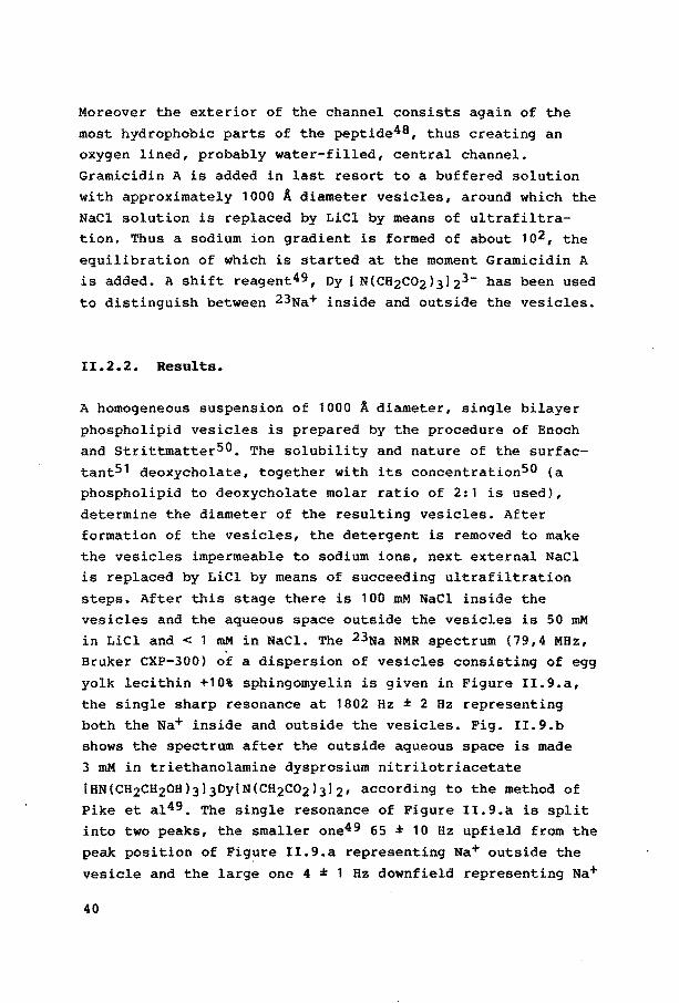
II.2.2. Results.
A homogeneous suspension of 1000 Ä diameter, single bilayer
phospholipid vesicles is prepared by the procedure of Enoch
and Strittmatter50. The solubility and nature of the surfactant51 deoxycholate, together with its concentration50 (a phospholipid to deoxycholate molar ratio of 2:1 is used),
determine the diameter of the resulting vesicles. After formation of the vesicles, the detergent is removed to make
the vesicles impermeable to sodium ions, next external NaCl is replaced by LiCl by means of succeeding ultrafiltration
steps. After this stage there is 100 mM NaCl inside the
vesicles and the aqueous space outside the vesicles is 50 mM in LiCl and < 1 mM in NaCl. The 23Na NMR spectrum (79,4 MHz, Bruker CXP-300) of a dispersion of vesicles consisting of egg
yolk lecithin +10% sphingomyelin is given in Figure II.9.a,
the single sharp resonance at 1802 Hz ± 2 Bz repreaenting both the Na+ inside and outside the vesicles. Fig. II.9.b shows the spectrum after the outside aqueous space is made
3 mM in triethanolamine dysprosium nitrilotriacetate
[HN(CH2CH20H)3)3Dy[N(CH2C02)3]2, according to the metbod of Pike et al49. The single resonance of Figure II.9.a is split into two peaks, the smaller one49 65 ± 10 Hz upfield from the
peak position of Figure II.9.a repreaenting Na+ outside the
vesicle and the large one 4 ± 1 Hz downfield repreaenting Na+
40
1.0
PC trom
0.9 egg yol k
0.8 6 + 50 •;. OMPC 7 • so·~ OPPC
0.7
R + , oo;. OPPC
1
5 . 10% OSPC 0.6
3 • 10% OMPC 0.5
0.4
2 + 10% sphingo-mye I in
0.3 9 •10% P inOSitOI 10 + 1 0°/o OPPE
PC pure
fro m egg yol k
0.2
0.1
8 + 10o/o P serine 6
3 5
-tfhrl
Figure II.10. The time of the re~ative integra~s of the two
peaks of Figure II.9. ten different differing in ~ipid com-
position of the vesicle ceZZ-waZl.
41
inside, the absolute magnitude of the shift being dependent
on the exact concentratien of the probe49. The fraction of
the total integral, due to the inside resonance varied from
0.77 to 0.92 for the various samples. The value for the
fraction of the total aqueous volume inside the vesicles is
calculated, assuming an internal volume of 2960 ml per mole of lipid52 and a final lipid concentratien of 10 mM, to be
0.05. There could be some error in this ratio because of inaccuracy in the knowledge of the final lipid concentration.
Bowever, other studies using the method of Enoch and StrittmatterSO, where phosphate analyses was conducted on the final
solutions, were in good agreement with the above-mentioned
fraction of lipid left after synthesis of the vesicles.
Combination of these numbers with 100 mM Na+in• yields a Na+out concentratien of 0.27 mM in the case the fraction of the total integral due to inside resonance at t = 0 is 0.92
or 0.92 mM in the case this fraction is 0.77. Since any
1eakiness of the vesicles would affect the observed ratio, all starting spectra were obtained between 0.5 and 3 hrs
after the last ultrafiltratien step, and samples with a
fraction due to inside resonance smaller than 0.77 were not used further. E.g. for the sample of egg yolk lecithin with 50% DSPC it was not possible, even after several attempts, to
obtain a sample with a fraction due to internal resonance greater than 0.30. Immediately after the spectrum of sample + probe was obtained, the salution was made 0.04 pM in the ionophore Gramicidin53-55. This amounts to ca. 3 Gramicidin
channels per vesicle49 and induces a rapid efflux of Na+ down
its ccncentration gradient, as can be seen in the Figures
II.9.c-l. They depiet some of the spectra obtained and show the time evolution of the spectrum measured in minutes from
the time of addition of Gramicidin, for a sample displaying
an intermediate time course in the total series measured (egg
yolk + 10% spingomyelin). The spectra given are power spectra in which the integral is proportional to the square of the number of the sodium nuclei56. The power spectra are taken to
cancel the influence of the phase-correction, that will vary
42
with time during the period the automated measurements are
recorded. A plot of the logarithm of ratio R, of the frac
tional integral (inner/total) at t = t to the fractional
integral at t = 0, against time is shown in Figure II.10. The
time during which the spectra were recorded after addition of
Gramicidin varied from 2 to 13 hrs, dependent on how fast the
ion transport took place. For 50% DPPC not the total time
course is given, since between 5 and 13 hrs after addition of
Gramicidin the slope of the plot was identical to that of the
part shown between 0 and 5 hrs.
II.2.3. Discussion.
The efflux of Na+ ions shows at least two stages49. Directly
after addition of Gramicidin a passive one-for-one Na+ for
Li+ exchange out of and into the vesicles takes place, both
ions moving down their concentratien gradients, although the
cl- transport may play a role at this stage (vide infra}.
This stage will end when the Li+ gradient is dissipated
(47.5 mM both inside and outside). Since the Na+ gradient
still exists at this point (52.5 mM inside, : 3 mM outside,
R = 0.52), during the second stage the Na+ gradient will be
further dissipated at the expense of creating a new Li+
gradient, still through a one-for-one exchange. This stage
will end when the ratio of inside concentratien to outside concentratien has the same value for both Na+ and Li+, so
that the same diffusion potential for both ions is reached.
Here Na+ in is 10 mM, Na+ out is 5.3 mM, Li+ in is 90 mM
and Li+ out is 45.3 mM, the fraction of total Na+ inside is
0.09. True equilibrium will only be obtained after a third
stage, namely passive nonfacilitated cl- transport out of the
vesicles. This stage will end when the fraction of total Na+
inside is equal to the analogous volume fraction, 0.05. In
these rough calculations osmotic swelling, a possible pH
gradient due to permeation of the counter ion of the probe
HN(CH2CH20H)3+ and the Donnan effect caused by the impermeant
43
Dy[N(CH2C02)3l23- ar~ ignorea50. The transitions of the
curves, shown in Figure II.10, are located between R = 0.5 and R = 0.3, the transitions being at lower R if ion trans
port is faster. This could be an indication of leakage before
Gramicidin is added, although measurements of ten different
blancos (after addition of the probe) of the same sample in a time period of half an hour showed no significant decrease of
the ratio of Na+in to Na+out• The fast process between R = 1 and R = 0.5, corresponding to
the one-for-one exchange of Na+ and Li+ down to their gradient, is believed to be limited by the Gramicidin induced Li+ transport, which is ca. 1/6th as fast as that of Na+ 57.
Thus the slow process, below ca. R = 0.5, would correspond to the essentially simultaneous occurrence of the second and third stages (vide supra) implying that they have very similar permeability constants. This is supported by measurements
of permeability coefficients of pass!ve nonfacilitated trans~ port of cl- 52. So both stages befare and after R = 0.5, are involved in sodium ion transport.
As can beseen in Figure II.10, the relative sequence in velocity of ion transport for the various samples is not
interchanged when passing R = 0.5. The reference sample, phosphatidylcholine from egg yolk
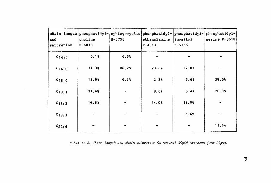
exists of predominantly C16 (34.3%) and C1a chains (59.8%
C18:0• C18:1 and c18:2)58, as can beseen in Table II.2. Ion transport over a vesicle cell wall consisting of egg yolk phosphatidylcholine is relatively fast, compared to
most of the other samples (2-10 in Figure II.10.). Clearly, the relative quantities of different chain lengtbs and
saturation is such, that the rigidity of the matrix is ideal to accommodate for the chain shift that results from the
formation of a five co-ordinated phosphorus intermediate. Increasing the percentage of synthetic saturated ebains
(samples 3-6), the matrix will adopt a more gel-like character, in which the packing of the optimally ordered ebains is
tighter, so the chain shift is more difficult. When increasing the chain lengtbs of the saturated chains, ion transport
44
becomes slower, although the difference between ~he sample
with 10% DSPC and with 10% DPPC is too smal!, compared to the
deviation in slope due to measuring faults, to be called
significant. Addition of more (50%)'of the same saturated
chains again gives slower ion transport, in which the slopes
of the plots of 50% DMPC and 50% DPPC are indistinguisable.
Addition of 10% of sphingomyelin from egg yolk, containing
primarily saturated palmitic acid chains at the sn-2 position
results in ion transport that is slower than that of the
reference sample of phosphatidylcholine from egg yolk (1),
but faster than that of the samples 3-6. The slower ion
transport of sample 2 compared to sample 1 could stem from
the restricted repulsion of the sn-2 nitrogen (see Figure
II.11.) through which the sn-1 chain is bound to the glycerol
backbone. The significantly faster ion transport compared to
the samples 3-6 can be explained by the fact that the sn-1
chain is significantly shorter (two atoms less than in DPPC,
viz. -CH=CH-(CH2l12-CH3 directly bound to the glycerol back
bene, and the appearance of a double bond), thus creating a
molecule comparable to MPPC (see Table II.1.).
Moreover, addition of a clearly different lipid will enhance
the heterogenity of the matrix, making the matrix more fluid,
and enabling lipids other than sphingomyelin to translate a
five co-ordinated phosphorus intermediate more easily in a
hydrocarbon chain shift.
Samples 8, 9 and 10 contain additions with primarily C16 or
C18:0 to C18:2 (see Table II.2.) as lipid alkyl chains,
thus changing the overall fluidity of the matrix not too
much, compared to 1, on additions of 10%. Egg yolk phosphati
dylcholine with 10% phosphatidylserine shows considerably
faster ion transport compared to the reference sample. The
availibility of two ligands within the serine, one as fifth
ligand to build up a five co-ordinated intermediate and one
to polarize the original P = 0 bond, could cause an appreci
ably increased life-time of the five co-ordinated inter
mediate, which explains the fast ion transport observed (see
11 -o,,,, I ......... + p .,,,, ····P-O ,.....N / "''o- -oV ~o. o
~N o, H 3 / ' rN 2 H :f
OH 0 ,,
~ rN OH ( b) )( ...
~ ~ ~ -
~ Figure II,ll. Compared to phosphatidyLcholine (a), in sphingomyelin (b)
chain following from the formation of a five co-ordinated
intermediate, is blocked.
47
FiguPe II.l2. VePy fast build-up of a five ao-ordinated intermediate in
the phosphatidyZsePine headgPoup, due to twofold stabilization.
Phosphatidylinositol possesses a large ligand in the phospho
lipid headgroup, the diameter of which can hardly be changed by the transition from the four to the five co-ordinated intermediate. A possibly formed five co-ordinated intermediate will not be stabilized by intramolecular ring formation
so its life-time will not be long enough to induce cluster
formation. Phosphatidylethanolamine already possesses a very
small headgroup in stretched conformation (see II.1.1.), so intramolecular ring-formation will not be able to influence
the angle of tilt of the hydrocarbon chains. Both samples 9
and 10 display a more or less expected rate of ion transport, somewhat slower than that of phosphatidylcholine (sample 1). Addition of these lipids hardly influences the overall lipid fluidity (the ebains display about the same degree of
saturation) and will not be able to induce cluster formation
48
7
(a)
( b)
Figure II.13. (a) Formation a five co-oPdinated intermediate will not
be able to influence hydracaPbon chain tilt in phosphatidylethanolamine.
(b). A five co-ordinated phosphatidylinositol intermediate will not be
stabilized by intramolecular ringformation.
49
via a P(V) TBP intermediate. So the number of phospholipids
that can induce cluster fo·rmation (the phosphatidylcholines} is simply reduced with ± 10%.
11.2.4. Conclusion.
The measurements of ion-fluxes through Gramicidin channels
over the walls of phospholipid vesicles, enabled us to
establish ion transport of different rates, if all the experimental conditions were left constant except the lipid
composition. This can be considered as a support for the
model, indicating lipid mediated influence of membrane
proteins and not just direct influence of proteins by external factors. To test several aspects of the model, in which
the transition of a four to a five co-ordinated phospholipid
intermediate is the trigger to cluster formation and protein
activation, phospholipids with various headgroups and hydrocar.bon ebains were added to a reference lipid. The results
can be considered as a support of the proposed model.
II.2.5. Bxperimental.
- Preparation of vesicles.
The vesicles are-prepared according to metbod II of Enoch and
Strittmatte.r50, using the same concentrations noted there. SUbsequently, nine· ultrafiltration steps (Millipore, Immer
sibie CX-30) with a concentration of 10 to 1 ml in every
step, are necessary to reach a fractional integral of inter
na! resonance relative to internal + outside resonance of
approx. 0.90. This is an indication that Na+ is strongly
bound to the external phospholipidmonolayer and that a high
concentration ratio between the external phospholipid mono
layer and the external solution is needed for the Na+ ions to desorb. After the last concentrating step to 1 ml buffered
50
vesicle solution 1 ml 020 is added to get the internal
reference for the NMR measurements •
.- Preparatien of the probe.
The probe is synthesized according to the double heterogeneous reaction59-63:
of 0.746 g Dy203 (dysprosiumoxide, Sigma No. D-0381) and
1.530 g H3NTA (nitrilotriacetate, Sigma No. N-9877) in 25 ml
water with 1.60 ml TEA (triethanolamine, Brocades, s.d. 1.12
1.13) to produce 0.16 Mof shift reagent59. The pH must
never be allowed to rise above 6 or 7 (even transiently)
during early stages, lest oy3+ hydrolysis and precipitation
becomes a problem. So at first 1.0 ml TEA is added toa
stirred suspension of Dy203 and H3NTA at a temperature of 40•c over a time-period of two hours. After heating the
solution becomes clear at 73"C, the pH is 3.5 at that moment.
After cooling to room temper.ature the solution is clowdy
again after standing over one night. Heating to 1o•c and
addition of the last 0.6 rol TEA will give a clear solution
again of pH = 4.5, that stays clear after cooling. Part of
this stock solution is brought to pH = 7 with LiOH just
before addition of the probe to the vesicle suspension, to
keep the pH of that solution as constant as possible and
diluted 1 : 1 with DzO to obtain a final concentratien of
0.065 M of the probe. From this solution 90 ~1 is added to
2 rol of vesicle suspension during the measurements •
.- Gramicidin solution.
2.3 rog Gramicidin no. G-5002 (Sigma) per 100 ml methanol
gives a stock solution of 1.23 x 1o-5 M. 6 ~lof this
solution is added to 2 ml vesicles suspension in 50% 020 I 50% H20. The concentratien of the stock solution is made such
51
that only very little methanol is added to the sample, since
methanol is an anesthetic that perturbs the bilayers strongly64.
~ NMR measurements.
The 23Na NMR {79.4 MHz) spectra are measured on a 300 MHz
Bruker CXP-300 spectrometer. For each measurement 256
free-induction decays (FID) are accumulated in 128 s. For each series an average of 8 blanks at t = 0 (after addition
of the probe) is taken as reference Na+in/Na+total,t=O• After addition of Gramicidin A, the spectra are taken automatically with a computer program, taking successively 30 measurements every 2 minutes, 10 every 4 minutes, 10 every 6
minutes, 10 every 16 minutes and 15 every 31 minutes. If the
ratio Na+in, t=t/Na+in, t=O falls below ± 0.3 the recording is stopped because of too small signal/noise ratio
for Na+in, t=O· Most series are stopped too if t ~ 3 hours. As internal loek 020 is used, as described by Pike et a150.
However, measurements of 2 mM NaCl in H20 or H20/D20, with
acetone or 020 as an external loek, show that the Dys-reagent is a 0-shift reagent as well. Since the shift with 020 as
loek is appreciably greater than without, the choice for 020 is maintained.
52
References and notes.
1. I.I. Merkelbach and H.M. Buck, Reel. Trav. Chim.
Pays-Bas, 1983, 102, 283-284.
2. M. Sundaralingam, Ann. N.Y. Acad. Sci., 1972, 195,
324-355.
3. (a) A. Tardieu, V. Luzzati and F.C. Reman, J. Mol.
Biol. 1973, 75, 711-733; (b) A. Blume, R.J. Wittebort,
s.K. Das Gupta and R.G. Griffin, Biochemistry, 1982, 21,
6243-6253.
4. S.H. Wu and H.M. McConnell, Biochemistry, 1975, 14,
847-854.
5. E.J. Shimshick and H.M. McConnell, Biochemistry, 1973,
12, 2351-2360.
6. F.T. Presti, R.J. Pace and S.I. Chan, Biochemistry,
1982, 21, 3831-3835.
7. T. Lookman, D.A. Pink, E.W. Grundke, M.J. Zückermann and
F. de Verteuil, Biochemistry, 1982, 21, 5593-5601.
8. R.P. Rand, D. Chapman and K. Larsson, Biophys. J., 1975,
15, 1117-1124.
9. M.J. Janiak, O.M. Small and G.G. Shipley, Biochemistry,
1976, 15, 4575-4580.
10. s.c. Chen and J.M. Sturtevant, Biochemistry, 1981, 20,
713-718.
11. P.L. Yeagle, w.c. Hutton, c. Huang and R.B. Martin,
Biochemistry, 1976, 15, 2121-2124.
12. D. Lichtenberg, S. Amselem and I. Tamir, Biochemistry,
1979, 18, 4169-4172.
13. B. Hille, Progr. Biophys. Mol. Biol., 1970, 21, 1-32.
14. B. Hille, Ann. Rev. Physiol., 1978, 38, 139-152.
15. D.J. Vaughan and K.M. Keough, FEBS Lett., 1974, 47,
158-161.
16. S.A. Bone, s. Trippettand P.J. Whittle, J. Chem. Soc.,
Perkin I, 1977, 80-84.
17. H.C. Lüttgau and H.G. Glitsch, Fortschr. Zool., 1976,
24, 1-131, p. 31,33.
53
18. A.M.C.F. Castelijns, Ph. D. Thesis, 'Reaction Mechanisms
in Organophosphorus Chemistry', Eindhoven University of
Technology, 1979.
19. F. Jähnig, K. Harlos, H. Vogel and H. Eibl, Biochemistry, 1979, 18, 1459-1468.
20. J. Seelig, Biochem. Soc. Trans., 1978, 6, 40-42. 21. A. Seelig and J. Seelig, Biochem. Biophys. Acta, 1975,
406, 1-5.
22. G. BÜldt and J. Seelig, Biochemistry, 1980, 19,
6170-6175. 23. H. Träuble, Naturwissensch., 1971, 58, 277-284.
24. A.G. Lee, N.J.N. Birdsall, J.C. Metcalfe, P.A. Toon and
G.B. Warren, Biochemistry, 1974, 13, 3699-3705.
25. G. Inesi, M. Millman and S. Eletr, J. Mol. Biol., 1973, 81, 483-504.
26. J. Seelig and w. Hasselbach, Europ. J. Biochem., 1971, 21, 17-21.
27. P. Woolley and H. Eibl, FEBS Lett., 1977, 74, 14-16. 28. P.R. Cullis, B. De Kruyff. A.E. McGrath, C.G. Morgan and
G.K. Radda, Nobel Symposium, 1977, 34, 389-407. 29. G. Boheim, W. Hanke, S. Oberschär and H. Eibl, Transp.
Biomembr. Model Syst. Reconstr., 1982, 135-143. 30. P.R. Cullis, B. De Kruyff, Biochim. Biophys. Acta, 1979,
559, 399-420.
31. R. Barrison and G. Lunt, Biologica! Membranes. Their
Structure and Function, Blackie, Glasgow, 1980, p. 116, 118.
32. s. Tokutomi, K. Ohki and S.I. Ohnishi, Biochim.
Biophys. Acta, 1980, 596, 192-200.
33. G. Boheim, w. Hanke and B. Eibl, Proc. Natl. Acad. Sci. USA, 1980, 77, 3403-3407.
III. The formation of a carbanionic intermediate in the
carbon-skeleton rearrangement step.
III.l Structure and function of coenayme B12-
Vitamin 812 is an essential nutritional element for the liver
to cure a special form of anaemial, a disease which is
characterized by a disturbed ripening and accelerated
degradation of the erythrocytes. Vitamin 812 itself is cyano
cobalamin, whose structure was determined by Hodgkin et alf,
using X-ray crystallography (see Figure III.1.). This struc
ture is composed of two principal parts, the highly substitu
ted, reduced, porphyrin-like corrin ring and the nucleotide,
which, unlike those obtained from nucleic acids, contains an
a-glycosidic linkage. The corrin ring contains tervalent
cobalt chelated to the four nitrogen atoms of this ring, to a
nitrogen atom of the 5,6-dimethylbenzimidazole ring and to a
cyanide ion, which is an artifact of the isolation procedure.
The entire structure, except for the cyanide, is termed
cobalamin. In addition to cyanide, hydroxide, water and
nitrous acid can be bound to the 6'-position. Vitamin 812
itself is not active as a coenzyme for any known enzymatic
reaction, but there exist two coenzymatically active
derivatives, 5'-deoxyadenosylcobalamin and aquocobalamin.
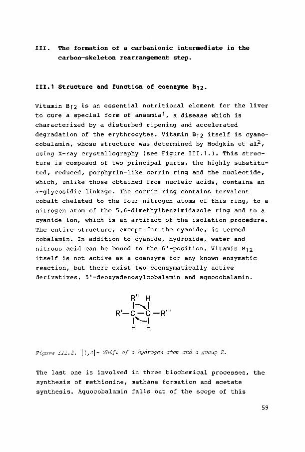
R" H ,,1 R'-C -C-R"'
1'--1 H H
Figure III.2. \1,2]- Shift of a hydragen atom and a group R.
The last one is involved in three biochemica! processes, the
synthesis of methionine, methane formation and acetate
synthesis. Aquocobalamin falls out of the scope of this
59
Figure III.l. Structure of vitamin
X-ray diffraction studies. as dete~ined by
thesis. Of the known enzymatic reactions which require
5'-deoxyadenosylcobalamin as cofactor, all but one (ribo
nucleotide reductase} involve an intramolecular [1,2] -shift
of hydrogen coupled with a [1,2]-shift of some other group,
as shown in Figure III.2. The eleven rearrangement reactions
known until now are divided in three groups, the carbon
skeleton rearrangements, the hydroxyl and the amine
migrations, according to the bond that is broken during the
reaction: a c-c, c-o or C-N bond3,4.
In this study special attention is given to the carbon
skeleton rearrangements, i.e. the isomerization of L-methyl
malonyl-Coenzyme A to succinyl-Coenzyme A, threo-B-methylene
glutarate to L-glutamate and B-methylitaconate to a-methyl
eneglutarate, where hydrogens for sake of clarity deuterons
with R=
o=( SCoA
methyl
malonyl-Co A
isomerization
----retention
NH~><' - )=o HO
methyl
aspartate
isomerization
or
inversion
HO
methyl
itaconate
isomerization
Figure III.3. The three carbon-skeleton rearrangements dependent on
coenzyme B12
.
61
are used in Figure III.3.) and a carbon-centered group R
migrate in an intramolecular (1,2]-shift. Onder enzymatic
conditions the hydragen (deuteron in Figure III.3.) is transferred via the 5'-methylene group of coenzyme B12S-8 and migrates in methylmalonyl-Co A with retention of configur
ation9-11 (i.e. the incoming hydragen and the leaving group
R occupy the same position). The migration in methylaspartate
occurs with inversion of configuration12,13, while the
stereochemistry of the methylitaconate isomerization is still
unknown.
111.2. Mechanism of action of the carbon-skeleton rearrange
ments.
In gene~al for the coenzyme B12-dependent rearrangements a
radiaal mechanism is proposed, as was recently summarized by Rétey14, based upon data arising from isotape labeling15,16
electron paramagnetic resonance17-19 and UV/VIS spectro
scopie measurements20. As Rétey describes in this
HRe H ~COSCoA
Hs · ''1 ""-, l'cH 3 cooHI _
HS. ( ,H 1 /,, ,_• ----1\:-....r-;;:.-' COS Co A
c~ 'cooH / _
enzyme enzyme
COSCoA H
Hs i~~--~{:::-·' H Re
I CH3 COOH -enzyme enzyme
Figur>e III.4. 3etent·ion of aonfigur>ation in t7w r>adiaal meahanism for> the
methylmalonyl-Co A rearrangement as pr>oposed by ;?étey.
62
article14, this radical mechanism is put forward independent
ly of the stereochemical results published, while in particu
lar the steric course of a reaction is extremely useful to
draw conclusions as to the mechanism. In order to integrate
the stereochemical results known and the radical mechanism,
he assumes14,21 a very intimate and unambiguous interaction
between enzyme and substrate, that prevents rotatien around
the C1-C3 bond (see Figure III.4.).
While the Retey model emphasizes the role of the enzyme, it
does not take into account the intrinsic properties of the
substrate, i.e. an enzyme can only lead a substrate through a
reaction-path that is pre-set in (allowed by} the substrate.
Moreover, no explanation is given of the modifications of the
enzyme needed to achieve inversion in the methyl aspartate
isomerization22.
From enzymatic data it has been shown by Pratt23, that the
three groups of rearrangements demonstrate some remarkably
distinct features. The enzymes of the carbon-skeleton
rearrangements require no other cofactors23, while those of
amine migrations all apparently require pyridoxal phosphate
and sametimes other cofactors such as K+, Mg+ and ATP. The
enzymes of the hydroxyl migrations all require simple ions
such as K+. Although the role of some of those factors, e.g.
pyriQOXal phosphate, is uncertain, the question is raised
whether there is a common denominator to the mechanism of
reaction of the different groups of substrates.
Another indication that the three groups of coenzyme 812-de
pendent reactions differ originates from ESR spectra24. The
enzymes that catalyze the isomerization of diols, glycerol
ethanolamine and the reduction of ribonucleotides give very
unusual and characteristic ESR spectra in the presence of
substrates (i.e. when frezen during catalysis) or substrate
analogues and even, in the case of ehanolamine ammonia lyase,
in the absense of any substrate or substrate analogue25,26.
In all cases, the ESR spectrum consists of two components,
namely a braad resonance at g - 2.3 due to the Co(II) ion
and a narrow doublet centered about g 2 due to an organic
63
radical, the splitting being explained by interaction with
the Co(II) ion. The fact that these paramagnetic species may
account for up to 65% of the coenzyme present depending on the enzyme and the substrate and, where detectable, are formed at a rate comparable to, or greater than, the turnover number of the enzyme strongly suggests that they repcesent
intermediates in the catalytic cycle25. No such signal could, however, be observed with methylmalonyl-Co A mutase27.
Though a radical mechanism might be appropriate for hydroxyl and amine migrations, the probability of such a mechanism
with respect to the carbon-skeleton rearrangements becomes
less. The only artiele publisbed to date indicating a free radical
rearrangement invalving the [1,2]-migration of a thioester
group as a model for the methylmalonyl Co A mutase reaction, is recently publisbed by Halpern et al28. This article,
however, is not very convincing. The kinetic measurements are presentea in such a way that radical and anionic processes
are not directly comparable. As far as one can see from the data presented, the uncatalyzed rearrangement rate of the radical is 235 sec-1 at 60.5"C and 2.5 sec-1 at 30"C, while
kcat for the. enzymatic methylmalonyl-Co A reaction has been estimated to be 102 sec-1. Rearrangement·of the anion gave
after 2 min. more thioester rearranged product (42% versus 1-9% in the radical rearrangement) at a lower temperature (-78"C versus 30"C) which comes closer to the enzymatic
process than the radical rearrangement. Moreover, no reason
is given why Halpern considers the formation of such a
substrate carbanion to be highly unfavorable and much less likely than the alternative free radical rearrangement,
although contributions from pathways invalving carbanion
intermediates cannot be definitively excluded!
Not only there is lot of literature suggesting the three
groups of carbon-skeleton rearrangements have d~fferent
mechanisms of action, there is also accuroulating evidence29-38 that in methylmal9nyl-Co A mutase reactions
the Co-C bond assists in the formation of a substrate carban-
64
ion in the rearrangement step. In the next Chapter we will
try to make plausible, why this could be the mechanism of
action for all the carbon-skeleton rearrangements.
III.3 The stereochemistry of the carbon-skeleton
rearrangements as 'test' for the carbanionic
mechanism. Scope of the second part of this thesis.
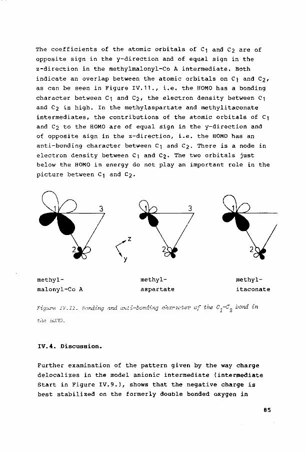
The few attempts made to date to include the stereochemical
results known for the carbon-skeleton rearrangements in the
back-side
ethylene glycol
0 H 11 I c---c L CoAS/ èf)::L
front-side
front-side
0 11 H c I
CoAS/ '-c L
\~ / Co
L'\1 "L CH
I COOH
COOH I H
/CH I NH2 'c L
\" / Co L~I"-L
CH
I COOH
Figure III. 5. 'l'he stereoc:hemistry of the
aspartate rearrangement~ according to the mechanism proposed by Corey et al.
65
mechanisme proposed, were incomplete (Rétey14, see Chapter
11.2.) or simply incorrect (Corey et alf9). The keystep Corey
proposes for the inversion of configuration in the conversion
of ethylene glycol to acetaldehyde29 is the migration of a
hydroxylgroup (with electron pair) to an electrophilic adjacent carbon and simultaneous bonding of cabalt (through
its vacant co-ordination side) to the methylene carbon in a back-side displacement of OH (see Figure !!!.5.). This
back-side displacement will lead to inversion of configuration. 1f we suppose that this mechanism is also
applicable to the carbon-skeleton rearrangements, then the -cosco A migration in the methylmalonyl-Co A isomerization to
a nucleophilic adjacent carbon and simultaneous bonding of cabalt to the methylene carbon in a front-side displacement
of the -cosco A group explains the retention of contiguration found for this rearrangement. However, the -CHNH2COOH group in the methyl-aspartate isomerization migrates to the same
nucleophilic carbon as found for the methylmalonyl Co A
rearrangement, which is in contradiction to the inversion of
contiguration known for the methylaspartate isomerization.
As indicated in Chapter 11.2. model studies suggest29-38
that in methylmalonyl-Co A mutase reactions the Co-C bond
assists in the formation of a substrate carbanion in the rearrangement step. 1f this model description (carbanion-formation) could predict the stereochemistry of
the carbon-skeleton rearrangements in general, this could be considered as an extra indication, in addition to the literature known to date29-38, towards a rearrangement of a
anionic intermediate. So the scope of this part of the
thesis is to find a correlation between the evolution of the
coefficients of the atomie orbitals in the Highest Occupied Molecular Orbital (HOMO) of anionic intermediates, and the
stereochemistry known for these reactions.
66
III.4 The nature of the hydrogen transferred temporarily to
coenzyme B12 during the carbon-skeleton rearrangements.
The way in which the anions can be generated is subject to a
lot of speculation. Three possible ways are summarized below.
All three should obey the observation of Miller et al~o,
showing the hydrogen which migrates during the isomerization
of methylmalonyl-Co A to succinyl-Co A becomes one of three
equivalent hydrogens on Cs' of coenzyme B12• before a hydrogen is returned to the substrate. The first two,
suggested by those who hold to initia! radical generation for
all the coenzyme B12-dependent rearrangements consist of radical generation in the substrate, foliowed by either elec
tron transfer from cobalt to the substrate:
R· ;+- Co(II)·:;;::!':Co(III)+ + R-, or by charge transfer from
protein basic and acidic sites to the substrate radical, a suggestion put forward by Finke41,42. A third possibility
is proton loss from the substrate to Cs' of the coenzyme (see
Figure III.6.), whereby one of the three hydrogens of the
S1.1bstrate &.Ad
+
+
I N
Substrate-
TH.6. Tr>ansfer of a proton :rom the substrate to C/ of coenzyme
of a Co-H-C bPidge.
substrate methylgroup originated becomes covalently bonded to
cobalt via an agostic M(H)C interaction, as proposed by Broekhart et al43,44. They state44 that carbon-hydrogen
honds, especially those of saturated (sp3) carbon centres,
are normally regarded as being chemically inert. Generally,
the C-H group is not thought of as a potential ligand which
can have a structural role or play an energetically
67
significant part in ground statea or in reaction intermedi~ ates. They review recent observations which show that the~
are in fact many circumstances in which a carbon-hydrogen group will interact with a transition metal centre with
formation of a two-electron three-centre bond,and that the \
effect of the interaction is such as to have ~'marked effect
on the molecular and electrooie structure and hence reactivity of the molecule. Once the anionic intermediate is formed, substrate-enzyme interaction can accommodate for the charge build-up in the substrate at the various stages of the rearrangements.
68
References and notes.
1. H.R. Mahler and E.H. Cordes, 'Biological Chemistry',
Harper and Row Publishers, New York, 1971, p. 424.
2. D.C. Hodgkin, J. Kamper, J. Lindsey, M. MacKay, J.
R.J. Prosen and K.N. Trueblood, Proc. Roy. Soc. (London)
A, 1957, 242, 228-263.
3. D. Dolphin, 'B12'• J. Wiley and Sons, New York, 1982,
volurne 1, p. 328.
4. B. Zagalak and W. Friedrich, 'Vitarnin B12', W. de
Gruyter, Berlin, 1979, p. 557.
5. J. Rétey and D. Arigoni, Experientia 1966, 22, 783-784.
6. G.J. Cardinale and R.H. Abeles, Biochirn. Biophys. Acta,
1967, 132, 517-518.
7. R.L. Switzer, B.G. Baltirnare and H.A. Barker, J. Biol.
Chern., 1969, 244, 5263-5268.
8. H.F. Kung and L. Tsai, J. Biol. Chern., 1971, 246,
6436-6443.
9. M. Sprecher, M.J. Clark and D.B. Sprinson, Biochern.
Biophys. Res. Comrn., 1964, 15, 581-587.
10. M. Sprecher, M.J. Clark and D.B. Sprinson, J. Biol.
Chern., 1966, 241, 872-877.
11. J. Rétey and B. Zagalak, Angew. Chern., 1973, 85,
721-722.
12. M. Sprecher and D.B. Sprinson, Ann. N.Y. Acad. Sci.,
1964, 112, 665-660.
13. M. Sprecher,·R.L. Switzer and D.B. Sprinson, J. Biol.
Chern., 1966, 241, 864-867.
14. J. Rétey, Recent Adv. Phytochern., 1979, 13, 1-27.
15. P.A. Frey and R.H. Abeles, J. Biol. Chern., 1966, 241,
2732-2733.
16. J. Rétey, A. Urnani Ronchi, J. Seibl and D. Arigoni,
Experientia, 1966, 22, 502-503.
17. B.M. Babior, T.H. Moss, W.H. Orrna-Johnson and H.
Beinert, J. Biol. Chern., 1974, 249, 4537-4544.
69
18. S.A. Cockle, H.A.O. Hill, R.J.P. Williams, S.P. Davies
and M.A. Foster, J. Am. Chem. Soc., 1972, 94, 275-277.
19. T.H. Finlay, J. Valinsky, A.S. Mildvan and R.H. Abeles,
J. Biol. Chem., 1973, 248, 1285-1290.
20. K.N. Joblin, A.W. Johnson, M.F. Lappert, M.R. Hollaway and H.A. White, FEBS Lett., 1975, 53, 193-198.
21. J. Rétey, Stud. Phys. Theor. Chem., 1982, 18, 367-389.
22. Reference 14, p. 19.
23. D. Dolphin, 'B12'• J. Wiley and Sons, New York, 1982, Volume 1; J.M. Pratt, p. 331.
24. D. Dolphin, 'B12'• J. Wiley· and Sons, New York, 1982, Volume 1; J.M. Pratt, p. 381 •.
25. B. Babior, Acc. Chem. Res., 1975, 8, 376-384.
26. J.F. Boas, P.R. Hicks, J.R. Pillbrow and T.D. Smith, J.
Chem. Soc., Faraday Trans. II, 1978, 417-431. 27. B. Zagalak and w. Friedrich, 'Vitamin B12'• w. de
Gruyter, Berlin, 1979; J. Rétey, p. 440. 28. s. Wollowitz and J. Halpern, J. Am. Chem. Soc., 1984,
106, 8319-8321.
29. P. Dowd and M. Shapiro, J. Am. Chem. Soc., 1976, 98, 3724-3725.
30. B. Zagalak and w. Friedlich, 'Vitamin B12'• W. de
Gruyter, Berlin, 1979; P. Dowd, p. 557. 31. A.I. Scott, J. Kang, D. Dalton and S.K. Chung, J. Am.
Chem. Soc., 1978, 100, 3603-3604.
32. A.I. Scott and K. Kang, J. Am. Chem. Soc., 1977, 99,
1997-1999. 33. G. Bidlingmaier, H. Flohr, O.M. Kempe, T. Krebs and
J. Rétey, Angew. Chem., 1975, 87, 877-8.
34. H. Flohr, W. Pannhorst and J. Rétey, Angew. Chem., 1976,
88, 613-614.
35. H. Flohr, u.w. Kempe, W. Pannhorst and J. Rétey, Angew. Chem., 1976, 88, 443-444.
36. M. Fountoulakis and J. Rétey, Chem. Ber., 1980, 113,
650-668.
37. B. Zagalak, w. Friedlich, 'Vitamin B12'• W. de Gruyter, Berlin, 1979; J. Rétey, p. 439.
70
38. J.H. Grate, J.W. Grate and G.N. Schrauzer, J. Am. Chem.
Soc., 1982, 104, 1588-1594.
39. E.J. Corey, N.J. Cooper and M.L.H. Green, Proc. Natl.
Acad. Sci. USA, 1977, 74, 811-815.
40. w.w. Millerand J.H. Richards, J. Am. Chem. Soc., 1969,
91, 1498-1507.
41. Personal communication of R.G. Finke.
42. R.G. Finke, O.A. Schiraldi and B.J. Mayer, Coord. Chem.
Rev., 1984, 54, 1-22.
43. M. Brookhart, M.L.H. Green and R.B.A. Pardy, J. Chem.
Soc., Chem. Commun., 1983, 691-693.
44. M. Brookhart and M.L.H. Green, J. Organomet, Chem.,
1983, 250, 395-408.
71
72
IV. Quantum chemica! calculations.
IV.l Introduction.
In order to test the model description, suggesting the
formation of a substrate carbanion in the rearrangement step
(see Chapter III.), for the stereochemistry of the
carbon-skeleton rearrangements in general, we selected the
anionic cyclopropane intermediates as giv~n in Figure IV.l.
for the quanturn chemica! calculations.
with R=
o=( SCoA
methyl
malonyl-Co A isomerization
0
OH H
H R
NH~><' ~ )=o HO
methyl
aspartate isomerization
methyl
itaconate isomerization
Figure IV.l. The anionia ayalopropane intermediates which account for the
stereoahemistry of the aarbon-skeleton rearrangements.
For sake of simplicity the calculations were confined to the
substrate system without introducing enzyme or coenzyme
specific sites. The key intermediate as illustrated in Figure
IV.1. is formed by proton abstraction from the methyl group
73
(C3) followed by an approach of group Rand the now negatively charged methylene group. The intermediate ring closure
during the isomerization of methylmalonyl-Co A and methyl
itaconate is facilitated by polarization of the C=O and C=C
bond in group R respectively. In the isomerization of methylaspartate a keto-enol tautomerization can provide an
analogous structure, able to accommodate negative charge (see
Figure IV. 2. )
OH
HO
OH
0=
•' ,\H
CH 3
/OH HO
Figure IV.2. The keto-enol tautomerization in methylaspartate resulting
in a structure which accommodates the negative charge after ring closure,
Instead of one enzyme essential for the isomerization of
methylmalonyl-Co A and methylitaconate, the enzyme complex for the isomerization of methylasparate consists of two proteins. The fully optimized MNDO structure of the enol form
is only 2 kcal/mole higher in energy than the keto form, an
energy difference which is smaller than the unpredictable error of the MNDO method1.
The final rearrangement product is formed by proton addition
at the acid substituted c, and rupture of the c,-R bond.
IV.2. The choice of the calculational method.
As indicated by Meiver and Komornicki2, a detailed understanding of the dynamics and stereochemistry of organic
74
reactions requires, above all, a knowledge of the many
dimensional potential energy surface. The very dimensionality
of this surface, however, precludes its evaluation for all
but the simplest systems. To reduce this problem to one of a
tractable size, two general approaches have been employed.
The first type of approach seeks to reduce the dimensionality
of the surface by eliminating eertaio degrees of freedom, for
example the length of a carbon-hydrogen bond. A related
technique involves choosing one or two degrees of freedom as
independent variables of the potential energy and to allow
the system to relax by optimizing the remaining degrees of
freedom for each value of the independent variables.
The second type of general approach involves consideration of
all the degrees of freedom of a system, but seeks only to
locate eertaio chemically interesting points on the potential
energy surface. For a one-step reaction, these points would
be the local minima corresponding to the equilibrium
geometries of reaetauts and products and a col or saddle
point which separates the local minima.
For a number of reasons this last method, which is preferred
by the author of the cited article, cannot be followed in
this case. The main reasen is, that our approach to the
problem is a dynamic one. Just localizing stationary points
on the curve would not elucidate effects such as fast electron
flow and orbital inversion (vide infra). In the 'product'
minimum these effects will be damped out, while comparison of
the points on the reaction path somewhere in between the
transition state (whose exact position is not important here)
and the 'product' minimum (called 'End' further on in the
article) will give the desired information. An extra handicap
is the definition of a stationary point at the 'Start' (vide
infra) of the ring closure. 'Start' is defined as the linear
molecule with tetrahedral carbon angles just after proton
abstraction. This is not a stationary point, but a rather
unstable intermediate. Reasons why is chosen for the first
type of approach indicated above.
75
IV.3. Results
The formation of the cyclopropane internediatea of Figure
IV.J. bas been studled with MNDO calculationsl.
OH OH OH H
H
OH
Figure IV.3. The three inteTmediates of uhiah the ring alosure has been
studied.
Of course MNDO results cannot give the final proof of the
assumed reaction mechanism, but a detailed anderstanding of the dynamica and stereochemietry of organic reactions requires, above all, a knowledge of the potentlal energy surface2. The energy profile of all three ring closure reactions is calculated by optimization of all distances, angles and
torsion angles to minimal heat of formation at a number of fixed values of r1 and r2 (see Figure IV.J.), ranging between 1.35 A and 1.80 A for r1 and between 1.40 A and 2.50 A for
r2. In this way the angle CJ-C1-C2 changes from tetrabedral (in the linear molecule direct after proton abstraction) to triangular (at the end of the ring closure). As initial values for distances and bond angles, those optimized by Dewar et al. for the MNDO program are used. The reaction path is drawn along the line of minimal energy. The results of these calculations are given in the Figures IV.4., IV.S. and IV.6. Following the reaction path, the heat of formation as a
function of the reaction co-ordinate r2 is given for all three ring closure reactions in Figure IV.S. The graphs are closely related, except that the transition state of the
76
2.30 • • I
I
2.20 I • • I I • I • 2.1 0 I • • • • • • • I • I
• I
2.00 • •
1.9 0
1.80
1.70
1.60 ' -130 ' ' •,
' 1.50 ' • •
1.40 1.50 1.60 1.70 1.80 .... r
1 ( Ä)
Fi~~re IV.4. Heat of formation in kcaZ/moZe as a function of r1
and r2
for intermediates during ring cZosure for the methyZmaZonyZ-Co A isome-
rization. 77
1.40 1.50 1.60 1.70 1.80 __ ..... .,_ r
1 (A)
Figure IV.5. Heat of formation in kaat/mote as a fUnation of r1
and r2
for intermediates during ring alosure for the methylaspartate isomerization.
78
r2 { Ä l -145
~· -140 2.40 • •
• • • 2.30
135
• 2.20
• • 2.1 0 •• • I - 13 5 • I • • I
2.00 .~ • I
• ~ • • 1.90 I •• \
\ • , . 1.8 0
\
• \ • 135
-140
1.70 -145
- 1 50
1.60
1.50 • •
1.40 1.50 1.60 1.70 1.80
---~- r 1 ( Ä } IV. 6. Heat kcal/mole as a function of and
for intermediates ring closure for• the methylitaconate isomere-
zat ion. 79
~Hf
(kc a 1/ mole}
• 11 0 *140 0 1 50
• 1 1 5 *145 0155
• 1 2 0 'lf.150 o160
• 1 25 * 1 55 0 1 65.
•130 *160 0 1 70
1
Start End
l
o Methylmalonyl- CoA
• Methylaspartate
*Methyl i toconate
0
\o
' 0-(
2.30 220 2.10 2.00 1.90 1.80 1.70 1.60 r2(A)
Figure IV.?. Heat of formation in kaaZ/moZe as a funetion of the reaetion
ao-ordinate r 2 for the three aarbon-skeZeton rearrangements.
80
methylitaconate ring closure is situated later on the
reaction co-ordinate. A minimum in energy is reached for
values of r2 between 1.60 and 1.50 Ä. The calculations are
not extended beyond this value because in our model the
proton addition to Ct takes place before this minimum in
energy is reached. The charge density accumulated on the
various groups of the intermediate structures varies with the
reaction co-ordinate r2 in a quite similar way for the three
rearrangements, with the exception of the charge density on
the group which stahilizes the negative charge by polariz
ation of a double bond, i.e. C=O, C=C(OH)2 and C=CH2 (C=X in
Figure IV.8.). In the isomerizations of methylaspartate and
methylitaconate, the charge accommodated by this group in the
beginning of the reaction co-ordinate is high in comparison
with the isomerization of methylmalonyl-Co A, as can be seen
in Figure IV.8.
0 Methylmalonyl-Co A . Methylaspartate e
1
0 . Methylitaconate ' * . ' . ' *-.. .,
*-.. . ' 0.2 *-..
* .,
' ., * . '* ' '*
., '*
., . -0.4 O-o
'* ..... -o-o '
., '0,0 *
. "" -o_ ' o, *
o_o '* -o_ '*--*-*'* o-o
-0.6 -o-o-o
2.2 2.0 1.8 1.6 -r2 !.&.l
Figure IV.B. Charge on group =X as a function of r2
for the carbon-skele
ton rear1•angements.
81
-0.25 -0.16 -0,21
-aot,~~H H I H
-M>( COOH H
A. ~ r3 1.43 3 -0.63 1.46 3 -0,57 1.53 .... ~
CQ H / H H :;:::
/ \1) ":i " /
~ \1)
" r2 / ~ f..; ..
/ /2.15 ti- :0::: .. 2.35 (0 ;c
-M>( Q
/ ti- Start TS _,.,( End ~
CQ
l _",( 0 -\l) (0
-0.39 SH 0 SH ~ SH -0-44
\l) -0.18 -0.58 -0.28 - 0.17
":i' ~ .... Jl ......
C>
Q 2 -0,29 ...... ...... -G.20 -0.17 C> ....
II ( COOH H
-M<( COOH H
_", ( COOH H (0 ~ I :;::: ;::)
~ <i- - o.oa H 1.47 3 -0.52 1.52 .... 3 o.oo C> +0.23 H / H ;:-! H H H C> / <::: ..
The charge distribution over the intermediate structures at
different stages of the ring closure is given in Figure IV.9.
The definition of the intermediates Start, TS (transition
state) and End is given in Figure IV.7.
The evolution of the coefficients of the atomie orbitals in
the HOMO of the cyclopropane intermediates along the reaction
co-ordinate is followed. The two atomie orbitals with the
highest coefficient in the HOMO are given in Figure IV.lO. as
a function of r1 and r2.
In all three reactions the overall picture is the same. In
the beginning of the ring closure the contribution of the
atomie orbitals on C3, the carbon from which the proton is
abstracted, is dominant. After the transition state the
atomie orbital on the formerly double bonded oxygen
I ',~ I
x
2.1
1.9 ~ x I
\
1.7
1.5
\
\~ .... 2
' x
1.4 1.6 1.8
Figure IV.lO. The two atomie orbitaZs with the
HOUO for all three carbon-skeleton rearrangements.
83
respectively carbon, in the direction of C3 becomes important. Near the end of the ring formation the orbital on
c, in the direction of c2 will have a large coefficient in the HOMO, as can beseen in Figure IV.10. A very important difference between the three reactions becomes clear, if also atomie orbitals with smaller coefficients in the HOMO are taken into account. If the intermediate is situated in the y-z plane, with the c,-c2 bond on the y-axis, the contribution of the atomie orbitals of the three carbons constituting the ring is given in Table IV.1. for rt=1.7 A and r2=l. 6 A.
methyl- methyl- methyl-malonyl-Co A aspartate itaconate
c, s - 0.15 - 0.12 - 0.14 x + 0.08 + 0.03 + 0.04 y + 0.50 + 0.31 + 0.38 z - 0.30 - 0.20 - 0.25
c2 s + 0.03 + 0.04 + 0.04 x + 0.01 + 0.03 + 0.03 y - 0.09 + 0.07 + o. 11
z - 0.21 + 0.03 + 0.03
c3 s + 0.11 + 0.06 + 0.06 x + 0.00 + o.oo + o.oo y - 0.31 - 0.18 - 0.22 z + 0.22 + 0.08 + 0.09
TabZe IV.l. The aoeffieients of the atomie orbitaZs of the ring aarbons
at t/he moment of proton addition to the ayaZopropane intermediates.
84
The coefficients of the atomie orbitals of Ct and c2 are of opposite sign in the y-direction and of equal sign in the
z-direction in the methylmalonyl-Co A intermediate. Both
indicate an overlap between the atomie orbitals on Ct and c2, as can beseen in Figure IV.11., i.e. the HOMO has a bonding
character between Ct and c2, the electron density between Ct and C2 is high. In the methylaspartate and methylitaconate
intermediates, the contributions of the atomie orbitals of Ct and C2 to the HOMO are of equal sign in the y-direction and of opposite sign in the z-direction, i.e. the HOMO has an
anti-bonding character between Ct and c2. There is a node in
electron density between Ct and c2. The two orbitals just below the HOMO in energy do not play an important role in the
picture between Ct and c2.
methyl
malonyl-Co A
methyl
aspartate
methyl
itaconate
IV.ll. mn'uJ.?,ruJ and anti-bonding character of the c1-c2 bond in
h0,10.
IV.4. Discussion.
Further examination of the pattern given by the way charge
delocalizes in the model anionic intermediate {intermediate Start in Figure IV.9.), shows that the negative charge is
best stabilized on the formerly double bonded oxygen in
85
methylmalonyl-Co A, in comparison with the way the -C(OB)2
group accommodates the negative charge in methylaspartate.
The methylene group of methylitaconate behaves intermediate in accommodation of the negative charge. The relative small
initia! accommodation of negative charge by the diol group in methylaspartate, can be considered as a prerequisite for a
larger charge migration during ring closure. Such a large
charge migration from C3 over C2 to C1 might force the electron density to pass momentarily beyond c,. Subsequent
orbital inversion at C2 will prevent the electron density to
flow back via the c,-c2 bond and to delocalize over the cyclopropane ring. Proton addition at that moment on the reaction co-ordinate will lead to inversion of configuration on c,, i.e. the proton comes in at the opposite side of the
leaving glycyl group. The relative small charge migration during ring closure of methylmalonyl-Co A will not be strong enough to force the electron density to pass c,, resulting in
retention of contiguration at c,. This rather new concept of
orbital inversion to prevent electron back-donation to c2 is
strongly supported by the picture originating from the
development of the coefficients of the atomie orbitals in the HOMO. Figure IV.10. shows charge migration via C2 and not
directly from c3 to c,. This direction of migration is necessary for the electron density to pass c, in line of the
c,-c2 bond, which will lead to inversion of configuration on c,. Figure IV.11. indicates an anti-bonding character for the
c,-c2 bond of the methylaspartate intermediate, which
prevents the electron density to flow back to C2 and the proton to add to the c,-c2 bond. The bonding character of the
c,-c2 bond in the methylmalonyl-Co A intermediate will cause proton addition witn retention of configuration at c,, due to
the high electron density between c, and C2. The anti-bonding character of the c1-c2 bond in the HOMO of the methylitaconate intermediate suggests inversion of configuration in
this rearrangement. Finally it may be of interest to note that in the case of methylmalonyl-Co A mutase using ethyl
malonyl-Co A as substrate instead of methylmalonyl-Co A only
86
partial inversion is observed4. Besides the role of the
enzyme in the enzyme-substrate binding (ethylmalonyl reacts
at only one thousandth the rate of the natural substrate),
the loss of stereospecificity in the case of ethylmalonyl
Co A may be also electronic in nature.
IV.5. Conclusion.
Energy profiles of the carbon-skeleton rearrangements with
cationic or radical intermediates in the rearrangement step
are required to discuss a possible preferenee for the anionic
pathway. However, the fact that an anionic intermediate can
explain the known stereochemistry, can be considered as an
extra indicator in addition to the chemica! evidence sugges
ting a carbanion in the rearrangement step of the methyl
malonyl-Co A isomerization.
87
References eand notes
1. J.S. Dewar and W. Thiel, J. Am. Chem. Soc., 1977, 99,
4899-4907.
2. J.W. Meiver and A. Komornicki, J. Am. Chem. Soc., 1972, 94, 2625-2633.
3. J.S. De war and w. Thiel, J. Am. Chem. Soc., 1977, 99,
4907-4917.
4. J. Rétey and B. Zagalak, Angew. Chem., 1973, 85, 721-2.
5. l.I. M.erkelbach, H.G.M.. Becht and H.M.. Buck, accepted for
publication in J. Am. Chem. Soc., 1985, 107.
88
Summary.
The first part of this thesis describes investigations con
cerning the role of penta co-ordinated phospholipid inter
mediates ~n the activatien of membrane embedded proteins,
viz. ion channels. On the basis of extensive literature
search a model is drawn in which a conformational change in
the phospholipid headgroup from a four co-ordinated tetra
gonal (P(IV)) toa five co-ordinated trigonal bipyramidal
(P(V) TBP) structure results in a change of conformation of
the lipid hydracarbon region. The conformational change in
the phospholipid headgroup is induced via external factors as
cation concentration, potentlal field and binding of e.g. a
water molecule. Enhanced electronegativity on the axial phos
phate oxygen of the five co-ordinated intermediate, leads to
repulsion of the sn-2 oxygen bound via an 0-C-C-0 sequence to
the phosphate group. The resulting adaptation in the hydra
carbon chain region will lead to cluster formation in the
sense of a region of different density compared to the
surrounding matrix. Cluster formation around a membrane
protein (viz. ion channel) will activate that ion channel to
open. To test several aspects of this model description,
phospholipid vesicles are synthetized of varying lipid
composition. Over their wall an ion gradient is created,
whose equilibration is followed with 23Na NMR after addition
of Gramicidin A. Leaving all the experimental conditlans as
constant as possible, except the lipid composition of the
vesicle wall, passive ion transport was measured of different
rates. The relative rate of ion transport as predicted on the
basis of the described model, was established by the measure
ments.
89
The second part of this thesis uses the stereochemistry of
the carbon-skeleton rearrangements dependent on coenzyme B12 as a 'test' for the mechanism of action postulated for these
isomerizations. An anionic mechanism is given, based on
extensive literature search, suggesting an anionic mechanism for the methylmalonyl-Co A rearrangement. With the help of
MNDO calculations, energy profiles are constructed for the
three ring closure reactions towards anionic enzyme stabilized cyclopropane intermediates. Following the reaction path,
charge distribution and migration in the substrates are monitored, as well as the evolution of the coefficients of the
atomie orbitals in the highest occupied molecular orbital (HOMO) of the cyclopropane intermediates. Large charge migra
tion as a consequence of small charge stabilization by a
certain group in the anionic intermediate is found to be a prerequisite for the inversion of configuration known for the methylaspartate isomerization. Conversely small charge migration as a consequence of good
charge stabilization in the methylmalonyl-Co A rearrangement
will lead to retention of configuration. For the methylitaconate isomerization inversion of configuration is suggested.
90
Samenvatting.
Het eerste gedeelte van dit proefschrift beschrijft het
onderzoek naar de rol van vijf-gecoördineerde fosfolipide
intermediairen bij de activering van in het membraan opgeno
men eiwitten, zoals ion-kanalen. Op basis van een uitgebreid
literatuur onderzoek is een model opgesteld, waarin een con
formatie verandering in de fosfolipide hoofdgroep van een
vier-gecoördineerde tetragonale (P(IV)) naar een vijf-gecoör
teert in een conformatie verandering in de koolwaterstof
ketens. De conformatie verandering in de fosfolipide hoofd
groep wordt geïnduceerd door externe factoren als kation-con
centratie, potentiaal veld en de binding van bijvoorbeeld een
water molecule. Een verhoogde electronegativiteit op de axi
ale fosfaat zuurstof van het vijf-gecoördineerde intermedi
air, leidt tot afstoting van de sn-2 zuurstof, die via een
o-e-c-o keten gebonden is aan de fosfaat groep. De resulte
rende aanpassing in de koolwaterstof ketens induceert cluster
vorming. De term 'cluster vorming' wordt hier gebruikt voor
een gebied met een dichtheid, verschillend van die van de
omgeving. Cluster vorming rond een membraan eiwit (het ion
kanaal) zal het ion-kanaal activeren, waardoor het ion-trans
port start. Om verschillende aspecten van het model te tes
ten, zijn fosfolipide 'vesicles' gesynthetiseerd met varie
erende lipide samenstelling. Over de wand van deze vesicles
is een ion-gradient gecreëerd. Het verdwijnen van deze
gradient na toevoeging van Gramicidine A is gevolgd met
behulp van 23Na NMR. Tijdens deze metingen zijn alle experi
mentele omstandigheden, behalve de lipide samenstelling van
de vesicles, zo constant mogelijk gehouden. Zo kan ion-trans
port gemeten worden van verschillende snelheid. De met behulp
van het beschreven model voorspelbare relatieve snelheid van
ion-transport is bevestigd door de metingen.
91
Het tweede gedeelte van dit proefschrift gebruikt de stereochemie van de coenzyme 8·12 afhankelijke koolstofskelet isome
risaties als een 'test' voor het werkingamechanisme dat gepostuleerd is voo• deze omleggingen. Een anionisch mechanisme wordt voorgesteld, gebaseerd op een groeiende hoeveelheid literatuur, die een anionisch mechanisme voor de methylmalonyl-Co A omlegging suggereert. Met behulp van MNDO berekeningen, zijn energieprofielen opgesteld voor de drie ringsluitingareacties naar anionische enzyme-gestabiliseerde cyclopropaan intermediairen. Ladingsverdeling en migratie zijn gevolgd gedurende de reactie, alsook de verandering van
de coëfficiënten van de atomaire orbitalen in de hoogst bezette moleculaire orbital (= HOMO) van de cyclopropaan
intermediairen. Sterke ladingsmigratie, als gevolg van een geringe ladingastabilisatie door een bepaalde groep in het anionisch intermediair, blijkt een voorwaarde te zijn voor de inversie van configuratie, gevonden voor de methylaspartaat isomerisatie. In tegenstelling hiermee leidt een geringe ladingsmigratie, als gevolg van een goede ladingsstabilisatie in de methylmalonyl-Co A omlegging, tot retentie van configuratie. Voor de methylitaconaat isomerisatie wordt op grond van de berekeningen een inversie van configuratie verondersteld.
92
Curriculum vitae.
De schrijfster van dit proefschrift werd op 3 augustus 1954
geboren te Mozambique. Na het behalen van het diploma HBS-B
aan het Lorentz Lyceum te Eindhoven in 1971, werd hetzelfde
jaar begonnen met de opleiding tot Chemisch Analist A aan het
Instituut voor Hoger Beroeps Onderwijs te Eindhoven. In 1973
werd dit diploma behaald, waarna begonnen werd met de studie
op de afdeling der Scheikundige Technologie aan de Technische
Hogeschool te Eindhoven. Het afstudeerwerk werd verricht bij
de vakgroep Organische Chemie, onder leiding van Prof. Dr.
H.M. Buck en Dr. Ir. D. van Aken. In novémber 1980 werd het
ingenieursexamen met lof afgelegd.
Van 1 december 1980 tot 1 mei 1985 was zij werkzaam bij de
vakgroep Organische Chemie van de Technische Hogeschool
Eindhoven als wetenschappelijk assistente. Tijdens deze
periode werd het onderzoek, beschreven in dit proefschrift,
uitgevoerd onder leiding van Prof. Dr. H.M. Buck.
93
Dankwoord.
Gedurende het onderzoek, dat geleid heeft tot dit proefschrift, heb ik van velen steun ondervonden. Alhoewel ik niet allen bij naam kan noemen, wil ik toch enkele van hen zeer
specifiek bedanken. Als eerste wil ik bedanken het technisch en administratief personeel, zowel binnen als buiten de vakgroep. Zij hebben
mij geholpen de in tijden van bezuiniging verouderde appa
ratuur op te knappen. Ten tweede wil ik bedanken degenen die mij moreel ondersteund hebben, waarbij ik mijn echtgenoot, familie, vrienden en de koffie-club uit de rookvrije kantine graag expliciet wil
noemen. Bij het tot stand komen van de 23Na NMR heb ik steun ondervonden van Jan de Haan en Leo van de Ven.
Bij het rekenwerk heb ik veel steun ondervonden van Maarten Donkersloot. Hanneke Becht wil ik bedanken voor het werk dat zij tijdens haar afstuderen op het gebied van Vitamine B12 heeft verricht.
Tenslotte wil ik Ria Hoozemans hartelijk bedanken voor het typewerk, dat leidde tot de uiteindelijke vormgeving van dit proefschrift. De mogelijkheid die zij creëerde, dit proefschrift met behulp van de tekstverwerker tot stand te laten komen, gaf mij de gelegenheid deze laatste fase van de promo
tie overeenkomstig de eisen van deze tijd te volbrengen. De bijzonder fraaie tekeningen in dit proefschrift zijn gemaakt door Henk Eding en Cas Bijdevier.
Financial support for this investigation was provided by
Unilever Research, Vlaardingen, The Netherlands.
94
I. De bewering van Roberts en medewerkers dat de 3p én de 3p orbital x y
deel uitmaken van de enkel bezette MO in TBP fosforanylradicalen, moet
op grond van symmetrie overwegingen verworpen worden.
J.W. Cooper, M.J. Parrotten B.P. Roberts,
J. Chem. Soc., Perkin II, 1977, 730-741.
2. Modelverbindingen voor koolstofskelet isomerisaties onder invloed van
Vitamine B12
, waarin het substraat gebonden is aan het centrale cobalt-
atoom, kunnen niet worden gebruikt om conclusies te trekken met betrek-
king tot waterstof abstractie van niet geactiveerde koolstofatomen.
P. Dowd en B.K. Trivedi, J. Org. Chem. 1985, 50, 206-217.
3. Het is de vraag of de ontwikkeling van nat naar droog ontwikkelbare
lakken in de lithografie ten behoeve van de I.C. technologie, analoog
aan de ontwikkeling van nat naar plasma etsen, zal leiden tot een be-
tere patroondefinitie.
4. Het inschatten van de mogelijkheid korter te gaan werken en een deel
van de eigen functie over te dragen aan anderen, is leiding geven-
de functionarissen mede afhankelijk van het vermogen de eigen complexe
functie te analyseren en van het vermogen tot delegeren,
Mr. H. Luik, Intermediair 35, augustus 1984, 1-5.
5. De conclusie van Bock en medewerkers, dat bij de isomerisatie van D-
fructose naar D-glucose onder invloed van het enzym glucose-isomerase,
het a-anomeer het begin van de omzetting met een hogere prefe-
rentie gevormd wordt dan uit de evenwichtsteestand blijkt, is een
overschatting van de mogelijkheden van de NMR techniek.
K. Bock, M. Meldal, B. Meyer en L. Wiebe,
Acta Chem. Scand. B, 1983, 37, 101-108.
6. De constatering dat sommige proteinen goed functioneren in een milieu
bestaande uit enkel detergentia, behoeft niet in strijd te zijn met
de bewering dat membraanproteinen een specifieke lipide-omgeving nodig
hebben om geactiveerd te worden.
P.R. Cullis en B. de Kruyff, Biochim. Biophys •. Acta, 1979,
559, 399-420.
7. De 'closed-shell' 1A1 overgangstoestand met c2v symmetrie van de ther
mische [1,5] - H shift in pentadiëen, zoals berekend doorHessen Schaad,
is een aangeslagen toestand en niet een grondtoestand.
B.A. Hess en L.J. Schaad, J. Am Chem. Soc. 1983, 105, 7185-7186.
8. Het wekt op zijn minst enige verwondering, dat het vrijkomen van for
maldehydgas uit spaanplaat bekend is en tot klachten leidt bij een aan
zienlijk gedeelte van de Nederlandse bevolking, terwijl de aanwezigheid
van ditzelfde gas in sigaretterook systematisch genegeerd wordt.
9. Bij de productie van (Al, Ga) As - halfgeleiderlasers, geschikt voor
toepassing in 'compact-disk' spelers, met behulp van organometaalgas
fase epitaxie, verdient het aanbeveling meer aandacht te besteden aan
de epitaxiale groei van (Al, Ga) As - lagen waarbij de groeisnelheid
kinetisch bepaald is.
International Conference on HOVPE II, april 1984,
J. Cryst. Growth, 1984, 68.
JO. Bij de bestudering van conformatieveranderingen in DNA structuren,
die optreden ten gevolge van complexering met metaalionen, wordt een
onjuist beeld van de fosfaat-metaal complexatie verkregen, indien ge
bruik wordt gemaakt van 5'-nucleotiden die enkelvoudig zijn veresterd
op de fosfaatgroep.
S.K. Hiller, D.G. VanDerveer en L.G. Marzilli, J. Am. Chem.
Soc., 1985, 107, 1048-1055.
ll. Bedrijven en overheidsinstellingen zullen via de wet gedwongen moeten
worden om een bepaald percentage vrouwen in dienst te nemen.
Mr. F.H.A.M. Kruse, directeur-generaal Arbeidsvoorzieningen,
i'finis ter ie van Sociale Zaken, januari 1985, op het congres