Abstract Tunneling nanotubes (TNTs) have previosly been observed as long andthin transient structures forming between cells and intercellular protein transferthrough them has been experimentally verified. It is hypothesized that this may bea physiologically important means of cell–cell communication. This paper attemptsto give a simple model for the rates of transfer of molecules across these TNTs atdifferent distances. We describe the transfer of both cytosolic and membrane boundmolecules between neighboring populations of cells and argue how the lifetime ofthe TNT, the diffusion rate, distance between cells, and the size of the moleculesmay affect their transfer. The model described makes certain predictions and opens anumber of questions to be explored experimentally.
1 Introduction
Cell–cell communication plays an important role in coordinating collective cell de-cisions. It is also critical to maintain both structural and functional homeostasis ina tissue. Since coordination between cells is an essential requirement for the suc-cessful functioning of a multicellular organism, many mechanisms have evolved toallow cells to communicate with each other bearing important outcomes in bothnormal functioning of the tissues and pathology. For example, high twitch muscle
Y. Suhail and Kshitiz contributed equally to the manuscript.
Y. Suhail · Kshitiz · M. Walker · M.D. Brennan · J.S. Bader · A. Levchenko (�)Department of Biomedical Engineering, Johns Hopkins University, Baltimore, MD 21205, USAe-mail: [email protected]
Y. Suhail · J.S. BaderHigh-Throughput Biology Center, Johns Hopkins University, Baltimore, MD 21205, USA
Kshitiz · J. Lee · D.-H. KimDepartment of Bioengineering, University of Washington, Seattle, WA 98195, USA
Modeling Intercellular Transfer of Biomolecules Through Tunneling 1401
cells are in close proximity with blood vessels, and nerve endings, and their closeinteractions are essential for the correct muscle functioning (Behnke et al. 2011;Vikne et al. 2012). Cell–cell interactions between cancer cells and endothelial cellsoccur within solid tumor, and metastatic cancer cells extravasating the endothelium(Weis and Cheresh 2011; Qin et al. 2012; Stine et al. 2011). Extensive research hasexplored the mechanisms of these cell–cell interactions, resulting in extensive infor-mation on the chemical cell–cell signaling pathways occurring in autocrine (Licht-enberger et al. 2010), paracrine (Abou-Khalil et al. 2009), and juxtracrinemanner(Bosenberg and Massague 1993; Singh and Harris 2005). However, recent evidencehas also suggested another form of cell–cell interaction that occurs via direct trans-fer of cellular components from one cell to another, thus transferring informationwithout involvement of traditionally implicated chemical mechanisms (Niu et al.2009; Ahmed and Xiang 2011; Li et al. 2010; Pap et al. 2009; Prochiantz 2011;Mack et al. 2000). Although the degree of such intercellular transfer of cellularcomponents and its role in defining cell and tissue behavior in vivo remain lessunderstood, the evidence for existence of this novel communication mechanism isoverwhelming, suggesting that it could potentially have a significant effect in in-fluencing the recipient cell phenotypes in such diverse processes as cancer pro-gression (Ambudkar et al. 2005), immunity (Baba et al. 2001; Carlin et al. 2001;Quah et al. 2008), HIV infection (Mack et al. 2000), transfer of drug resistance(Levchenko et al. 2005), and ribosomal recruitment in neuronal axons (Twiss andFainzilber 2009). Direct protein–protein transfer is therefore important to understandin greater detail, both experimentally and computationally.
Previous studies have reported multiple examples of transfer of membrane pro-teins between cells (Levchenko et al. 2005; Guescini et al. 2012; Agnati et al. 2011;Al-Nedawi et al. 2008; Davis 2007). In addition, small cytoplasmic biochemical com-ponents have also been shown to be transferred between cells in a size-dependentmanner (Niu et al. 2009). However, intercellular transfer of large cytoplasmic pro-teins has not been yet examined with conclusive results. Various mechanisms havebeen suggested for intercellular transfer of cellular components, including formationof tunneling nanotubes (TNTs) between cells (Guescini et al. 2012; Rustom et al.2004), spontaneous secretion and integration of microvesicles (Valadi et al. 2007;Denzer et al. 2000), and transient cell–cell fusion (Driesen et al. 2005). As is fre-quently the case with poorly understood biological phenomena, it is not easy to dis-criminate between putative mechanistic details and generate most plausible modelsof this cell communication phenomenon. It is also possible that the mechanisms maybe cell-type specific and multiple mechanisms might coexist in diverse physiologi-cal and patho-physiological contexts. However, certain findings have been suggestiveof the constraints that can be placed on the mechanistic models of this process. Forinstance, the reports of membrane protein transfer are much more frequent and bet-ter supported than the reports of transfer of large cytosolic components, including ofproteins (Agnati et al. 2011; Camussi et al. 2010). We questioned whether a reasonfor this discrepancy might lie in the properties of the transfer process itself. Another,potentially more revealing constraint comes from the observation that transfer of cy-tosolic, but not membrane components is strongly dependent on the molecular weightof the transferred molecules (Niu et al. 2009). Thus, a plausible model of the trans-
1402 Y. Suhail et al.
fer process has to be able to explain these particular well-established features of theintracellular transfer of different cellular components.
Here, we propose a mathematical model to explain passive protein transfer be-tween cells via formation of tunneling nanotubes (TNTs), which have been observedin various studies to be responsible for intercellular protein transfer (Guescini et al.2012; Rustom et al. 2004; Bukoreshtliev et al. 2009). Our steady state model explainsthat while membrane protein transfer may be unrelated to the mass of the protein, cy-toplasmic proteins may follow an inverse correlation with size. Though no existingreport conclusively shows the transfer of cytoplasmic proteins between cells, smallercytoplasmic components have been shown to be transferred between cells in a size-dependent manner (Niu et al. 2009), as predicted by the model. The model explainsthat while transfer of cytoplasmic proteins may occur between cells, it would be inrelatively smaller amounts in comparison to smaller biochemical components presentin the cytosol, or membrane proteins. Further, we predict that protein transfer may de-pend on the stabilization of TNTs for longer duration.
2 Methods
2.1 Basic Assumptions
Previous studies have revealed that proteins and other cellular components can trans-fer between cocultured cells (Niu et al. 2009; Li et al. 2010; Prochiantz 2011;Davis 2007). Typically donor and recipient cells are defined according to the cri-terion of observation for the transfer. Commonly, these observations are specific tothe transferred component, e.g., by using an antibody or fluorescent tag to observethe dynamics of transfer of a biochemical molecule from one cell to another. Theschematic in Fig. 1 details a typical experimental setup used to detect transfer of cel-lular components between cells. For simplicity, in the schematic and in the model, weassume that both membrane and cytosolic components are transferred from a donorpopulation to a second recipient population.
Variables and constants used in the model are described in Table 1. We assume thatcells are cultured as adherent cells in a dish. Consider a cell located at the origin of asystem of coordinates superimposed onto the cell adhesion substratum. The cell canexchange proteins or other molecules with cells around it by sampling the space insome manner, by means of tunneling nanotubes (TNTs) protruding into the extracel-lular space (Fig. 2). The maximal length of TNTs will be limited by the physical andenergetic constraints of the cell. The growth of the exploring TNT can be expected tobe driven by some sort of polymeric growth, like the filopodia or actin growth. In anygiven direction, this growing nanotube can only expect to make a connection with theclosest cell. If the cells are uniformly distributed points, and the placement of one cellis independent of another, the distance of any cell to it’s nearest cell will follow anexponential distribution. The physical limit of the TNT growth, however, limits theexponential at its tail and most of the distribution thus lies in the linear regime of theexponential. As a first-order approximation, we can thus approximate the abundance
Modeling Intercellular Transfer of Biomolecules Through Tunneling 1403
Fig. 1 Schematics showing the experimental observations of molecule transfer and the TNTs. (A) Trans-fer of membrane and cytosolic protein transfer between cells in coculture. The donor and recipient cellsare defined according to the observation criterion. After coculture, both membrane and cytosolic proteinsare transferred to the recipient cells from the donor cells at a relatively slow rate in comparison to therate of production of proteins in the donor cells. Observation post coculture depicts a small population ofrecipient cells that received transferred protein that can now be detected. (B) Schematic showing transferof membrane and cytosolic components from acceptor to donor cells via tunneling nanotubes (TNTs). Inthis model, donor cells contains higher amount of cytosolic component (shaded), and membrane boundcomponent (dots) than the recipient cell. Coculture results in formation of TNTs from donor cell that cantransiently connect with the recipient cell, resulting in transfer of both cytosolic component, and mem-brane-bound component. In the model, the membrane composition of TNT remains similar to the rest ofthe donor cell membrane, the cytosol within the TNT shaft contains a gradient of cytosolic components tillsteady state is reached. Since most TNTs are transient (i.e., their lifetime is smaller than that required forthe concentration of cytosolic components within the TNT shaft to attain steady state), the transfer of cy-tosolic components to the recipient cell is determined by the concentration of the component at the site ofconnection between the TNT and the recipient cell (The cytosolic component is green and the membraneproteins are red dots in the color version of the figure online)
of the TNT lengths to fall linearly with length. Denoting the maximum length as l,we assume that the length r of such TNTs follows a distribution:
p(r) ∝{
l − r, if r < l,0, otherwise.
(1)
1404 Y. Suhail et al.
Table 1 Table describing the constants and variables used in the mathematical model describing intercel-lular transfer of molecules through nanotubes
Description Symbolused
Parametervalue used inthe analysis
Source of theparametervalue
Comments Dimensions
Radius of a cell b No value used,analysis donealgebraically
Length
Maximumlength of TNT
l 50 µm Inexactestimate inthe range ofobservationsreported in(Rustom et al.2004)
Length
Diffusioncoefficient
D See Table 2 Length2/time
Stoke’s radius r See Table 2 Length
Constant relatedto membranebound moleculetransfer
A No value used,analysis donealgebraically
Related to the area ofmembrane transferredon each connection,the frequency withwhich a cell sends outTNTs, and the activetransport of themolecule to donorcell membrane
Molecules/length3
Constant relatedto cytosolicmoleculetransfer
B No value used,analysis donealgebraically
Related to thefrequency with whicha cell sends out TNTsand the concentrationof the molecule in thedonor cell
Molecules/length3
Cell density ρ No value used,analysis donealgebraically
Cells/length2
Hence, the probability of forming a connection to transfer protein with a part of an-other cell located within the infinitesimal region (r dr , dθ ) at the polar co-ordinates(r, θ ) within some time unit is
dp(Connection|r, θ) ∝{
(r − l)r dr dθ, if r − l,0, otherwise.
(2)
Now consider another cell of radius b located distance r away from the donor cellof interest sending out TNTs. Assuming that the region of sampling by the TNTs ismuch larger than the dimensions of the cells (l � b and r � b), the probability offorming such a connection will be p(Connection|r) ∝ (l − r)πb2.
Once such a connection is formed, we consider the cases of cytoplasmic and mem-brane proteins transferred from a region of donor cells to a region of acceptor cells.
Modeling Intercellular Transfer of Biomolecules Through Tunneling 1405
Fig. 2 Schematic showing thecalculation of the probability ofa tunneling nanotube (TNT)connection between an acceptorand donor cell. As explained inEq. (3), consider r as the lengthof the TNT, and l the maximumlength. For a cell located at adistance x from the boundary,there is an arc of angle2 arccos(x/r) with cells locatedat a distance r . This correspondsto the illustrated infinitesimalarea 2 arccos(x/r)r dr , whichcan be integrated from r = x to l
for the total applicable area
2.2 Transfer of Cellular Components by TNT
We assume that when a TNT from a donor cell reaches the recipient cell, and con-nects with the membrane of a recipient cell, it can “donate” a small portion of themembrane to the donor cell. This process may actually occur as “exchange” of mem-brane portions, but here we describe only the transfer of observable membrane andcytoplasmic components present exclusively in the donor cells. Furthermore, we as-sume that TNTs are open to diffusive transport of donor cell components and theconcentrations of the transferred components are not necessarily at the steady statein the TNTs. Due to more extensive reservoirs of the potentially transferable cytoso-lic vs. membrane components (e.g., cytosolic proteins) and the potential for lowerdiffusivity through the cytosolic vs. membrane parts of TNTs, the diffusion of themembrane components may lead to a more effective exchange vs. that of the cy-tosolic ones during the transient, TNT-mediated cell–cell fusion. Thus, the transferof membrane components may be limited by the rate of their access to an individ-ual TNT on the donor cell side, with the membrane density otherwise reaching asteady state within the TNT. On the other hand, the transfer of cytosolic componentsmay be limited by the rate of reaching the steady state in the TNT, with transportmostly resulting in and dependent on the spatial gradient of the component withinthe TNT.
1406 Y. Suhail et al.
2.3 Transfer of Membrane Proteins
Consider an acceptor cell located at a distance x from the region of donor cells, eachof radius b. There is an arc of radius r subtending an angle of θ radians from theacceptor cell that falls on the region of the donor cells where θ = 2 arccos(x/r).Assuming a cell density of ρ cells per unit area, the probability of our cell making aconnection with any cell located in the donor region at the distance x away will thusbe
p(Connection|x) ∝ ρπb2∫ l
x
(l − r)2 arccos(x/r)r dr. (3)
Membrane bound molecules are actively transferred to the membrane by the cellularmachinery. Every time a TNT connection is formed there is a merging of the mem-branes of the two cells at one end of the TNT. We assume that a small amount ofmembrane protein is transferred to the acceptor cell due to the TNT-cell membranecontact. The total amount of membrane bound molecules (ϕ molecules per cell) trans-ferred into an acceptor cell at a distance x from the region of the donor cells will bethe frequency or abundance TNT connections (represented by quantity of Eq. (3))multiplied by a constant related to the amount of molecules of interest transferred ineach TNT connection:
dφ
dt Transfer
= Aρπb2{∫ l
x(l − r) arccos(x/r)r dr, if x < l,
0 otherwise
={
Aρπb2
3 (−2lx√
l2 − x2 + l3 arccos[ xl] − x3 log[ x
l+√
l2−x2]), if x < l,
0 otherwise,(4)
where A is a constant with dimensions of molecules/m3 collapsing all the unknownssuch as density of the protein on the donor cell membrane, efficiency of transferacross cells, etc.
With a protein degradation rate of β s−1, we have the dynamics
dφ
dt= dφ
dt Transfer− βφ. (5)
This leads to the steady state condition of
φ(x) = 1
β
dφ
dt Transfer
={
Aρπb2
3β(−2lx
√l2 − x2 + l3 arccos[ x
l] − x3 log[ x
l+√
l2−x2]) if x < l,
0 otherwise
=
⎧⎪⎨⎪⎩
Al3ρπb2
3β(−2
√1 − (x/ l)2 + arccos[ x
l]
− (x/ l)3 log[ (x/ l)
l+√
1−(x/ l)2]) if x < l,
0 otherwise.
(6)
Modeling Intercellular Transfer of Biomolecules Through Tunneling 1407
As expected, the protein is transferred up to a distance equal to the maximum lengthof the TNTs, l. The rate of decline of the protein levels with distance at the boundaryis [
dφ(x)
dx
]x→0
= −Al2ρπb2
β. (7)
2.4 Transfer of Cytoplasmic Proteins
We consider an identical arrangement of donor and acceptor cells in this case. Thechances of TNT connections formed between the donor and acceptor cells is
p(Connection|x) ∝ ρπb2∫ l
x
(l − r)2 arccos(x/r)r dr. (8)
Here, we assume that the diffusive transport through a TNT is the rate limiting step.According to Fick’s Law, in one-dimensional diffusion from a source of density η,the density at a distance r at time t is η Erfc(r/2
√Dt) where D is the coefficient
of diffusion and Erfc is the complimentary error function. We assume that once aconnection is made, the transfer of a cytosolic component occurs due to diffusion fora certain amount of time (effective connection time).
The amount of protein transfer per connection is thus proportional toErfc(r/2
√Dt). Now the rate of protein transferred will be proportional to the num-
ber of connections made per unit time and the amount of protein transferred perconnection:
dφ
dt Transfer= Bρπb2
{∫ l
x(l − r)2 arccos(x/r)Erfc(r/C)r dr, if x < l,
0 otherwise,(9)
where B is a constant of dimension molecules/m3 (corresponding to A for the mem-brane bound case) incorporating the chemical unknowns and C is the mean diffusionlength
C = 2√
Dt (10)
collapsing the diffusion coefficient and effective mean connection time of the TNTconnections. The integral with the error function can be computed numerically but isanalytically cumbersome.
Since we have already used the one-dimensional approximation and assumed nodiffusion within the TNT before the formation of the connection, we can make onemore simplifying approximation and use a linearized approximation to the error func-tion,
Erfc(x/C) ={
1 − 2xπC
if x < π2C
,
0 otherwise.(11)
Similarly, solving for the steady state with the approximation of Eq. (11), we have
+ 16Cπx3 log[Cπ + √C2π2 − 4x2]), if 2x/π < C < 2l/π ,
13Cπ
(l2x√
l2 − x2 − 2Clπx√
l2 − x2
− 2x3√
l2 − x2 − l4 arccos[ xl]
+ Cl3π arccos[ xl] − 2lx3 log[ x
l+√
l2−x2]
− Cπx3 log[ x
l+√
l2−x2]), if C > 2l/π ,
0 otherwise.
(12)
Therefore, the region where the acceptor cells receive the protein is limited byboth the maximum length of the TNTs and also by the mean diffusion length. Themaximum protein levels evaluated from Eq. (12) seen at the boundary are
φ(x)x→0 =(
Bρπb2
β
){Cl3π−l4
6Cif C ≥ 2l
π,
π3
96 (4C2l − C3π) if C < 2lπ
.(13)
The rate of decline of protein levels with distance is also dependent on both the max-imum length of the TNTs and the mean diffusion length.
[dφ(x)
dx
]x→0
=(
Bl2ρπb2
β
){(1 − 2l
3Cπ) if C ≥ 2l
π,
Cπ(Cπ−6l)
4l2if C < 2l
π.
(14)
Interestingly, we see that while greater mean diffusion length increases the observedlevels of the transferred molecules transferred adjacent to the donor cells (Eq. (13)),it also sharpens the fall in the concentration of the molecule as we move farther fromthe donor cells (Eq. (14)).
To calculate the mean diffusion lengths, we assume that the diffusion coefficientfollows the Einstein–Stokes equation D = kBT
6πηr, where r is the radius (or the effective
Stokes radius for nonspherical particles) of the diffusing molecule; kB is the Boltz-mann’s constant; T the absolute temperature, and η the viscosity of the medium. Weconsidered a few representative molecules with varying sizes, the diffusion coeffi-cients of which are tabulated in Table 2.
According to Gregor et al. (2005), and Luby-Phelps et al. (1986), the viscosityof the cytosol is approximately 4 times of that of water, therefore, we simulated ourmodel with the value of value of η = 4.2 × 10⁻³ kg m−1 s−1.
Modeling Intercellular Transfer of Biomolecules Through Tunneling 1409
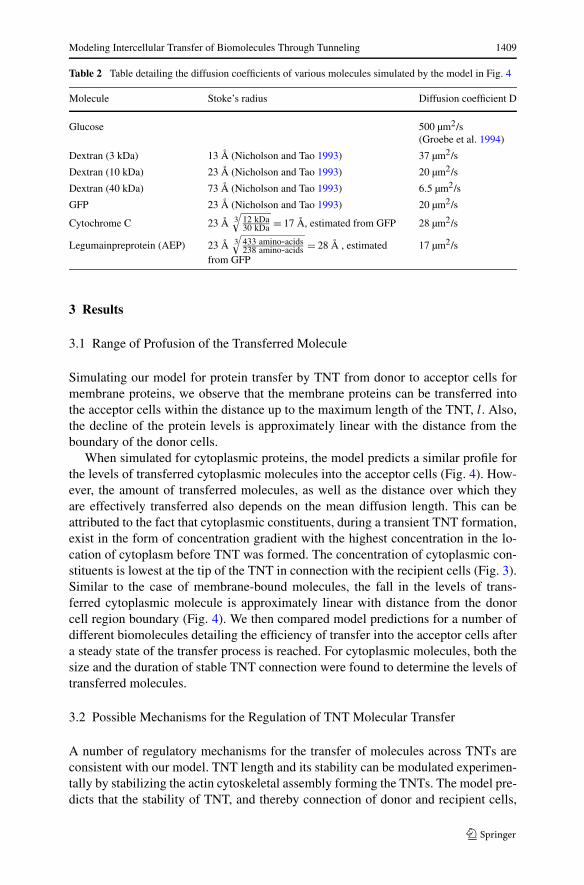
Table 2 Table detailing the diffusion coefficients of various molecules simulated by the model in Fig. 4
Molecule Stoke’s radius Diffusion coefficient D
Glucose 500 µm2/s(Groebe et al. 1994)
Dextran (3 kDa) 13 Å (Nicholson and Tao 1993) 37 µm2/s
Dextran (10 kDa) 23 Å (Nicholson and Tao 1993) 20 µm2/s
Dextran (40 kDa) 73 Å (Nicholson and Tao 1993) 6.5 µm2/s
GFP 23 Å (Nicholson and Tao 1993) 20 µm2/s
Cytochrome C 23 Å 3√
12 kDa30 kDa = 17 Å, estimated from GFP 28 µm2/s
Legumainpreprotein (AEP) 23 Å 3√
433 amino-acids238 amino-acids = 28 Å , estimated
from GFP17 µm2/s
3 Results
3.1 Range of Profusion of the Transferred Molecule
Simulating our model for protein transfer by TNT from donor to acceptor cells formembrane proteins, we observe that the membrane proteins can be transferred intothe acceptor cells within the distance up to the maximum length of the TNT, l. Also,the decline of the protein levels is approximately linear with the distance from theboundary of the donor cells.
When simulated for cytoplasmic proteins, the model predicts a similar profile forthe levels of transferred cytoplasmic molecules into the acceptor cells (Fig. 4). How-ever, the amount of transferred molecules, as well as the distance over which theyare effectively transferred also depends on the mean diffusion length. This can beattributed to the fact that cytoplasmic constituents, during a transient TNT formation,exist in the form of concentration gradient with the highest concentration in the lo-cation of cytoplasm before TNT was formed. The concentration of cytoplasmic con-stituents is lowest at the tip of the TNT in connection with the recipient cells (Fig. 3).Similar to the case of membrane-bound molecules, the fall in the levels of trans-ferred cytoplasmic molecule is approximately linear with distance from the donorcell region boundary (Fig. 4). We then compared model predictions for a number ofdifferent biomolecules detailing the efficiency of transfer into the acceptor cells aftera steady state of the transfer process is reached. For cytoplasmic molecules, both thesize and the duration of stable TNT connection were found to determine the levels oftransferred molecules.
3.2 Possible Mechanisms for the Regulation of TNT Molecular Transfer
A number of regulatory mechanisms for the transfer of molecules across TNTs areconsistent with our model. TNT length and its stability can be modulated experimen-tally by stabilizing the actin cytoskeletal assembly forming the TNTs. The model pre-dicts that the stability of TNT, and thereby connection of donor and recipient cells,
1410 Y. Suhail et al.
Fig. 3 Simulated transfer of molecules from donor to accepted cells via TNT. (A) Transferred mem-brane-bound molecule levels in acceptor cells at a distance x from the boundary of the donor cells region,calculated from Eq. (6). The distances used in the plot are in units of the maximum TNT length l. The
levels of transferred membrane molecules are given in units of Al3ρπb2
3βwhere A is a constant related to
the physics of the membrane contact and level in the donor cells, ρ the density of the donor cells, and b theradius of the cells. The level is maximal at the boundary and gradually decreases to zero at a distance equalto the maximum TNT length l, after which no TNT connection can be made between the donor and accep-tor cells. (B) Transferred cytoplasmic molecule levels in acceptor cells at a distance x from the boundaryof the donor cells region, calculated from Eq. (12) for various values of the mean diffusion length (C). Thedistances (x) used in the plot are in units of the maximum TNT length l. The level of transferred molecule
levels are given in units of Bl3ρπb2
3βwhere B is a constant related to the level in the donor cells, ρ the
density of the donor cells, and b the radius of the cells. The values of the mean diffusion length C consid-ered are specified as fractional multiples of the maximum TNT length l. More of the cytosolic molecule istransferred for larger mean diffusion lengths. Both the amount of molecules transferred to a particular dis-tance and the maximal distance to which it is transferred is limited by the mean diffusion length. The meandiffusion length itself may depend on both the diffusion constant and mean time of stable TNT formations(Eq. (10)), which is explored further in Fig. 4
will have a significant effect on the level of cytoplasmic molecules in donor cells.The length of time TNT connections are made will also influence the transfer ofmembrane bound molecules, with longer stable connections leading to higher mem-
Modeling Intercellular Transfer of Biomolecules Through Tunneling 1411
Fig. 4 Stability of TNTconnection determines extent ofcytoplasmic protein transfer in asize dependent manner. Level oftransferred moleculestransferred from donor toacceptor cells via TNTformation, simulated to bestabilized for mean durations of(A) 1 second, (B) 10 seconds,and (C) 100 seconds. In allcases, levels of transferredmolecules in the recipient cellsare plotted against the distance x
from the region of donor cells,calculated from Eqs. (6)and (12). The levels oftransferred molecules are given
in units of Al3ρπb2
3βfor
membrane bound molecules,
and Bl3ρπb2
3βfor cytosolic
molecules similar to Fig. 3.In all cases, size of TNT isconsidered to be 50 µm
brane molecule transfer. Changes in the transport of the molecule through the Golgiapparatus, micro-vesicles, and endosomes to and from the membrane and the effectof post translational control of its binding with the membrane in the donor cells willalso determine the amount of membrane-bound molecule available for transfer on the
1412 Y. Suhail et al.
TNTs. These changes in membrane recruitment and binding could be modulated inthe cells by various pathways.
3.3 Effect of the Size of the Transferred Molecules
The model predicts that small molecules are quite robust in their transfer across theTNTs while larger proteins require favorable conditions, for example, stable TNT thatretract after longer durations. Our model predicts that, in general, in a typical TNTthat exists for a few seconds to tens of seconds, transfer of membrane proteins will beappreciably higher than cytoplasmic molecules. Among cytoplasmic molecules, largemolecules will have an extremely low transfer efficiency, with transfer occuring onlywithin cells that are extremely close to each other (Fig. 4A). The difference of extentof transfer between small and large molecules is quite pronounced, suggesting thatthe size of molecules plays a significant role in cytoplasmic protein transfer betweencells.
Small cytosolic molecules, for example, glucose (Groebe et al. 1994) or othermetabolites, transfer at a much faster rate. Thus the model explains previous observa-tions that dextran molecules of different sizes showed a size dependent intercellulartransfer amounts between dextran containing Chinese Hamster Ovarian (CHO) cellsto those without dextran (Figs. 4A, B). Since typical cytosolic proteins are muchlarger than smaller metabolites (e.g., green fluorescent protein used as a reporterprobe in cell biology experiments, which has a Stoke’s radius of 23 Å, similar toa 10 kDa Dextran (Phillips 1997)), the extent of their transfer is expected to be muchlower, suggesting a possible reason why the detection of cytosolic protein transferhas been rare, or has remained unreported.
However, as the TNT becomes more stabilized, the diffusion of cytoplasmicmolecules within the TNT shaft shifts more towards a steady state, and becomes shal-lower. This results in higher transfer of cytoplasmic molecules (Fig. 4B). As TNTsare stabilize even further, the extent of transfer of cytoplasmic molecules approachesthe transfer of membrane proteins for all distances between donor and recipient cells(Fig. 4C). The difference of transfer within cytoplasmic molecules of different sizesbecomes less pronounced, suggesting that size remains a smaller factor in cytoplas-mic protein transfer as TNTs stabilize (Fig. 4C).
4 Discussion
Intercellular transfer of cellular components is a fascinating phenomenon largely be-cause it is understood so little, and does not seem to obey any known classical cell–cell communication mechanisms. This transfer also seems surprising since it sug-gests a phenomenon over which cells can only have partial or no active control. Eventhen, it has been implicated as important in various physiological and pathologicalcontexts. For example, while heterotypic and homotypic cell–cell interactions occurfrequently in various tissues, including cancer cells and blood vessels, muscles, andnerve fibers, etc., potentially allowing transfer of molecules between cells, patholog-ical contexts present new avenues for intercellular transfer of biomolecules to play
Modeling Intercellular Transfer of Biomolecules Through Tunneling 1413
significant roles. For example, drug-resistant cancer cells could transfer small drug-resistant molecules to neighboring drug-susceptible cells causing lateral transfer ofresistance. However, in spite of the importance of this passive form of intercellularcommunication, our present understanding about it is limited.
In spite of the relatively few reports of intercellular transfer of biomolecules,largely due to the small amounts of transfer that can frequently go undetected, afew trends stand out in the reported studies. It has been observed that the mem-brane bound molecules could transfer more readily from one cell to another, but cy-toplasmic molecules transfer has been reported less frequently. Interestingly, whilethere are no reports that the efficiency of transfer of membrane-integrated moleculesis dependent on the molecular weight of the transferred components, cytoplasmicmolecules have been reported to transfer in a size dependent manner. In addition,there has not been any conclusive demonstration of cytoplasmic protein transfer be-tween cells. Here, we propose a mathematical model describing intercellular transferof biomolecules via TNTs that explains these observations and makes useful predic-tions.
The model makes a critical assumption about distinct characteristics of transportof membrane vs. cytosolic components through TNTs. In particular, it is assumed thatlarge cytosolic components, such intracellular proteins, can diffuse over the length ofTNT much slower that the membrane components. Thus the rate limiting step in thetransfer of the membrane components is the rate of their access to TNTs on the donorcell side, whereas the rate limiting step of the transport of cytoslic components istheir diffusion over TNT. As a consequence and due to the transient nature of TNTs,membrane but not cytosolic components would reach a steady state distribution overthe length of a TNT, with cytoslic components forming a diffusion based gradient.Since diffusion is dependent on the size of the molecule, and consequently results ina size/mass dependent transfer of cytosolic molecules.
For both the membrane bound and cytosolic molecules, the transfer is limitedby the spatial separation between an acceptor cell and the nearest donor cells. Dueto the size dependence of the cytosolic molecular transfer, both the amount of themolecule transferred and the maximum separation between the cells that allows forany observable transfer becomes negligible for large proteins. A consequence of themodel then is that in most physiological contexts, any signaling happening acrosscells in this fashion is limited to either membrane bound molecules or small cytosolicmolecules. Thus, our model provides a physical basis for the observation of signalingby membrane proteins and small cytosolic molecules.
The model predictions regarding the importance of the length of the TNTs thetime scale of TNT lifetimes open new avenues for of the analysis of intercellularcommunication through individual TNT formations. In addition, it raises the ques-tion of whether there could be specific pathways regulating the formation and behav-ior of such TNTs. For example, it has been reported that HIV induces the formationof TNTs in macrophages (Eugenina et al. 2009). This hypothesis can be tested bymodulating the frequency of TNT formation by cells, achievable by chemical andenvironmental means (Lou et al. 2012). Another hypothesis generated by this modelis that increased stability of TNTs could reduce the differential transfer of moleculesof different sizes, and this can be tested by modulating TNT stability (Marzo et al.
1414 Y. Suhail et al.
2012). Recent reports of transfer of endocytotic organelles due to TNTs, which can becontrolled by a number of molecular signals (Gurke et al. 2008), suggest a scope formore detailed theoretical models than the one presented here, taking into account theactive and modulated TNT formation frequency and dynamics and how they affectthe transfer of components in response to specific biological signals. It is plausiblethat TNT formation may be regulated by cells as response to various stresses or otherstimuli, resulting in a controlled selection of the nature, size, and amount of the trans-ferred components and the corresponding phenotypes.
5 Directions for experimental future studies
The model presented here, in the absence of precise values for the multitude of phys-ical parameters involved in the process, makes a number of assumptions in order toprovide some qualitative predictions. Careful experimental studies may validate orcorrect certain aspects of this model. These predictions and assumptions should helpto tease out the role the transfer of molecules across TNTs plays physiologically.
We have a simplistic linear relation between the length of TNTs and their abun-dance in a uniform density of cells (Eq. (1)). Imaging a large number of cells formingTNTs could help to provide us with a better understanding of the dynamic of TNTformation and their static distribution.
Experiments measuring the profusion of cytosolic and membrane bound moleculesof differing sizes can shed light on the validity of our models of diffusive transfer ofcytosolic molecules and membrane transfer at the tip of the TNTs.
As mentioned earlier, experiments perturbing the frequency and stability of theTNTs provide another avenue for testing the model and at least one possible regula-tory mechanism.
Once certain quantitative characteristics of the transfer of these molecules havebeen verified for some control molecules, any deviation from these transfer ratesfor physiologically important proteins opens the way for investigating the signalingpathways the cells employ for regulating this intracellular traffic.
References
Abou-Khalil, R., et al. (2009). Autocrine and paracrine angiopoietin 1/Tie-2 signaling promotes musclesatellite cell self-renewal. Cell Stem Cell, 5(3), 298–309.
Agnati, L. F., et al. (2011). Possible new targets for GPCR modulation: allosteric interactions, plasmamembrane domains, intercellular transfer and epigenetic mechanisms. J. Recept. Signal Transduct.Res., 31(5), 315–331.
Ahmed, K. A., & Xiang, J. (2011). Mechanisms of cellular communication through intercellular proteintransfer. J. Cell. Mol. Med., 15(7), 1458–1473.
Al-Nedawi, K., et al. (2008). Intercellular transfer of the oncogenic receptor EGFRvIII by microvesiclesderived from tumour cells. Nat. Cell Biol., 10(5), 619–624.
Ambudkar, S. V., Sauna, Z. E., Gottesman, M. M., & Szakacs, G. (2005). A novel way to spread drug resis-tance in tumor cells: functional intercellular transfer of P-glycoprotein (ABCB1). Trends Pharmacol.Sci., 26(8), 385–387.
Baba, E., et al. (2001). Functional CD4 T cells after intercellular molecular transfer of 0X40 ligand. J. Im-munol., 167(2), 875–883.
Modeling Intercellular Transfer of Biomolecules Through Tunneling 1415
Behnke, B. J., Armstrong, R. B., & Delp, M. D. (2011). Adrenergic control of vascular resistance variesin muscles composed of different fiber types: influence of the vascular endothelium. Am. J. Physiol.,Regul. Integr. Comp. Physiol., 301(3), R783–790.
Bosenberg, M. W., & Massague, J. (1993). Juxtacrine cell signaling molecules. Curr. Opin. Cell Biol.,5(5), 832–838.
Bukoreshtliev, N. V., et al. (2009). Selective block of tunneling nanotube (TNT) formation inhibits inter-cellular organelle transfer between PC12 cells. FEBS Lett., 583(9), 1481–1488.
Camussi, G., Deregibus, M. C., Bruno, S., Cantaluppi, V., & Biancone, L. (2010). Exosomes/microvesiclesas a mechanism of cell-to-cell communication. Kidney Int., 78(9), 838–848.
Carlin, L. M., Eleme, K., McCann, F. E., & Davis, D. M. (2001). Intercellular transfer and supramolecularorganization of human leukocyte antigen C at inhibitory natural killer cell immune synapses. J. Exp.Med., 194(10), 1507–1517.
Davis, D. M. (2007). Intercellular transfer of cell-surface proteins is common and can affect many stagesof an immune response. Nat. Rev. Immunol., 7(3), 238–243.
Denzer, K., Kleijmeer, M. J., Heijnen, H. F., Stoorvogel, W., & Geuze, H. J. (2000). Exosome: frominternal vesicle of the multivesicular body to intercellular signaling device. J. Cell Sci., 113(19),3365–3374.
Driesen, R. B., et al. (2005). Partial cell fusion: a newly recognized type of communication between ded-ifferentiating cardiomyocytes and fibroblasts. Cardiovasc. Res., 68(1), 37–46.
Eugenina, E. A., Gaskilla, P. J., & Bermana, J. W. (2009). Tunneling nanotubes (TNT) are induced by HIV-infection of macrophages: A potential mechanism for intercellular HIV trafficking. Cell. Immunol.,254(2), 142–148.
Gregor, T., Bialek, W., de Ruyter van Steveninck, R. R., Tank, T. D., & Wieschaus, E. F. (2005). Diffusionand scaling during early embryonic pattern formation. Proc. Natl. Acad. Sci. USA, 102(51), 18403–18407.
Groebe, K., Erz, S., & Mueller-Klieser, W. (1994). Glucose diffusion coefficients determined from con-centration profiles in EMT6 tumor spheroids incubated in radioactively labeled L-glucose. Adv. Exp.Med. Biol., 361, 619–625.
Guescini, M., et al. (2012). Microvesicle and tunneling nanotube mediated intercellular transfer of g-protein coupled receptors in cell cultures. In Experimental cell research.
Gurke, S., et al. (2008). Tunneling nanotube (TNT)-like structures facilitate a constitutive, actomyosin-dependent exchange of endocytic organelles between normal rat kidney cells. Exp. Cell Res., 314(20),3669–3683.
Levchenko, A., et al. (2005). Intercellular transfer of P-glycoprotein mediates acquired multidrug resis-tance in tumor cells. Proc. Natl. Acad. Sci. USA, 102(6), 1933–1938.
Li, M., et al. (2010). Intercellular transfer of proteins as identified by stable isotope labeling of amino acidsin cell culture. J. Biol. Chem., 285(9), 6285–6297.
Lichtenberger, B. M., et al. (2010). Autocrine VEGF signaling synergizes with EGFR in tumor cells topromote epithelial cancer development. Cell, 140(2), 268–279.
Lou, E., et al. (2012). Tunneling nanotubes provide a unique conduit for intercellular transfer of cellularcontents in human malignant pleural mesothelioma. PLoS ONE, 7(3), e33093.
Luby-Phelps, K., Taylor, D. L., & Lanni, F. (1986). Probing the structure of cytoplasm. J. Cell Biol.,102(6), 2015–2022.
Mack, M., et al. (2000). Transfer of the chemokine receptor CCR5 between cells by membrane-derivedmicroparticles: a mechanism for cellular human immunodeficiency virus 1 infection. Nat. Med., 6(7),769–775.
Marzo, L., Gousset, K., & Zurzolo, C. (2012). Multifaceted roles of tunneling nanotubes in intercellularcommunication. Front. Physiol., 3, 72.
Nicholson, C., & Tao, L. (1993). Hindered diffusion of high molecular weight compounds in brain extra-cellular microenvironment measured with integrative optical imaging. Biophys. J., 65(6), 2277–2290.
Niu, X., Gupta, K., Yang, J. T., Shamblott, M. J., & Levchenko, A. (2009). Physical transfer of membraneand cytoplasmic components as a general mechanism of cell–cell communication. J. Cell Sci., 122(5),600–610.
Pap, E., Pallinger, E., Pasztoi, M., & Falus, A. (2009). Highlights of a new type of intercellular communi-cation: microvesicle-based information transfer. Inflamm. Res., 58(1), 1–8.
Phillips, G. N. Jr. (1997). Structure and dynamics of green fluorescent protein. Curr. Opin. Struct. Biol.,7(6), 821–827.
Prochiantz, A. (2011). Homeoprotein intercellular transfer, the hidden face of cell-penetrating peptides.Methods Mol. Biol., 683, 249–257.
1416 Y. Suhail et al.
Qin, L., Bromberg-White, J. L., & Qian, C. N. (2012). Opportunities and challenges in tumor angiogenesisresearch: back and forth between bench and bed. Adv. Cancer Res., 113, 191–239.
Quah, B. J., et al. (2008). Bystander B cells rapidly acquire antigen receptors from activated B cells bymembrane transfer. Proc. Natl. Acad. Sci. USA, 105(11), 4259–4264.
Rustom, A., Saffrich, R., Markovic, I., Walther, P., & Gerdes, H. H. (2004). Nanotubular highways forintercellular organelle transport. Science, 303(5660), 1007–1010.
Singh, A. B., & Harris, R. C. (2005). Autocrine, paracrine and juxtacrine signaling by EGFR ligands. Cell.Signal., 17(10), 1183–1193.
Stine, M. J., et al. (2011). Integration of genotypic and phenotypic screening reveals molecular mediatorsof melanoma-stromal interaction. Cancer Res., 71(7), 2433–2444.
Twiss, J. L., & Fainzilber, M. (2009). Ribosomes in axons—scrounging from the neighbors? Trends CellBiol., 19(5), 236–243.
Valadi, H., et al. (2007). Exosome-mediated transfer of mRNAs and microRNAs is a novel mechanism ofgenetic exchange between cells. Nat. Cell Biol., 9(6), 654–659.
Vikne, H., Gundersen, K., Liestol, K., Maelen, J., & Vollestad, N. (2012). Intermuscular relationship ofhuman muscle fiber type proportions: slow leg muscles predict slow neck muscles. Muscle Nerve,45(4), 527–535.
Weis, S. M., & Cheresh, D. A. (2011). Tumor angiogenesis: molecular pathways and therapeutic targets.Nat. Med., 17(11), 1359–1370.