35
Modeling of Methyl Transfer Reactions in S-Adenosyl-L-Methionine Dependent Enzymes Polina Velichkova Theoretical Chemistry Royal Institute of Technology Stockholm 2006

Modeling of Methyl Transfer Reactions inS-Adenosyl-L-Methionine Dependent
Enzymes
Polina Velichkova
Theoretical ChemistryRoyal Institute of Technology
Stockholm 2006

c© Polina VelichkovaISBN 91-7178-289-3 pp i-iii, 1-30Printed by Universitetsservice US-AB,Stockholm, Sweden, 2006

Abstract
A very important trend for studying biomolecules is computational chemistry.In particular, nowadays it is possible to use theoretical methods to figure out thecatalytic mechanism of enzyme reactions. Quantum chemistry has become a pow-erful tool to achieve a description of biological processes in enzymes active sitesand to model reaction mechanisms.
The present thesis uses Density Functional Theory (DFT) to investigate cat-alytic mechanism of methyltransferase enzymes. Two enzymes were studied –Glycine N-MethylTransferase (GNMT) and Guanidinoacetate Methyltransferase(GAMT). Different models of the enzyme active sites, consisting of 20 to 100atoms, are employed. The computed energetics are compared and are used tojudge the feasibility of the reaction mechanisms under investigation.
For the GNMT enzyme, the methyl transfer reaction was found to follow anSN2 reaction mechanism. The calculations demonstrate that the mechanism isthermodynamically reasonable. Based on the calculations it was concluded thathydrogen bonds to the amino group of the glycine substrate lower the reactionbarrier, while hydrogen bonds to carboxylate group raise the barrier.
In the GAMT enzyme the methyl transfer reaction was found to follow a con-certed asynchronous mechanism which includes transfer of a methyl group ac-companied by a proton transfer taking place simultaneously in the same kineticstep. The calculated barrier agrees well with the experimental rate constant.
i

List of Papers
• Methyl Transfer in Glycine N-Methyltransferase, a Theoretical Study.Polina Velichkova, Fahmi Himo,J. Phys. Chem. B 109 (16), 8216-8219 (2005).
• Theoretical Study of the Methyl Transfer in Guanidinoacetate Methyl-transferase.Polina Velichkova, Fahmi Himo,J. Phys. Chem. B 110 (1), 16-19 (2006).
ii

Acknowledgments
There is a long list of people that I wold like to thank for their support duringthe work presented in this thesis.
First of all I would like to express my sincere gratitude to my supervisor Dr.Fahmi Himo for the guidance, helpfulness, and encouragements. Without him thisthesis would not have been possible. Under your guidance I have learned a lot.You have always taken your time to answer and discuss all my questions. Thankyou for your help and friendly atmosphere during these years!
I would like to express my gratitude to Prof. Hans Ågren for accepting me inthe Theoretical Chemistry group and giving me a warm welcome in Stockholm.
I will be forever grateful to Prof. Alia Tadjer (from the University of Sofia,Bulgaria) and the Quantum Chemistry group there – Galia, Anela for their beliefin me. They opened my eyes to science, and gave me a lot of support. Apartfrom science you are excellent persons to work with and talk about other things.Million thanks to all of you!
One of the most important people to whom I would like to thank is youValentin. Thank you for your love and support, thank you for encouraging me,and being by my side every day through the last hard years. I am infinitely grate-ful to Valentin, with all my heart.
Ivo, we had much fun together, and still we have. You helped me solve manyof my problems as they appeared. Thank you! I am in particular thankful to mypast and present officemates – Oscar, Luca, Kathrin, Ivo, Freddy for being alwayswilling to talk to me, and making my time very pleasant.
Huge thanks go to my friends from Bulgaria (some of them are somewherearound this world) - Ivo, Mary, Pepi, Slavy, Did, Stancho, Petka, Borko, Deni etc.Thanks for the friendship and the good moments we still share.
Last, but not least, warm thanks to my family who have not been directlyinvolved in the thesis, and who won’t read this thesis but love me, and have beenon my side all the time. Thank you! Thanks to Valentin’s family for the hospitalityand love.
It is impossible to thank one by one all other people who in all sorts of wayscontributed to this thesis. The atmosphere in the group has been most accom-modating, and I cordially appreciate the company of all the present and previousmembers of the group.
THANK YOU ALL!
iii

Contents
Preface 2
1 Enzymes and life processes. An Introduction. 3
2 How to study these remarkable biomolecules? 52.1 Choosing the theoretical method – Density Functional Theory.. . 62.2 Why DFT (B3LYP)? . . . . . . . . . . . . . . . . . . . . . . . . 62.3 Transition State Theory.. . . . . . . . . . . . . . . . . . . . . . . 7
3 How do we do enzyme modeling? 93.1 From enzyme to “small” and good model.. . . . . . . . . . . . . 93.2 Geometry optimization.. . . . . . . . . . . . . . . . . . . . . . . 103.3 Single-point energy calculations.. . . . . . . . . . . . . . . . . . 113.4 Dielectric effects. . . . . . . . . . . . . . . . . . . . . . . . . . . 113.5 Frequency calculations.. . . . . . . . . . . . . . . . . . . . . . . 12
4 SAM dependent enzymes. 134.1 Methyl transfer reactions catalyzed by SAM dependent enzymes. . 14
4.1.1 Glycine N-MethylTransferase.. . . . . . . . . . . . . . . 154.1.2 Guanidinoacetate Methyltransferase.. . . . . . . . . . . . 20
5 Concluding Remarks 25
Abbreviations and Acronyms 26
One and three letter abbreviation for amino acids 27
Bibliography 28
1

Preface
The work presented in this thesis has been carried out at Laboratory of TheoreticalChemistry, Royal Institute of Technology, Stockholm, Sweden.
The first chapter of this thesis presents a general introduction about enzymes.What do they do? And why are they so important for life processes?
The second chapter presents the methodology of our investigations. This chap-ter does not contain a detailed description of Density Functional Theory methods.Instead, there is a section explaining why this method was used. Transition StateTheory is also briefly outlined due to its fundamental role in the analysis of chem-ical reactivity.
Chapter three quickly sets up the details of the approach adopted to build thecomputational models. This chapter presents computational details and some oftheir advantages and limitations in our study.
Finally, chapter four presents a brief summary of the main results produced inthe articles on which this thesis is based. The thesis finishes with some conclu-sions for the SAM-dependent enzymes.
2

Chapter 1
Enzymes and life processes. AnIntroduction.
When we speak of Nature it is wrong to forget that we are ourselves apart of Nature.We ought to view ourselves with the same curiosity andopenness with which we study a tree, the sky or a thought, because wetoo are linked to the entire universe.
Henri Matisse
Enzymes are present in every living organism. These amazing nature struc-tures are responsible for the normal undergoing of the processes responsible forlife. They are involved in every biochemical reaction, like break down the foodwe eat into nutrients, they can convert one molecule to another, which is used asa therapeutic agent. In short, we can describe the enzymes like a crucial elementof life.
What makes enzymes so special? From a structural point of view, enzymes aredefined as protein molecules. They consist of long chains of amino acids boundtogether by peptide bonds [1]. The most important part of the enzyme moleculeis the active site. It is the place where the catalysis occurs. This part of theenzymes is responsible for their physiological role. The active site of an enzymeis designed to fit with only one type of substrate molecules which meaning thatthe enzyme is selective. If the active site of an enzyme can fit with only one typeof substrate this means that there are thousands of different enzymes, as thereare a lot of substrate molecules. From this point of view, enzymes are describedas specific molecules – for every substrate molecule Nature designed an enzymemolecule. Another property defining the enzyme functionality is their effectivity.They catalyze biological reactions, which in normal conditions would proceedwith negligible rate.
3
4
The specific properties of the enzymes can be illustrated by describing the wayan enzyme catalytic reaction goes. There are a lot of theories explaining how theenzymes do their work. One of the most widely used is the lock-and-key theory.The enzyme-catalyzed reaction starts with binding the substrate molecule intothe active site. The substrate molecule is oriented in a particular, optimal spatialposition and then “locked” into the active site with hydrogen bonds and electro-static interactions. Once the enzyme-substrate complex is formed, the catalysisoccurs and the product molecule is formed. With the transformation to the prod-uct molecule the hydrogen bonds and electrostatic interactions are destroyed andthe product molecule is free to leave the active site [2]. This is a very crude picturewhich is used only for illustrational purposes.
The major driving force for studying catalytic mechanisms of enzymes is obvi-ous: understanding the reaction pathways and enzyme structure is very helpful inareas like drug design, designing synthetic catalysts, pharmaceutical industry, etc.The problem is that in Nature most of the chemical reactions happen in systems oflarge size including thousands of atoms, which makes accurate theoretical treat-ment of these systems impossible. Fortunately, advances in computer technologyand quantum theory provide a possibility for carrying out research on enzymecatalysis.

Chapter 2
How to study these remarkablebiomolecules?
Thus, the task is, not so much to see what no one has yet seen; but tothink what nobody has yet thought, about that which everybody sees.
Erwin Schrodinger
Talking about enzyme modeling, the first question that arises is “How canthese big molecules be studied computationally, and how precise are the calcula-tions?” The theoretical study of the catalytic effects in enzymes needs appropriatetheory and computational methods.
Nowadays, plenty of theoretical methods exist, and choosing a relevant one forcharacterization of enzyme catalytic mechanisms is an important question [6, 7].The best way is to perform full ab initio calculations. The problem is that whenwe have large systems, consisting of thousands of atoms, performing ab initio cal-culations with the computer power of today is impossible. There are few methods,for instance Molecular Dynamics (MD) and Molecular Mechanics (MM), whichare developed for studying models consisting of thousands of atoms. These meth-ods are based on classical mechanics and are very cheap computationally, whichmakes them able to describe the whole enzyme molecule. However, these methodsare very approximate and can not be used to study reactions, which involve bondmaking and breaking. On the other hand, there are many Quantum Mechanical(QM) methods which can be used for analyzing the enzyme catalytic mechanismusing relatively small models.
The main information obtained from a QM study is the energy of the differentstructures in the investigated system. These energies are used to figure out the en-ergy barrier of a given reaction, which is enough to exclude or allow the suggestedcatalytic mechanism.
5

2.1. Choosing the theoretical method – Density Functional Theory. 6
2.1 Choosing the theoretical method – Density Func-tional Theory.
A widely used QM method is the DFT. The foundation of this theory was laidby Hohenberg and Kohn in 1964 [8], providing a new approach of solving theSchrodinger equation for many-electron systems. Instead of wave functions, thismethod puts the electron density of the system into a Schrodinger-like equation.The energy obtained from solving the equation is a functional of the total electrondensity.
A major problem with DFT is finding sufficiently accurate density functionals.The most popular functional is the hybrid B3LYP functional [6], which in generalform can be written as:
FB3LYPxc = (1− A)FS later
x + AFHFx + BFBecke
x + (1−C)FVWNc + CFLYP
c ,
where FS laterx is the Dirac-Slater exchange,FHF
x is the Hartree-Fock exchangeterm, FBecke
x is the gradient part of the exchange functional of Becke [9],FVWNc
andFLYPc are the correlation functionals of Vosko, Wilk, and Nusair [10] and Lee,
Yang, and Parr [11] respectively. The parameters A, B and C are related to theHartree-Fock exchange and Coulomb correlation. They were determined empiri-cally by Becke [9], and have the values of A=0.20, B=0.72, and C=0.81.
2.2 Why DFT (B3LYP)?
The most popular method used in quantum chemical studies of enzyme catalysisis DFT based on the hybrid B3LYP functional. The reason for its wide popularitycan be explained with the accuracy of this method. Based on the comparisonbetween computational results and experimental data, the accuracy of a certaintheoretical method can be established. For enzyme catalysis, our main interest isfocused on the errors in the total electronic energies and geometrical parameters.
The accuracy of B3LYP was tested against the standard G2 benchmark [12]set of molecules. The G2 set includes 55 molecules for which very accurate ex-perimental data is available. Since 1998 the G3 benchmark [13] set has also beenavailable and B3LYP is compared to the G3 results as well. The accuracy of theB3LYP calculations depends on the choice of basis set. The smallest basis setwhich is recommended for calculations is 6-31G* or equivalent of double-zeta

7 2.3. Transition State Theory.
quality. Comparison with experimental data [14] reveals that this rather small ba-sis set works quite well for geometry optimizations (Table. 2.1). The average errorin the atomization energies using this basis set appears to be 5.18 kcal/mol.
Table 2.1:Mean absolute error for DFT(B3LYP) method compared to the G2 benchmark set[14, 15, 12].
error 6-31G* 6-311+G(3df,2p)average 0.013 0.008Bond Lengths [Å]
maximum 0.055 0.039average 0.62◦ 0.61◦Bond Angles
maximum 1.69◦ 1.85◦
average 0.35◦ 3.66◦Dihedral Anglesmaximum 0.63◦ 6.61◦
average 5.18 2.20Atomization Energy [kcal/mol]maximum 31.5 8.4
However, using a large basis set, as 6-311+G(3df,2p) [15, 12], contributes verylittle in geometry accuracy when added, but the average error for the atomizationenergies is much lower, 2.20 kcal/mol (Table. 2.1).
Based on the values presented inTable. 2.1and previous experience [16,17, 18] it can be concluded that results obtained from B3LYP calculations haveenough accuracy for our purpose. The atomization energies appear to be a fewkilocalories per mole off the experimental values. This makes DFT calculationsvery attractive – to get high accuracy at relatively low computational cost.
An explanation of why B3LYP is so useful for studying enzyme reactionmechanisms is the combination of speed and accuracy provided. It is commonto consider few different hypothetical mechanisms of the same enzyme reaction.One of these mechanism is the right one which is realized in nature. B3LYPcalculations on all mechanisms usually produce different energy barriers for thedifferent hypothetical mechanisms. It is then assumed that the mechanism withthe lowest energy barrier is most probably realized in the cell.
2.3 Transition State Theory.
An indispensable tool for understanding the enzyme reaction mechanism is Tran-sition State Theory (TST) [3, 4, 5]. TST is based on the Arrhenius equation anddescribes chemical transformations which convert reactants to products. These

2.3. Transition State Theory. 8
transformations pass through different chemically stable and unstable structures(intermediates, transition states, etc.) with various energies. All chemical trans-formations that happen in an enzyme active site pass through an unstable structurecalled Transition State (TS). This TS structure is poised between the reactant andthe products. One of the main goals of the theoretical study of enzyme catalyzedreactions is to calculate the Potential Energy Surface (PES) for the reaction mech-anism which involves locating and characterizing of the TS.
TST postulates an equilibrium (Boltzmann) energy distribution at all stableand unstable states along the reaction coordinate. Based on the equilibrium en-ergy distribution, and taking into account the Arrhenius equation, the followingexpression for the rate constantk can be derived:
k =kBTh
exp
(−∆G,
RT
),
wherekB is the Boltzmann’s constant;h is the Planck’s constant;T is the abso-lute temperature;R is the universal gas constant; and∆G, corresponds to the freeenergy of activation. The exponential Eyring equation presented above gives a re-lation between the experiment and the theory. Experiments measure rate constantsand calculations produce the energy barriers. When the rate constant for a certainreaction mechanism is available, the calculated rate constant from the receivedenergies is good enough to exclude or allow the suggested catalytic mechanism.
Based on the Eyring equation, rate constant of ca. 1 s−1 at room temperaturecorresponds to a barrier of ca. 18 kcal/mol. When the energy barrier decreases orincreases with ca. 1.4 kcal/mol the rate constant increases or decreases with oneorder of magnitude, respectively.

Chapter 3
How do we do enzyme modeling?
If we knew what it was we were doing, it would not be called research,would it?
Albert Einstein
The theoretical study of enzymes comes along with a lot of obstacles. Themain problem is that the systems studied experimentally are too large for a quantum-chemical treatment. One way to solve this problem is using a small model con-sisting of about a hundred atoms to represent the whole enzyme molecule whichis built from thousands of atoms. The limit on the size of the model is imposedby the speed of today’s computers for performing calculations with the availableDFT methods. This approximation looks very crude at first sight – how is it pos-sible to get a correct description of the reaction mechanism by neglecting a majorpart of the molecule and calculating only a tiny bit of the enzyme [19, 20, 21]?Important part of a quantum chemical study is to select a good chemical modeland also to understand its limitations.
3.1 From enzyme to “small” and good model.
An essential part of the theoretical study of an enzyme is the choice of a model.From a computational point of view, a good model should be rather small in orderto reduce the computational cost. Nonetheless, this small model needs to containall the parts that are important for the catalysis.
The first step in solving the catalytic mechanism is constructing a good modelsystem of interest. Starting point for modeling is usually the X-ray crystal struc-ture of the investigated enzyme. These structures are deposited at different databases,one of the most popular being the Protein Data Bank (PDB).
9

3.2. Geometry optimization. 10
When constructing the model of interest a major part of the enzyme must beexcluded and only a small part (determined by the available computer power) isleft. Based on the investigations of many researchers in this area, the usefulness ofsuch modeling has been proved. It is assumed that in many cases including onlythe active site residues, the substrate molecule, and the cofactor molecules into thecomputational model is enough for an accurate description of the catalytic mech-anism. The active site consists of a lot of amino acid residues and in the model itis impossible to include all of them. The trick is that the residues directly involvedin the catalysis are just a few and these are most important in the catalytic process.Without these amino residues in the model important electrostatic interactions canbe missed and the final calculated energies will not be comparable with the experi-ment. On the other hand, these few important amino acid residues can be reducedto smaller molecules. Looking at the structure of the amino acids it is obviousthat they possess the same pattern – a central, so calledα-carbon atom, amino andcarboxyl groups attached to it, a hydrogen atom and a side chain (R) attached alsoto theα-carbon. In most of the cases we can keep only the side chain motif inthe model and remove the rest of the residue. Cases when the amino and carboxylgroups attached to theα-carbon are essential for the catalysis cannot be excludedand in this case it is important to keep them modeled as a peptide bond.
Once the model is done, different reaction mechanism can be probed. To figureout a reaction path connecting all stable and unstable structures involved one needsto optimize the reactants, the products, and to find the TS. The complicated partof a theoretical study of enzyme catalysis is locating the TS. Reaction path scancalculations are required as the best way to reach the TS structure. They are doneby systematically freezing one or more internal coordinates along the reactionpath, and optimizing the other degrees of freedom. The energy maximum in thescanned surface provides a good guess for the TS. This point is the best startingstructure for the TS optimization.
3.2 Geometry optimization.
Geometry optimizations of the reactants, the products and the transition state are anecessary part for any theoretical examination of reaction mechanism. During thegeometry optimizations, some atoms are fixed in order to keep the model struc-ture closer to the available X-ray data. For all models described in this thesis, theenergy calculations were performed using the B3LYP method as implemented inthe Gaussian03 [22] program package. Geometry optimizations were performedin gas phase with the 6-31G(d,p) basis set. According to the data presented inSec-tion 2.2andTable 2.1this double zeta basis set is good enough to get reasonablegeometric structures, but the error in the energies is quite large.

11 3.3. Single-point energy calculations.
3.3 Single-point energy calculations.
To obtain more accurate energy, single point calculations with a large basis set,6-311+G(2d,2p), were done.
3.4 Dielectric effects.
Geometry optimizations are usually performed in a gas phase model, but the en-zyme reactions occur in protein environment. Thus, an important part in studyingbiomolecular problems is to consider the effect of the environment. The final en-ergies should account for the protein environment and include correction to thegas phase energies [23].
A common way to compute the influence of the surrounding environment isby performing a single point calculation using the polarizable continuum model(PCM) [24]. Usually, a dielectric constant equal to 4 is used to set up the sur-rounding environment. Based on experimental data this value for the dielectricconstant of the protein has been empirically determined.ε = 4 corresponds to adielectric constant of about 3 for the protein itself and 80 for the water mediumsurrounding the protein [25].
In the PCM method each atom is surrounded with a spherical cavity. The sol-vent cavity is then formed as a surface of constant charge density of the solvatedmolecule. An important factor in the PCM method is the use of proper boundaryconditions on the surface of the cavity and the solute. Inside the cavity the dielec-tric constant is the same as in vacuum, outside it takes the values of the relevantsolvent (ε = 4). Such a modeling of the solvent surrounding accounts for severalcontributions to the solvation energy [26, 27]. The first one is the energy to createthe cavity. Interactions between the solvent and the solute corresponding to thevan der Waals interactions are also accounted as a dispersion energy. Betweenthe solute and the medium polarized by its charge distribution exists electrostaticstabilization which is included as electrostatic energy in the PCM model.
Usually, the dielectric effect, i.e. the difference in solvation energy betweenreactant and TS or between different minima is around a few kcal/mol. A largeeffect is considered as a sign of a bad model. On the other hand there are caseswhere larger effects can be explained. For example when we have electron or pro-ton transfer over long distances the dielectric effect becomes large due to increaseddipole-dipole interactions with the solvent. It is important to note that the dielec-tric continuum theory cannot account for short-range solute-solvent interactionssuch as hydrogen bonds [28], these must be included explicitly.

3.5. Frequency calculations. 12
3.5 Frequency calculations.
Once we have an optimized stationary point, minimum or TS, a frequency is cal-culated. The reason for doing this calculation is to confirm that the optimized TSstructure has only one imaginary frequency. The TS is defined as a structure whichhas a minimal energy with respect to all coordinates except one – namely the re-action coordinate. Along the reaction coordinate the energy takes maximum andthus the second derivative in the Hessian is negative. In this sense the imaginaryfrequency represents movement along the reaction coordinate. Also, calculationof the molecular Hessian of the reactant, the TS, the intermediates, and the prod-ucts, is used to estimate the zero-point vibration contributions to the total energy.
In the present thesis, frequency calculations have been performed on all struc-tures. These were performed at the B3LYP/6-31G(d,p) level of theory to confirmthe nature of the stationary points and to evaluate the zero-point effects.

Chapter 4
SAM dependent enzymes.
I am always doing things I can’t do, that’s how I get to do them.
Pablo Picasso
Methyl transfer, or methylation, represents a simple process of adding or re-moving a CH3 group. This simple reaction undergoing in all cells has a huge num-ber of effects in the body. The methylation process requires methyl donor agents.Only a few types of methyl donors are present in the body and S-adenosylmethionine(SAM) is the most common of them. So far more than 120 different SAM-dependent methyltransferases are known to exist in the cell, and each of themcatalyzing the synthesis of an essential product – sarcosine, creatine (important formuscle energy metabolism), melatonine (the so called “sleep” hormone), adrenaline,acethylcholine (a neurotransmitter), carnitine (important for fat burning in mitho-chondria), choline (important for fat mobilization and cell membrane fluidity), etc.Hence, irregular functioning of the methyl transferase enzymes can cause severediseases, such as brain disease, mental retardation, epilepsy etc. Understandingthe mechanism of these enzymes can thus aid in the development of treatment forthese diseases.
The present thesis is focused on studying the methyl transfer reaction mech-anisms in two SAM-dependent enzymes – Glycine N-Methyltrasferase (GNMT)and Guanidinoacetate Methyltransferase (GAMT).
GNMT is responsible for the formation of sarcosine by methylation of glycine.So far the product sarcosine has no known physiological role. Then why does thisreaction occur in cells? A lot of biochemical and structural studies show that theconversion of glycine to sarcosine, and from sarcosine to glycine, play an impor-tant role in the regulation of methyl group metabolism in the liver and pancreas,by regulating the ratio between SAM and S-Adenosylhomocysteine (SAH) [29].Furthermore, GNMT is important for the folate metabolism [29].
13

4.1. Methyl transfer reactions catalyzed by SAM dependent enzymes. 14
In recent years there has been a huge interest in the product of the reactioncatalyzed by the enzyme GAMT – creatine. GAMT catalyzes the final step ofthe creatine biosynthesis. Creatine is a natural energy compound used to supplyenergy to our muscles [30, 31]. It is produced in the liver, pancreas, and kidneys,and then transported to the body muscles through the bloodstream. Once it reachesthe muscles, it is converted into phosphocreatine (creatine phosphate), which isthen used to regenerate the muscles energy source – adenosine triphosphate (ATP)[31]. GAMT deficiency may lead to creatine deficiency, which can cause differentmental diseases. Nowadays, ingesting creatine supplements has become a fashionbecause it helps increasing fat-free mass and improve the anaerobic, and possiblythe aerobic, performance. Creatine is one of the most popular and commonly usedsports supplements available today.
4.1 Methyl transfer reactions catalyzed by SAM de-pendent enzymes.
SAM is a natural substance present in the cells of the body. The role of this com-pound is to donate a methyl group in variety reactions catalyzed by the enzymes(Fig. 4.1).
COO-
NH3+S
HO
HO
O
adenine
CH3
R
X H
B
COO-
NH3+S
HO
HO
O
adenine
R
X CH3
HB
SAM SAH
substrate molecule product
enzy
me
enzy
me...
...
......
Figure 4.1:Generic mechanism for SAM dependent enzymes.
The substrate molecule enters the active site of the enzyme and bind there bya number of hydrogen bonds to different protein residues. The nucleophilic en-tity, having a lone pair of electrons or possessing partial negative charge, pulls the

15 4.1. Methyl transfer reactions catalyzed by SAM dependent enzymes.
methyl group, while the positivly charged sulfur atom of SAM attracts electrondensity from the methyl group. In this way the methyl transfer reaction can oc-curs.Figure 4.1shows a general reaction mechanism, in which a base is neededto abstract a proton from the substrate.
4.1.1 Glycine N-MethylTransferase.
As a SAM-dependent enzyme, GNMT follows the mechanism described above inSection 4.1to transfer a methyl group (Fig. 4.2), [32].
COO-
NH3+SD
HO
HO
O
adenine
CH3
COO-
NH3+SD
HO
HO
O
adenine
N
O
O
glycine
H
H
N
O
OH
H
CH3
SAM SAH
sarcosine
Figure 4.2:Reaction catalyzed by GNMT.
The reaction starts with the binding of SAM and the glycine substrates, instrict order. In this particular case the glycine has a lone-pair of electrons andbinds in such a way that this lone-pair of the amino nitrogen is directed towardto the CE methyl carbon of SAM. A single SN2 methyl transfer step occurs fromSAM to glycine, resulting in products SAH and sarcosine. In the case of GNMT,there is no base at the active site that can abstract a proton, so one has to assumethat the glycine substrate is bound to the enzyme in a deprotoneted amine form.
To study the reaction mechanism of GNMT described above we used the re-cent X-ray crystal structure. The structure was solved in complex with SAM andan acetate molecule at a 2.0 Å resolution by Takataet al. (Fig. 4.3), [32].
As described inSection 3.1, one of the most important parts of enzyme model-ing is creating a good model for investigation. Looking inside the GNMT enzyme

4.1. Methyl transfer reactions catalyzed by SAM dependent enzymes. 16
Figure 4.3:X-ray crystal structure of the GNMT active site.
it can be seen that a number of hydrogen bonds are formed between the active siteresidues and the substrate molecule. These bonds are non-covalent, electrostaticinteractions and they set up the substrate molecule for the reaction that the en-zyme catalyzes. The positively charged guanidino group of Arg175 forms a pairof hydrogen bonds with the carboxylate group of the acetate. Other groups thatform hydrogen bonds to the substrate are Tyr33, Asn138 and Gly137.
The largest model used for studying the SN2 mechanism catalyzed by GNMTconsists of 98 atoms (Fig. 4.4). This model includes the SAM molecule – trun-cated two carbons away in each direction from the sulfur center. The glycinesubstrate, which was modeled based on the structure of the acetate, to which anamino group was added. The side chain of Arg175 forms strong hydrogen bondsto the carboxylate of glycine and is hence essential to bind the substrate and sta-bilize its charge. The phenol group of Tyr21 was included to test the proposalthat this group polarizes the S-C bond of SAM. Parts of Gly137 and Asn138, arepresent as these groups are found to form hydrogen bonds to both the amino andthe carboxylate groups of the glycine substrate. The phenol group of Tyr194, wasincluded since this group forms hydrogen bonds to both the glycine substrate andto Gly137. Hydrogen atoms were added manually.

17 4.1. Methyl transfer reactions catalyzed by SAM dependent enzymes.
Figure 4.4:Optimized structure of the reactant of GNMT. Distances are given in Ångstroms.Stars indicate atoms that are fixed to their X-ray positions.
A B
Figure 4.5:Optimized transition state A, and product B, structures of the GNMT.

4.1. Methyl transfer reactions catalyzed by SAM dependent enzymes. 18
It was found that the methyl group is transferred to the glycine molecule in asingle SN2 reaction that involves the displacement of the leaving group (SAH), bythe nucleophile (glycine). The structure of the optimized TS is displayed inFig.4.5 A.
At the transition state the bond to the glycine is partially formed while thebond to the SAM is partially broken. The critical SD–CE and CE–N bond dis-tances are 2.28 and 2.18 Å, respectively. The barrier for the methyl transfer inGNMT is calculated to be 15.0 kcal/mol. No experimental data for a rate constantis available, but an energy barrier of 15 kcal/mol for enzyme reaction is consid-ered energetically feasible (see aboveSection 2.3). The reaction was found to beexergonic by 14.1 kcal/mol. The optimized structure of the product is shown inFig. 4.5 B.
A B C D
E F G
Figure 4.6:Optimized transition-state structures for models A-G.

19 4.1. Methyl transfer reactions catalyzed by SAM dependent enzymes.
To investigate the role of the various active site residues, we created a smallmodel composed of glycine, a truncated model of SAM, and truncated residueArg175 (Fig. 4.6 A). The roles of the other amino acid residues were tested byadding the residues to the small model A one at the time, in order to isolate thecontribution of each group. The optimized TS structures are shown inFig. 4.5,and the energetic results are summarized inTable. 4.1.
Table 4.1:Calculated Barriers and Reaction Energies (kcal/mol) for the different models.
model parts included barrier reaction energyA SAM + Glycine+ Arg175 11.2 -20.1B A + Tyr21 13.5 -16.8C A + Tyr21+ His 142 11.4 -21.2D A + Gly137 9.9 -24.8E A + Asn 138 15.1 -12.6F A + Tyr194 17.5 -14.5G A + Tyr194+ Gly137 10.5 -16.6
largest A + Tyr21+ Gly137+ Asn 138+ Tyr194 15.0 -14.1
Model A has a barrier of 11.2 kcal/mol and is exothermic by 20.1 kcal/mol.Adding the phenol group of Tyr21 to this model results in a slight increase in thebarrier, to 13.5 kcal/mol, and a decrease in the exothermicity, to 16.8 kcal/mol(Fig. 4.6 B). When both Tyr21 and the imidazole ring of His142 are added (Fig.4.6 C) the barrier is found to be almost identical toModel A, 11.4 kcal/mol. Theseresults speak against the suggestion that Tyr21 polarizes the S-C bond to cause adecrease of the barrier [32]. As also seen for the largest model discussed above(Fig. 4.5), the phenolic proton of Tyr21 was found to point away from SAM,despite attempts to make it point toward the sulfur center.
Adding the peptide bond of Gly137, which forms a hydrogen bond to theamino group of the substrate (Fig. 4.6 D), results in a decrease of the barrierby 1.2 kcal/mol to 9.9 kcal/mol. The hydrogen bond to the carbonyl moiety ofGly137 makes the nitrogen center of the substrate slightly more negative, whichwould make the transfer of the positively charged methyl group slightly easier.
Adding the side chain of Asn138, which forms a hydrogen bond to the car-boxylate moiety of the substrate leads to a higher barrier, calculated to 15.1 kcal/mol(Fig. 4.6 E). Adding the Tyr194 residue to Model A leads to a dramatic increasein the barrier, from 11.2 to 17.5 kcal/mol (Fig. 4.6 F). This is easily explained ifwe note that the phenolic proton forms a hydrogen bond to the nitrogen atom of

4.1. Methyl transfer reactions catalyzed by SAM dependent enzymes. 20
the substrate in the reactant species. This hydrogen bond will be lost when themethyl is transferred to the nitrogen, resulting in the barrier raise. On the otherhand, if both Tyr194 and the peptide bond of Gly137 are added at the same time(Fig. 4.6 G), the tyrosine will form a hydrogen bond to the carbonyl of the glycineinstead and the barrier is lowered to 10.5 kcal/mol.
By adding or eliminating various groups at the active site, we showed thathydrogen bonds to the amino group of the substrate lower the reaction barrier,whereas hydrogen bonds to carboxylate group of the substrate raise the barrier.
4.1.2 Guanidinoacetate Methyltransferase.
The GAMT enzyme catalyzes the transfer of a methyl group from SAM to Guani-dinoacetate (GAA), resulting in the formation of creatine and SAH (Fig. 4.7),[33, 34, 35, 36, 37, 38]. The way how this reaction occurs is similar to the genericmechanism described inSection 4.1
COO-
NH3+SD
HO
HO
O
adenine
CEH3
COO-
NH3+SD
HO
HO
O
adenine
SAM SAH
O
O
NE
N
NH2
guanidinoacetate
H
H
H
COD1
O
Asp134
O
O
NE
N
NH2
H
H
H
COD1
O
Asp134
CEH3
creatine
Figure 4.7:Reaction catalyzed by GAMT enzyme.
With information about the structure of GAMT, crystallized with SAH andGAA, (Fig. 4.8) in 2004 by Komotoet al. [39], the reaction mechanism wastheoretically investigated.

21 4.1. Methyl transfer reactions catalyzed by SAM dependent enzymes.
Figure 4.8:X-ray crystal structure of GAMT active site.
The substrates are attached to the active site by a number of hydrogen bonds.Glu45 and Asp134 form hydrogen bonds with the guanidino group of GAA. Theamide groups of Leu170 and Thr171 form hydrogen bonds with the carboxylategroup of GAA. All these hydrogen bonds facilitate the orientation of GAA inGAMT. In the GAMT:(SAH+GAA) structure, the distance between the sulfurcenter of SAH (SD) and the NE of GAA is found to be 3.9 Å. The model of SAMwas built by attaching a methyl group at SD of SAH [39]. When the CE methylgroup of SAM was added to SAH, the CE – NE distance became ca 2.2 Å, and theSD – CE – NE centers were almost linear [39].
The model system used to reproduce the GAMT active site and to elucidatethe reaction mechanism was prepared based on the crystallographic structure andcontained 92 atoms. The model includes a part of the cofactor SAM, which wasbuilt on the basis of the SAH structure by adding a methyl group to the sulfuratom. Furthermore, SAM was truncated in both directions relative to the sulfurcenter – at the adenine group an one side and three carbons away an the otherside. This is sufficient to model the properties of the SD – CE bond and to grantsome flexibility to the SAM-model. The GAA substrate molecule was included inthe model without any changes. Five amino acids were furthermore included in
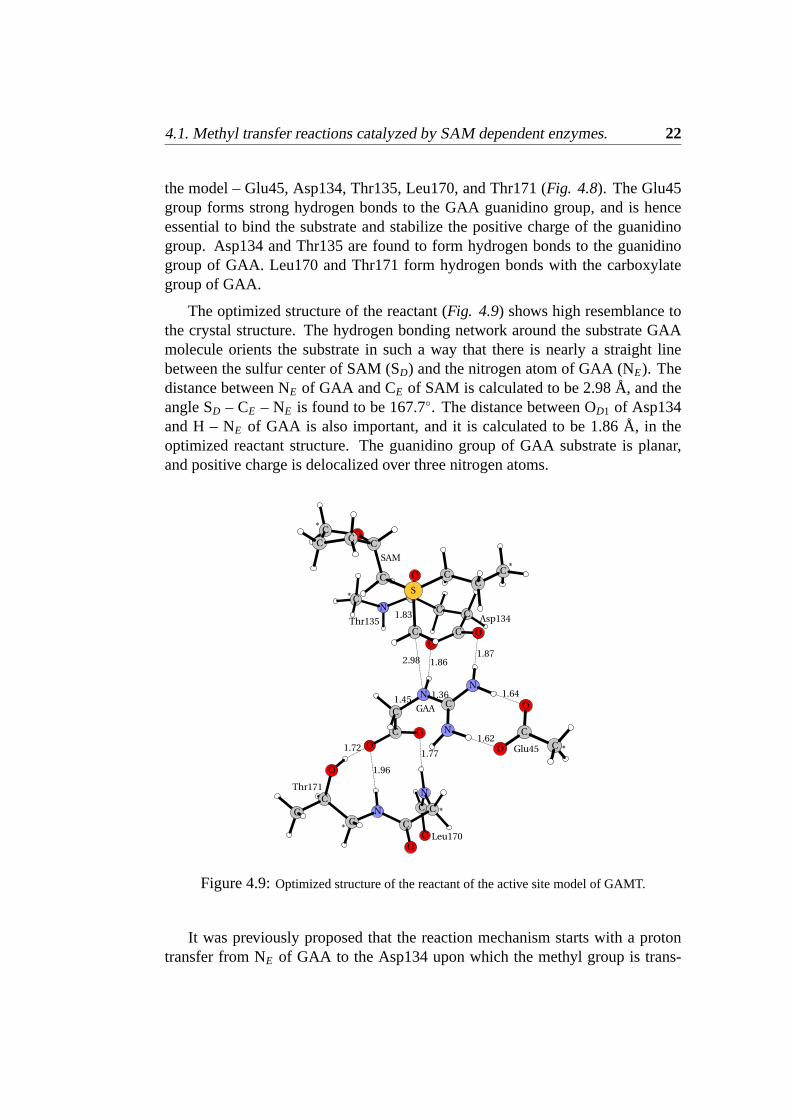
4.1. Methyl transfer reactions catalyzed by SAM dependent enzymes. 22
the model – Glu45, Asp134, Thr135, Leu170, and Thr171 (Fig. 4.8). The Glu45group forms strong hydrogen bonds to the GAA guanidino group, and is henceessential to bind the substrate and stabilize the positive charge of the guanidinogroup. Asp134 and Thr135 are found to form hydrogen bonds to the guanidinogroup of GAA. Leu170 and Thr171 form hydrogen bonds with the carboxylategroup of GAA.
The optimized structure of the reactant (Fig. 4.9) shows high resemblance tothe crystal structure. The hydrogen bonding network around the substrate GAAmolecule orients the substrate in such a way that there is nearly a straight linebetween the sulfur center of SAM (SD) and the nitrogen atom of GAA (NE). Thedistance between NE of GAA and CE of SAM is calculated to be 2.98 Å, and theangle SD – CE – NE is found to be 167.7◦. The distance between OD1 of Asp134and H – NE of GAA is also important, and it is calculated to be 1.86 Å, in theoptimized reactant structure. The guanidino group of GAA substrate is planar,and positive charge is delocalized over three nitrogen atoms.
Figure 4.9:Optimized structure of the reactant of the active site model of GAMT.
It was previously proposed that the reaction mechanism starts with a protontransfer from NE of GAA to the Asp134 upon which the methyl group is trans-

23 4.1. Methyl transfer reactions catalyzed by SAM dependent enzymes.
ferred from SAM to the deprotonated GAA [39].
To check this hypothesis a linear transit scan of moving the proton from NE
of the substrate to Asp134 was done. The H – OD1 distance was kept fixed insteps between 1.86 Å and 1.00 Å, while all other degrees of freedom were op-timized. As seen fromFig. 4.10 Athe energy increases monotonously, and noenergy minimum could be found corresponding to an intermediate where the pro-ton is transferred to the Asp134. Transferring a proton from GAA costs in gasphase 15.9 kcal/mol, while employing protein environment yields a value of 12.6kcal/mol. This results speaks against the stepwise mechanism, where the protonis transferred first.
A B
Figure 4.10:Potential energy curves for: A) moving the proton from NE of the substrate to OD1
of Asp134, and B) moving the methyl group from SAM to NE of GAA substrate, the H – OD1
distance is given in brackets.
A linear transit scan to move the methyl group from SAM to GAA was used tofind the TS structure. The CE – NE distance was kept fixed in steps starting from2.98 Å, which is the distance in the reactant, to 1.46 Å, which is the distance in theproduct (Fig. 4.10 B). As the methyl approaches the nitrogen center, the energyincreases up to a distance of 2.2 – 2.0 Å, after which it starts to drop. The NE
proton moves toward Asp134 and the nitrogen center becomes more pyramidal.At a CE – NE distance of 1.8 Å the proton has transferred completely.
The unconstrained TS for this reaction was located (Fig. 4.11 A). It shows thatthe methyl and the proton transfer take place in one concerted asynchronous step.At the TS the critical SD – CE and CE – NE distances are calculated to be 2.29Å and 2.16 Å, respectively. The other two important distances NE – H and H –

4.1. Methyl transfer reactions catalyzed by SAM dependent enzymes. 24
OD1 in the TS structure are found to be 1.05 Å and 1.73 Å, respectively. To besure that the suggested transition state is the right one, it was identified with oneimaginary frequency, -446 cm−1. The activation barrier for this process is foundto be 14.9 kcal/mol in gas phase, and 19.7 kcal/mol in protein environment withε = 4. The calculated energy barrier is in good agreement with the experimentalrate constant of 3.8± 0.2 min−1, which corresponds to ca. 19 kcal/mol. Thereaction was calculated to be exergonic by as much as 36.2 kcal/mol in gas phaseand 24.0 kcal/mol usingε = 4.
A B
Figure 4.11:Optimized transition state A, and product B, structures of GAMT enzyme model.
The optimized structure of the product is displayed inFig. 4.11 B. The proto-nated Asp134 rotates in such a way that the hydrogen bond to the guanidino groupof GAA is broken, while a hydrogen bond to acetate of GAA is formed.
We tried to optimize the intermediate structure in which the substrate is methy-lated, but the proton is not transferred to Asp134, i.e. corresponding to a stepwisemechanism, but this was not possible. We tried also to optimize the intermediatein which the proton is transferred to Asp134 before the methyl transfer, but it wasnot possible either.

Chapter 5
Concluding Remarks
The investigations presented in this thesis try to shed some light on the methyltransfer reactions in SAM-dependent enzymes. We have used DFT methods tomodel two enzymes from methyltransferase family, namely Glycine N-Methyl-transferase (GNMT) and Guanidinoacetate Methyltransferase (GAMT). The mod-els consist of up to one hundred atoms.
For GNMT it was found that hydrogen bonds to the substrate molecule (glycine)play an important role. The calculations show that hydrogen bonds to the aminogroup of glycine lower the barrier, and hydrogen bonds to the carboxylate groupof glycine raise the barrier.
For GAMT, a concerted asynchronous mechanism of methyl and proton trans-fers was found. The reaction proceeds with a barrier of 19.7 kcal/mol, whichreproduces very well the experimental rate constant (3.8± 0.2 min−1, ca. 19.0kcal/mol )
Both studies show that a rather small model of enzyme active site can be veryuseful for studying the reaction mechanism.
25

Abbreviations and Acronyms
B3LYP Becke 3 parameter Lee-Yang-Parr functionalDFT Density Functional TheoryGAA GuanidinoAcetateGAMT GuanidinoAcetate MethylTransferaseGNMT Glycine N-MethylTransferaseMD Molecular DynamicsPCM Polarizable Continuum ModelPDB Protein Data BankPES Potential Energy SurfaceQM Quantum MechanicsSAM S-AdenosylMethionineSAH S-AdenosylHomocysteineTS Transition StateTST Transition State Theory
26

One and three letter abbreviationsof amino acids
G Gly GlycineA Ala AlanineV Val ValineL Leu LeucineI Ile IsoleucineS Ser SerineC Cys CysteineT Thr ThreonineM Met MethionineP Pro ProlineF Phe PhenylalanineY Tyr TyrosineW Trp TryptophanH His HistidineK Lys LysineR Arg ArginineD Asp Aspartic acidE Glu Glutamic acidN Asn AspargineQ Gln Glutamine
27

Bibliography
[1] Christopher, K. M.; van Holde, K. E.; Kevin, G. A.; Biochemistry Ben-jamin/Cummings, an imprint of Addison Wesley Longman, 1999.
[2] Silverman, R. B.; The organic chemistry of enzyme-Catalyzed reactionsAcademic press, An Elsevier Science Imprint, 2002.
[3] Eyring, H.; Stern, A.;Chem. Rev.24, 2, 1939
[4] Anderson, J.;Adv. Chem. Phys.91, 381, 1995
[5] Truhlar, D.; Garrett, B.; Klippenstein, S.;J. Phys. Chem.100, 12771, 1996
[6] Jensen, F.; Introduction to Computational Chemistry John Wiley & SonsLtd, 2002.
[7] Young, D. C.; Computational Chemistry John Wiley & Sons Ltd, 2001.
[8] a)Parr, R.; Yang, W.; Density Functiona Theory of Atoms and MoleculesOxford University Press, 1989. b)Hohenberg, P.; Kohn, W.;Phys. Rev.136,B864-B887, 1964 c)Kohn, W.; Sham, L.;Phys. Rev.140, A1133-A1138,1965
[9] a)Becke, A.D.;Phys. Rev., A38, 3098, 1988. b)Becke, A.D.;J. Chem. Phys.,96, 2155, 1992. c)Becke, A.D.;J. Chem. Phys., 97, 9173, 1992. d)Becke,A.D.; J. Chem. Phys., 98, 5648, 1993.
[10] Vosko, S. H.; Wilk, L.; Nusair, M.;Can. J. Phys., 58, 1200, 1980.
[11] Lee, C.; Yang, W.; Parr, R. G.;Phys. Rev., B37, 785, 1988.
[12] Curtiss, L. A.; Raghavachari, K.; Trucks, G. W.; Pople, J. A.;J. Chem. Phys.94, 7221-7230, 1991
[13] Curtiss, L. A.; Raghavachari, K.; Redfern, P. C.; Pople, J. A.;J. Chem. Phys.109, 7764-7776, 1998
28

29 BIBLIOGRAPHY
[14] Bauschlicher, C.; Partridge, H.;Chem. Phys. Lett.240, 533-540, 1995
[15] Bauschlicher, C.;Chem. Phys. Lett.246, 40-44, 1995
[16] Siegbahn, P.;Inorg. Chem.39(13), 2923 -2935, 2000.
[17] Siegbahn, P. E. M.;Quarterly Reviews of Biophysics., 36, 91-145, 2003.
[18] Lundberg, M.; PhD thesis, Stockholm University, Department of Physics,2005
[19] Jan, F.; Goodman, M.; Warshel, A.;PNAS, 102, 6819-6824, 2005.
[20] Philipe, A.; Schlegel, B.;J. Chem. Phys., 107, 375-384, 1997.
[21] Garcia-Viloca M.; Gao, J.; Karplus, M.; Truhlar D.;Science, 303, 186-195,2004.
[22] Gaussian 03, Revision C.02, M. J. Frisch, G. W. Trucks, H. B. Schlegel,G. E. Scuseria, M. A. Robb, J. R. Cheeseman, J. A. Montgomery, Jr., T.Vreven, K. N. Kudin, J. C. Burant, J. M. Millam, S. S. Iyengar, J. Tomasi,V. Barone, B. Mennucci, M. Cossi, G. Scalmani, N. Rega, G. A. Petersson,H. Nakatsuji, M. Hada, M. Ehara, K. Toyota, R. Fukuda, J. Hasegawa, M.Ishida, T. Nakajima, Y. Honda, O. Kitao, H. Nakai, M. Klene, X. Li, J. E.Knox, H. P. Hratchian, J. B. Cross, V. Bakken, C. Adamo, J. Jaramillo, R.Gomperts, R. E. Stratmann, O. Yazyev, A. J. Austin, R. Cammi, C. Pomelli,J. W. Ochterski, P. Y. Ayala, K. Morokuma, G. A. Voth, P. Salvador, J. J.Dannenberg, V. G. Zakrzewski, S. Dapprich, A. D. Daniels, M. C. Strain,O. Farkas, D. K. Malick, A. D. Rabuck, K. Raghavachari, J. B. Foresman,J. V. Ortiz, Q. Cui, A. G. Baboul, S. Clifford, J. Cioslowski, B. B. Stefanov,G. Liu, A. Liashenko, P. Piskorz, I. Komaromi, R. L. Martin, D. J. Fox, T.Keith, M. A. Al-Laham, C. Y. Peng, A. Nanayakkara, M. Challacombe, P. M.W. Gill, B. Johnson, W. Chen, M. W. Wong, C. Gonzalez, and J. A. Pople,Gaussian, Inc., Wallingford CT, 2004.
[23] Warshel, A.;Theor. Chem. Acc., 103, 337-339, 2000.
[24] a) Cammi, R.; Mennucci, B.; Tomasi, J.;J. Phys. Chem. A, 103, 9100, 2000.b) Cammi, R.; Mennucci, B.; Tomasi, J.;J. Phys. Chem. A, 104, 5631, 2000.c) Cossi, M.; Rega, N.; Scalmani, G.; Barone, V.;J. Chem. Phys., 114, 5691,2001. d) Cossi, M.; Scalmani, G.; Rega, N.; Barone, V.;J. Chem. Phys., 117,43, 2002.
[25] Blomberg, M.; Siegbahn, P.; Babcock, G.;J. Am. Chem. Soc., 120(34),8812-8824, 1998.

BIBLIOGRAPHY 30
[26] Takano Y.; Houk, K. N.;J. Chem. Theory Comput., 1, 70-71, 2005.
[27] Qiang Cui;J. Phys. Chem., 117(10), 4720-4728, 2002.
[28] a) Tomasi, J.; Persico, M.;Chem. Rev, 94, 2027-2094, 1994. b) Cramer, C.J.; Truhlar, D. G.;Chem. Rev, 99, 2161-2200, 1999. c) Chipman, D. M.;J.Phys. Chem. A, 106, 7413-7422, 2002. d) Chipman, D. M.;J. Phys. Chem.A, 118, 9937-9942, 2003.
[29] Rowling, M.G.; Schalinske, K.L.;J. Nutr., 133, 3392-3398, 2003.
[30] Lee, H.; Ogawa, H.; Fujioka, M.; Gerton, G.;Physiological Reviews., 50,152-162, 1994.
[31] Wyss, M.; Kaddurah-Daouk, R.;Physiological Reviews., 80, 1107-1213,2000.
[32] Takata, Y.; Huang, Y.; Komoto, J.; Yamada, T.; Konishi, K.; Ogawa, H.;Gomi, T.; Fujioka, M.; Takusagawa, F.;Biochemistry, 42, 8394, 2003.
[33] Fujioka, M.; Konishi, K.; Takata, Y.;Biochemistry, 27, 7658, 1988.
[34] Fujioka, M.; Konishi, K.; Gomi, T.;Biochem. Biophys., 285, 181, 1991.
[35] Takata, Y.; Fujioka, M.;Int. J. Biochem., 22, 1333, 1990.
[36] Takata, Y.; Date, T.; Fujioka, M.;Biochem. J., 277, 399, 1991.
[37] Takata, Y.; Fujioka, M.;Biochemistry, 31, 4369, 1992.
[38] Takata, Y.; Konishi, K.; Gomi, T.; Fujioka, M.;J. Biol. Chem., 269, 5537,1994.
[39] Komoto, J.; Yamada, T.; Takata, Y.; Konishi, K.; Ogawa, H.; Gomi, T.;Fujioka, M.; Takusagawa, F.;Biochemistry, 43, 14385-14394, 2004.

![Alkenylphosphonates: unexpected products from reactions of methyl … · Supporting information 1 Alkenylphosphonates: unexpected products from reactions of methyl 2-[(diethoxyphosphoryl)methyl]benzoate](https://static.documents.pub/doc/80x56/5d2087c588c993ea218c3cb8/alkenylphosphonates-unexpected-products-from-reactions-of-methyl-supporting.jpg)


![Ab-initio-MO-Studie Methyl- und Phenyl-substituierter ... · [4.1.0.02,4.03,5]heptane (Benzvalene Sulfide) - Synthesis and Reactions 3213 Maringgele Walter: Reaction of Metal and](https://static.documents.pub/doc/80x56/5e4568eaa71e0d62712632f6/ab-initio-mo-studie-methyl-und-phenyl-substituierter-410024035heptane.jpg)

