Page 1
ORIGINAL PAPER
Modelling for rearrangement of fusiform initials during radialgrowth of the vascular cambium in Pinus sylvestris L.
Wiesław Włoch • Anna Wilczek • Joanna Jura-Morawiec •
Paweł Kojs • Muhammad Iqbal
Received: 9 October 2012 / Revised: 20 December 2012 / Accepted: 8 January 2013 / Published online: 13 February 2013
� The Author(s) 2013. This article is published with open access at Springerlink.com
Abstract In contrast to common belief, recent studies have
confirmed that intrusive growth of fusiform cambial initials
has a significant role in the rearrangement of the initials, but
does not contribute to the cambial circumference increment.
We observed a rapid rearrangement of cambial initials on a
long series of transverse sections of the vascular cambium
and the wood of a 50-year-old pine (Pinus sylvestris L.) tree.
A comparison of cell arrangement in consecutive sections, as
well as a critical analysis of tangential reconstructions, has
confirmed that changes in cell locations in a group of cells on
the tangential surface caused no change in the total tangential
width of the whole group. Models illustrating changes in
locations of the initials have been proposed, assuming that
intrusive growth, which makes the growing initials intrude
between the neighbouring initials and their immediate
derivatives, is localized on the longitudinal edges of cells.
We infer that intrusive growth of the cambial initials in
P. sylvestris is not involved in the cambial circumference
increment, but plays a significant role in the rearrangement of
the initials, probably allowing for a relaxation of shearing
strains generated during radial growth. The relationship of
intrusive growth with the elimination of initials has been
discussed with reference to the frequency of anticlinal divi-
sions. It has been proposed that the occurrence of anticlinal
divisions in excess over the actual requirement for increase
in the cambial circumference could be due to internal
shearing strains.
Keywords Vascular cambium � Circumference �Rearrangement � Intrusive growth � Models �Pinus sylvestris
Introduction
In the world, there are millions of square kilometres of
vascular cambium—a fine layer of meristematic cells that
form wood, layer after layer, just like the tiny corals that
form a huge coral reef. Wood is one of the most fascinating
natural resources that forms the backbone of several
industries all over the globe. Although much research has
focused on wood, the vascular cambium, which is
responsible for producing each cell in the wood, has hardly
received enough attention. It is now known that the rear-
rangement of cambial initials influences the properties of
wood (e.g. the grain in wood) and, therefore, understanding
the mechanism of this cambial cell rearrangement becomes
immensely important.
In this paper the cambium is considered in its broad
sense as a multilayered meristematic cylinder around the
wood core. The characteristic divisions, intrusive growth,
as well as symplastic growth of cambial initials contribute
markedly to the formation of wood patterns. Intrusive
growth is peculiarly characteristic of cambial initials and
the developing fibres and vessel elements, and occurs only
occasionally in other plant cells (Lev-Yadun 2001). In
Communicated by R. Aloni.
W. Włoch � A. Wilczek (&)
Department of Biosystematics, University of Opole,
Oleska 22, 40-052 Opole, Poland
e-mail: [email protected] ; [email protected]
W. Włoch � J. Jura-Morawiec � P. Kojs
Polish Academy of Sciences Botanical Garden,
Centre for Biological Diversity Conservation, Prawdziwka 2,
Powsin, 02-973 Warsaw 76, Poland
M. Iqbal
Department of Botany, Jamia Hamdard (Hamdard University),
New Delhi 110062, India
123
Trees (2013) 27:879–893
DOI 10.1007/s00468-013-0842-8
Page 2
general, intrusive growth of cambial initials is believed to
contribute to the circumferential growth of the cambium
(Sinnot and Bloch 1939; Majmudar 1941; Bannan 1950,
1956; Hejnowicz 1961, 1968; Iqbal 1990; Larson 1994;
Evert 2006). If that is so, the intrusively growing initial
should grow between the anticlinal walls of its neigh-
bouring initials. But this assumption is unable to explain
phenomena such as the rapid rearrangement of initials in
the old stem cambia (Włoch and Połap 1994; Włoch et al.
2002; Karczewska et al. 2009) or the tumorous cambia
(Włoch 1976; Włoch et al. 2001). According to newer
findings, intrusive growth of the cambial initial occurs
between the tangential walls of neighbouring initial cells
and their immediate derivatives and, therefore, does not
contribute to the circumferential enlargement of the cam-
bial cylinder (Włoch et al. 2001, 2002, 2009; Kojs et al.
2004a, b; Jura et al. 2006; Karczewska et al. 2009; Wilczek
et al. 2011). In fact, two adjacent initials (one growing
intrusively, the other being eliminated) temporarily com-
pete for the same area of initial surface and their sub-
sequent periclinal divisions give rise to two unequal cells in
each of these radial files (Jura et al. 2006; Karczewska et al.
2009; Włoch et al. 2009; Wilczek 2012). In the case of the
intrusively growing initial, the longer of the two sister cells
includes the intrusively growing tip, and maintains the
status of initial, whereas the shorter one, which represents
the dimensions of the cell before the intrusive growth,
becomes a secondary-phloem or secondary-xylem-mother
cell. An unequal periclinal division of the partially elimi-
nated initial also results in the formation of two cells:
the shorter cell preserves its initial status and remains in the
initial surface, whereas the longer one (resembling the
original size of the mother initial) moves away to become
the secondary phloem- or secondary xylem-mother cell. In
the total elimination of the initial, the whole cell loses its
initial status (because it is moved away from the initials
surface by the intrusively growing tip of a neighbour ini-
tial), and becomes the xylem- or phloem-mother cell. The
intrusive growth and elimination, which have been
described mostly as separate events, are actually two
aspects of the same event (Kojs et al. 2004a, b; Jura et al.
2006; Włoch et al. 2009). In transverse view, the incidence
of intrusive growth may compel the tangential walls to
occupy an anticlinal position and appear as a slanted wall
(Jura et al. 2006; Włoch et al. 2009; Wilczek et al. 2011).
Recent investigations indicate that the main role of intru-
sive growth in the vascular cambium of trees pertains to
relaxation of tensional strains generated during the growth
of tree trunks and branches (Kojs and Rusin 2011). Intru-
sive growth helps in readjusting the initials according to the
direction of shearing strains, and this readjustment is
recorded as such in the consecutive layers of secondary
phloem and secondary xylem.
None of the various models hitherto proposed for cir-
cumferential growth of the cambium, has considered the
phenomenon of intrusive growth (Barlow et al. 2002; Kra-
mer 2002), although one of these (Forest and Demongeot
2006) has referred to the elimination of the initials. The aim
of the present work is to explain the mechanism of cambial
initials’ rearrangement through models constructed on the
basis of events characteristic to the cambium and its
derivative tissues in Pinus sylvestris L., taking into account
the occurrence of intrusive growth and eliminations of the
cambial cells. Those events were interpreted according to
the recent hypothesis regarding the incidence of intrusive
growth of the cambial initial along the tangential walls of a
neighbouring initial and its immediate derivative.
Materials and methods
Sampling and material preparation
The study was carried out with samples of cambium, col-
lected in October 2010 from the trunk of a 50-year-old
P. sylvestris tree growing in a pine forest in southern Poland.
The diameter of trunk at breast height was about 25 cm. Ten
blocks (2 mm long 9 1 mm wide) including cambium and
developing secondary xylem and phloem were collected,
fixed in 2.5 % glutaraldehyde, and embedded in Epon
(Meek 1976). These blocks were later cut with an ultrami-
crotome (Tesla BS490A), each block into a long series of
3 lm thin transverse (about 100 pieces) and tangential
(about 30 pieces) sections and processed as described earlier
(Włoch et al. 2001, 2002). The sections, glued to glass slides
with Haupt adhesive (1 % gelatine in water with 2 % phenol
crystals and 15 % glycerine), were stained with Periodic Acid
Schiff Reagent (Schiff Reagent—C19H18N3Cl ? H2SO3) and
toluidine blue (C15H16N3SCl), and finally mounted in Euparal
(Karczewska et al. 2009).
The series of successive sections, presenting the slanted
walls in radial files either in the cambial zone or in the
secondary xylem and phloem carrying records of rear-
rangement of cambial cells, were photographed with an
Olympus camera. Reconstructions of cells were made on
the basis of these photographs, using Corel Draw X3
software, to show new adjustments and the consequent
rearrangement of growing cells.
Modelling of the rearrangement of cambial initials
due to intrusive growth
On the basis of the rearrangement of initial cells, displayed
as slanted walls in radial cell files, certain models have
been developed to explain the most probable course of
events at different points of time. For the models proposed,
880 Trees (2013) 27:879–893
123
Page 3
we assumed that the cambial cells in a cross section are
rectangles (with a longer tangential wall and shorter radial
walls), having equally and evenly thickened radial and
tangential walls. The models are based on the following
assumptions:
1. The radial walls of cambial cells expand by means of
symplastic growth;
2. The radial growth involving addition of new cell layers
occurs with different intensity on the phloem and
xylem sides, resulting in differential radial increment
(in proportion of 1:3 respectively);
3. Occurrence of periclinal divisions in neighbouring
radial files is non-synchronous;
4. The initial grows intrusively along the tangential walls
of the neighbouring cells.
5. Each radial file contains one initial that forms part of
the initial surface.
Results
New concept of intrusive growth of cambial initials
and the formation of slanted walls
The P. sylvestris trunk examined had a right-hand spiral
grain in the wood. On transverse sections of the cam-
bium, we observed numerous slanted cell walls repre-
senting the tips of intrusively growing cambial initials
and indicating that rapid rearrangement of cambial ini-
tials was in progress (Fig. 1a–f). The old concept of the
cambial-cell rearrangement would consider these situa-
tions as separate events of intrusive growth (between
radial walls of adjacent initials) (Fig. 1a–c) and cell
elimination (Fig. 1d–f). The new concept of the cambial
initials’ rearrangement has explained the appearance of
slanted cell walls in transverse sections as an outcome of
the intrusive growth along tangential walls and the cell
elimination, considering these two events as different
aspects of the same process. This process may result in
the formation of a new radial file, initially visible in trans-
verse view as the characteristic triangular or rhomboidal
figure (Fig. 1a–c), or in the elimination of a radial file
(Fig. 1d–f).
Figure 1g–i presents certain locations on transverse
sections of the wood, where numerous slanted cell walls
are visible, indicating the occurrence of intrusive growth on
tips of the cambial initials, at the time when the concerned
layers of wood cells were formed. Analysis of the radial
files’ arrangement revealed that the slanted cell walls
appeared in the case of both (i) the eliminating and (ii) the
newly emerging radial files. The total number of the radial
files examined did not change during the time period
involved in the deposition of those wood layers, despite a
significant change in the arrangement of the files.
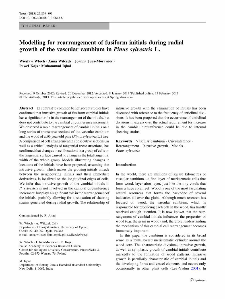
Analysis of the rearrangement of the cambial initials,
carried out with a series of 75 semi-thin transverse sections,
obtained from a sample of 263 lm length in axial direction
(Fig. 2), revealed that we cannot distinguish on the basis of
single sections only whether the radial file examined has
been eliminated or is actually growing intrusively. There-
fore tangential reconstructions of cells from the xylem,
cambium and phloem regions were made. Superimposing
the reconstructions of the xylem and phloem layers revealed
that the arrangement of the radial walls of cells in these
layers was precisely the same. Also, overlaying the recon-
structions of cambial cell layers versus phloem cell layers,
and cambial cell layers versus xylem cell layers revealed
that the initials of radial files #2 and 3 had changed their
positions in an S (right) direction, whereas the initials of
radial files 1 and 4 hardly changed their position during this
time span. Consequent upon this rearrangement, the initial
of radial file 2 was transferred to radial file 3, and simul-
taneously, the initial cell of radial file 3 was partly elimi-
nated (Fig. 2ii). Careful examination of transverse sections
[(Fig. 2i (a–h)] has revealed that the initial cell of radial file
3 had been eliminated, as this radial file was not visible in
the cambium in sections f–h. The tip of this initial is still
visible in a 4-celled intruding radial file in the cambium
[Fig. 2i (e)]. Interestingly, the tip of this partially eliminated
initial experienced slight intrusive growth after the process
of elimination was over; this appeared in sections d and e as
the slanted walls of the 4-celled intruding radial file. Thus, it
could be deduced from the above that the elimination of this
initial had taken place before the four layers of its imme-
diate derivatives were produced.
If we analyze the arrangement of radial files only in a
single section, e.g. in Fig. 2i (f) of this series, we would
interpret it as a typical example of elimination of radial file
2, whereas a detailed analysis of the series of transverse
sections has brought out that in fact it was the radial file #3
that underwent elimination. The initial of radial file 2 did
not eliminate, but only moved to the right.
Models for rearrangement of the fusiform initials
The model presented in Fig. 3 is based on a detailed
analysis of mutual contacts of cells in three (two already
existing and one newly emerging) radial files, as seen at
four points of time, showing the gradual elimination of an
initial of radial file #1 by an intrusively growing initial in
the newly emerging file. In the area where intrusive growth
is in progress, the process of gradual shifting of periclinal
walls of some fusiform initials to a radial position becomes
visible in the form of slanted walls. The intrusively
growing fusiform initial intrudes between the tangential
Trees (2013) 27:879–893 881
123
Page 4
walls of the neighbouring initial and its closest derivative
(t1), which, with the passage of time (t2–t3), leads to the
development of slants, similar to those shown in Fig. 1a–c.
The fixed position of corresponding filled circles placed on
the contiguous tangential-to-radial transforming walls
(x–x0, y–y0, z–z0) indicates that the intrusive growth takes
place on longitudinal edges of the cell, resulting in intru-
sion of the elongating cell tip along the tangential walls of
the adjacent initial and its closest derivative. This rear-
rangement of cells is concurrent with the radial expansion
of cell walls by symplastic growth and the occurrence of
periclinal divisions of the cells so enlarged, and causes no
increase in the cambial circumference.
Three radial files (two already existing and one newly
emerging cell files) chosen to demonstrate the rearrange-
ment of cambial initials due to their apical intrusive
growth, as detected at six points over a given time span of
radial growth of the stem, are presented in Fig. 4. Both the
initial cells and their derivatives grow in a radial direction
in a coordinated way by symplastic growth, i.e. the radial
walls of adjacent cells in neighbouring radial files grow
together and these growing cells do not change their con-
tacts. Thus cells in neighbouring files grow simultaneously
and this is why the lines marked according to location of
edges between tangential and radial walls of the given three
radial files do not cross each other. The rearrangement
Fig. 1 Transverse sections
from different positions of
cambium (a–f) and wood
(g–i) of P. sylvestris, showing
the intrusive growth of the
cambial initial, marked with an
arrow (in a, b and c), and the
elimination of radial file 2 in
(d) and radial file 3 (in e and f).Rearrangement of cambial
initials as recorded in the wood
can be seen in transverse
sections (g, h and i) obtained
from different levels and
locations in the wood sample.
Time–vector indicates the
sequence of consecutively
deposited layers of wood, from
the oldest to the newest layer.
The dashed lines indicate the
arrangement of radial files.
Eliminated files are marked by
the termination of dashed lines.
Intrusion of new radial files is
marked with dotted lines. The
split of one dashed line into two
indicates the occurrence of
anticlinal division (i)
882 Trees (2013) 27:879–893
123
Page 5
caused no increment of cambial circumference, as the total
tangential width of the presented group of three radial files
did not change. At the time point t0, the tip of the intru-
sively growing fusiform initial of radial file II is seen
protruding between the initial cell and its derivative sec-
ondary-phloem-mother cells of radial file I. The views
taken at the other points of time (t1–t5) present the con-
secutive changes in the cell arrangement in those radial
files, which occur together with the concurrent symplastic
radial enlargement as well as the periclinal divisions of the
cells concerned.
Figure 5 analyzes the varied pace of intrusive growth
in circumferential direction, between tangential walls of
an initial cell in the neighbouring radial file and its
immediate derivative. The intrusive growth in circumfer-
ential direction takes place in conjunction with a significant
Fig. 2 Rearrangement of cambial initials in P. sylvestris stem.
Section (i) includes eight views chosen from a long series of 75
transverse sections covering a distance of 263 lm in axial direction.
In the tangential reconstructions of section (ii), 1, 2, 3 and 4 are the
serial numbers of radial files examined, while a–h are different
positions along the given axial length of the stem axis from where the
selected eight transverse sections were obtained. The four tangential
views of (ii) present the superimposed reconstructions of cells of the
four radial files in different combinations. Letters X, C and Ph
indicate the cells of secondary xylem, vascular cambium and
secondary phloem, and are drawn by dashed line, dotted line and
continuous line, respectively
Trees (2013) 27:879–893 883
123
Page 6
intrusive growth in axial direction, but this cannot be
observed on a single transverse section, and hence is not
presented in this model. When the intrusive growth is
slow, the angle between slant and radial walls is small
(Fig. 5a); as shown also in the actual transverse section in
Fig. 1g. A rapid cell growth gives rise to a large angle
between slant and radial cell walls, with the same inten-
sity of symplastic radial growth (Fig. 5b). A similar
example is shown in Fig. 1h.
The arrangement of slanted walls, as observed in
transverse sections, depends on the extent of intrusive
growth in circumferential direction, as presented in Fig. 6,
depicting three different models for identical pace of
intrusive growth (the angles between slants and radial cell
walls being equal). If the extent of intrusive growth is
limited to half of the neighbouring initial’s width, after the
cessation of intrusive growth the cell walls transform from
slanted to radial ones, and a new additional radial file is
formed (Fig. 6i). If the extent of intrusive growth is large
enough, the intrusively growing cell may eliminate the
whole of the neighbouring initial (Fig. 6ii). In this case,
one radial file replaces another radial file. If the initial
grows intrusively in one direction, it results in a triangular
arrangement of slanted walls, similar to those shown in
Fig. 1b, c. However, if the intrusive growth occurs in both
(left and right) directions, the slanted walls form a
rhomboidal figure, similar to one presented in Fig. 1a. In
this case a new, an additional radial file is formed.
Fig. 3 A model of intrusive growth of fusiform initial covering
situations at four time points (t0–t3): enlarged views of the intrusively
growing initial and the eliminating initial and their closest derivatives
are presented below arrows. At the time point t0, two radial files
(1 and 2) are considered. At t1–t3, in addition to these two radial files a
third one also becomes visible, which has been derived from the
initial growing intrusively between the neighbouring initial and its
closest derivative in radial file 1. The cells marked by dark grey
(initials) and bright grey (derivatives) undergo periclinal divisions
(dotted line) during the time span involved. Black circles marked with
letters xyz and x0 y0 z0 and placed in pairs on opposite cell walls, which
are transforming their orientation from tangential to slanted, exhibit
constant cell-wall contacts. Letters ‘a’ and ‘b’ correspond with the
occurrence of successive periclinal divisions; their combinations thus
indicate the sequence of derivatives formation. The fusiform cells in
the first point of time are numbered in each radial file, from xylem to
phloem as a, b, c, following the number of radial file (1, 2). The next
letters (a and b) indicate the sequence of periclinal divisions: for
instance, periclinal division of cell 1a results in the formation of two
cell; 1aa, which stays on the initial surface, and cell 1ab, which
becomes a xylem-mother-cell. Similarly, periclinal division of cell
1ab forms two cells: 1aba (closer to initial surface) and 1abb (closer
to mature xylem). The cells formed by periclinal divisions, which are
closer to initial surface, are marked by addition of letter ‘a’ at the end
of previous label, whereas those farther from initial surface are
marked by addition of letter ‘b’ (on time points t1–t3)
884 Trees (2013) 27:879–893
123
Page 7
The next three models (Figs. 7, 8, 9) analyze different
situations of the cambial initial rearrangement, both in the
tangential and transverse views. They establish the rela-
tionship between intrusive growth and elimination of ini-
tials, as per the recent hypothesis of intrusive growth
occurring between tangential walls of neighbouring initial
and its immediate derivative. Moreover, these models
exhibit the simultaneous occurrence of intrusive growth in
an axial direction as well as a circumferential direction, a
feature not included in the previous models.
Figure 7 presents a model of the rearrangement of
cambial initials, comparing the transverse and tangential
views. Three fusiform cells (1, 2, 3) are shown before the
occurrence of intrusive growth (Fig. 7a) and after the
occurrence of intrusive growth of initial #3 (Fig. 7b, c, d).
The extent of intrusive growth is varied. It eliminates either
a part of initial #2 (Fig. 7b), corresponding with the situ-
ation presented in Fig. 6 (i), or the whole initial # 2
(Fig. 7c), corresponding with the situation presented in
Fig. 6 (ii), or parts of initials 1 and 2 (Fig. 7d), corre-
sponding with the situation presented on Fig. 6 (iii). The
model reveals that together with the occurrence of intrusive
growth in circumferential direction, which can be seen in
single transverse section (as in Figs. 5, 6), there also occurs
intrusive growth in axial direction, which can be seen in
tangential sections.
In Fig. 8, scheme ‘a’ shows the fusiform cells before the
occurrence of intrusive growth, whereas schemes ‘b’ and
‘c’ exhibit fusiform initials of the same radial files, as in
scheme ‘a’, but after the occurrence of intrusive growth. It
can be seen that tips of the growing initials (cell # 1 in
scheme b and cells #1 & 2 in scheme c) partially eliminated
the initial #3. There are also models of transverse sections
showing the situation at the two levels marked on tan-
gential sections by lines X and Y. The models for plane
X exhibit the rearrangement commonly described as ‘total
elimination’, whereas those for plane Y demonstrate what is
described as ‘partial elimination’, i.e. narrowing of the
radial file. These models reveal that the event of cell
elimination, shown by single transverse sections, is actu-
ally a result of intrusive growth of the neighbouring initial
or initials. Similar examples of eliminations of radial files
are shown in the actual transverse sections in Fig. 1d, e &
f. The main feature in which this model differs from the
previous one (Fig. 7) is the location of transverse sections
on the tangential view in relation to growing tip of the
initial. In Fig. 7, transverse section (a) presents the view of
a location above the tip of the growing initial #3. In this
situation the axial intrusive growth of the initial resulted in
the formation of a new, intruded radial file. In Fig. 8,
transverse section (a) has been made at a different level,
which includes the tips of initials (#1 and 2) that were
supposed to grow; so these cells were visible even before
the occurrence of intrusive growth. This condition dem-
onstrates that the axially growing initials would look and
be described in transverse sections as the circumferential
expansion of these initials, causing partial (Y) or total
(X) elimination of radial file #3. Thus the analysis of a
single transverse section (X) would be described just as the
‘‘total elimination’’, though it is only a ‘‘partial elimina-
tion’’ of the tip of the initial #3.
Figure 9 presents a model developed to emphasise the
location of the growing tip of a fusiform initial in relation
to positions along the plant axis from where the transverse
sections have been obtained. This model also exhibits
rearrangement of cambial initials in tangential view, and
compares it with three views seen in transverse sections
(Fig. 9 iii) taken from positions indicated on Fig. 9 i, ii
with lines X, Y and Z. The first of these transverse sections
(X) presents the situation described commonly as elimi-
nation of radial file (Fig. 1d, e, f). Similar examples are
shown in Fig. 1d, e, f. In the next two transverse sections
(Y and Z), elimination of radial file is located next to the
intrusively growing tip of cell (1). Examples similar to
transverse section Z have been presented in Fig. 1a, b, c. If
we observe the same cells in superposition of tangential
sections as in Fig. 9[i(a ? b)] and their enlarged view in
Fig. 9(ii), we would describe it as intrusive growth of
initials 1 and 2. So, depending on the plane and the position
from where the section has been obtained, the same rear-
rangement would be described as an outcome of elimina-
tion of one initial or intrusive growth of the other. This
clearly shows that elimination of an initial is a result of
intrusive growth of the neighbouring initials.
Discussion
Intrusive growth and elimination of cambial initials
Cumbie (1963) highlighted the significance of longitudinal
elongation of sister fusiform initials produced by the
pseudotransverse anticlinal divisions in the non-storeyed
cambium. Apical intrusive growth of these initials was
considered to be an important mechanism of increase in the
cambial circumference (Hejnowicz and Branski 1966; Iq-
bal 1990; Larson 1994; Evert 2006). The exact localization
of intrusive growth of the fusiform cambial initials is still a
subject of debate. Hejnowicz and Zagorska–Marek (1974)
identified occurrence of intrusive growth at radial edges of
the cambial initials and suggested that the cell-tip intrusion
takes place between radial walls of the neighbouring ini-
tials (Larson 1994). If the intrusive growth of the initials
increases the cambial circumference, obviously it has to
occur between the radial walls of adjacent initials. Thus all
those authors who consider intrusive growth as the main
Trees (2013) 27:879–893 885
123
Page 8
mechanism of circumferential enlargement of the cambial
cylinder, in principle assume that intrusive growth occurs
along radial walls, even if they do not confess it
categorically.
The intensity of anticlinal divisions in the non-storeyed
cambia, followed by intrusive growth of the initials pro-
duced, was found to be much higher than actually required
for due increment of the cambial circumference; this excess
cell production was supposed to be compensated through
elimination of the initials (Bannan 1950, 1957, 1960a, b;
Evert 1961; Hejnowicz 1961; Hejnowicz and Branski
1966; Cumbie 1967; Srivastava 1973; Romberger et al.
1993; Larson 1994). Such a phenomenon has not been
observed in storeyed cambia, and it has yet to be explained
why the excessive generation of initials is confined to the
non-storeyed cambia alone.
Elimination of extra initials is believed to occur through
their decline (Hejnowicz and Branski (1966), their
886 Trees (2013) 27:879–893
123
Page 9
differentiation into the phloem or xylem element (Bannan
1952; Cumbie 1967; Larson 1994), or by the domination of
neighbouring initials (Forest and Demongeot 2006). Inter-
estingly, none of the above explanations takes into account
the occurrence of intrusive growth of cambial initials. In
the words of Forest and Demongeot (2006), elimination
stems from the progressive domination of cells of one file
over those of the other, causing decrease in perimeters of
the latter’s cells. These authors regarded this domination as
being caused by the contact strength exerted by the
neighbouring files as a consequence of competition. Such
an assumption, however, is not consistent with the recorded
observations, given that the cambium is always in a state of
considerable tensile stress in tangential direction (He-
jnowicz 1980; Kojs and Rusin 2011; Kwiatkowska and
Nakielski 2011).
The above explanations suggest that intrusive growth
should occur when the cambium is tangentially stretched,
i.e. under tensile stress (Romberger et al. 1993; Kwiat-
kowska and Nakielski 2011), whereas a precise mechanism
of elimination has not yet been determined. It is difficult to
explain the occurrence of competition for space between
the cambial initials, as postulated by Forest and Demongeot
(2006), when the tissue is tangentially stretched. On the
contrary, such a competition may be expected when the
tissue would be locally compressed in tangential direction,
a situation not yet reported about the cambium. The above
interpretation also leads to the assumption that intrusive
growth and elimination do not occur together, in the same
area of the cambium, as they would be an outcome of
contradictory mechanical conditions. However, observa-
tions confirm that intrusive growth occurs commonly in the
neighbourhood of eliminations, as reviewed by Larson
(1994). Long back, Nageli (1864) mentioned that incre-
ment of one radial file explains the ‘‘shortening and death’’
of the adjacent file, a condition later described as elimi-
nation of initials. Bannan (1957) also enunciated that fol-
lowing the pseudotransverse anticlinal division, one of the
sister initials would predominantly be eliminated, whereas
the other one would grow intrusively.
Kojs et al. (2004a, b) regarded the intrusive growth as
occurring on longitudinal (tangential) edges, thus sug-
gesting the cell intrusion between tangential walls of the
neighbouring initial and its immediate derivative. A similar
Fig. 5 Models of intrusive growth of fusiform initials in circumfer-
ential direction occurring at different pace over a time span (t0–t2) as
seen in a transverse view. The middle drawing (t0) shows the cell
pattern at the beginning of intrusive growth. Toward its left is the
model (A) of slow growth of the initial whereas to its right is the
model (B) of rapid growth during the same time span and in
conjunction with the same rate of the symplastic radial growth. The
initials are marked with darker grey than their derivatives. Cells in the
radial file marked with bright grey display a case of elimination,
whereas those marked with middle grey are the derivatives of the
intrusively growing (dark grey) initial
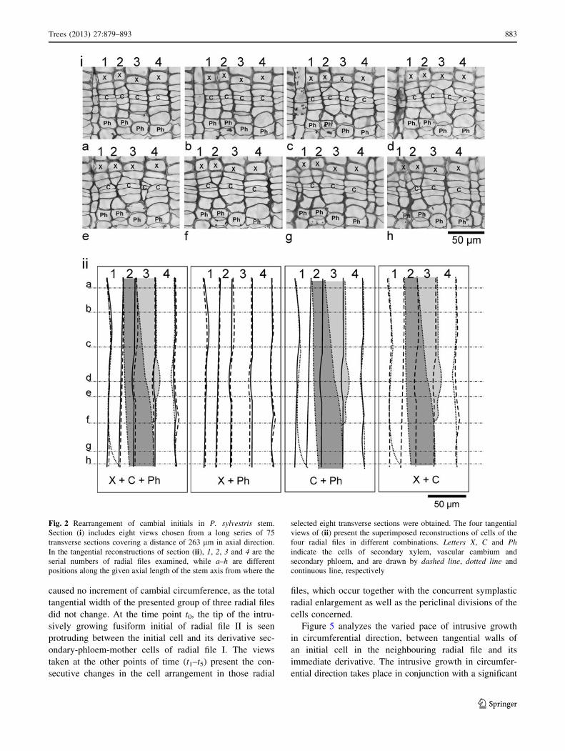
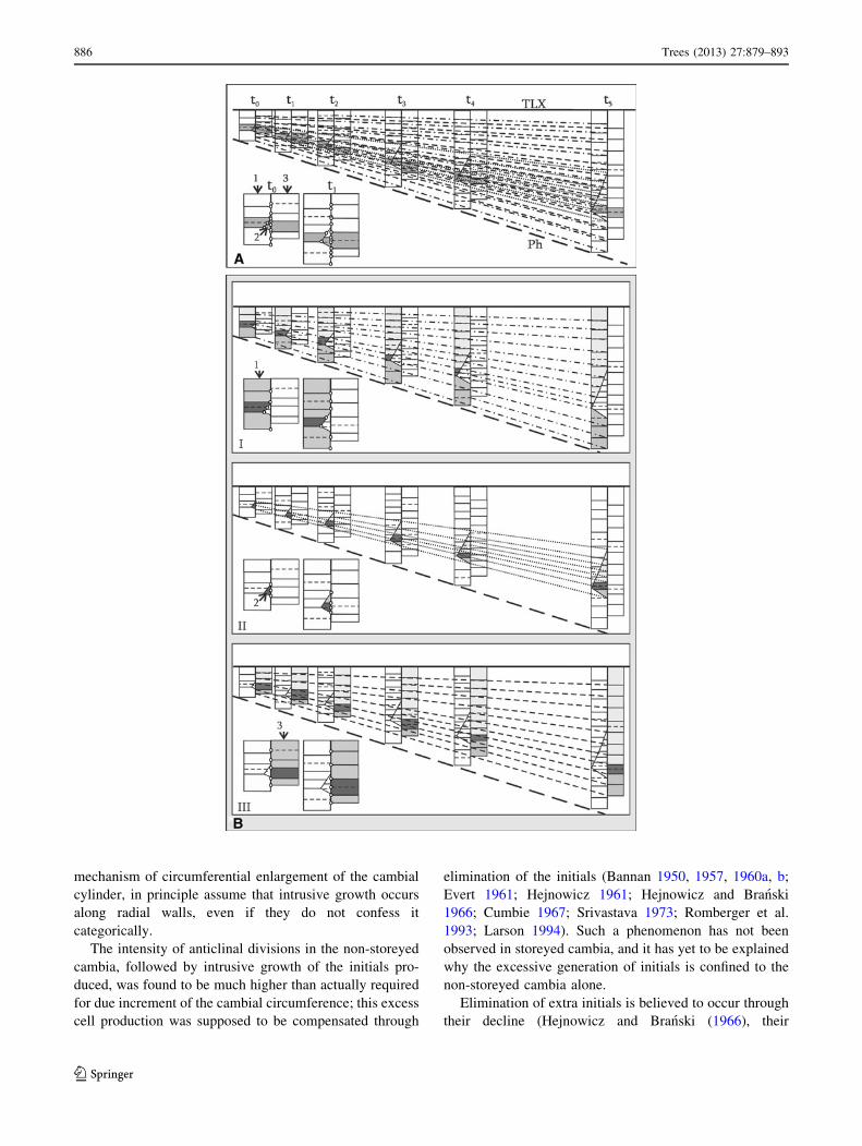
Fig. 4 A model of radial growth of three radial files (1–3) of fusiform
cambial cells (files # 1 and 3 already exist, while file # 2 is emerging
due to intrusive growth of an initial), considered in transverse plain: A
single transverse section was seen at six time points (t0–t5). The initial
cell of radial file 2 is currently growing intrusively. The thick,
continuous horizontal line represents the border between the current
annual ring and the last layer of the previous annual ring (terminal
late xylem) marked as TLX, as a reference surface, which is in a
stable position during the radial growth. The arrangement of cambial
cells in three radial files at all the six time points indicates the same
position of TLX. On the lower side, the thick dashed line represents
the border between the last layer of the mature phloem derived in the
previous year (marked as Ph) and the current year increment of the
phloem. This border is pushed outward by radial growth of stem due
to addition of new derivatives on the inner side of cambium through
repeated periclinal divisions. Periclinal divisions of cambial cells are
marked with horizontal, dashed lines. A In the left bottom corner of
the scheme (A), two points of time are enlarged and circles have been
marked indicating cell wall edges between tangential and radial walls
(also periclinal divisions) between the three radial files analyzed. In
the upper scheme the corresponding edges have been connected in
radial files 1–3 between all six points of time with lines: 1-dashed and
dotted, 2-dotted and 3-dashed lines. The initials are marked in grey.
B Lines drawn to mark the location of edges between tangential and
radial cell walls in each of the radial files 1, 2 and 3 are shown in three
separate drawings (I, II, III respectively). In the left bottom corner of
the schemes situations at two points of time (t0 and t1) are shown in
enlarged view, marking the cell wall edges between tangential and
radial walls (also the periclinal divisions) with small circles. The
radial file analyzed is marked in grey shades; the phloem-mother cells
and xylem-mother cells are marked in medium grey and bright grey
respectively, whereas the initials are shown in dark grey. The changes
in location of cell wall edges between the tangential and radial walls
occurring in radial files 1, 2 and 3 are shown in illustrations I, II and
III of section B, respectively
b
Trees (2013) 27:879–893 887
123
Page 10
situation was described earlier for tumorous cambia
(Włoch 1976; Larson 1994; Włoch et al. 2001). Later it
was generalised as a common mechanism of intrusive
growth of the cambial initials (Kojs et al. 2004a, b; Jura
et al. 2006; Karczewska et al. 2009; Włoch et al. 2009).
This hypothesis of the occurrence of intrusive growth along
tangential walls explains why intrusive growth occurs
always in conjunction with eliminations. This also confirms
that neither the elimination nor the intrusive growth has an
influence on cambial circumference (Jura et al. 2006;
Włoch et al. 2009; Wilczek et al. 2011). The occurrence of
intrusive growth in one initial means an equal elimination
of the adjacent initial, thus proving that the two events are
complementry.
The rearrangement of cambial initials can hardly be
understood by examining a single transverse or tangential
section. Analysis of long series of transverse sections is
similar to data collection from tomography and allows for
precise reconstruction of cells’ arrangement. The models
presented in this paper allow for taking into consideration
the occurrence of intrusive growth in the modelling for
symplastic growth in radial direction. The initials, after
completion of intrusive growth, continue their growth by
symplastic mode only. New observations have revealed
that the growing tips of the cambial initials intrude between
the tangential walls of the neighbour initials and their
immediate derivatives and, therefore, have no influence on
the cambial-circumference enlargement.
Anticlinal divisions versus circumferential growth
of cambium
Anticlinal divisions increase the number of cambial ini-
tials. The percentage of added fusiform cells remains
considerably higher during the first few years of cambial
activity, when the radius of the cambial cylinder is very
little; it later decreases markedly when the stem grows
thicker (Bailey 1923; Butterfield 1972; Iqbal 1994; Kojs
et al. 2004b). In an old, thick stem the overall number of
fusiform initials added to the cambial circumference is very
small, although the anticlinal divisions are more frequent.
Predominantly, after the anticlinal division of a fusiform
initial, one of the resultant sister initials is eliminated
(Bannan 1950; Evert 1961; Srivastava 1973; Cumbie 1967;
Lim and Soh 1997a, b).
The linear diagram of tracheids’ radial files from the
secondary xylem of Chamaecyparis nootkatensis (Bannan
1950) revealed that 16 fusiform initials identified on the
circumference of cambial cylinder at the 6 mm radius
divided anticlinally 71 times during the radial increment of
2 mm (from 6 to 8 mm radius), thus adding 71 new cells to
the already existing 16 cells and making a total of 87 ini-
tials. However, over the radius of 8 mm, the chosen seg-
ment possessed only 26 fusiform initials instead of 87.
This means that only 10 fusiform initials (14 %) were
added, and 61 of them (86 %) eliminated. The number
of anticlinal divisions was 7 times higher than the actual
Fig. 6 A model of three different extents of intrusive growth in
circumferential direction over a given time span (t0–t2), as observed in
transverse section. The pattern of the slanted cell walls depends on
layers of cells deposited during the progress of intrusive growth. In
diagram (1), intrusive growth occurs in one (left) direction, initiating
the triangular arrangement of slants (at t0), and ceases when the
growing cell has reached the middle of the partly eliminated radial
file, thus causing the formation of a new radial file (t1–t2). In diagram
(2), intrusive growth occurs in one direction (left) resulting in
triangular arrangement of slants (t0–t1), and ceases when the
intrusively growing cell has reached the next radial file, thus leading
to the elimination of one radial file through consistent narrowing of its
initial (at t2). In diagram (3), the cell grows intrusively in two (left and
right) directions, resulting in a rhomboidal figure marked with slanted
walls of newly formed radial file with intrusively growing initial (at
t0), ultimately causing a partial elimination of two neighbouring radial
files (at t2)
888 Trees (2013) 27:879–893
123
Page 11
requirement for due increase in the cambial circumference.
Analysis of the same diagram at a different magnitude of
cambial radius (between 3 and 6 mm) revealed that despite
a very high number (83) of anticlinal divisions occurring
during the time span involved with this radial increment,
the number of fusiform initials in the given sector had not
changed at all. There were 16 initials both at the beginning
and at the end of this 3 mm radial increment (Bannan
1950). If anticlinal divisions occur only to increase the
number of initials in the cambial circumference, it is
incredible that all the added initials were eliminated in this
instance. It showed, however, that anticlinal divisions took
almost no part in increasing the cambial circumference.
In 1964, Bannan’s study of Pseudotsuga menziensii
wood, sampled from a trunk of 11-inch diameter, brought
out a curious example of excess anticlinal divisions. By
examining 8 mm of radial increment, caused by cambial
activity in 4 years, he traced the history of anticlinal
divisions and eliminations of 10 fusiform initials. The 10
initials divided anticlinally 18 times, adding 18 new ini-
tials. However, after the last annual increment examined,
there were only 11 fusiform initials in total (Bannan 1964).
Fig. 7 A model to compare a
given situation of cell
arrangement in tangential (1)
and transverse (2) sections: The
horizontal, dashed line drawn
on tangential sections indicates
the position of transverse
sections presented below (b,
c and d). The horizontal, dotted
lines drawn on transverse
sections (b, c, d) correspond to
the positions of tangential
sections (a, b, c and d,
respectively) presented above
a) A tangential view showing
two radial files (1–2) at the
position indicated by the dotted
line, which is marked as ‘a’ on
all the three transverse sections
(b, c & d), indicates the
situation prior to the occurrence
of intrusive growth. Radial file
#3 of this drawing is not visible
in the transverse view, because
its upper end is still below the
dashed, horizontal line b–
d) Three examples of cell
rearrangement, showing varied
extent of intrusive growth of the
initial of radial file #3, both in
tangential (above) and
transverse (below) views. The
initial #3 and its derivatives are
marked with dotted line a ? b,
a ? c and a ? d are the
superpositions of the respective
tangential views and reveal the
occurrence of intrusive growth
of initial # 3
Trees (2013) 27:879–893 889
123
Page 12
This means that 17 newly added initials were eliminated
(94.4 %) and only 1 initial (5.6 %) was added to the
cambial circumference.
Such examples are numerous in the literature. Evert
(1961), for instance, examined the wood of pear tree trunk
in 7 consecutive annual increments. During this time span,
20 fusiform initials divided anticlinally 35 times, thus
forming 35 new initials altogether. Of these, seven cells
(i.e. 20 %) transformed into rays, 23 cells (i.e. 65.7 %)
were eliminated and only 5 cells (i.e. 14.3 %) maintained
the fusiform initial status. Thus, the number of eliminations
was more than 4 times higher than the number of actually
added initials. Srivastava (1973) proposed a model illus-
trating the frequency of anticlinal divisions over a 50-year
span of cambial activity, assuming that the tree-trunk
diameter grew from 20 to 200 cm (10 times increase). In
his model, each fusiform initial should divide 9 times,
forming 10 fusiform initials altogether, in order to meet the
actual requirement of the expanding cambial circumfer-
ence. He observed that the actual number of anticlinal
divisions in the trunk was much higher (26 divisions).
From the newly added 26 fusiform initials, 17 initials (i.e.
65.4 %) were eliminated and only 10 (i.e. 38.5 %)
contributed to the cambial-circumference increment. Sev-
eral other studies (Cumbie 1967; Lim and Soh 1997a, b)
have given similar results.
Since the classical concept of rearrangement of cambial
initials in non-storeyed cambia considers occurrence of
anticlinal divisions, followed by intrusive growth of the
resultant initials, in excessively higher intensity than the
actual requirement of the expanding cambial circumfer-
ence, the excess of the cambial circumference increment
was assumed to be balanced through their eliminations.
Recent reports have revealed that intrusive growth and
eliminations of the initials are two facets of the same
process, and none of these takes part in the increase of
cambial circumference. Therefore, the new concept does
not refer to any excess of cambial circumference incre-
ment, which would have to be balanced by cell elimina-
tions. Our models (Figs. 3, 4, 5, 6, 7, 8, 9) confirm that
intrusive growth and eliminations of the initials do not
influence the cambial circumference, as proposed in some
earlier studies (Jura et al. 2006; Włoch et al. 2009).
In the examples discussed above, only a small percentage
of newly formed initials, e.g. 11.5 % (Bannan 1950) or
9.1 % (Evert 1961), took part in increasing the cambial
Fig. 8 A model comparing the arrangement of three radial files of
cambial cells (1–3) observed in tangential and transverse sections: On
the left, the arrangement of cells has been shown in tangential
sections before the occurrence (a) and after the occurrence (b and
c) of intrusive growth i) Superposition (a ? b) shows intrusive
growth of initial #1, resulting in a partial elimination of initial #3,
whereas superposition (a ? c) demonstrates the concomitant intru-
sive growth of initials #1 and #2, again causing a partial elimination
of initial #3 ii) The schemes illustrate an enlarged view of
superpositions a ? b and a ? c (i), showing the cell overlap by
intrusive growth. The eliminated part of initial #3 is marked in dark
grey. The cells #1 and 2, before the occurrence of intrusive growth,
are shown in bright grey. The tips of these intrusively growing initials
have also eliminated slightly the small parts of neighbouring radial
files outside the area originally covered by the initials 1, 2 and 3. The
initial #3, partly eliminated but still appearing on the initial surface, is
shown as a white cell iii) The models illustrate reconstructions of
transverse sections made at positions marked with dotted horizontal
lines X and Y drawn on tangential sections (i, ii). Dotted lines (a, b,
c) indicate the plane in which tangential sections a, b and c respec-
tively, have been made
890 Trees (2013) 27:879–893
123
Page 13
circumference. This is typical for thick trunks, where the
relative increment of cambial circumference is minute.
According to Karczewska et al. (2009), in a trunk of 78.5 m
circumference concurrent with the radial increment of
100 lm, the relative circumferential increment is only
0.08 %. This means that only 31 initials were added to the
circumference of this trunk, though the total number of ini-
tials was almost 40,000. In order to meet the actual necessity,
only 1 out of 1,266 initials will have to divide anticlinally.
Thus, if the anticlinal divisions are meant only for contrib-
uting to the increase of the cambial circumference, these
should hardly occur in the cambium of a thick trunk.
A reasonable conclusion that can be drawn from the
above analysis is that the high frequency of anticlinal
divisions is not related to the mechanism of the cambial
circumference increment, but it helps in the rearrangement
of cambial initials, as does the directed intrusive growth of
anticlinally divided fusiform initials, which occur along the
tangential walls of the neighbouring initial and its imme-
diate derivative. The actual increase of cambial circum-
ference results from symplastic growth of the resultant
initials after anticlinal divisions.
Studies of mechanical forces acting through tissues
(Lintilhac and Vesecky 1984; Lynch and Lintilhac 1997)
have shown that orientation of division plate in dividing
cells is determined by growth strains in the tissue. An ideal
model of the cambium suggests that the radial tensional
strains, occurring as a result of diurnal strains (Kojs and
Rusin 2011), would cause periclinal divisions, whereas the
tangential tensional strains, due to increase in the girth of
wood cylinder would cause the sparse radial longitudinal
divisions (Srivastava 1973; Karczewska et al. 2009). The
pattern of cell divisions in double-storeyed cambia con-
forms to the ideal situation, whereas the non-storeyed
cambia experience mostly oblique anticlinal divisions, the
frequency of which surpasses the actual need of the
Fig. 9 A model comparing the
arrangement of cells observed in
tangential and transverse
sections. As in previous figures,
the drawings indicate the
arrangement of cells before
(a) and after (b) the occurrence
of intrusive growth of initials #1
and #2, which results in partial
elimination of initial #3.
Module (i) illustrates the
tangential sections in drawings
a and b and their superposition
(a ? b). Module (ii) illustrates
enlarged area of intrusive cell
growth marked with a
rectangular frame on the
superposition a ? b on module
(i). The eliminated part of initial
#3 is shown with dark grey; the
cell in radial files #1 and 2,
before occurrence of intrusive
growth, are bright grey, while
the initial cell #3, partly
eliminated but still remaining in
initial surface, is white. Module
(iii) illustrates three transverse
sections taken from positions
indicated on modules (i) and (ii)by the horizontal lines X, Y and
Z
Trees (2013) 27:879–893 891
123
Page 14
expanding cambial-circumference. This excess of oblique
anticlinal divisions, in our opinion, is a consequence of
significant shearing strains produced in this type of cam-
bium. These strains may possibly generate in storeyed
cambia also, but are likely to be relaxed quickly by the
directed intrusive growth of fusiform initials, which is
meagre in amount and localized only around the storey
borders. This results in a rapid and coordinated change of
inclination and orientation of the initials in the whole
storey (Włoch and Połap 1994; Kojs et al. 2003; 2004a, b;
Jura et al. 2006). This type of cell rearrangement is
impossible in the non-storeyed cambium, mostly because
the ends of fusiform initials are located at different height
levels. Apical intrusive growth has to be far greater in
magnitude in this type of cambium (Kojs et al. 2004a).
In view of the observed correlation of strains in the
tissue with the orientation of division plates (Lintilhac and
Vesecky 1984; Lynch and Lintilhac 1997), it could be
possible that shearing strains in non-storeyed cambium are
not relaxed quickly and overpass some threshold level,
which might cause the occurrence of oblique anticlinal
divisions. If so, the occurrence of oblique anticlinal divi-
sions may not be related to cambial circumference incre-
ment. The high frequency of anticlinal divisions exceeding
the need of cambial-circumference increment confirms this
inference. According to the new concept, intrusive growth
of fusiform cambial initials is the main mechanism for
allowing relaxation of shearing strains (Kojs and Rusin
2011). Therefore, the cells exposed to shearing strains
would presumably divide anticlinally (as a result of
shearing strains), and then grow intrusively (as the mech-
anism for relaxation of shearing strains).
We propose that the oblique anticlinal divisions are the
result of shearing strains occurring locally in the tissue at a
level optimal for occurrence cell divisions; the specific
pattern of these strains determines also the orientation of
division plates. Excessive occurrence of pseudotransverse
divisions is, therefore, not related to the expansion of
cambial circumference. Oblique anticlinal divisions adjust
the length of fusiform initials through apical intrusive
growth to the extent typical for the species. Further studies
to understand the cell-growth dynamics in the cambial zone
should confirm these hypotheses.
Conclusions
1. The new hypothesis for intrusive growth of cambial
initials suggests that intensities of elimination and
intrusive growth of the initials are equal.
2. The excess of fusiform initials, observed frequently in
the non-storeyed cambia, is eliminated by intrusive
growth, unlike the repeatedly asserted over-speculative
concept of the two events, with an obscure explanation
of the mechanism of cell elimination.
3. Instead of identifying the high frequency of anticlinal
divisions with high intensity of cell eliminations, it
should be identified with high intensity of intrusive
growth.
4. Occurrence of intrusive growth in storeyed cambia is
widespread, i.e. many cells on the initial surface
undergo intrusive growth simultaneously, though its
extent is quite small and confined to the borders of
storeys. This allows for a rapid rearrangement without
a high frequency of anticlinal divisions of fusiform
initials.
5. Elimination of initials cannot be defined satisfactorily
as a separate process, even though the process of
intrusive growth is well defined. Intrusive growth and
eliminations of the cambial initials should essentially
be regarded as two facets of one and the same process.
Acknowledgments M. I. was a Visiting Professor at Plant Pro-
duction Department of the College of Food and Agricultural Sciences,
King Saud University of Riyadh, Saudi Arabia, during the course of
this study.
Open Access This article is distributed under the terms of the
Creative Commons Attribution License which permits any use, dis-
tribution, and reproduction in any medium, provided the original
author(s) and the source are credited.
References
Bailey IW (1923) The cambium and its derivative tissues IV. The
increase in girth of the cambium. Am J Bot 10:499–509
Bannan MW (1950) The frequency of anticlinal divisions in fusiform
cambial cells of Chamaecyparis. Am J Bot 37:511–519
Bannan MW (1952) Further observations on the reduction of fusiform
cambial cells in Thuja occidentalis L. Can J Bot 31:63–74
Bannan MW (1956) Some aspects of the elongation of fusiform
cambial cells in Thuja occidentalis. Can J Bot 34:175–194
Bannan MW (1957) The relative frequency of the different types of
anticlinal divisions in conifer cambium. Can J Bot 35:875–884
Bannan MW (1960a) Ontogenetic trends in conifer cambium with
respect to frequency of anticlinal division and cell length. Can J
Bot 38:795–802
Bannan MW (1960b) Cambial behaviour with reference to cell length
and ring width in Thuja occidentalis L. Can J Bot 40:1057–1062
Bannan MW (1964) Tracheid size and anticlinal divisions in the
cambium of Pseudotsuga. Can J Bot 42:603–631
Barlow PW, Brain P, Powers SJ (2002) Estimation of directional
division frequencies of vascular cambium and in marginal
meristematic cells of plants. Cell Prolif 35:49–68
Butterfield BG (1972) Developmental changes in the vascular
cambium of Aeschynomene hispida Willd. N Z J Bot 10:373–
386
Cumbie BG (1963) The vascular cambium and xylem development in
Hibiscus lasiocarpus. Am J Bot 50:944–951
Cumbie BG (1967) Developmental changes in Leitneria floridana.
Am J Bot 54:414–424
892 Trees (2013) 27:879–893
123
Page 15
Evert RF (1961) Some aspects of cambial development in Pyrus
communis. Am J Bot 48:479–488
Evert RF (2006) Esau’s plant anatomy; Meristems, cells and tissues of
the plant body; their structure, function and development, 3rd
edn. Wiley, Hoboken
Forest L, Demongeot J (2006) Cellular modeling of secondary radial
growth in conifer trees: application to Pinus radiata (D. Don).
Bull Math Biol 68:753–784
Hejnowicz Z (1961) Anticlinal divisions, intrusive growth and loss of
fusiform initials in nonstoried cambium. Acta Soc Bot Pol 30:
729–758
Hejnowicz Z (1968) The structural mechanism involved in the
changes of grain in timber. Acta Soc Bot Pol 37:347–365
Hejnowicz Z (1980) Tensional stress in the cambium and its
developmental significance. Am J Bot 67(1):1–5
Hejnowicz Z, Branski S (1966) Quantitative analysis of cambium
growth in Thuja. Acta Soc Bot Pol 35:395–400
Hejnowicz Z, Zagorska–Marek B (1974) Mechanism of changes in
grain inclination in wood produced by storeyed cambium. Acta
Soc Bot Pol 43:381–398
Iqbal M (1990) The vascular cambium. Wiley, New York
Iqbal M (1994) Structural and operational specializations of the
vascular cambium of seed plants. In: Iqbal M (ed) Growth patterns
in vascular plants. Dioscorides Press, Portland, pp 211–271
Jura J, Kojs P, Iqbal M, Szymanowska-Pułka J, Włoch W (2006)
Apical intrusive growth of cambial fusiform initial along the
tangential walls of adjacent fusiform initials: evidence for a new
concept. Aust J Bot 54:493–504
Karczewska D, Karczewski J, Włoch W, Jura-Morawiec J, Kojs P,
Iqbal M, Krawczyszyn J (2009) Mathematical modelling of
intrusive growth of fusiform initials in relation to radial growth
and expanding cambial circumference in Pinus sylvestris L. Acta
Biotheor 57:331–348
Kojs P, Rusin T (2011) Diurnal strains in plants. In: Glinski J,
Horabik J, Lipiec J (eds) Encyclopedia of agrophysics. Springer,
Berlin, pp 220–224
Kojs P, Włoch W, Rusin A, Szendera W (2003) Storeyed structure of
cambium as an adaptative strategy to environmental conditions
in trees forming canopy and the emergent layer of the tropical
rain forests. Bull Bot Gardens 12:23–29
Kojs P, Włoch W, Rusin A (2004a) Rearrangement of cells in
storeyed cambium of Lonchocarpus sericeus (Poir.) DC.
connected with formation of interlocked in the xylem. Trees
18:136–144
Kojs P, Włoch W, Iqbal M, Rusin A, Jura J (2004b) Readjustment of
cambial initials in Wisteria floribunda (Willd.) DC to ensure the
development of storeyed structure. New Phytol 163:287–297
Kramer EM (2002) A mathematical model of pattern formation in the
vascular cambium of trees. J Theor Biol 216:147–158
Kwiatkowska D, Nakielski J (2011) Mechanics of the meristems. In:
Wojtaszek P (ed) Mechanical integration of plant cells and
plants. Springer, Heidelberg, pp 133–172
Larson PR (1994) The vascular cambium: development and structure.
Springer series in wood science. Springer, New York
Lev-Yadun S (2001) Intrusive growth—the plant analog of dendrite
and axon growth in animals. New Phytol 150:508–512
Lim DO, Soh WY (1997a) Cambial development and tracheid length
of Dwarf Pines (Pinus densiflora and P. thunbergii). IAWA J
18(3):301–310
Lim DO, Soh WY (1997b) Development of cambium and length of
vessel elements and fibres in dwarf Alnus hirsuta (Spach) Rupr.
J Plant Biol 40(4):245–248
Lintilhac PM, Vesecky TB (1984) Stress-induced alignment of
division plane in plant tissues grown in vitro. Nature 307:363–
364
Lynch TM, Lintilhac PM (1997) Mechanical signals in plant
development: a new method for single cell studies. Dev Biol
181:246–256
Majumdar GP (1941) The fine structure of collenchymas cells in
Heracleum sphondylium L. Proc R Soc Lond B 130:201–217
Meek GA (1976) Practical electron microscopy for biologists. Wiley,
London
Nageli C (1864) Dickenwachstum des Stengels und Anordnung der
Gefassstrange bei den Sapindaceen. Beitr Wiss Bot 4:1–72
Romberger JA, Hejnowicz Z, Hill F (1993) Plant Structure: functionand Development. A treatise on anatomy and vegetative
development, with special reference to woody plants. Springer,
Berlin
Sinnott EW, Bloch R (1939) Changes in intercellular relationships
during the growth and differentiation of living plant tissues. Am
J Bot 26:625–634
Srivastava LM (1973) Cambial activity in trees. Arnoldia 33:46–66
Wilczek A (2012) The formation of heterogeneous storeys in
cambium on example of Laburnum anagyroides Medik. Acta
Agrobotanica 65(2):47–56
Wilczek A, Jura-Morawiec J, Kojs P, Iqbal M, Włoch W (2011)
Correlation of intrusive growth of cambial initials to rearrange-
ment of rays in vascular cambium. IAWA J 32(3):313–332
Włoch W (1976) Cell events in cambium, connected with the
formation and existence of a whirled cell arrangement. Acta Soc
Bot Pol 45:313–326
Włoch W, Połap E (1994) Zdarzenia komorkowe i ich lokalizacja
w trakcie tworzenia wzoru domenowego w kambium Tilia
cordata Mill. Acta Soc Bot Pol 63(2):215–228
Włoch W, Mazur E, Kojs P (2001) Intensive change of inclination of
cambial initials in Picea abies (L.) Karst. Tumours. Tress Struct
Funct 15:498–502
Włoch W, Mazur E, Bełtowski M (2002) Formation of spiral grain in
the wood of Pinus sylvestris L. Trees Struct Funct 16:306–312
Włoch W, Jura-Morawiec J, Kojs P, Iqbal M, Krawczyszyn J (2009)
Does intrusive growth of fusiform initials really contribute to
circumferential growth of vascular cambium? Botany 87:154–
163
Trees (2013) 27:879–893 893
123