Models for the orientation of chemotactic cells and growth cones For the orientation of chemotactic cells, growth cones etc. it is crucial that cells can measure minute concentration differences across their diameter. In the following it is shown that a corresponding patterning is possible if a self- enhancing reaction is antagonized by two antagonistic reactions: a long-ranging antagonist is responsible for the competition around the entire cell cortex to allow only small active patches. The other acts cell-local and has a longer time constant. It quenches the local signals shortly after its generation, allowing new peaks to form an a updated
Transcript
Models for the orientation of chemotactic cells and growth cones
For the orientation of chemotactic cells, growth cones etc. it is crucial that cells can measure minute concentration differences across their diameter. In the following it is shown that a corresponding patterning is possible if a self-enhancing reaction is antagonized by two antagonistic reactions: a long-ranging antagonist is responsible for the competition around the entire cell cortex to allow only small active patches. The other acts cell-local and has a longer time constant. It quenches the local signals shortly after its generation, allowing new peaks to form an a updated position.
Meinhardt, H. (1999). Orientation of chemotactic cells and growth cones: Models and mechanisms. J. Cell Sci. 112, 2867
Pattern-forming reactions can be very sensitive for directional cues
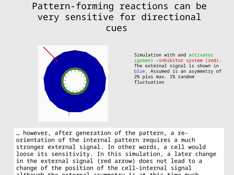
At the homogeneous steady state, a pattern-forming systems is in an instable situation. Any external asymmetry or even random fluctuations can trigger the onset of pattern formation. Thus, minute external influences determine the position at which a maximum will form within the cell. However…
… however, after generation of the pattern, a re-orientation of the internal pattern requires a much stronger external signal. In other words, a cell would loose its sensitivity. In this simulation, a later change in the external signal (red arrow) does not lead to a change of the position of the cell-internal signal although the external asymmetry is at this time much stronger
Simulation with and activator (green) -inhibitor system (red). The external signal is shown in blue. Assumed is an asymmetry of 2% plus max. 1% random fluctuation
Meinhardt and Gierer (1974). J. Cell Sci. 15,321-346
If the antagonist has a longer half-life than the activator, the system will oscillate. After each burst, the activation collapses, providing a chance that the next maximum appears at an updated position. In this model, the antagonist has a dual role: generating the pattern in time and the pattern in space. This model was inspired by the oscillation observed in Dictyostelium.However…
Meinhardt and Gierer (1974). J. Cell Sci. 15,321-346
… there are severe problems:
1. The time window in which the system is sensitive is very short. It is restricted to a short period before the next burst occurs. No reorientation can take place during the refractory period. This is in contrast with the observation in Dictyostelium that the cells remain sensitive permanently.
2. In many chemotactic cells, several protrusions may exist at a given time. While some stretch out, other may retract, indicating that they are not under control of a global oscillation.
Further, such a mechanism requires an unrealistic rapid spread of the antagonist since the competition all around the cell cortex has to be maintained during the burst.
Multiple peaks cannot result from a limited range of the antagonist
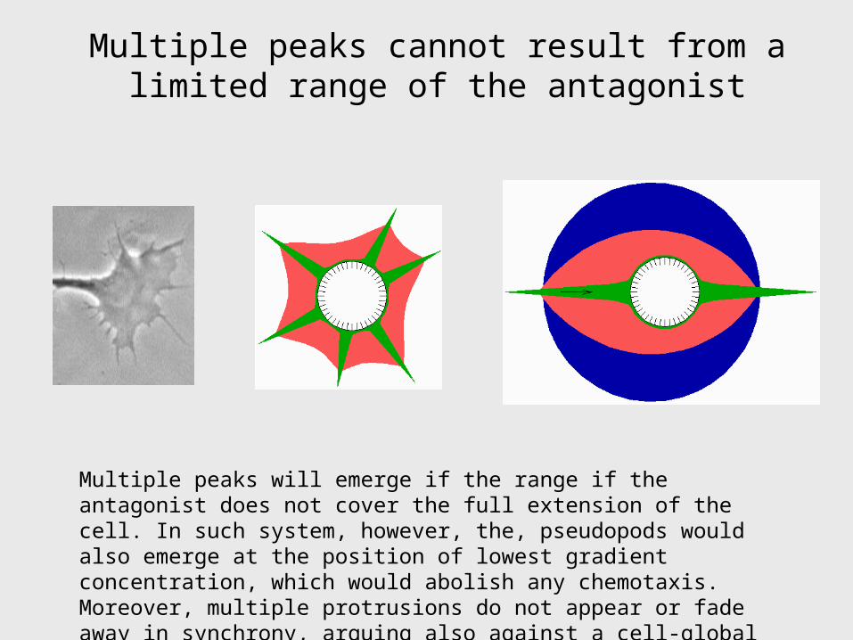
Multiple peaks will emerge if the range if the antagonist does not cover the full extension of the cell. In such system, however, the, pseudopods would also emerge at the position of lowest gradient concentration, which would abolish any chemotaxis. Moreover, multiple protrusions do not appear or fade away in synchrony, arguing also against a cell-global oscillation
In the following, the proposed mechanism is introduced step by step
Without diffusion of the activator and without a saturation of the autocatalysis, the peak would be very narrow and locally fixed.
With saturation (sa > 0), the maximum level would be lower but the peak would be much broader…
…if the activator does not diffuse, the activated regions need not to be coherent; random fluctuations can lead to multiple peaks…
…and without an external asymmetry, the peaks would appear at random position
2
2....
(1 )a
a a
t s ah
Permanent sensitivity and multiple signals can be achieved by an additional antagonist: the newly
formed signals become quenched after some time.
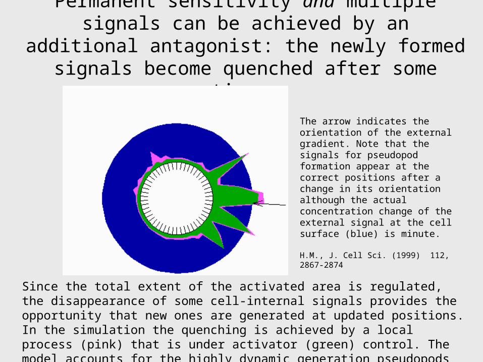
The arrow indicates the orientation of the external gradient. Note that the signals for pseudopod formation appear at the correct positions after a change in its orientation although the actual concentration change of the external signal at the cell surface (blue) is minute.
H.M., J. Cell Sci. (1999) 112, 2867-2874
Since the total extent of the activated area is regulated, the disappearance of some cell-internal signals provides the opportunity that new ones are generated at updated positions. In the simulation the quenching is achieved by a local process (pink) that is under activator (green) control. The model accounts for the highly dynamic generation pseudopods
… the activated area can also be a coherent region..
A somewhat elevated diffusion of the self-enhancing reaction(s) causes that the activated region is more coherent. Nevertheless, a change in the external gradient (black arrow) leads to a rapid reorientation.
Prediction: the highly dynamic patterning does not depend on an external signal
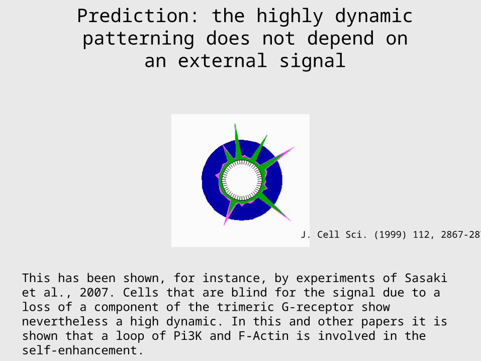
J. Cell Sci. (1999) 112, 2867-2874
This has been shown, for instance, by experiments of Sasaki et al., 2007. Cells that are blind for the signal due to a loss of a component of the trimeric G-receptor show nevertheless a high dynamic. In this and other papers it is shown that a loop of Pi3K and F-Actin is involved in the self-enhancement.
Sasaki et. al., 2007: G protein-independent Ras/PI3K/F-actin circuit regulates basis cell motility. J. Cell Biol. 178,185-191
Chemotaxis by splitting of filopods
In many chemotaxing cells, branching occurs by a Y-like splitting of pseudopods. First an extension of both branches occur, followed by a retraction of one of these branches. Usually, the branch that is exposed to the highest level of attraction survives.
A key paper: Andrew and Insall (2007). Chemotaxis in shallow gradients is mediated independently of PtdIns 3-kinase by biased choices Nature Cell Biol. 9, 193-200
Such splitting also occurs if no external signal can be detected, for instance, if the ß-unit of the G-receptor is defect
For instance: Sasaki et. al., 2007: G protein-independent Ras/PI3K/F-actin circuit regulates basis cell motility. J. Cell Biol. 178,185-191
Chemotaxis by splitting of filopods
In this simulation, a separate system is assumed (red) that quenches the signal (green). This leads to splitting. In agreement with the observation, the signal scatters around the optimal orientation. After a change of the orientation of the external signal, the internal signal moves around the cell until it has, on average, the optimal orientation, in agreement with the observation (Andrew and Insall, 2007). Whether a separate quenching signal really exists is not yet clear
For pattern formation within a cell the activator-depletion mechanism is more appropriate
22
02a a
a as a a D
t x
22
2s s
s ss a s D
t x
For cell-internal pattern-forming processes the activator-depleted substrate mechanism seems to be more appropriate. The self-enhancing process occurs by a cooperative aggregation at the membrane. This proceeds at the expense of monomers that spread more readily in the cytoplasm (the modeling of the out-of-phase oscillation of the MinD protein in E.coli [1] strongly suggested such a mechanism). Another example is the formation of Cdc42 maxima in budding yeast [2].
[1] Meinhardt, H. and de Boer, P.A.J. (2001). Pattern formation in E.coli: a model for the pole-to-pole oscillations of Min proteins and the localization of the division site. PNAS 98, 14202-14207.
[2] Irazoqui, et al. (2003): Scaffold-mediated symmetry breaking by Cdc42p. Nat. Cell Biol. 5,1062
Conclusion
The high sensitivity of chemotactic cells can be achieved by pattern-forming systems that operate within the cell. Local cooperative aggregation at the cell cortex together with a depletion of monomers in the plasma is an appropriate scenario.
To maintain the sensitivity against minute external signals it is required that the signals, shortly after their generation become locally quenched, allowing new signals to emerge. The nature of the quenching is not yet know. A possible candidate is the effect of the dense mesh of actin itself.
This model accounts for the high dynamic pattern as observed, for instance, in the Actin - PIP3 patterning within cells.
In this model, this highly dynamic patterning can proceed also in the absence of an external signal, in agreement with the observation.
Meinhardt, H. (1999). Orientation of chemotactic cells and growth cones: Models and mechanisms. J. Cell Sci. 112, 2867-2874. (also on our website, http://www.eb.tuebingen.mpg.de/meinhardt)