Workshop Draft Report: Molecular and Cellular Biology of Moderate Dose (1- 10 Sv) Radiation and Potential Mechanisms of Radiation Protection Bethesda, MD December 17-18, 2001 [**Draft report prepared for the Workshop participants by the Radiation Research Program, Division of Cancer Treatment, National Cancer Institute, National Institutes of Health. Contact individuals include: C. Norman Coleman , Philip Tofilon, Helen Stone, Rosemary Wong. Phone: 301-496-6111 or 301-496-6360; email addresses : [email protected]; [email protected]; [email protected]; [email protected] ] EXECUTIVE SUMMARY Normal tissue response and injury after exposure to ionizing radiation are of great importance to patients with cancer, populations potentially subjected to military, accidental or intentional exposure, and workers in the nuclear power industry. In these situations exposure is likely to include the moderate radiation dose range (1 – 10 Sievert, Sv). Exposure of limited tissue volumes to higher doses during cancer treatment has been the subject of research by the National Cancer Institute (NCI) which has also supported research into fundamental radiobiology, DNA damage and repair and epidemiology of people exposed to ionizing radiation. Exposure to low radiation doses such as that from nuclear fallout has been of interest to the Department of Energy (DOE) and exposure of astronauts to cosmic irradiation has been studied by NASA. Protection of members of the armed forces against intentional exposure has been studied by the Department of Defense (DOD) and Armed Forces Radiobiology Research Institute (AFRRI). Given the wide range of expertise involved, an interdisciplinary scientific workshop was convened to address the recent scientific progress in molecular, cellular and whole animal radiobiology, biodosimetry, and current and future treatments to prevent or ameliorate radiation damage to normal tissues. This workshop focused on these topics as they pertain to moderate doses defined as 1- 10 Sv (Sievert), a range that was not the topic of recent scientific workshops on low dose radiation and radiation oncology. The broad term “radioprotectors” was used to include chemical and/or biological treatments that might be administered before or after exposure. Understanding the molecular, cellular and tissue changes that can result from moderate dose radiation exposure necessitates input from experts in a number of fields including radiation biology, wound healing and clinical medicine. The development of radioprotector strategies for a single radiation exposure will differ from that for radiation oncology in which treatment is delivered over a multi-week course, a notable exception being the short course for total body irradiation for immunosuppression and transplantation. Additionally, in cancer treatment, the radioprotector should not protect the tumor cells from radiation-induced killing to an appreciable extent. Treatment of populations exposed to a single radiation dose requires accurate and rapid biodosimetry to determine an individual’s exposure level and risk for morbidity and mortality as a result of the exposure, and the availability of appropriate therapeutic agents/strategies and expertise in treatment.
Transcript
Workshop Draft Report:
Molecular and Cellular Biology of Moderate Dose (1- 10 Sv) Radiation and Potential Mechanisms of Radiation Protection
Bethesda, MD December 17-18, 2001
[**Draft report prepared for the Workshop participants by the Radiation Research Program, Division of Cancer Treatment, National Cancer Institute, National Institutes of Health. Contact individuals include: C. Norman Coleman , Philip Tofilon, Helen Stone, Rosemary Wong. Phone: 301-496-6111 or 301-496-6360; email addresses : [email protected]; [email protected]; [email protected]; [email protected] ]
EXECUTIVE SUMMARY
Normal tissue response and injury after exposure to ionizing radiation are of great importance to patients with cancer, populations potentially subjected to military, accidental or intentional exposure, and workers in the nuclear power industry. In these situations exposure is likely to include the moderate radiation dose range (1 – 10 Sievert, Sv). Exposure of limited tissue volumes to higher doses during cancer treatment has been the subject of research by the National Cancer Institute (NCI) which has also supported research into fundamental radiobiology, DNA damage and repair and epidemiology of people exposed to ionizing radiation. Exposure to low radiation doses such as that from nuclear fallout has been of interest to the Department of Energy (DOE) and exposure of astronauts to cosmic irradiation has been studied by NASA. Protection of members of the armed forces against intentional exposure has been studied by the Department of Defense (DOD) and Armed Forces Radiobiology Research Institute (AFRRI). Given the wide range of expertise involved, an interdisciplinary scientific workshop was convened to address the recent scientific progress in molecular, cellular and whole animal radiobiology, biodosimetry, and current and future treatments to prevent or ameliorate radiation damage to normal tissues. This workshop focused on these topics as they pertain to moderate doses defined as 1- 10 Sv (Sievert), a range that was not the topic of recent scientific workshops on low dose radiation and radiation oncology. The broad term “radioprotectors” was used to include chemical and/or biological treatments that might be administered before or after exposure. Understanding the molecular, cellular and tissue changes that can result from moderate dose radiation exposure necessitates input from experts in a number of fields including radiation biology, wound healing and clinical medicine. The development of radioprotector strategies for a single radiation exposure will differ from that for radiation oncology in which treatment is delivered over a multi-week course, a notable exception being the short course for total body irradiation for immunosuppression and transplantation. Additionally, in cancer treatment, the radioprotector should not protect the tumor cells from radiation- induced killing to an appreciable extent. Treatment of populations exposed to a single radiation dose requires accurate and rapid biodosimetry to determine an individual’s exposure level and risk for morbidity and mortality as a result of the exposure, and the availability of appropriate therapeutic agents/strategies and expertise in treatment.
The goals of the interdisciplinary workshop were to define the current state-of-the-science and research opportunities. The conclusions are those of the workshop participants and not those of the individual agencies. The following are the highlights with additional detail provided at the end of the Report. 1. Research
The biological changes elicited in the moderate dose range involve the cells that are irradiated, their non-irradiated neighbors (bystander effect) and the complex interactions among cells, tissues and organs. Research is needed to identify the key molecular, cellular and tissue pathways that lead from the initial molecular lesions to immediate and delayed injury, the latter being a chronic progressive process for which post-exposure treatment may now be possible. In addition to increased support for basic mechanistic studies by individual investigators, consideration should be given to a new program studying radiation toxicology of normal tissues, which involves long-term toxicity and radioprotector studies.
2. Technology High throughput technology will greatly enhance the study of the basic mechanisms of normal tissue injury (for example, a “normal tissue” gene and/or protein chip) and, as molecular targets are defined, will identify agents for normal tissue radioprotection for pre- and post- irradiation treatment. Biomarkers of radiation exposure and rapid and accurate techniques for analyzing multiple samples need to be identified and va lidated to allow for the prompt delivery of the most appropriate treatment.
3. Treatment strategies At present there are a limited number of pre- and post-exposure therapeutic agents and there is a need for research to identify additional biological targets and effective treatments. This is optimally done by collaboration among researchers, industry and governmental agencies. As effective agents are defined tested, and approved for human use, sufficient quantities will be needed.
4. Ensuring sufficient expertise
Over the last decade or so, the number of investigators studying radiation dosimetry, radiation biology and normal tissue injury has declined substantially. It is critical to maintain an interdisciplinary effort and train and recruit investigators from such fields as radiation biology, molecular biology, cellular biology and wound healing. Communication of the current state of the knowledge of the effects of radiation exposure, of which a great deal is known, is important for investigators and policy makers. The timely preparation of a more detailed, user-friendly summary document for the public by workshop participants is recommended.
INTRODUCTION Goals of the workshop Define the state-of-the-science in normal tissue radiobiology, radioprotection and biodosimetry; Describe currently available treatments for preventing and reducing radiation- induced injury; Determine the research opportunities and resources required; Develop a research-action plan for further discussion and implementation.
Background There is an extensive body of research relevant to cancer therapy on radiation exposures higher than that in the range covered in this Workshop and also on lower doses of irradiation relevant to environmental exposure and specific aspects of nuclear fallout. Normal tissue injury resulting from traditional radiotherapy was the topic of a recent NCI- Radiation Research Program Workshop, which has been summarized (Appendix 4). Uniquely, this workshop focused on the “moderate dose” range of 1-10Sv which could be received either in fractionated doses for radiation therapy or in a single dose from accidental or intentional exposure. Workshop and report logistics: Experts (Appendix 2) with a breadth of scientific expertise were invited to discuss the scientific topics of: a) radiation- induced genetic and epigenetic effects in cells and tissues, and whole-body effects; b) biological dosimetry; and c) treatment approaches for radiation protection (Appendix 3). Radiogenic DNA repair and effects of radiation damage on the regulation of the cell cycle were touched on in several sessions but were not a main focus at the workshop. The attendees worked in two Breakout Groups- Detection & Biology and Protection- and discussed the final recommendations as a group. This report was prepared by a subset of the Workshop participants. The recommendations for research were divided, somewhat arbitrarily, into that which could be completed within the following time frames: immediate - within 1 year; medium term - 1 to 3 years; and longer term - greater than 3 years.
DEFINING THE EXPOSURES Units of exposure and dose: Sieverts (Sv) and Gray (Gy): The units Sv will be used in this Report. Sv are units of radiation dose-equivalent that account for the different biological effects of the different types of radiation, i.e., photons, neutrons or particles. For most radiobiology experiments, low photon energy is used so that dose in Sv = dose in Gy. Similarly, for clinical radiation therapy where there is little or no neutron exposure, Sv and Gy are the same.
Appendices: Appendix 1 Glossary of abbreviations Appendix 2 Workshop agenda Appendix 3 Participants and attendees Appendix 4 A summary of Workshop on Normal Tissue injury Appendix 5 Additional reading by topic
Potential radiation exposure during IMRT In cancer treatment, exposure of normal tissues to the moderate dose range is increasingly likely with the use of Intensity Modulated Radiation Therapy (IMRT). IMRT is an evolving radiation therapy technique that allows the radiation oncologist to “sculpt” the dose so that there may be a higher dose given to the tumor and a lower dose to nearby normal tissue. Foci of higher doses can also be produced within the tumor, with the theory that the higher dose will improve local tumor control. The implementation of IMRT depends on complex imaging, computerized treatment planning and treatment delivery. The radiation beam sweeps through large arcs and/or is delivered with multiple fields, thereby exposing larger volumes of normal tissue to lower doses while focusing higher doses within the tumor compared to that achieved with traditional radiotherapy. To accomplish this, the linear accelerator is “on” for a longer time period and the multiple fields of entry spread out dose delivery to more tissues resulting in larger volumes of normal tissues receiving some radiation dose, including the accumulation of a higher whole-body dose compared to traditional radiation therapy. The dose of radiation to the patient from the linear accelerator depends on the X-ray energy and the technique used. The higher-energy linear accelerators, (>10 MV and especially ? 12 MV), produce neutron contamination that adds to the whole body equivalent dose. Because of the “quality factor” multiplier for neutrons, there would be an increased risk of a patient developing a fatal secondary cancer many years after treatment. It should be emphasized that lifetime risk estimates of excess cancers with the lower energy linear accelerators (? 10 MV) is low, below 2%. For this reason IMRT is best performed with 6 or 10 MV nominal energy. The volume of normal tissue treated to a certain dose is limited in IMRT by the design of the treatment plan that is aimed at avoiding clinically apparent organ dysfunction. This treatment planning is based on the existing knowledge of organ tolerance, which depends on the organ involved and the dose distribution within that organ, as well as treatment schedule. What is not known is the impact of the dose distributions from IMRT (large volumes at moderate doses) on long-term organ function and susceptibility to damage from other causes years later. Late tissue responses and the development of agents that might reduce latent injury following radiation therapy were the topic of a recent NCI Radiation Research Program Workshop entitled “Modifying normal tissue damage post- irradiation.” The recommendations of that workshop are summarized in Appendix 4. Acute effects of total-body irradiation The effects of ionizing radiation on animals have been studied in the laboratory. Data on human exposures have been obtained from the Japanese survivors of Hiroshima and Nagasaki and from accidental exposures. To briefly summarize the extensive literature on whole body irradiation, there are three general classes of radiation lethality, which depend on dose, exposure rate and quality of irradiation, (i.e., photons, neutrons or particles). The single dose exposure syndromes are: Cerebrovascular syndrome, >100 Sv, death within 24 to 48 hours; Gastrointestinal syndrome, 5 to 12 Sv (primarily >10 Sv), death within 3 to 10 days ; survival possible in lower end of the range; and Hematopoietic syndrome, 2.5 to 8 Sv, death within 1-2 months; survival possible.
The dose range in this Workshop encompasses the hematopoietic syndrome and the lower range of the gastrointestinal syndrome. However, at longer times after exposure in this moderate dose range, there is also the potential for tumor development as well as the expression of injury in other tissues such as the kidney and central nervous system. As our ability to deal with the acute effects of moderate dose exposure improves, the potential for these late effects is of increasing concern. The effects of an accidental or intentional nuclear event are complex interactions of the immediate blast and the irradiation. To place whole body exposure in context for scientific discussion, data regarding a potential nuclear event were reviewed. The consequences for this scenario were partitioned into what are called “blast-prompt” and “fallout-area delayed” effects. These fallout-area casualties were stratified into several medical-care and dose-ranges categories (Table 1) recognizing that age or concomitant illness could have a significant impact on a particular individual’s outcome.
Table 1
The LD50, a concept used to quantify radiation mortality in a population, is defined as the dose of radiation that will cause death in half (50%) of the people (or animals) exposed. The time of death depends on the dose, as noted above, being almost immediate for the cerebrovascular syndrome, approximately 3–10 days for the gastrointestinal syndrome, and 30-60 days for the hematopoietic syndrome. Therefore, the term for hematopoietic death is the LD50/30 (it is also known as LD50/60, because the marrow failure may occur up to 2 months or 60 days). Human LD50/30
values are estimated to be about 4.5 Sv (approximate range of 3 – 6 Sv) based on the experience of the Japanese atomic bombs and other studies. Medical interventions such as blood cell replacements, antibiotics, cytokines, and in high-dose cases, stem cell transplants, could increase survival to the extent of doubling the LD50 value. The largest proportion of people (47% in Table 1) would represent both worried-well patients (no radiation exposure) and individuals exposed to non- lethal radiation doses (i.e., ? 1.5 Sv). In the other extreme, some 22% of people (Table 1) would represent both those lethally exposed and those requiring intensive care. The ability to identify and triage people exposed to intermediate doses (1.5–5.3 Sv), which represent 31% of this casualty component, can result in reductions in acute casualties and possibly a reduction in cancer incidence in these survivors should effective treatments be developed and utilized. To optimize treatment, biodosimetry is essential. The radius and range of significant injuries from a nuclear event depend on the yield. The 4 Sv dose is within the moderate dose range of this workshop (Table 2).
MOLECULAR AND CELLULAR BIOLOGY AND DETECTION OF RADIATION DAMAGE
Summary of critical information Classical radiobiology is based on the paradigm that cell death results from DNA damage that occurs both directly in the form of strand breaks and indirectly as a result of oxidative reactions. In cells that survive, there is the potential for DNA mutations and chromosomal aberrations. Mutations and to some extent chromosomal alterations can be characterized at the molecular level, although their mechanisms of formation following radiation exposure remain to be fully defined. New techniques, especially those based on fluorescence in situ hybridization (FISH) allow for a more complete assessment of the genomic changes following radiation exposure. In addition, FISH should allow for the identification of informative biomarkers following exposure. However, it is now clear that radiation induces a variety of additional effects that can be expressed at cellular and tissue levels. These effects include the generation of oxidative stress, alterations in gene transcription, changes in signal transduction, and a number of epigenetic phenomena. The latter, to be described in more detail below, involve alterations in cells and tissue not directly related to a change in the structure of the DNA per se. Although a wide variety of events occur, their specific role in tissue radioresponse requires further investigation using a variety of model systems ranging from single cell mechanisms to complex multicellular models to in vivo organ and whole animal studies In addition to contributing to the fundamental understanding of radiation effects within tissue, evaluation of specific changes in gene expression or protein profiles in irradiated cells will likely provide a practical means of defining tissue exposure. Such information may identify sentinel genes or proteins that can serve as in vivo ‘bio-dosimeters’. This type of research is in its infancy. However, its advancement would likely provide a powerful tool for the accurate assessment of the individual risk within an exposed population and possibly determine appropriate pre- and post-exposure interventions. To clearly understand non-carcinogenic and carcinogenic radiation effects, it is necessary to understand multiple response pathways, including cell-cell and cell-microenvironment
interactions. Although less is known about epigenetically-mediated responses, it is becoming clear that there are complex sets of cell-cell interactions so that irradiating one cell may induce transformation, mutation and transcriptional activation in neighboring unirradiated cells, a phenomenon known as the “bystander effect”. This effect enlarges the apparent target size from that predicted by physical dose distribution. Thus, the bystander effect, discussed in more detail below, can be expected to contribute to tissue level response. These types of cell-cell interactions again serve to highlight the need to address radiation responses at the level of the tissue and whole animal in addition to that of single cells. An understanding of each level of response along with the translation of in vitro systems to in vivo and clinical studies will be needed to predict adverse health outcomes following radiation exposure and to develop interventions to prevent and ameliorate injury. Chromosomal damage Chromosomal aberrations are important indicators of radiation exposure and have been used extensively to investigate the mechanisms of radiation action. Aneuploidy, mutagenesis, and carcinogenesis are significant outcomes from chromosomal damage. Measurement of chromosomal abnormalities can be by classical scoring of Giemsa stained metaphase cells or by the use of FISH, multiplex FISH (mFISH) or spectral karyotypic analysis (SKY). Symmetrical exchanges, which by definition are considered to be relatively stable, do not involve the production of acentric fragments and, therefore, are not usually lethal to cells. Such abnormalities are generally cumulative over a lifetime. The use of mFISH has demonstrated that with gamma-ray exposure in the 1 to 4 Sv dose range, up to 25-30% of abnormalities are complex, i.e., involve three or more break-points in two or more chromosomes. For densely ionizing radiation such as charged particles, a much higher proportion of aberrations are complex. Better understanding of mechanisms of chromosomal aberration formation will help elucidate the pathways involved in mutagenesis and carcinogenesis. Chromosomal aberrations also serve as a sensitive biodosimeter. There is a need for methods to analyze chromosomal aberrations in cells from tissues other than blood. In addition to aberration scoring in metaphase cells, induction of premature chromosome condensation (PCC) in interphase cells is a sensitive methodology for measuring radiation damage. It is now possible to induce high PCC yields in proliferating cells. For example, a recently developed alternative PCC technique employs a phosphatase inhibitor (e.g., okadaic acid or calyculin A) combined with p34cdc2/cyclin B kinase to induce high yields of PCC in resting human peripheral blood lymphocytes, producing spreads suitable for biodosimetry applications. Detection of cells with translocations by specific chromosome painting allows evaluation of a broad range of radia tion doses using automated cytological systems. Mutagenesis and carcinogenesis Ionizing radiation in the range of 1–10 Sv causes mutagenesis and carcinogenesis. Cancer has been associated with exposures in the 1 Sv range in approximately 4.5% of patients; approximately a fourth of these patients, or 1% overall, will contract leukemia. Data from Hiroshima indicate that the frequency of chromosomal mutation increased substantially in lymphocytes in ionizing radiation-exposed residents. In addition, a 20% increase in mutation frequency was observed in workers involved in the clean up at Chernobyl. Studies in mice report
that with a 1 Sv exposure, there is an increase in mutation frequency in spermatogonia indicating that stem cells also are sensitive to ionizing radiation exposure. Tissue effects: non-carcinogenic alterations In most tissues, relatively large radiation doses are required to induce overt tissue injury or organ failure. Although there are notable exceptions (e.g., bone marrow), single doses of >10 Sv are generally required to induce significant tissue dysfunction. However, after exposure to doses of 1 to 10 Sv, measurable effects can be detected in many tissues, including persistent and transient alterations in protein expression, growth factor activity, and normal cell and tissue function. Although the significance of such changes with respect to normal tissue radioresponse after moderate doses has not been specifically determined, similar tissue changes have been observed in a number of other pathologic conditions. Thus, it is likely that such changes can contribute to radiation response. Our knowledge in this area is, however, incomplete and further studies, particularly the long-term studies, are needed to evaluate the health impact of such tissue effects of irradiation. It must be emphasized that the target cell for tissue damage may include stromal and endothelial cells as well as epithelial cells and stem cells. Over the past decade, molecular biological approaches have been employed to define subcellular and biochemical events occurring after irradiation. Much of this work has relied on in vitro model systems in which cells are considered as autonomous units, responding to damage as independent entities. However, tissues are highly integrated systems in which cell-cell interactions play major functional roles under physiological and pathological conditions. Thus, the response of individual cells in an artificially isolated situation can be misleading when determining what occurs in situ. Moreover, the history of cells and their microenvironment directly affect how they respond to stimuli. Not only do irradiated cells modify the tissue microenvironment, but the irradiated microenvironment also influences subsequent cell/tissue responses. Application of the technique of laser capture microdissection (LCM), which allows for the in situ analysis of specific cell populations within normal and tumor tissue, should provide relevant information in this research area. Currently, critical deficiencies exist in our understanding of how irradiated cells and the microenvironment interact and function. In certain tissues, stem and precursor cells, which are critical targets for radiation, can undergo rapid apoptosis after exposure and are particularly sensitive to moderate radiation doses. Cell populations that undergo a non-apoptotic death as they divide (“mitotic death”) are also affected by moderate radiation doses, but generally to a lesser extent. Although in the small intestine rapid stem cell loss through apoptosis is not associated with immediate functional deficits, this may not be true for other tissues. For example, in the central nervous system, radiation doses as low as 0.5 Sv induce significant apoptotic cell death in neural precursor cell populations, with a very steep dose response between 0-2 Sv in both rats and mice. Subsequent to the apoptosis there is a persistent reduction in overall cell proliferation. Given that the reduction in neural precursor cells results in cognitive impairment it is reasonable to speculate that radiation- induced changes in these cells might have similar effects. Ionizing radiation induces cognitive impairment, including functions associated with memory, but as yet the pathogenesis of these changes has not been elucidated. In the brain, as in other tissues, critical, but as yet unanswered questions include how microenvironmental factors influence the outcome following irradiation, and how irradiation affects fate decisions of cells, that is, differentiation or mitogenesis. Understanding
these relationships is critical in developing protective strategies, stem cell transplantation, etc., to ameliorate the consequences of radiation exposure of tissues. In the absence of overt tissue damage, persistent radiation effects may contribute to evolving pathology, or response to subsequent trauma, disease or the aging process. For example, radiation has been shown to produce chronic oxidative stress, and there are a number of degenerative conditions as well as aging that have been associated with decreased antioxidant status and increased oxidative stress. In addition, persistent changes in growth factor activity after irradiation may initiate a cascade of events resulting in delayed injury in susceptible tissues or individuals. Given the potential significance of the interaction of low dose irradiation with various forms of tissue pathology as well as trauma or stress on the tissue, considerable research is required to define the potential risks and understand the mechanisms responsible. To address the many factors involved in moderate dose, non-carcinogenic effects in tissues, it will be necessary to employ new and existing in vivo experimental models, develop new in vitro models/approaches and/or adapt models used to study tissue injury after other types of insults. The use of mice genetically modified in their expression of potentially critical molecules (e.g., TGF? and SOD) in various pathways relevant to specific disease end-points would facilitate investigation of the role of these molecules in response to radiation. Co-culture models, in which cells from different types of tissue and/or cells plus matrix are grown together, can be used to delineate functional and molecular analyses of tissue radiation response, which depends upon individual cell response, cell-cell interaction and microenvironmental factors. Assessing gene expression and encoded or modified proteins In addition to DNA damage, it is now well established that ionizing radiation induces a complex pattern of gene expression that depends on cell type. Specific patterns of radiation- induced gene expression can now be analyzed in detail using micro-array gene chip technology. This technology is being applied to irradiated cell culture models as well as to in vivo experimental systems. Data are emerging suggesting that signatures of radiation-induced gene expression may ultimately aid in identifying genetic determinants responsible for the variations in radiation sensitivity within a population, defining molecular targets for radioprotective strategies and serving as biomarkers for human radiation exposure. Radiation-induced gene expression can also be evaluated using real- time polymerase chain reaction assays. Radiation responsive proteins, which may be easier to detect than radiation-induced gene expression, have considerable potential as biodosimeters. Such proteins may be the result of gene expression or possibly a protein directly altered by radiation. Tissue specific protein biomarkers detected in peripheral blood have been described for an in vivo murine system, which suggests the possibility of providing diagnostic information of organ specific radiation injury. Radiation-induced gene and protein expression are active areas of research that will contribute to both the fundamental and applied levels of normal tissue radiobiology. Oxidative stress and tissue fibrosis Reactive oxygen species (ROS) and reactive nitrogen species (RNS) are formed and degraded by all aerobic organisms. In normal cells, ROS are believed to play an important role in intracellular signaling and redox regulation; ROS/RNS generation and removal are in balance in the presence of effective antioxidant defenses (antioxidants and antioxidant enzymes). Any
imbalance between ROS/RNS generation and antioxidant defenses in favor of ROS/RNS generation can create cellular stress. A sufficient degree of stress can initiate mitochondrial changes that, in turn, can lead to an irreversible damage cascade. Indicators of oxidative stress have been detected in in vitro models after irradiation as well as in the kidney and central nervous system after irradiation of rodents. Fibrosis, a debilitating late response occurring in a number of critical normal tissues, is an example of radiation- induced injury that may involve oxidative stress. Radiation- induced fibrosis has been viewed as a chronic, progressive, untreatable injury. However, this view is being challenged by a new paradigm of fibrosis as a wound-healing response involving complex and dynamic interactions among several cell types and the extracellular matrix. This suggests the possibility of developing therapies that inhibit or reverse the fibrotic process induced by radiation exposure. A growing body of evidence suggests that chronic oxidative stress is an important factor in the etiology and development of fibrosis. Antioxidants, particularly SOD (superoxide dismutase), have proven to be effective for inhibiting and reversing fibrosis in preclinical models, an observation that supports the contention that it may be possible to intervene in the chronic-progressive process. Recent development of novel SOD mimetics offer the promise of improved clinical therapies for ROS-mediated injury. Bystander effects The “bystander effect” is the induction of biological effects in cells not directly hit by radiation. These bystander cells may have been in close proximity to the cells that were hit and sustained damage, or may have been cultured in medium from irradiated cells. The bystander effect has been demonstrated by three different techniques; media transfer, a low fluence of alpha particles and single-cell microbeams. It has been observed using a number of biological endpoints including cell lethality, micronuclei formation, mutation, oncogenic transformation, sister chromatid exchange, and gene expression. Two mechanisms have been hypothesized. The first is signal transmission via cell- to-cell gap junction communication and the second is a release of signal into the extracellular space. The bystander effect appears to predominate at very low doses of radiation. A single nuclear traversal by a high LET particle such as an ? -particle or, for low LET gamma rays, doses as low as 0.01Sv can induce bystander responses. In general, the majority of effects described are detrimental to the affected cells. This suggests that at low doses of radiation, induced bystander effects may amplify the biological effectiveness of a given radiation dose by increasing the number of cells injured beyond those directly exposed to radiation. To date, the information on radiation-induced bystander effects comes almost exclusively from in vitro tissue culture experiments. It is not clear what types of bystander effects might be observed in three-dimensiona l tissues or intact organisms, or how these effects might be modulated by moderate (1 – 10 Sv) radiation doses. However, a second related epigenetic phenotype associated with in vivo and in vitro exposure to radiation has also been described, the induction of “clastogenic factors” which can be found in plasma from irradiated humans. Culturing normal human peripheral blood lymphocytes in medium containing plasma from irradiated individuals can result in significantly more chromosomal aberrations than in lymphocytes cultured with plasma from non- irradiated individuals. Clastogenic factors have been described after irradiation over a range of doses, and include such diverse exposures as radiotherapy patients, Japanese A-bomb survivors, salvage personnel at Chernobyl, and children
exposed at Chernobyl. These factors appear to be extremely persistent in irradiated individuals, with clastogenic activity observed >30 years after the initial exposure. Mechanisms of susceptibility to carcinogenesis and tissue injury In a nuclear accident or intentional exposure, the vast majority of an irradiated population will likely receive a dose of <1.5 Sv and will not develop any acute radiation symptoms. Cured cancer patients are likely to survive for many years. Although the risks are low, survivors in all radiation exposure groups will be at some increased risk for development of a radiation- induced malignancy. Ionizing radiation is an established mutagenic and carcinogenic agent albeit a weak one; however, the underlying mechanisms responsible remain to be fully determined. Radiation is known to induce chronic inflammation, genomic instability, and expression of genes involved in antiapoptosis. As the pathways involved in radiation- induced oncogenesis are elucidated and the mechanisms of non-carcinogenic late tissue damage are defined, treatments to prevent secondary malignancy or injury could be conceived. Furthermore, because individuals vary in their susceptibility to such complications, research is needed to develop biological markers/assays that can determine individual risk. In regard to non-carcinogenic tissue damage, animal models and human studies suggest that individual subjects have naturally differing expression of cytokines that have significant effects on the expression of radiation toxicity. Clarification of these mechanisms and development of suitable biomarkers would provide important information for determining long-term risk and potential preventative treatment.
BIODOSIMETRY AND BIOMARKERS Summary of critical information. In accidental or intentional exposure, life-threatening injuries must be treated immediately. Biodosimetry combined with physical dosimetry then becomes a priority, because individuals may respond differently to the same dose. The underlying concepts of biodosimetry and biomarkers are summarized in Figure 1, which relates the concept of exposure to ultimate biological effect (i.e., a disease or illness).
Figure 1. The relationship between exposure and effect. The molecular, cellular and tissue responses vary among individuals; appropriate biomarkers can then be useful in determining an individual’s risk and, therefore, possible therapeutic intervention. [Figure provided by A.L. Brooks]. Biomarkers At exposures of 1–10 Sv, there currently are a number of useful biomarkers that have the sensitivity to expeditiously quantify exposure. Medical response for radiation accidents involves the use of multiple parameters of physical dose, biological dosimetry and clinical diagnostics as illustrated in Figure 2.
Figure 2. Biodosimetry for clinical use: current state-of-the-science. Because a “biomarker” is an indicator of biological processes it depends on the type of tissue to be sampled, the time at which it should be sampled depends on the type of exposure and the endpoint. Table 3 illustrates how current biomarkers would be used in situations of external (Table 3a) or internal exposure (Table 3b).
Table 3a. Biomarkers of external exposure Exposure type
Blood count and molecular and cellular changes in tissue at early time after exposure ------------------------------- ESR (electron spin resonance)- any time after exposure
Table 3b. Biomarkers of internal exposure Exposure type
Biological samples Sampling time
Beta-gamma emitters Partial (including target organ) and whole-body counting ------------------------------- Body fluids (blood, urine, saliva, etc.), expired air, nasal swipes, and fecal samples ------------------------------- Cells or tissue from target organ
Early time and multiple counts post-exposure ------------------------------- Multiple counts post-exposure. ------------------------------- Any time post-exposure
Alpha emitters Body fluids (blood, urine, saliva, etc.), expired air, nasal swipes, and fecal samples ------------------------------- Cells or tissue from target organ
Early time and multiple counts post-exposure ------------------------------ Any time post-exposure
The “gold standard” for external exposure has been dicentric chromosome aberrations scored in peripheral blood lymphocytes. Blood sampling should be performed 1 day after exposure to ensure adequate circulation of blood in order to obtain a representative sample. However, other markers of exposure in lymphocytes are available including premature chromosome condensation (PCC), changes in the expression of well-defined genes and the number and characterization of lymphocytes. For the moderate radia tion doses considered in this report (1-10 Sv), the frequency of dicentrics per cell would be very high and thus would not require scoring many cells to estimate the level of the radiation exposure. Lymphocytes from peripheral blood would be available for cytogenetic biodosimetry analysis for several days after exposure to doses up to 4 Sv. However, the problem with higher doses is that blood lymphocytes are very sensitive to cell killing so that the lymphocyte cell population is depleted as a function of both dose and time after exposure. In the 1.5 – 7 Sv dose range, dose estimates can also be obtained from measurement of lymphocyte depletion kinetics from peripheral blood cell counts in this early time phase (1-7 days) after exposure. In the dose region where lymphocytes are depleted, biomarkers in other tissues need to be considered and further developed. Another tissue that is readily available, easily sampled and that provides a source of epithelial cells is the buccal epithelium. It is possible to sample viable cells, score micronuclei and obtain
RNA and DNA samples. Additional research is needed on other potential biomarkers that can be employed using this cell type, such as PCC and FISH. Detection of changes in electron spin resonance (ESR), being studied in the teeth of rats, provides a very sensitive indicator of dose into the 30 mSv range. Fallout could result in non-uniform exposure from internally deposited radioactive materials as well as the uniform external radiation. In a nuclear accident or bioterrorism event, despite efforts to prevent or minimize ingestion and inhalation, internal deposition of radioactive isotopes may still occur. Long- lived ingested isotopes will cause less acute lethality even after high doses because of their protracted exposures but could still cause late tissue damage and an increased risk of cancer. Biokinetic models using input data based on whole-body and target-organ counting and measurements of samples of blood, urine and feces can be used to determine the dose from internally deposited radioactive materials and to determine if intervention is needed. Most biomarkers of tissue damage have limited usefulness for internally deposited radioactive materials since the tissue in which the radiation is concentrated is not usually available for evaluation. This is especially true for alpha emitting radionuclides where the range in the tissue is only from a few tens of microns.
RADIATION PROTECTORS AND TREATMENT OF RADIATION EXPOSURE Summary of critical information The treatment of individuals exposed to whole body radiation will, of course, depend upon dose estimations, but should include the use of standard supportive procedures that have been developed in bone marrow transplant regimens. This includes the use of antibiotics and anti-emetics, perhaps supplemented by the use of chelators for specific isotopes to which the individual may have been exposed. However, current therapeutics are limited and effective prophylaxis and treatment of radiation injuries will require novel strategies to prevent hematopoietic, gastrointestinal, pulmonary, renal, and cutaneous syndromes and their associated long-and short-term effects. Historically, considerable scientific effort has been put into the development of chemical radioprotectors with anti-oxidant properties that might be taken prior to entry into a radioactively contaminated site. Current limitations of such radioprotectors are that they are most effective when administered prior to exposure to radiation, their radioprotective effects are not long lasting, and there is toxicity associated with their use at cytoprotective doses. Growth factors and cytokines have also been investigated for their ability to prevent radiation- induced damage and to accelerate recovery of stem/precursor cells post-radiation exposure. The most promising are the hematopoietic growth factors (e.g. G-CSF, GM-CSF, SCF, IL-11, MGDF, Flt-3 ligand, IL-7) and new epithelial cell specific growth factors (e.g. keratinocyte growth factor, KGF). As our ability to treat syndromes associated with acute radiation toxicity improves, late damage to other organ systems will become evident and will need to be addressed. This is also relevant to cancer treatment with radiation alone and/or combined with chemotherapeutic or biologic agents, as well as to other types of radiation exposures. Recently, strategies have been developed aimed at reversing certain long-term radiation-induced physiological imbalances in tissues, with some success. Although the mechanisms by which this can be achieved are not fully known, the role of
free radicals and redox state in mutagenesis, carcinogenesis, and normal tissue injury following radiation exposure is a highly promising area of research that needs to be explored expeditiously. Chemical radiation protectors In the past, drug development for use in radioprotection focused on chemicals possessing anti-oxidant properties. At present the phosphorothioate, amifostine (Ethyol? ), is the only radioprotector drug that has been approved by the FDA and is applicable for decreasing the incidence of moderate-to-severe xerostomia (dry mouth) in patients undergoing postoperative radiation therapy for the treatment of head and neck cancer. This agent is the most studied of the radioprotector drugs developed by the Antiradiation Drug Development Program of the U.S. Army Medical Research and Development Command. However, toxicity may limit its general applicability in that it often requires co-administration with an anti-emetic agent. Clearly, there is a need for the development of additional agents that can prevent and ameliorate radiation injury.
Other agents are under development in the laboratory. Nitroxides, represented by the lead compound Tempol? , scavenge free radicals formed by ionizing radiation. Both aminothiols (amifostine) and nitroxides have been found to be effective in protecting against radiation toxicity to cells and tissues and appear to reduce mutagenesis and carcinogenesis in rodents. It is unknown whether they will have similar anticarcinogenic effects in humans. An important limitation of the current radioprotectors is the requirement that they be administered intravenously. Although this may be achieved under controlled clinical conditions, such as with radiotherapy patients, this route dependency limits its applicability under emergency conditions in the field. There is ongoing research into the administration of radioprotectors via a subcutaneous route. Amifostine, even at low non-cytoprotective doses, is effective in protecting against radiation induced mutagenesis and carcinogenesis in rodents. Because the dose of amifostine in mice needed to protect against radiation- induced mutagenesis is about one-twentieth that required to protect against cell killing, it may be possible to develop both oral and topical forms of drug administration for use in an anti-mutational and/or anti-carcinogenesis application. A lower drug dose is likely to exhibit less toxicity. Another potential radioprotector that currently is being studied is the anti-oxidant enzyme superoxide dismutase (SOD). This may be considered a biological agent in that SOD has been modulated via gene therapy. However, other chemical radioprotector treatments may act by inducing SOD, as noted below. Both superoxide and hydroxyl radicals generated by ionizing radiation are rapidly destroyed by SOD with the generation of hydrogen peroxide, which is converted by intracellular catalase to oxygen and water. Overexpression of intracellular manganese superoxide dismutase (MnSOD) has been demonstrated to be radioprotective in rodents. A gene therapy approach has been demonstrated to be effective in preclinical testing and clinical trials currently are planned for further evaluation using radiation doses >10 Sv. Anti-oxidants must be administered prior to radiation exposure to be effective protectors because the half- life of radiation- induced free radicals is so short that free radical damage is essentially complete by 10-3 sec. Although anti-oxidants generally work best if given around the time of irradiation, recent observations that thiol-containing drugs such as N-acetylcysteine, oltipraz, Captopril? , and
amifostine, as well as cytokines such as KGF (keratinocyte growth factor), TNF (tumor necrosis factor), and IL-1 (Interleukin-1) can induce manganese superoxide dismutase (MnSOD) production may change this concept and it may be worth examining these compounds in post-radiation settings. For example, amifostine can increase MnSOD 24 hours after administration; resistance to 2 Sv is similar at this time point whether the amifostine is present or has been removed. Although the prolonged radioprotective effects could be advantageous for post-exposure treatment in an environmental radiation exposure, this might not necessarily be suitable for radiotherapy because treatments are given daily and the previous day’s radioprotector dose may impact the next day’s radiation effect. The potential of any radioprotective agent for cancer treatment will require attention to dose, schedule and mechanisms of protection and avoidance of tumor protection. Additional classes of radioprotectors under development in the laboratory include a group of agents called “neutraceuticals,” that includes genistein and vitamin E analogs, as well as a new class of agents, the androstene steroids (i.e., 5-androstenediol, AED). Biological agents The use of biological agents to limit damage following radiation exposure draws heavily on clinical and preclinical experience with hematopoietic cytokines and other growth factors. In contradistinction to chemical agents that protect all or most tissues, growth factors target specific cell populations and their use is best considered in the context of specific radiation-induced syndromes. Treatment of the hematopoietic syndrome Strategies to counter this syndrome come from the field of bone marrow transplantation. Options include the use of cytokines that have been shown to expand specified stem and progenitor cell populations in vivo and in vitro, as well as the use of stem cell transplants. Numerous cytokines have been demonstrated to prevent radiation- induced hematopoietic deficiency in animal models. There is sufficient clinical experience using these agents in the treatment of chemotherapy-induced myelosuppression to be able to assess their probable utility in a setting of acute whole body exposure to moderate radiation doses. The primary goal in such situations is to eliminate the obligate periods of neutropenia and thrombocytopenia (low white blood cell and platelet counts). Most preclinical and clinical experience has been obtained with G-CSF and GM-CSF, which are approved for use by the FDA and have been proven to shorten the duration of neutropenia and time to recovery of neutrophils in myelosuppressed patients subsequent to chemotherapy or myeloablative (marrow ablative) conditioning prior to stem cell transplant. These benefits translate into fewer days on antibiotics, less risk of infection and significantly less morbidity. G- and GM-CSF have been safely administered to hundreds of thousands of patients.
Numerous other cytokines have been tested in preclinical models, but few have entered into common clinical usage. They may however be of value as radioprotectors within the framework under consideration in this report. The following agents may be effective if given either before or after irradiation. Stem-cell factor (SCF) acts on both primitive and mature progenitor cells and is best given before exposure. It is approved for clinical use in Europe but not in the U.S. Preclinical studies have shown that recombinant SCF can protect against lethal irradiation, elicit multilineage hematopoietic responses and increases in bone marrow cellularity, and increase the number of circulating peripheral blood progenitor cells (PBPCs) in a dose-dependent manner. Both preclinical and early clinical studies using recombinant methionyl human SCF plus
recombinant methionyl human granulocyte colony-stimulating factor (Filgrastim? ) have demonstrated increased PBPC mobilization as compared with the use of either factor alone. Thrombocytopenia has been more difficult to combat than neutropenia, but is perhaps less of an immediate problem following chemotherapy or radiation exposure. Currently there is only one cytokine, IL-11 (Neumega? , Oprelvekin? ) that is FDA-approved for reducing chemotherapy-induced thrombocytopenia. Unfortunately, IL-11 has only modest clinical efficacy and has an uncertain safety profile. Thrombopoietin and megakaryocyte growth and development factor (MGDF) continue in clinical trials, but require further investigation. MGDF, although not being evaluated in the U.S., remains under clinical evaluation in several other countries. Cytokines that aim to reconstitute the immune system, such as IL-7 and Flt-3 ligand, are under development and may prove of value in treatment after radiation exposure. Moderate dose radiation exposure of the magnitude associated with neutropenia and thrombocytopenia will lead to the subacute development of anemia approximately 3 months after the exposure. This condition can be treated with blood transfusion in emergency settings but can be more effectively addressed over the long term with cytokines including erythropoietin (Epogen? , Procrit? ) and novel erythropoiesis stimulating protein (NESP, Darbepoetin? ). Stem cells and immune function Cytokine-based therapy of radiation injury has fewer logistical problems and is less technically demanding than stem cell transfer (either auto- or allo-transplants), although the latter may be advantageous under specific conditions, and the approaches are not mutually exclusive. For example, banking of autologous cells may be desirable prior to entry of personnel into a possible exposure situation. Cytokine-mobilized peripheral blood and umbilical cord blood are the most easily available sources of stem and progenitor cells for autologous or allogeneic transplantation. Cord blood is rather low in cell numbers for transplantation in adults, but methods to expand hematopoietic stem and progenitor cells in vitro using combinations of cytokines and cell selection technologies may make this a valuable resource in the future. Because of the paucity of compatible HLA matched stem cell donors and the length of time to find them, allogeneic stem cell transplants will have a very limited application for accidental and intentional exposures.
The need exists for novel strategies to counter defects in immune function and increased mortality associated with the hematopoietic syndrome despite the probable utility of the therapies mentioned above. One new approach uses the steroid 5-androstene-3beta, 17beta-diol (5-androstenediol, AED). In rodents, AED stimulated myelopoiesis, ameliorated neutropenia and thrombocytopenia, and enhanced resistance to infection after exposure to ionizing radiation. Further preclinical research is needed using large animals to confirm efficacy and to define the best setting for evaluating this drug in humans.
Other organ systems As our ability to treat the hematopoietic syndrome improves, damage to other organ systems will become evident and need to be addressed. This is very relevant to clinical cancer treatment with radiation and with combined modality therapy with radiation plus chemotherapeutic or biologic agents.
Treatment of Gastro intestinal Syndrome Whole body radiation doses in the 2 to 6 Sv range are sufficient to produce severe leukopenia and predispose to death from infection. Moderately higher doses (7 to 12 Sv) cause a more acute death attributed to the gastrointestinal (GI) syndrome. Crypt cell death and possibly endothelial cell death in the submucosal vessels occur in the higher end of this range and above. Histopathologically, the crypts and villi of the small bowel are affected with damage appearing within a few days following irradiation. Thus, deaths that occur in less than 10 days following exposure are usually attributed to the gastrointestinal syndrome. Loss of the integrity of the mucosal surface predisposes to sepsis and malabsorption. Supportive measures that inc lude the use of antibiotics and fluid administration are important. A unique feature of the GI tract is the option for use of oral and non-absorbable therapies, in addition to intravenous therapies. Altering sub-clinical effects of GI syndrome in the lower dose range is likely to reduce lethality from bone marrow syndrome, even at doses less than 7 Sv. Non-absorbed orally administered antibiotics are of proven benefit in immunosuppressed patients. The use of hematopoietic growth factors are of value, not only to counteract hematopoitic death and infection, but because some of these appear to protect against gastrointestinal syndrome itself, although the mechanism is unclear. Agents that specifically protect epithelial surfaces need to be explored in more detail and new agents developed. Keratinocyte growth factor (KGF) is the only epithelial-specific growth factor currently available. It mediates proliferation, differentiation, and homeostasis in a wide variety of epithelial cells, including type II pneumocytes, keratinocytes, hepatocytes, gastrointestinal epithelial cells, and urothelial cells. In preclinical models, KGF has been shown to prevent oral and lower GI tract mucositis, hemorrhagic cystitis, pulmonary injury and alopecia and can be effective if given before or after irradiation. Recombinant human KGF is currently in clinical trials for mucositis. Kidney and lung Chronic renal failure is an acknowledged late complication of exposure to radiation in the myeloablative dose range and there is a need for better understanding of this syndrome. Radiation- induced chronic renal failure can evolve to end-stage renal disease requiring chronic dialysis or renal transplantation and result in a shortened life span. There is growing evidence that the renin-angiotensin system is important in the expression of renal radiation injury. Progression of established radiation nephropathy in rats was delayed by continuous treatment with Captopril? , an angiotensin-converting-enzyme (ACE) inhibitor, or an angiotensin II type-1 (AT1) receptor antagonist (AII blocker, e.g., Losartan? ). There is extensive clinical experience with these agents and they are well tolerated.
In the rat, these interventions are particularly important between 3 and 10 weeks after irradiation, which supports the concept that there are specific and sequential events in the pathogenesis of kidney failure. The underlying mechanisms require investigation to enhance our understanding of their optimal use in this context. Nonetheless, these agents are promising and are already available for clinical use.
Besides protecting against radiation nephropathy, both ACE inhibitors and AII blockers have also been found to protect rats against radiation-induced pneumonitis and fibrosis. There are biological reasons to suggest that they might also protect the central nervous system. Keratinocyte growth factor (KGF) stimulates the differentiation of type II pneumocytes into type
I pneumocytes (responsible for gas exchange in the lung). Currently, no clinical data are available on post-radiation exposure use of these drugs to ameliorate radiation-induced pneumonitis, and they should be investigated in this regard.
Radiation fibrosis The concept that late effects can be ameliorated by treatments given some time after irradiation has been supported by the findings that pentoxifylline with tocopherol can reverse fibrosis in humans. The mechanisms of these effects are not understood, as pentoxifylline has multiple effects on cytokine production, red cell deformability, and cell cycle effects. Cu/Zn and MnSOD have similar effects in treating radiation-induced fibrosis in pigs and in patients, and also reduce the incidence of radiation- induced cystitis, suggesting that some aspect of oxidative stress is involved (see above). Studies with ACE inhibition, pentoxifylline, and SOD have provided clear evidence that late consequences of irradiation can be reversed, even if treatment is initiated some time after exposure. Studies as to the underlying mechanisms are urgently required so that the pathways that are involved can be specifically targeted and new drugs developed. New approaches to normal tissue protector drug development: high-throughput screening High throughput screening (HTS) has been used for a number of years by academia and the pharmaceutical industry as a tool for drug discovery. HTS can also be applied to the identification of novel radioprotectors and, in a broader sense, protectors against normal tissue injury from a variety of stresses. For this to occur there are three basic requirements:
a) agents to test, that is, combinatorial libraries composed of synthetic small molecules and/or libraries of natural products;
b) assay systems amenable to automation; and c) appropriate normal tissue targets.
A number of libraries are currently available and more are being developed; assays amenable for high throughput analysis can be developed based on a compound’s ability to alter the function of a specific protein or modify a biological process. The most difficult task will be determining the specific protein or process to target in the HTS approach. This will require an increase in the understanding of the cellular and molecular events that regulate normal tissue radioresponse. However, based on the current understanding of normal tissue radiobiology, possible targets for use in HTS include apoptosis, cell cycle progression, DNA repair, oxidative stress, TGF? -mediated gene transcription and activity of various other cytokines. The discovery of compounds that inhibit these events may not only lead to the identification of radioprotectors, but may also provide insight into the mechanisms regulating the radioresponse of normal tissues. Approaches to radioprotection Timelines for the development of effective new therapies cannot be stated with certainty. To help conceptualize the state-of-the-science, approaches were arbitrarily divided into three categories:
a) Immediate indicates drugs and biologics that have been used in patients (this is illustrated in Figure 3, below). Analogues of these drugs would require further development over a longer time frame;
b) Medium term, estimated in the 1-3 year range, indicates approaches that are already under development in the laboratory but require additional research; and
c) Long-term are concepts that are earlier in development in the laboratory and may lead to new treatments in several years.
Figure 3 illustrates the Immediate Approach divided into agents that are given either before exposure for prophylaxis or after radiation exposure as therapy to ameliorate damage.
Immediate approach to radioprotectors
Prophylaxis
Pre-exposure
Therapeutics
Post-exposure
Radiationexposure
Chelating/blocking agents
Anti-emetics
Stem cells/ factors
Chemical agents- amifostine
Antibiotics
Cytokines (G-, GM-CSF)
ACE inhibitors
Anti-oxidants
Agents for radioprotection: summary of critical information There is clearly a pressing need for developing better agents using both empiric and mechanistic approaches. Interdisciplinary strategies and coordination will be essential in effectively achieving the scientific and population-based goals. The underlying general principles for development include attention to:
?? basic research into biological mechanisms ranging from molecular biology through whole animal studies;
?? establishment of appropriate animal models and research facilities to study normal tissue injury and radiation protectors, which are long-term experiments;
?? high throughput screening and evaluation of normal tissue targets; ?? ongoing interaction and dialogue between scientists, industry and regulatory agencies; ?? adequate supply of effective drugs ; and ?? for clinical radiation therapy, the assessment of whether a given radioprotector affects
Available Radioprotective Agents For the individual categories, the target tissue and pertinent research questions are included.
PROPHYLACTIC ADMINISTRATION (PRE-EXPOSURE)
Immediate
Amifostine and other aminothiols ?? Target tissue: bone marrow, GI tract, salivary gland (FDA approved), lung, kidney,
liver, spermatogonia, hair follicles (amifostine is not effective for central nervous system) ?? Research needs: explore and develop additional agents including those with oral/topical
delivery potential; protection of renal function; protection of lung function; protection of central nervous system (with newer agents);
-For radiation therapy, concomitant evaluation of the effect of new agents on tumor radioprotection
KGF (Keratinocyte growth factor) ?? Target tissue: epithelial tissue, hair follicles ?? Research needs: schedule/dose; study effect on gut immunity and bacterial infection;
- For radiation therapy, study effect on tumor radioprotection
Anti-emetics ?? Target tissue: GI tract, central nervous system related nausea ?? Research: none
Stem cell banking
?? Target tissue: bone marrow ?? Research needs: means of in vitro expansion; potential use of umbilical cord blood
Medium term
Nitroxides ?? Target tissue: whole body ?? Research needs: time/dose/efficacy, toxicity, pharmacokinetics; explore mechanism of
effect; explore role in post-treatment protection and anticarcinogenesis
MnSOD ?? Target tissue: mitochondria (therefore, potentially all tissues) ?? Research needs: schedule/dose; in vivo studies of different organs; duration and
magnitude of effect; induction of MnSOD by reducing and other agents, delivery (gene therapy)- can it reach target? -For radiation therapy, study effect on tumor radioprotection
AED (5-androstenediol) ?? Target tissue: bone marrow, thymus/lymphocytes ?? Research needs: effects on bone marrow biology;
- For radiation therapy, study effects on tumor radioprotection
SCF (stem cell factor) ?? Target tissue: bone marrow ?? Research needs: combination with other growth factors/radioprotectors; toxicity
Anti-oxidants
(vitamin E, selenium, N-acetyl cysteine, Captopril, MESNA, Oltipraz) ?? Target tissue: whole body; or specific tissues ?? Research needs: localization- tissue specific protection? long-term effects,
- For radiation therapy, study effects on tumor radioprotection
THERAPEUTIC ADMINISTRATION (POST-EXPOSURE) [Current recommendations for treatment of acute radiation exposure that includes accidental and intentional exposure are available in publications and reports from a number of agencies (Appendix 5)].
Immediate
ACE Inhibitors (other receptor blockers)
?? Target tissue: kidney, lung, possibly central nervous system ?? Research needs: Animal studies; mechanisms; clinical trials for radiation therapy
Growth factors (G-CSF, GM-CSF, KGF, Epo)
?? Target tissue: bone marrow, whole body ?? Research needs: time of delivery post-exposure,
- For radiation therapy, study effect on tumor radioprotection
Chelating and isotope competing agents (Prussian blue, DTPA, EDTA, potassium iodide, penacillamine, alginates) ?? Target tissue: thyroid, bone marrow ?? Research needs: question of isotope specificity
?? Target process: fibrosis ?? Research needs: Mechanism, schedules; further clinical trials
Anti-emetics
?? Target tissue: Gut, CNS ?? Research needs: none
Medium term
Pentoxifylline ?? Target process: fibrosis ?? Research needs: derivatives; mechanism, effects on tumor
Amifostine (anti-carcinogen effects)
?? Target process: mutagenesis, carcinogenesis (given within 3 hours of exposure) ?? Research needed: mechanism; human model system; possibly future clinical trial
Tempol
?? Target tissue/process: whole body, fibrosis ?? Research needed: analogues; efficacy, in vivo studies, effect on tumors
RECOMMENDATIONS 1. Research is needed in the following areas to increase understanding of the
fundamental effect of ionizing radiation on human biological systems. ?? Determine genetic and epigenetic mechanisms that govern individual
susceptibility to radiation, including those involved in cell death, cancer induction, organ-specific damage and the fibrotic response.
?? Develop and characterize genetic, chromosomal, gene expression, and protein
biomarkers of exposure in the range of 1-10 Sv.
?? Define the functional effects of ionizing irradiation on tissue stem cells (proliferation, differentiation and migration). Both acute and long-term animal studies are essential to determine the consequences of radiation- induced stem cell dysfunction.
?? Define the functional effects of ionizing radiation on parenchymal cells of tissues
and organs that develop chronic radiation injuries (e.g., proliferation, apoptosis, cytokine response and production).
?? Conduct long-term animal studies to determine the consequences of radiation-
induced parenchymal cell dysfunction, including stromal and endothelial cell populations.
?? Continue long-term organ and animal toxicity studies of ionizing radiation alone
and in combination with radioprotector drugs and biologics.
?? Conduct epidemiologic studies of late normal tissue toxicity in people exposed to irradiation in cancer treatment and in accidental or intentional exposure.
?? Identify molecular targets for intervention in ionizing irradiation- induced injury.
?? Investigate the role of oxidative stress in the cellular and tissue response to
ionizing irradiation and the role of antioxidants for prevention and treatment of injury.
2. Technologies will be required for investigations of ionizing radiation-induced injury.
?? Develop systems for analysis of gene and protein expression of normal tissues (normal tissue “chips”).
?? Develop high throughput assays based on molecular targets to identify novel
protectors of normal tissue injury.
?? Develop detection technology for rapid analysis of molecular biomarkers of radiation exposure for large numbers of samples. Automate sample preparation and analysis of cytogenetic bioassays
3. Treatment strategies. ?? Develop treatment strategies for use before and after exposure based on
optimizing current approaches and on newly discovered molecular, cellular and tissue targets.
?? Facilitate cooperation and collaboration among industry, government agencies
and the academic communities for the development, testing and production of new agents.
4. Ensuring sufficient expertise.
?? Increase the pool of researchers with expertise in normal tissue and animal radiation biology. There is a very serious shortage of such individuals.
?? Increase the pool of experts in health physics, radiation protection and dosimetry.
?? Support long-term animal studies in radiation toxicology and effective protection
strategies.
?? Recruit individuals with expertise in cellular biology, molecular biology, physiology and wound healing to the normal-tissue radiobiology field.
?? Include training in long-term late effects of ionizing radiation, chemotherapy and
biotherapy in the education of oncologists.
?? National capabilities for medical radiological response need to be supported.
Agencies and organizations AFRRI Armed Forces Radiobiology Research Institute DHHS Department of Health and Human Services DOD Department of Defense DOE Department of Energy EPA Environmental Protection Agency FDA Food and Drug Administration NASA National Aeronautic and Space Administration NCI National Cancer Institute NIH National Institutes of Health NCRP National Council on Radiation Protection and Measurements REAC/TS Radiation Emergency Assistance Center/Training Site RRP Radiation Research Program (of the NCI) Scientific terminology ACE Angiotensin converting enzyme AED 5-androstenediol AII Angiotensin II AT1 Angiongensin II type 1 CSF (G- and GM-CSF) Colony stimulating factor (G-CSF- granulocyte stimulating factor); GM- granulocyte/macrophage stimulating factor) CNS Central nervous system COX Cyclooxygenase DNA Deoxyribonucleic acid ESR Electron spin resonance FISH (mFISH) Fluorescent in situ hybridization (mFISH-multiplex Fish) GI Gastrointestinal Gy Gray, unit of absorbed dose or radiation IL Interleukin (different interleukins have different numbers, e.g. IL-1, IL-ll) IMRT Intensity modulated radiotherapy In vitro In glass (or in the laboratory, but not in animals) In vivo In animal models IR Ionizing radiation KGF Keratinocyte growth factor LCM Laser capture microdissection LD50 Lethal dose, for 50% of people or animals exposed MGDF Megakarocyte growth and development factor MV Megavolt (unit of energy) PBPC Peripheral blood progenitor cells PCC Prematurely condensed chromosome or premature chromosome condensation RNA Ribonucleic acid RNS Reactive nitrogen species ROS Reactive oxygen species SCF Stem cell factor SKY Spectral karyotyping system SOD Superoxide dismutase Sv Sievert- unit of equivalent or effective dose used in radiation protection TGF? Transforming growth factor ?
Molecular and Cellular Biology of Moderate Dose Radiation and Potential Mechanisms of Radiation Protection
Bethesda, MD, December 17-18, 2001 December 17 Presenter Introduction and welcome Norm Coleman, Jim Deye William F. Blakely, Bruce Wachholz Genetic effects Moderator - Julian Preston Chromosomal damage Joel S. Bedford Mutation and carcinogenesis Howard L. Liber, Oxidative stress Mike Robbins, David Gius Gene expression Gayle E. Woloschak, Sally A. Amundson Protein expression Alexandra C. Miller, David Boothman Epigenetic effects Moderator - Noelle Metting, Richard Pelroy Bystander effect William F. Morgan, Eric Hall Cellular/tissue effects Mary-Helen Barcellos-Hoff, John Fike Biological dosimetry William F. Blakely, Antone L. Brooks Accidental medical exposure response Moderators - W.F. Blakely, Robert C. Ricks Assessment, Diagnosis, and Clinical Care W.F. Blakely, Ronald E. Goans Radiation protectors Moderators - William H. McBride, Helen Stone Radiation protector- amifostine David J. Grdina Radiation protector- nitroxides James B. Mitchell Radiation protector- SOD Joel Greenberger Radiation protector- Angiotensin II inhibitors John E. Moulder Radiation protector- growth factors and cytokines Paul Okunieff, Thomas M. Seed, Thomas MacVittie Use of stem cells and marrow transplantation Ian McNiece, Michael Bishop High throughput screens Phil Tofilon December 18 - Breakout groups I. Detection and Biology (Chair- Julian Preston, co-Chair John Fike, NCI- Rosemary Wong) II. Protection (Chair- Bill McBride, co-Chair- Dave Grdina, NCI- Helen Stone) Breakout reports- presentation of draft report/recommendations and group discussion
APPENDIX 3. Workshop participants and attendees. Participants Sally A. Amundson, NIH, NCI Col. Edward Baldwin, DOD, USAF Mary Helen Barcellos-Hoff, Lawrence Berkeley Laboratory Joel S. Bedford, Colorado State University Michael Bishop, NIH, NCI William F. Blakely, DOD, AFRRI David Boothman, Case Western Reserve University David Brizel, Duke University Antone Brooks, Washington State University, Tri-Cities C. Norman Coleman, NIH, NCI Curtis E. Cummings, DOD, AFRRI Nancy Daly, ASTRO John Fike, University of California, San Francisco Amato Giaccia, Stanford University David Gius, NIH, NCI Ronald Goans, Oak Ridge Associated Universities Mary Beth Grace, DOD, AFRRI David Grdina, University of Chicago Joel Greenberger, University of Pittsburgh Eric Hall, Columbia University Alan Huston, DOD,USN John M. Jacocks, DOD, AFRRI David G. Jarrett, DOD, USAMRIID K. Sree Kumar, DOD, AFRRI Michael R. Landauer, DOD, AFRRI Robert Leedham, FDA Howard Liber, Massachusetts General Hospital Richard S. Lofts, DOD, AFRRI Min Lu, FDA Thomas MacVittie, University of Maryland Kali Mather DOD, USAF William McBride, University of California, Los Angeles Ian McNiece, University of Colorado Noelle Metting, DOE Alexandra C. Miller, DOD, AFRRI James Mitchell, NIH, NCI William Morgan, University of Maryland John Moulder, Medical College of Wisconsin Ruth Neta, DOE Paul Okunieff, University of Rochester Richard Pelroy, NIH, NCI Pataje G. S. Prasanna, DOD, AFRRI Julian Preston, EPA Michael E. C. Robbins, Wake Forest University
Sara Rockwell, Radiation Research Society Amy Rosenberg, FDA Walter Schimmerling, NASA Thomas Seed, DOD, AFRRI Venkataraman Srinivasan, DOD, AFRRI Helen Stone, NIH, NCI Donald L. Thompson, FDA Horace Tsu, DOD, AFRRI Bruce Wachholz, NIH, NCI Joseph Weiss, DOE Mark Whitnall, DOD, AFRRI Gail Woloschak, Argonne National Laboratory Robert Yaes, FDA
Observers Richard Cumberlin, NIH, NCI James Deye, NIH, NCI Albert Fornace, NIH, NCI Peter Inskip, NIH, NCI Francis J. Mahoney, NIH, NCI Steven Simon, NIH, NCI Paul Strudler, NIH Acknowledgments: Kathleen Horvath, NIH, NCI, RRP, logistical assistance Darrell Anderson and Anna Small, editorial assistance
APPENDIX 4. Summary of NCI-RRP Workshop on Modifying Normal Tissue Damage Post-irradiation The Radiation Research Program of NCI held a workshop in September 2000, entitled “Modifying Normal Tissue Damage Postirradiation.” The group focused on the higher doses encountered in radiation therapy, but the underlying mechanistic studies are relevant to the current moderate dose workshop. The workshop brought together experts in radiation oncology and radiation biology with those outside the radiation field, including physiology, functional imaging, inflammation, wound healing, and molecular biology to identify research opportunities that could lead to development of treatments to prevent or reverse late effects. Late effects develop in the months to years following treatment, and include such problems as fibrosis, radionecrosis, stricture, fracture, and ulceration. The risk depends on the dose and schedule of irradiation, chemotherapeutic agents, the tissue or organ, the volume irradiated, the time after irradiation, precipitating factors such as surgery or dental extraction, and predisposing factors in the patient, such as genetic susceptibility and co-morbid conditions. Late effects were thought to be inevitable and irreversible, but we are now looking at the development of late effects as a process, similar to wound healing or inflammation, involving a series of steps that might be redirected toward more satisfactory healing. There are a number of studies that suggest this is possible. Key recommendations of the workshop are included in the table below. Long-term support Late effects develop months to years after therapy; long-
term pre-clinical and clinical studies will be necessary to track the process.
Multidisciplinary approach
Radiation biologists, physicists, and oncologists will need the assistance of pathologists, physiologists, and geneticists, as well as experts in functional imaging, would healing, burn injury, molecular biology, and medical oncology.
LENT/SOMA Scoring System
An effective scoring system is essential to assessing late effects in patients, and comparing treatments. Objective scoring systems must replace subjective systems as they are developed and validated. The system should be computerized and refined for ease of use.
Tissue sharing; tissue bank
A repository of irradiated and unirradiated normal tissues could be useful resources for research.
Mechanistic studies It will be necessary to identify potential targets for interventions and how they will be most useful in a clinical setting.
Dose modification factors Radiation dose response studies in clinically relevant dose ranges and treatment schedules will identify potential treatments and assist in choosing therapies for clinical trials.
Study models Models to study late effects and their mechanisms must be chosen carefully and, in some cases, new models should be developed.
APPENDIX 5. Additional reading This bibliography is not intended as a comprehensive scientific reference. It includes a limited number of references that are provided by the Workshop participants for additional information. The citations follow the sequence of the Report.
Introduction and Background D. Followill, P. Geis and A. Boyer, Estimates of whole-body dose equivalent produced by beam
intensity modulated conformal therapy. Int J Radiat Oncol Biol Phys 38, 667-672 (1997). Erratum: Int J Radiat Oncol Biol Phys 38, 783 (1997). E. J. Hall, Radiobiology for the Radiologist, pp. 124-135, Lippincott Williams & Wilkins,
Philadelphia, PA, 2000. Neutron contamination from medical electron accelerators, NCRP Report 79. National Council
on Radiation Protection and Measurements, Bethesda, MD, 1984. J. A. Deye and F. C. Young, Neutron production from a 10MV medical linac. Phys Med Biol 22,
90-94 (1977). H. B. Stone, W. H. McBride and C. N. Coleman, Meeting Report. Modifying normal tissue
damage postirradiation. Report of a workshop sponsored by the Radiation Research Program, National Cancer Institute, Bethesda, Maryland, September 6-8, 2000. Radiat Res 157, 204-233 (2001).
Management of terrorist events involving radioactive material, NCRP Report 138. National
Council on Radiation Protection and Measurements, Bethesda, MD, 2001. D. G. Jarrett, Medical management of radiological casualties, First edition. Bethesda, MD:
Armed Forces Radiobiology Research Institute, 1999. The Medical Basis for Radiation Accident Preparedness: The Clinical Care of Victims.
Proceedings of the Fourth International Conference REAC/TS Conference on the Medical Basis of Radiation-Accident Preparedness, March 2001, Orlando, FL (R. C. Ricks, M. E. Berger and F. M. O’Hara, Jr., Eds.), The Parthenon Publishing Group, New York, 2002.
21st Century Biodosimetry: Quantifying the Past and Predicting the Future, Vol. 97(1).
Arlington, VA: Nuclear Technology Publishing, 2001. R. H. Mole, The LD50 for uniform low LET irradiation of man. Br J Radiol 57, 355-369 (1984). G. H. Anno, S. J. Baum, H. R. Withers and R. W. Young, Symptomatology of acute radiation
effects in humans after exposure to doses of 0.5-30 Gy. Health Phys 56, 821-838 (1989).
Advances in the Treatment of Radiation Injuries, Proceedings of the 2nd Consensus Development Conference on the Treatment of Radiation Injuries, held in Bethesda, MD on April 1993, Found in: Advances in the Biosciences, Vol 94 (Editors: T.J. MacVittie, J.F. Weiss, and D. Browne) Pergamon/Elsevier Science Inc. Tarrytown, NY, 1996.
Defining the exposures and risk
D. A. Pierce and D. L. Preston, Radiation-related cancer risks at low doses among atomic bomb survivors. Radiat Res 154, 178-186 (2000).
C. R. Muirhead, Cancer after nuclear incidents. Occup Environ Med 58, 482-487; quiz 487-488,
431 (2001). Risk estimates for radiation protection, NCRP Report 115. National Council on Radiation
Protection and Measurements, Bethesda, MD, 1993. E. L. Alpen, P. Powers-Risius, S. B. Curtis and R. DeGuzman, Tumorigenic potential of high-Z,
high-LET charged-particle radiations. Radiat Res 136, 382-391 (1993). R. J. M. Fry, P. Powers-Risius, E. L. Alpen, E. J. Ainsworth and R. L. Ullrich, High-LET
radiation carcinogenesis. Adv Space Res 3, 241-248 (1983).
Molecular and cellular biology and detection of radiation damage Chromosomal damage
J. Bayani and J. A. Squire, Advances in the detection of chromosomal aberrations using spectral karyotyping. Clin Genet 59, 65-73 (2001).
D. C. Lloyd, A. A. Edwards, J. E. Moquet and Y. C. Guerrero-Carbajal, The role of cytogenetics
in early triage of radiation casualties. Appl Radiat Isot 52, 1107-1112 (2000). J. M. Brown and M. S. Kovacs, Visualization of nonreciprocal chromosome exchanges in
irradiated human fibroblasts by fluorescence in situ hybridization. Radiat Res 136, 71-76 (1993).
B. D. Loucas and M. N. Cornforth, Complex chromosome exchanges induced by gamma rays in
human lymphocytes: an mFISH study. Radiat Res 155, 660-671 (2001). R. M. Anderson, S. J. Marsden, E. G. Wright, M. A. Kadhim, D. T. Goodhead and C. S. Griffin,
Complex chromosome aberrations in peripheral blood lymphocytes as a potential biomarker of exposure to high-LET alpha-particles. Int J Radiat Biol 76, 31-42 (2000).
R. T. Johnson and P. N. Rao, Mammalian cell fusion: induction of premature chromosome
condensation in interphase nuclei. Nature 226, 717-722 (1970). C. A. Waldren and R. T. Johnson, Analysis of interphase chromosome damage by means of
premature chromosome condensation after X- and ultraviolet- irradiation. Proc Natl Acad Sci U S A 71, 1137-1141 (1974).
W. N. Hittelman and P. N. Rao, Premature chromosome condensation. I. Visualization of x-ray-induced chromosome damage in interphase cells. Mutat Res 23, 251-258 (1974).
M. N. Cornforth and J. S. Bedford, X-ray--induced breakage and rejoining of human interphase
chromosomes. Science 222, 1141-1143 (1983). M. N. Cornforth and J. S. Bedford, On the nature of a defect in cells from individuals with
ataxia-telangiectasia. Science 227, 1589-1591 (1985). G. E. Pantelias and H. D. Maillie, Direct analysis of radiation- induced chromosome fragments
and rings in unstimulated human peripheral blood lymphocytes by means of the premature chromosome condensation technique. Mutat Res 149, 67-72 (1985).
J. M. Coco-Martin and A. C. Begg, Detection of radiation- induced chromosome aberrations
using fluorescence in situ hybridization in drug- induced premature chromosome condensations of tumour cell lines with different radiosensitivities. Int J Radiat Biol 71, 265-273 (1997).
M. Durante, Y. Furusawa and E. Gotoh, A simple method for simultaneous interphase-
metaphase chromosome analysis in biodosimetry. Int J Radiat Biol 74, 457-462 (1998). I. Hayata, R. Kanda, M. Minamihisamatsu, A. Furukawa and M. S. Sasaki, Cytogenetical dose
estimation for 3 severely exposed patients in the JCO criticality accident in Tokai-mura. J. Radiat. Res. 42 (Suppl), S149-S155 (2001).
P. G. Prasanna, N. D. Escalada and W. F. Blakely, Induction of premature chromosome
condensation by a phosphatase inhibitor and a protein kinase in unstimulated human peripheral blood lymphocytes: a simple and rapid technique to study chromosome aberrations using specific whole-chromosome DNA hybridization probes for biological dosimetry. Mutat Res 466, 131-141 (2000).
Mutagenesis and carcinogenesis
Committee on the Biological Effects of Ionizing Radiation: The Effects of Exposure of Low Levels of Ionizing Radiations. Natl. Acad. Sci./Natl. Res. Council, Washington, D.C., 1990.
Sources and Effects of Ionizing Radiation. United Nations Scientific Committee on the Effects of
Atomic Radiation (UNSCEAR), New York, NY, 1988. Report to the General Assembly, Vol. II: Effects. DNA repair and mutagenesis, pp. 1-72. United
Nations Scientific Committee on the Effects of Atomic Radiation (UNSCEAR), New York, NY, 2000.
Report to the General Assembly, Vol. II: Effects. Biological effects at low radiation doses, pp.
73-175. United Nations Scientific Committee on the Effects of Atomic Radiation (UNSCEAR), New York, NY, 2000.
J. S. Greenberger, M. W. Epperly, A. Zeevi, K. W. Brunson, K. L. Goltry, K. L. Pogue-Geile, J. Bray and L. Berry, Stromal cell involvement in leukemogenesis and carcinogenesis. In Vivo 10, 1-17 (1996).
Tissue effects: non-carcinogenic alterations
M. H. Barcellos-Hoff, How do tissues respond to damage at the cellular level? The role of cytokines in irradiated tissues. Radiat Res 150, S109-S120 (1998).
C. S. Potten and H. K. Grant, The relationship between ionizing radiation- induced apoptosis and
stem cells in the small and large intestine. Br J Cancer 78, 993-1003 (1998). T. J. Shors, G. Miesegaes, A. Beylin, M. Zhao, T. Rydel and E. Gould, Neurogenesis in the adult
is involved in the formation of trace memories. Nature 410, 372-376 (2001). D. D. Roman and P. W. Sperduto, Neuropsychological effects of cranial radiation: current
knowledge and future directions. Int J Radiat Oncol Biol Phys 31, 983-998 (1995). C. Shinohara, G. T. Gobbel, K. R. Lamborn, E. Tada and J. R. Fike, Apoptosis in the
subependyma of young adult rats after single and fractionated doses of X-rays. Cancer Res 57, 2694-2702 (1997).
E. Tada, J. M. Parent, D. H. Lowenstein and J. R. Fike, X-irradiation causes a prolonged
reduction in cell proliferation in the dentate gyrus of adult rats. Neuroscience 99, 33-41 (2000).
E. Tada, C. Yang, G. T. Gobbel, K. R. Lamborn and J. R. Fike, Long-term impairment of
subependymal repopulation following damage by ionizing irradiation. Exp Neurol 160, 66-77 (1999).
P. J. Tofilon and J. R. Fike, The radioresponse of the central nervous system: a dynamic process.
Radiat Res 153, 357-370 (2000). F. Paris, Z. Fuks, A. Kang, P. Capodieci, G. Juan, D. Ehleiter, A. Haimovitz-Friedman, C.
Cordon-Cardo and R. Kolesnick, Endothelial apoptosis as the primary lesion initiating intestinal radiation damage in mice. Science 293, 293-297 (2001).
Assessing gene expression and encoded or modified proteins
S. A. Amundson, K. T. Do and A. J. Fornace, Jr., Induction of stress genes by low doses of gamma rays. Radiat Res 152, 225-231 (1999).
S. A. Amundson, K. T. Do, S. Shahab, M. Bittner, P. Meltzer, J. Trent and A. J. Fornace, Jr.,
Identification of potential mRNA biomarkers in peripheral blood lymphocytes for human exposure to ionizing radiation. Radiat Res 154, 342-346 (2000).
S. A. Amundson and A. J. Fornace, Jr., Gene expression profiles for monitoring radiation
S. A. Amundson, M. Bittner, P. Meltzer, J. Trent and A. J. Fornace, Jr., Induction of gene expression as a monitor of exposure to ionizing radiation. Radiat Res 156, 657-661 (2000).
W. F. Blakely, P. G. Prasanna, M. B. Grace and A. C. Miller, Radiation exposure assessment
using cytological and molecular biomarkers. Radiat Prot Dosimetry 97, 17-23 (2001). D. A. Boothman, I. Bouvard and E. N. Hughes, Identification and characterization of X-ray-
induced proteins in human cells. Cancer Res 49, 2871-2878 (1989). D. A. Boothman, M. Meyers, N. Fukunaga and S. W. Lee, Isolation of x-ray- inducible
transcripts from radioresistant human melanoma cells. Proc Natl Acad Sci U S A 90, 7200-7204 (1993).
C. R. Yang, S. Yeh, K. Leskov, E. Odegaard, H. L. Hsu, C. Chang, T. J. Kinsella, D. J. Chen and
D. A. Boothman, Isolation of Ku70-binding proteins (KUBs). Nucleic Acids Res 27, 2165-2174 (1999).
C. R. Yang, C. Wilson-Van Patten, S. M. Planchon, S. M. Wuerzberger-Davis, T. W. Davis, S.
Cuthill, S. Miyamoto and D. A. Boothman, Coordinate modulation of Sp1, NF-kappa B, and p53 in confluent human malignant melanoma cells after ionizing radiation. FASEB J 14, 379-390 (2000).
C. R. Yang, K. Leskov, K. Hosley-Eberlein, T. Criswell, J. J. Pink, T. J. Kinsella and D. A.
Boothman, Nuclear clusterin/XIP8, an x-ray-induced Ku70-binding protein that signals cell death. Proc Natl Acad Sci U S A 97, 5907-5912 (2000).
Oxidative stress and tissue fibrosis
P. J. Tofilon and J. R. Fike, The radioresponse of the central nervous system: a dynamic process. Radiat Res 153, 357-370 (2000).
W. Zhao, D. R. Spitz, L. W. Oberley and M. E. Robbins, Redox modulation of the pro-fibrogenic
mediator plasminogen activator inhibitor-1 following ionizing radiation. Cancer Res 61, 5537-5543 (2001).
J. K. Leach, G. Van Tuyle, P. S. Lin, R. Schmidt-Ullrich and R. B. Mikkelsen, Ionizing
radiation- induced, mitochondria-dependent generation of reactive oxygen/nitrogen. Cancer Res 61, 3894-3901 (2001).
R. C. Benyon and J. P. Iredale, Is liver fibrosis reversible? Gut 46, 443-446 (2000). G. Poli and M. Parola, Oxidative damage and fibrogenesis. Free Radic Biol Med 22, 287-305
(1997). S. Delanian, F. Baillet, J. Huart, J. L. Lefaix, C. Maulard and M. Housset, Successful treatment
of radiation- induced fibrosis using liposomal Cu/Zn superoxide dismutase: clinical trial. Radiother. Oncol. 32, 12-20 (1994).
M. E. C. Robbins, W. Zhao, C. S. Davis, S. Toyokuni and S. M. Bonsib, Radiation- induced
kidney injury: a role for chronic oxidative stress? MICRON 33, 133-141 (2002). M. Epperly, J. Bray, S. Kraeger, R. Zwacka, J. Engelhardt, E. Travis and J. Greenberger,
Prevention of late effects of irradiation lung damage by manganese superoxide dismutase gene therapy. Gene Ther 5, 196-208 (1998).
J. L. Lefaix, S. Delanian, J. J. Leplat, Y. Tricaud, M. Martin, A. Nimrod, F. Baillet and F.
Daburon, Successful treatment of radiation- induced fibrosis using Cu/Zn-SOD and Mn-SOD: an experimental study. Int J Radiat Oncol Biol Phys 35, 305-312 (1996).
Bystander effects
E. I. Azzam, S. M. de Toledo and J. B. Little, Direct evidence for the participation of gap junction-mediated intercellular communication in the transmission of damage signals from alpha -particle irradiated to nonirradiated cells. Proc Natl Acad Sci U S A 98, 473-478 (2001).
M. H. Barcellos-Hoff and A. L. Brooks, Extracellular signaling through the microenvironment: a
hypothesis relating carcinogenesis, bystander effects, and genomic instability. Radiat Res 156, 618-627 (2001).
O. V. Belyakov, A. M. Malcolmson, M. Folkard, K. M. Prise and B. D. Michael, Direct evidence
for a bystander effect of ionizing radiation in primary human fibroblasts. Br J Cancer 84, 674-679 (2001).
I. Emerit, A. Levy, L. Cernjavski, R. Arutyunyan, N. Oganesyan, A. Pogosian, H. Mejlumian, T.
Sarkisian, M. Gulkandanian, M. Quastel and et al., Transferable clastogenic activity in plasma from persons exposed as salvage personnel of the Chernobyl reactor. J Cancer Res Clin Oncol 120, 558-561 (1994).
I. Emerit, M. Quastel, J. Goldsmith, L. Merkin, A. Levy, L. Cernjavski, A. Alaoui-Youssefi, A.
Pogossian and E. Riklis, Clastogenic factors in the plasma of children exposed at Chernobyl. Mutat Res 373, 47-54 (1997).
K. O. Goh, Total-body irradiation and human chromosomes. V. Additional evidence of a
transferable substance in the plasma of irradiated persons to induce chromosomal breakages. J. Med. 6, 51-60 (1975).
J. G. Hollowell, Jr. and L. G. Littlefield, Chromosome damage induced by plasma of x-rayed
patients: an indirect effect of x-ray. Proc Soc Exp Biol Med 129, 240-244 (1968). C. Mothersill and C. Seymour, Medium from irradiated human epithelial cells but not human
fibroblasts reduces the clonogenic survival of unirradiated cells. Int J Radiat Biol 71, 421-427 (1997).
C. Mothersill and C. B. Seymour, Cell-cell contact during gamma irradiation is not required to induce a bystander effect in normal human keratinocytes: evidence for release during irradiation of a signal controlling survival into the medium. Radiat Res 149, 256-262 (1998).
H. Nagasawa and J. B. Little, Induction of sister chromatid exchanges by extremely low doses of
alpha-particles. Cancer Res 52, 6394-6396 (1992). G. S. Pant and N. Kamada, Chromosome aberrations in normal leukocytes induced by the plasma
of exposed individuals. Hiroshima J Med Sci 26, 149-154 (1977). K. M. Prise, O. V. Belyakov, M. Folkard and B. D. Michael, Studies of bystander effects in
human fibroblasts using a charged particle microbeam. Int J Radiat Biol 74, 793-798 (1998).
S. G. Sawant, G. Randers-Pehrson, C. R. Geard, D. J. Brenner and E. J. Hall, The bys tander
effect in radiation oncogenesis: I. Transformation in C3H 10T1/2 cells in vitro can be initiated in the unirradiated neighbors of irradiated cells. Radiat Res 155, 397-401 (2001).
S. G. Sawant, G. Randers-Pehrson, N. F. Metting and E. J. Hall, Adaptive response and the
bystander effect induced by radiation in C3H 10T(1/2) cells in culture. Radiat Res 156, 177-180 (2001).
C. B. Seymour and C. Mothersill, Relative contribution of bystander and targeted cell killing to
the low-dose region of the radiation dose-response curve. Radiat Res 153, 508-511 (2000).
H. Zhou, G. Randers-Pehrson, C. A. Waldren, D. Vannais, E. J. Hall and T. K. Hei, Induction of
a bystander mutagenic effect of alpha particles in mammalian cells. Proc Natl Acad Sci U S A 97, 2099-2104 (2000).
H. Zhou, M. Suzuki, G. Randers-Pehrson, D. Vannais, G. Chen, J. E. Trosko, C. A. Waldren and
T. K. Hei, Radiation risk to low fluences of alpha particles may be greater than we thought. Proc Natl Acad Sci U S A 98, 14410-14415 (2001).
Mechanisms of susceptibility to carcinogenesis and tissue injury
R. Neta and P. Okunieff, Cytokine-Induced Radiation Protection and Sensitization. Semin Radiat Oncol 6, 306-320 (1996).
Y. Chen, P. Rubin, J. Williams, E. Hernady, T. Smudzin and P. Okunieff, Circulating IL-6 as a
predictor of radiation pneumonitis. Int J Radiat Oncol Biol Phys 49, 641-648 (2001). X. L. Fu, H. Huang, G. Bentel, R. Clough, R. L. Jirtle, F. M. Kong, L. B. Marks and M. S.
Anscher, Predicting the risk of symptomatic radiation-induced lung injury using both the physical and biologic parameters V(30) and transforming growth factor beta. Int J Radiat Oncol Biol Phys 50, 899-908 (2001).
M. H. Barcellos-Hoff and S. A. Ravani, Irradiated mammary gland stroma promotes the expression of tumorigenic potential by unirradiated epithelial cells. Cancer Res 60, 1254-1260 (2000).
K. Kamiya, J. Yasukawa-Barnes, J. M. Mitchen, M. N. Gould and K. H. Clifton, Evidence that
carcinogenesis involves an imbalance between epigenetic high-frequency initiation and suppression of promotion. Proc Natl Acad Sci U S A 92, 1332-1336 (1995).
A. R. Kennedy and J. B. Little, Protease inhibitors suppress radiation- induced malignant
transformation in vitro. Nature 276, 825-826 (1978). C. R. Muirhead, Cancer after nuclear incidents. Occup Environ Med 58, 482-487; quiz 487-488,
431 (2001). D. A. Pierce and D. L. Preston, Radiation-related cancer risks at low doses among atomic bomb
survivors. Radiat Res 154, 178-186 (2000). B. Ponnaiya, M. N. Cornforth and R. L. Ullrich, Radiation- induced chromosomal instability in
BALB/c and C57BL/6 mice: the difference is as clear as black and white. Radiat Res 147, 121-125 (1997).
R. L. Ullrich, N. D. Bowles, L. C. Satterfield and C. M. Davis, Strain-dependent susceptibility to
radiation- induced mammary cancer is a result of differences in epithelial cell sensitivity to transformation. Radiat Res 146, 353-355 (1996).
Biodosimetry and biomarkers
M. A. Bender and P. C. Gooch, Persistent chromosome aberrations in irradiated human subjects. Radiat Res 16, 44-53 (1966).
M. A. Bender, A. A. Awa, A. L. Brooks, H. J. Evans, P. G. Groer, L. G. Littlefield, C. Pereira, R.
J. Preston and B. W. Wachholz, Current status of cytogenetic procedures to detect and quantify previous exposures to radiation. Mutat Res 196, 103-159 (1988).
A. L. Brooks, M. A. Khan, R. F. Jostes and F. T. Cross, Metaphase chromosome aberrations as
markers of radiation exposure and dose. J Toxicol Environ Health 40, 277-288 (1993). G. E. Pantelias and H. D. Maillie, Direct analysis of radiation- induced chromosome fragments
and rings in unstimulated human peripheral blood lymphocytes by means of the premature chromosome condensation technique. Mutat Res 149, 67-72 (1985).
M. Durante, Y. Furusawa and E. Gotoh, A simple method for simultaneous interphase-
metaphase chromosome analysis in biodosimetry. Int J Radiat Biol 74, 457-462 (1998). R. Kanda, I. Hayata and D. C. Lloyd, Easy biodosimetry for high-dose radiation exposures using
P. G. Prasanna, C. J. Kolanko, H. M. Gerstenberg and W. F. Blakely, Premature chromosome condensation assay for biodosimetry: studies with fission-neutrons. Health Phys 72, 594-600 (1997).
P. G. Prasanna, N. D. Escalada and W. F. Blakely, Induction of premature chromosome
condensation by a phosphatase inhibitor and a protein kinase in unstimulated human peripheral blood lymphocytes: a simple and rapid technique to study chromosome aberrations using specific whole-chromosome DNA hybridization probes for biological dosimetry. Mutat Res 466, 131-141 (2000).
A. T. Natarajan, F. Darroudi and A. T. Ramalho, Cytogenetic indicators of radiation exposure.
Adv Biosci 94, 263-269 (1994). F. Darroudi, A. T. Natarajan, P. A. Bentvelzen, P. J. Heidt, A. Van Rotterdam, J. Zoetelief and J.
J. Broerse, Detection of total- and partial-body irradiation in a monkey model: a comparative study of chromosomal aberration, micronucleus and premature chromosome condensation assays. Int J Radiat Biol 74, 207-215 (1998).
W. F. Blakely, P. G. S. Prasanna and A. C. Miller, Update on current and new developments in
biological dose assessment techniques. In 4th Intl. Conf. REAC/TS Conf. on the Medical Basis of Radiation Accident Preparedness, Boca Raton, FL, 2001.
D. C. Lloyd, A. A. Edwards, J. E. Moquet and Y. C. Guerrero-Carbajal, The role of cytogenetics
in early triage of radiation casualties. Appl Radiat Isot 52, 1107-1112 (2000). R. E. Goans, E. C. Holloway, M. E. Berger and R. C. Ricks, Early dose assessment following
severe radiation accidents. Health Phys 72, 513-518 (1997). R. E. Goans, E. C. Holloway, M. E. Berger and R. C. Ricks, Early dose assessment in criticality
accidents. Health Phys 81, 446-449 (2001). A. L. Brooks, Biomarkers of exposure and dose: state of the art. Radiat Prot Dosimetry 97, 39-
46 (2001). R. M. Anderson, S. J. Marsden, E. G. Wright, M. A. Kadhim, D. T. Goodhead and C. S. Griffin,
Complex chromosome aberrations in peripheral blood lymphocytes as a potential biomarker of exposure to high-LET alpha-particles. Int J Radiat Biol 76, 31-42 (2000).
B. D. Loucas and M. N. Cornforth, Complex chromosome exchanges induced by gamma rays in
human lymphocytes: an mFISH study. Radiat Res 155, 660-671 (2001).
Reducing radiation dose to humans in radionuclide -contaminated areas F. W. Whicker, G. Shaw, G. Voigt and E. Holm, Radioactive contamination: state of the science
and its application to predictive models. Envir. Pollution 100, 133-149 (1999).
Radiation protectors and treatment of radiation exposure Chemical radiation protectors
D. M. Brizel, T. H. Wasserman, M. Henke, V. Strnad, V. Rudat, A. Monnier, F. Eschwege, J. Zhang, L. Russell, W. Oster and R. Sauer, Phase III randomized trial of amifostine as a radioprotector in head and neck cancer. J Clin Oncol 18, 3339-3345 (2000).
C. R. Culy and C. M. Spencer, Amifostine: an update on its clinical status as a cytoprotectant in
patients with cancer receiving chemotherapy or radiotherapy and its potential therapeutic application in myelodysplastic syndrome. Drugs 61, 641-684 (2001).
D. J. Grdina, Y. Kataoka and J. S. Murley, Amifostine: mechanisms of action underlying
cytoprotection and chemoprevention. Drug Metabol Drug Interact 16, 237-279 (2000). M. L. Hensley, L. M. Schuchter, C. Lindley, N. J. Meropol, G. I. Cohen, G. Broder, W. J.
Gradishar, D. M. Green, R. J. Langdon, Jr. and D. G. Pfister, American Society of Clinical Oncology clinical practice guidelines for the use of chemotherapy and radiotherapy protectants. J Clin Oncol 17, 3333-3355 (1999).
J. B. Mitchell, A. Russo, P. Kuppusamy and M. C. Krishna, Radiation, radicals, and images. Ann
N Y Acad Sci 899, 28-43 (2000). J. B. Mitchell, W. DeGraff, D. Kaufman, M. C. Krishna, A. Samuni, E. Finkelstein, M. S. Ahn,
S. M. Hahn, J. Gamson and A. Russo, Inhibition of oxygen-dependent radiation- induced damage by the nitroxide superoxide dismutase mimic, tempol. Arch Biochem Biophys 289, 62-70 (1991).
S. M. Hahn, Z. Tochner, C. M. Krishna, J. Glass, L. Wilson, A. Samuni, M. Sprague, D. Venzon,
E. Glatstein, J. B. Mitchell and et al., Tempol, a stable free radical, is a novel murine radiation protector. Cancer Res 52, 1750-1753 (1992).
M. Epperly, J. Bray, S. Kraeger, R. Zwacka, J. Engelhardt, E. Travis and J. Greenberger,
Prevention of late effects of irradiation lung damage by manganese superoxide dismutase gene therapy. Gene Ther 5, 196-208 (1998).
M. W. Epperly, J. A. Gretton, S. J. DeFilippi, J. S. Greenberger, C. A. Sikora, D. Liggitt and G.
Koe, Modulation of Radiation-Induced Cytokine Elevation Associated with Esophagitis and Esophageal Stricture by Manganese Superoxide Dismutase-Plasmid/Liposome (SOD2-PL) Gene Therapy. Radiat Res 155, 2-14 (2001).
B. A. Carnes and D. J. Grdina, In vivo protection by the aminothiol WR-2721 against neutron-
induced carcinogenesis. Int J Radiat Biol 61, 567-576 (1992). J. S. Murley, Y. Kataoka, D. E. Hallahan, J. C. Roberts and D. J. Grdina, Activation of
NFkappaB and MnSOD gene expression by free radical scavengers in human microvascular endothelial cells. Free Radic Biol Med 30, 1426-1439 (2001).
J. F. Weiss, Pharmacologic approaches to protection against radiation- induced lethality and other damage. Environ Health Perspect 105 Suppl 6, 1473-1478 (1997).
P. G. Komarov, E. A. Komarova, R. V. Kondratov, K. Christov-Tselkov, J. S. Coon, M. V.
Chernov and A. V. Gudkov, A chemical inhibitor of p53 that protects mice from the side effects of cancer therapy. Science 285, 1733-1737 (1999).
C. S. Potten, D. Booth and J. D. Haley, Pretreatment with transforming growth factor beta-3
protects small intestinal stem cells against radiation damage in vivo. Br J Cancer 75, 1454-1459 (1997).
J. F. Weiss and M. R. Landauer, Radioprotection by antioxidants. Ann N Y Acad Sci 899, 44-60
(2000). M. H. Whitnall, T. B. Elliott, R. A. Harding, C. E. Inal, M. R. Landauer, C. L. Wilhelmsen, L.
McKinney, V. L. Miner, W. E. r. Jackson and T. M. Seed, Androstenediol stimulates myelopoiesis and enhances resistance to infection in gamma-irradiated mice. Int J Immunopharmacol 22, 1-14 (2000).
M. H. Whitnall, C. E. Inal, W. E. Jackson, 3rd, V. L. Miner, V. Villa and T. M. Seed, In vivo
radioprotection by 5-androstenediol: stimulation of the innate immune system. Radiat Res 156, 283-293 (2001).
S. Delanian, F. Baillet, J. Huart, J. L. Lefaix, C. Maulard and M. Housset, Successful treatment
of radiation- induced fibrosis using liposomal Cu/Zn superoxide dismutase: clinical trial. Radiother. Oncol. 32, 12-20 (1994).
J. L. Lefaix, S. Delanian, J. J. Leplat, Y. Tricaud, M. Martin, A. Nimrod, F. Baillet and F.
Daburon, Successful treatment of radiation- induced fibrosis using Cu/Zn-SOD and Mn-SOD: an experimental study. Int J Radiat Oncol Biol Phys 35, 305-312 (1996).
F. Sanchiz, A. Milla, N. Artola, J. C. Julia, L. M. Moya, A. Pedro and A. Vila, Prevention of
radioinduced cystitis by orgotein: a randomized study. Anticancer Res 16, 2025-2028 (1996).
Stem cells and treatment of the hematopoietic syndrome
Consensus summary on the treatment of radiation injuries. In Advances in the treatment of radiation injuries (T. J. MacVittie, J. F. Weiss and D. Browne, Eds.), pp. 325-346, Pergamon Press, Oxford, 1996.
D. Browne, J. F. Weiss, T. J. MacVittie and M. V. Pillai, Protocol for the treatment of radiation
injuries. Adv Space Res 12, 165-168 (1992). T. J. MacVittie and A. M. Farese, Experimental approaches for therapeutic treatment of
radiation- induced hematopoietic injury. In Radiation Toxicology: Bone Marrow and Leukaemia (J. H. Hendry and B. I. Lord, Eds.), pp. 141-193, Vol. 1, Taylor & Francis, London, 1995.
T. J. MacVittie, J. F. Weiss and D. Browne, Advances in the treatment of radiation injuries. Adv
Biosci 94, 3-14 (1996). M. S. Cairo and J. E. Wagner, Placental and/or umbilical cord blood: an alternative source of
hematopoietic stem cells for transplantation. Blood 90, 4665-4678 (1997). E. Gluckman, H. A. Broxmeyer, A. D. Auerbach, H. S. Friedman, G. W. Douglas, A. Devergie,
H. Esperou, D. Thierry, G. Socie, P. Lehn and et al., Hematopoietic reconstitution in a patient with Fanconi's anemia by means of umbilical-cord blood from an HLA-identical sibling. N Engl J Med 321, 1174-1178 (1989).
K. M. Zsebo, K. A. Smith, C. A. Hartley, M. Greenblatt, K. Cooke, W. Rich and I. K. McNiece,
Radioprotection of mice by recombinant rat stem cell factor. Proc Natl Acad Sci U S A 89, 9464-9468 (1992).
I. McNiece and R. Briddell, Ex vivo expansion of hematopoietic progenitor cells and mature
cells. Exp Hematol 29, 3-11 (2001).
Other organ systems W. Dorr, R. Noack, K. Spekl and C. L. Farrell, Modification of oral mucositis by keratinocyte
growth factor: single radiation exposure. Int J Radiat Biol 77, 341-347 (2001). C. L. Farrell, J. V. Bready, K. L. Rex, J. N. Chen, C. R. DiPalma, K. L. Whitcomb, S. Yin, D. C.
Hill, B. Wiemann and D. L. Lacey, Keratinocyte growth factor protects mice from chemotherapy and radiation-induced gastrointestinal injury and mortality. Cancer Res 58, 933-939 (1998).
E. S. Yi, S. T. Williams, H. Lee, D. M. Malicki, E. M. Chin, S. Yin, J. Tarpley and T. R. Ulich,
Keratinocyte growth factor ameliorates radiation- and bleomycin- induced lung injury and mortality. Am. J. Pathol. 149, 1963-1970 (1996).
M. J. Kelley, M. Pech, H. N. Seuanez, J. S. Rubin, S. J. O'Brien and S. A. Aaronson, Emergence
of the keratinocyte growth factor multigene family during the great ape radiation. Proc Natl Acad Sci U S A 89, 9287-9291 (1992).
C. L. Farrell, K. L. Rex, S. A. Kaufman, C. R. Dipalma, J. N. Chen, S. Scully and D. L. Lacey,
Effects of keratinocyte growth factor in the squamous epithelium of the upper aerodigestive tract of normal and irradiated mice. Int J Radiat Biol 75, 609-620 (1999).
A. Molteni, J. E. Moulder, E. F. Cohen, W. F. Ward, B. L. Fish, J. M. Taylor, L. F. Wolfe, L.
Brizio-Molteni and P. Veno, Control of radiation- induced pneumopathy and lung fibrosis by angiotensin-converting enzyme inhibitors and an angiotensin II type 1 receptor blocker. Int J Radiat Biol 76, 523-532 (2000).
P. Okunieff, Interactions between ascorbic acid and the radiation of bone marrow, skin, and


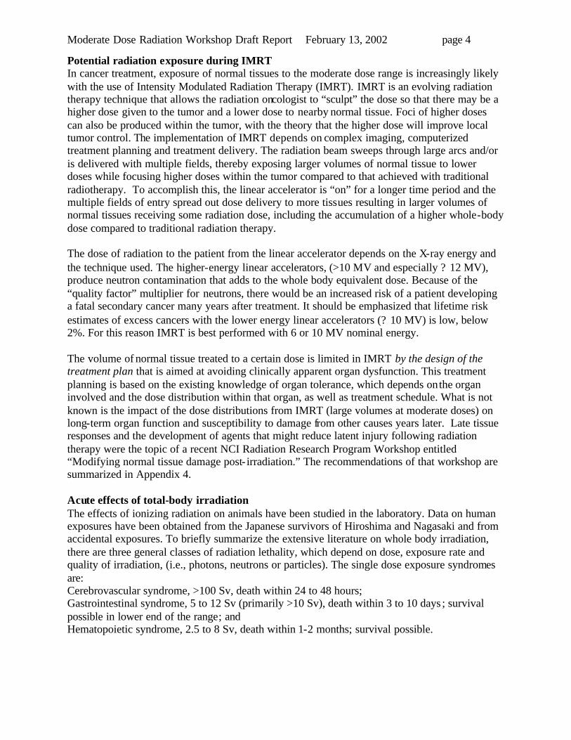

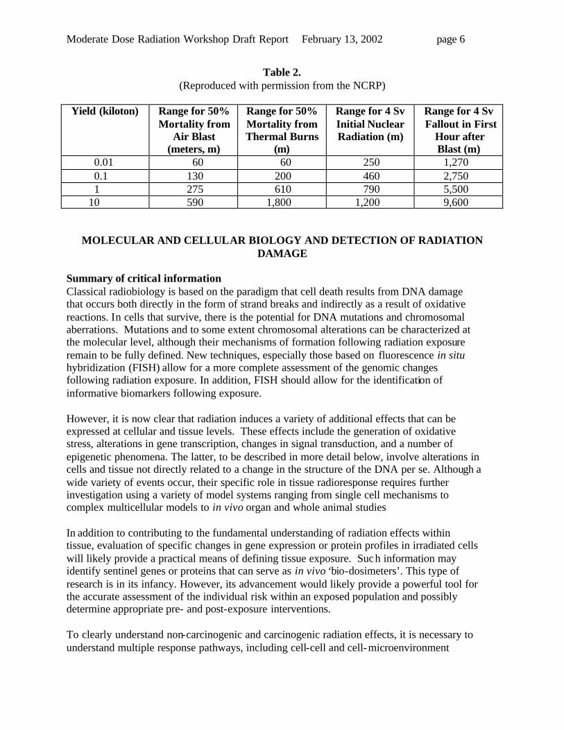


![Case Report Primary MALT Lymphoma of the Breast Treated ... · radiation therapy, such as involved eld radiation therapy, and a moderate dose of Gy are recommended [,]. Treatment](https://static.documents.pub/doc/80x56/6136877c0ad5d20676481606/case-report-primary-malt-lymphoma-of-the-breast-treated-radiation-therapy-such.jpg)


