Published: March 16, 2011 r2011 American Chemical Society 5346 dx.doi.org/10.1021/ja1097379 | J. Am. Chem. Soc. 2011, 133, 5346–5356 ARTICLE pubs.acs.org/JACS Modulation of the Photocycle of a LOV Domain Photoreceptor by the Hydrogen-Bonding Network Sarah Raffelberg, † Madina Mansurova, † Wolfgang G € artner, † and Aba Losi* ,†,§ † Max-Planck-Institute for Bioinorganic Chemistry, Stiftstrasse 34-36, 45470 M€ ulheim, Germany § Department of Physics, University of Parma, viale G.P. Usberti 7/A, 43100 Parma, Italy b S Supporting Information ’ INTRODUCTION The photocycle of LOV (light, oxygen, voltage) protein domains is chiefly dictated by the photophysical and photoche- mical properties of the bound flavin mononucleotide (FMN) chromophore 1 but also influenced by the surrounding microenvironment. 2,3 Weak interactions, such as hydrogen- bonding (HB), and spatial constraints imposed by the side chains of amino acids forming the FMN binding cavity are expected to affect the kinetics, the energetics, and the efficiency of the different steps of the photocycle. In this respect the hydrogen bonds formed around the polar groups of the FMN isoalloxazine could be of primary importance, besides being a major determinant in chromophore stabilization and the first proteinchromophore interface undergoing conformational changes upon light activation. 48 LOV domains are small photosensing protein modules of ca. 100 amino acids forming a quite compact and structurally conserved R/β core flanked by variable N- and C-terminal helical regions. 911 They host FMN as a non-covalently bound chro- mophore and absorb maximally around 450 nm (resting or dark- adapted state, referred to as LOV 447 ), and they constitute a subclass of the PAS (PerArntSim) superfamily. 12 According to a recently proposed nomenclature, 13 we assign the following secondary structure elements to the LOV core (from the N-ter- minal part): AβBβCRDRERFRGβHβI. LOV domains are the photochemically active moiety of LOV proteins, a class of blue- light (BL) photoreceptors comprising, among others, plant phototropins (phot), 9 the fungal protein VIVID, 14 and a growing number of prokaryotic photoresponsive proteins. 11,1521 The photochemistry of LOV domains, first elucidated for phot and afterward for a variety of bacterial and fungal proteins, involves the formation of a FMNcysteine C(4a)thiol adduct, significantly blue-shifted with respect to the dark state and non-fluorescent (referred to as LOV 390 ), generated via the short microsecond decay of the FMN triplet state. 1,22,23 In LOV 390 , a covalent bond is formed between the carbon atom at position 4a and the thiol group of a conserved cysteine localized in the DRER loop (Figure 1 provides structural views of the chromophore and its protein surroundings for both the parent state and the photo- adduct; amino acid numbering herein refers to YtvA from Bacillus subtilis). LOV 390 reverts in the dark to the unphotolyzed state (LOV 447 ) on a time scale ranging from a few seconds to many hours at room temperature. 1,2427 Importantly, the recovery is accelerated by imidazole, suggesting a base-catalyzed reaction. 28,29 Furthermore, pH, 23,29 deuterium effects, 22,30 and structural studies indicate that a proton-transfer reaction from N(5) is the rate-limiting step for the dark recovery reaction. Received: November 12, 2010 ABSTRACT: An extended hydrogen-bonding (HB) network stabilizes the isoalloxazine ring of the flavin mononucleotide (FMN) chromophore within the photosensing LOV domain of blue-light protein receptors, via interactions between the C- (2)dO, N(3)H, C(4)dO, and N(5) groups and conserved glutamine and asparagine residues. In this work we studied the influence of the HB network on the efficiency, kinetics, and energetics of a LOV protein photocycle, involving the reversible formation of a FMNcysteine covalent adduct. The following results were found for mutations of the conserved amino acids N94, N104, and Q123 in the Bacillus subtilis LOV protein YtvA: (i) Increased (N104D, N94D) or strongly reduced (N94A) rate of adduct formation; this latter mutation extends the lifetime of the flavin triplet state, i.e., adduct formation, more than 60-fold, from 2 μs for the wild-type (WT) protein to 129 μs. (ii) Acceleration of the overall photocycle for N94S, N94A, and Q123N, with recovery lifetimes 20, 45, and 85 times faster than for YtvA-WT, respectively. (iii) Slight modifications of FMN spectral features, correlated with the polarization of low-energy transitions. (iv) Strongly reduced (N94S) or suppressed (Q123N) structural volume changes accompanying adduct formation, as determined by optoacoustic spectroscopy. (v) Minor effects on the quantum yield, with the exception of a considerable reduction for Q123N, i.e., 0.22 vs 0.49 for YtvA-WT. The data stress the importance of the HB network in modulating the photocycle of LOV domains, while at the same time establishing a link with functional responses.
Transcript
Published: March 16, 2011
r 2011 American Chemical Society 5346 dx.doi.org/10.1021/ja1097379 | J. Am. Chem. Soc. 2011, 133, 5346–5356
ARTICLE
pubs.acs.org/JACS
Modulation of the Photocycle of a LOV Domain Photoreceptor by theHydrogen-Bonding NetworkSarah Raffelberg,† Madina Mansurova,† Wolfgang G€artner,† and Aba Losi*,†,§
†Max-Planck-Institute for Bioinorganic Chemistry, Stiftstrasse 34-36, 45470 M€ulheim, Germany§Department of Physics, University of Parma, viale G.P. Usberti 7/A, 43100 Parma, Italy
bS Supporting Information
’ INTRODUCTION
The photocycle of LOV (light, oxygen, voltage) proteindomains is chiefly dictated by the photophysical and photoche-mical properties of the bound flavin mononucleotide (FMN)chromophore1 but also influenced by the surroundingmicroenvironment.2,3 Weak interactions, such as hydrogen-bonding (HB), and spatial constraints imposed by the sidechains of amino acids forming the FMN binding cavity areexpected to affect the kinetics, the energetics, and the efficiencyof the different steps of the photocycle. In this respect thehydrogen bonds formed around the polar groups of the FMNisoalloxazine could be of primary importance, besides being amajor determinant in chromophore stabilization and the firstprotein�chromophore interface undergoing conformationalchanges upon light activation.4�8
LOV domains are small photosensing protein modules of ca.100 amino acids forming a quite compact and structurallyconserved R/β core flanked by variable N- and C-terminal helicalregions.9�11 They host FMN as a non-covalently bound chro-mophore and absorb maximally around 450 nm (resting or dark-adapted state, referred to as LOV447), and they constitute asubclass of the PAS (PerArntSim) superfamily.12 According to arecently proposed nomenclature,13 we assign the followingsecondary structure elements to the LOV core (from the N-ter-minal part): AβBβCRDRERFRGβHβI. LOV domains are the
photochemically active moiety of LOV proteins, a class of blue-light (BL) photoreceptors comprising, among others, plantphototropins (phot),9 the fungal protein VIVID,14 and a growingnumber of prokaryotic photoresponsive proteins.11,15�21 Thephotochemistry of LOV domains, first elucidated for phot andafterward for a variety of bacterial and fungal proteins, involves theformation of a FMN�cysteine C(4a)�thiol adduct, significantlyblue-shifted with respect to the dark state and non-fluorescent(referred to as LOV390), generated via the short microseconddecay of the FMN triplet state.1,22,23 In LOV390, a covalent bond isformed between the carbon atom at position 4a and the thiolgroup of a conserved cysteine localized in the DR�ER loop(Figure 1 provides structural views of the chromophore and itsprotein surroundings for both the parent state and the photo-adduct; amino acid numbering herein refers to YtvA from Bacillussubtilis). LOV390 reverts in the dark to the unphotolyzed state(LOV447) on a time scale ranging from a few seconds to manyhours at room temperature.1,24�27 Importantly, the recoveryis accelerated by imidazole, suggesting a base-catalyzedreaction.28,29 Furthermore, pH,23,29 deuterium effects,22,30 andstructural studies indicate that a proton-transfer reaction fromN(5) is the rate-limiting step for the dark recovery reaction.
Received: November 12, 2010
ABSTRACT: An extended hydrogen-bonding (HB) networkstabilizes the isoalloxazine ring of the flavin mononucleotide(FMN) chromophore within the photosensing LOV domain ofblue-light protein receptors, via interactions between the C-(2)dO, N(3)H, C(4)dO, and N(5) groups and conservedglutamine and asparagine residues. In this work we studied theinfluence of the HB network on the efficiency, kinetics, andenergetics of a LOV protein photocycle, involving the reversibleformation of a FMN�cysteine covalent adduct. The following results were found for mutations of the conserved amino acids N94,N104, andQ123 in the Bacillus subtilis LOVprotein YtvA: (i) Increased (N104D, N94D) or strongly reduced (N94A) rate of adductformation; this latter mutation extends the lifetime of the flavin triplet state, i.e., adduct formation, more than 60-fold, from 2 μs forthe wild-type (WT) protein to 129 μs. (ii) Acceleration of the overall photocycle for N94S, N94A, and Q123N, with recoverylifetimes 20, 45, and 85 times faster than for YtvA-WT, respectively. (iii) Slight modifications of FMN spectral features, correlatedwith the polarization of low-energy transitions. (iv) Strongly reduced (N94S) or suppressed (Q123N) structural volume changesaccompanying adduct formation, as determined by optoacoustic spectroscopy. (v) Minor effects on the quantum yield, with theexception of a considerable reduction for Q123N, i.e., 0.22 vs 0.49 for YtvA-WT. The data stress the importance of the HB networkin modulating the photocycle of LOV domains, while at the same time establishing a link with functional responses.
Nevertheless, despite this well-established photocycle, the detailsfor both LOV390 formation from the triplet state and its reversionare still a matter of debate,31�33 as is the molecular basis of thedramatically different photocycle kinetics in different proteins. Inthis respect, understanding how the microenvironment sur-rounding the chromophore influences the photocycle, i.e., theeffects of protein�chromophore interactions, is of crucial im-portance. Furthermore, modulation of the photocycle might turnout to be a very important aspect for BL photoreceptor-basedoptogenetic applications, by tuning the kinetics and yielding therequirements of the particular cell system and/or metabolicprocess of interest.34�37
Recently, a series of mutations have been designed to altersteric restrictions in the vicinity of the 8R-methyl group at thexylene side of FMN in Avena sativa phot1-LOV2 domain ofphototropin 1 (Asphot1-LOV2) and studied by means ofproton ENDOR and optical spectroscopy.3 Asn 425, localizedwithin the Aβ�Bβ loop, appears to have a significant influenceon the recovery lifetime of the adduct, which decreased from 48s in the wild-type (WT) protein to 7.5 s in Asphot1-LOV2-N425C. Interaction between Asn425 and the 8R-methyl groupappears to force the isoalloxazine ring into a precise conforma-tion, thus stabilizing the cysteinyl�C(4a) adduct. Anotherimportant residue influencing the kinetics of the photocyclethat stabilize the photoadduct through steric effects is Ile427(I39 in YtvA) on strand Bβ of Asphot1-LOV2.2 Nevertheless, inthat case the 2-fold slower formation and 10-fold faster decay ofthe photoadduct by removing the δ-carbon of the isoleucinechain (I427V mutation) are not due to direct interaction ofIle427 with FMN but rely on van der Waals contact between aCH2 group and the nearby sulfur of the reactive cysteine. Thecorresponding substitution I403V has been recently used toachieve a very fast photocycle in Arabidopsis thaliana (At)phot1-LOV2, in order to productively allow Fourier transforminfrared (FTIR) step-scan measurements of chromophore andprotein dynamics during formation of the photoadduct.32
Further, mutations of residues interacting with the ribityl chainof FMN have been shown to affect the recovery reaction38,39 byas much as 2 orders of magnitude.27 Recently, a doublemutation of YtvA, I39V/F46H, was demonstrated to accelerate75-fold the recovery reaction, although the two residues are notdirectly interacting with the chromophore.34 In particular, F46undergoes a light-induced flipping, suggesting that this regionof the protein is particularly flexible.7,34
An alternative approach has been recently reported by Man-surova et al., who exchanged the native chromophore withsterically modified flavins as chromophores via a chromophoreexchange protocol.40 Also in those experiments, the tight inter-actions between the chromophore and the protein were docu-mented as a change of this interplay that significantly alters thethermal recovery kinetics.40
Here we focus instead on the HB network between the N(5),C(4)dO, N(3)H, and C(2)dO positions of the isoalloxazinemoiety of FMN and an ensemble of conserved polar, unchargedamino acids (Figure 1). The hydrogen bonds at these positions ofthe chromophore undergo changes after blue-light absorption, aswas recently documented in detail by means of advanced infraredspectroscopy techniques.8,31,32,43 In particular, hydrogen bondsat C(2)dO and C(4)dO are weakened upon formation of theadduct, to somewhat different extents in different LOVsystems.8,44�46 In YtvA, the B. subtilis LOV protein investigatedhere, the three involved amino acids are Asn94, Asn104, andGln123 (Figure 1; in the following we will refer to the amino acidcoding of YtvA from B. subtilis).
Our study was prompted by formerly reported observationsthat hydrogen bonds affect not only the UV�vis absorptionspectrum of riboflavin but also its photoreactivity.47,48 In parti-cular, H-bonding at N(1), C(2)dO, N(3)H, and C(4)dO wasshown to increase the rate of hydrogen abstraction from a donorsubstrate, thus facilitating flavin reduction, when the riboflavinderivative was converted to its triplet state.48 A theoreticalinvestigation, based on quantum mechanics/molecular me-chanics (QM/MM) methods applied to lumiflavin, has contrib-uted to our knowledge of the influence that the HB network
Figure 1. Hydrogen-bonding network of YtvA-LOV investigated in thiswork, in the (top) dark-adapted and (bottom) light-activated(photoadduct) states (PDB coordinates from 2pr5 and 2pr6). For thedark state we show the calculated polarization directions of the low-energy electronic transitions: I, blue-light range, λmax∼450 nm; II, UVArange, λmax ∼350 nm (modified from ref 41, but see also ref 42 for anexperimental determination). The atoms of FMN involved in the HBnetwork studied here are shown in yellow.
exerts on the low-lying excited state of the flavin cofactor,indicating that the protein microenvironment enables participa-tion of the (nπ*) states in the decay processes of the lowest(ππ*) excited singlet state (transition I in Figure 1), i.e., byenhancing singlet�triplet spin�orbit coupling.49 Mutations ofthe three conserved polar residues that chiefly contribute to theHB network within the FMNbinding pocket are thus expected toaffect the photocycle kinetics, efficiency, and/or energetics.Indeed, it was shown recently that mutation of Q513 inAsphot1-LOV2 (Q123 in YtvA) can strongly affect the recoveryreaction kinetics.50 Furthermore, N94, N104, and Q123 belongto the β-scaffold of the LOV core (strands Gβ, Hβ, and Iβ,respectively), participating in light-to-signal transmission, prob-ably with the intervention of helical regions flanking the LOVcore.7,10,32,39,51,52 In particular, the conserved glutamine onstrand Iβ (here Q123) was recently identified as a potentialswitch to convey light-triggered conformational changes fromthe chromophore cavity to the LOV domain surface, e.g., Q575in Atphot1-LOV2,53 Q513 in Asphot1-LOV2,50 Q182 in thefungal protein VIVID,51 and Q1029 in Adiantum capillus venerisAcphy3-LOV2.43 Themechanism underlying the conformationalchange effects resides in the fact that formation of the thioetherbond in the adduct reduces the flavin ring and protonates N(5).31
In order tomaintainHBwithN(5)H, this requires that the lateralchain of Q123 must flip in the lit state, as suggested by thestructural data of several LOV domains.4,5,7,10,51 Time-resolvedFTIR spectroscopy has demonstrated that the Gln flipping, withloss of HB, is completed within a fewmicroseconds, concomitantwith formation of the adduct.32 This flipping is then reversed,with recovery of the dark state and breakage of the covalentFMN�Cys bond. A link between this glutamine-based confor-mational switch and in vivo effects is indicated by the lack of lightactivation of the stress factor σB with the mutated YtvA-Q123Nin B. subtilis.54 Furthermore, it has been reported that themutation Q575L (on LOV2) attenuates light-induced self-phos-phorylation in Atphot1.55 However, similar functional investiga-tions have not been carried out for the positions (in YtvA) Asn94and Asn104.
In this work we have investigated a series of mutations atQ123, N94, and N104 with the aim of characterizing thekinetics, efficiency, and energetics of the photocycle of YtvAproteins with an altered HB network around FMN, by means ofsteady-state spectroscopy, nanosecond flash photolysis, andtime-resolved calorimetry. The aim of our investigation wastwofold: first to assess the direct influence of the H-bondingsituation of the chromophore on the photochemical andthermally driven steps, and second to understand the linkbetween chromophore�protein interactions and signal propa-gations from the LOV core to domain/protein partners in thesignal transduction chain. The outcome of our investigation willalso be discussed in the frame of recently published experi-mental data on the mechanism of adduct formation and decay,and of possible extensions of the designed mutations forbiotechnology applications.
’MATERIALS AND METHODS
Mutagenesis, Protein Expression, and Purification. A totalof seven new mutations were generated: Q123N, N94S, N94D,N94A, N104S, N104D, and N104A. Mutagenesis was performed bythe QuikChange method (QuikChange II XL, Stratagene). Theprimer design was performed with PrimerX56 using the option
“DNA-based”. The primer sequences suggested by this programwere again inspected individually, before primers were ordered formutagenesis (see Supporting Information for the primersequencees). For all mutagenesis experiments, the obtained PCRproducts were treated with the restriction enzyme DpnI (NewEngland BioLabs). DpnI is specific for methylated and hemimethy-lated DNA (targeting sequence: 50-Gm6ATC-30) and is used to digestthe parental DNA template in the PCR products. A 0.5 μL portion ofDpnI (20 000U/mL) was added to each PCR product, and the digestionreaction was carried out at 37 �C for 30 min. In all cases, mutations wereconfirmed by sequencing.
Expression in E. coli (BL21) (Stratagene, Amsterdam, TheNetherlands) yielded His-tagged, mutated proteins, using IPTG(BioMol, Hamburg, Germany) induction and employing the pET28aplasmid (Novagen-Merck, Darmstadt, Germany), as described.57 Theproteins were purified by affinity chromatography on a Talon resin(Qiagen, Hilden, Germany) and finally concentrated in sodium phos-phate buffer 10 mM, NaCl 10 mM, pH = 8.Steady-State and Transient Optical Spectroscopy. Absor-
bance spectra were recorded with a Jasco 7850 UV/vis spectrophot-ometer. Steady-state fluorescence measurements were carried out with aPerkin-Elmer LS50 luminescence spectrometer. The output signal wasdivided by the fraction of absorbed energy (1�10�A, where A is theabsorbance at the excitation wavelength) in order to obtain a signal thatis proportional to the quantum yield.
Transient absorbance changes after nanosecond laser flash excitationwere recorded using an LFP111 detection system (Luzchem, Ontario,Canada). For excitation, a Nd:YAG-driven tunable OPO laser was used(Nd:YAG, Innolas, Garching, Germany; OPO, GWU Lasertechnik,Erftstadt, Germany). The single-shot experiments were performed inthe linear laser-energy dependence region of the transient absorbancechanges with λexc = 450 nm. All measurements were done at 20 �C using1 cm light-path quartz cuvettes. The data were handled and analyzedusing Origin Professional version 5.0 (Microcal Software, Inc., North-ampton, MA).
Arrhenius and Eyring plots for the dark recovery reaction of thephotoadduct were built by recording the recovery of FMN fluorescence(λex = 303 nm, λem = 500 nm). The kinetics traces were fitted with amono- or bi-exponential function, furnishing the recovery lifetime (τrec)as a function of temperature, in the range of the protein stability(11�25 �C). Excitation was at 303 nm, in order to minimize secondaryphotochemistry leading to the formation of the photoproduct duringrecording of the traces.39
Laser-InducedOptoacoustic Spectroscopy (LIOAS). For theLIOAS experiments, excitation at 450 nm was achieved by pumping thefrequency-tripled pulse of a Nd:YAG laser (SL 456G, 6-ns pulseduration, 355 nm, Spectron Laser System, Rugby, Great Britain) intoa β-barium borate optical parametric oscillator (OPO-C-355, bandwidth420�515 nm, GWU Lasertechnik, Erftstadt, Germany) as previouslydescribed.58,59 The FLASH 100 cuvette holder (Quantum NorthwestInc., Spokane,WA) was temperature-controlled to(0.02 �C. The signalwas detected by a V103-RM ultrasonic transducer and fed into a 5662preamplifier (Panametrics Inc., Waltham, MA). The pulse fluence wasvaried with a neutral density filter and measured with a pyroelectricenergy meter (RJP735 head connected to an RJ7620 meter from LaserPrecision Corp.). The beam was shaped by a 1� 12 mm slit, allowing atime resolution of ∼60 ns by using deconvolution techniques.60 Theexperiments were performed in the linear regime of amplitude versuslaser fluence, and the total incident energy normally used was ∼40 μJ/pulse (corresponding to 15 � 10�11 einstein for 450 nm excitation,photon energy 265.8 kJ/mol). The sample concentration was about 15μM, giving 1.8� 10�9 mol in the excitation volumeV0 = 0.12mL. Theseconditions correspond to a ratio of 0.08 photon per protein molecule.New coccine (Fluka, Neu-Ulm, Germany) was used as calorimetric
reference.61 The time evolution of the pressure wave was assumed to bea sum of monoexponential functions. The deconvolution analysisyielded the fractional amplitudes (ji) and the lifetimes (τi) of thetransients (Sound Analysis 3000, Quantum Northwest Inc.). The timewindowwas between 20 ns and 5 μs. At a given temperature and for eachresolved ith step, the fractional amplitudeji is the sum of the fraction ofabsorbed energy released as heat (Ri) and the structural volume changeper absorbed einstein (ΔVi), according to eq 1:62,63
ji ¼ Ri þΔVi
Eλ
cpFβ
ð1Þ
where Eλ is themolar excitation energy, β = (∂V/∂T)pΔV is the volumeexpansion coefficient, cp is the heat capacity at constant pressure, and Fis the mass density of the solvent. In this work we used the so-called“two temperature” method in order to separate Ri from ΔVi.
64 Thesample waveform was acquired at a temperature for which heattransport is zero, Tβ=0 = 3.2 �C, and at a slightly higher temperature,Tβ>0 = 10 �C. At Tβ=0, the LIOAS signal is only due to ΔVi. Thereference for deconvolution was recorded at Tβ>0, and eqs 2a and 2bwere then used to derive Ri and ΔVi:
ΦiΔVi ¼ jijTβ¼ 0� Eλ
β
cpF
�����Tβ > 0
ð2aÞ
Ri ¼ jijTβ > 0�jijTβ¼ 0
ð2bÞ
Photophysical Parameters and LIOAS Data Handling. TheLIOAS signals for YtvA-WT and the mutated proteins were best fitted(with some exceptions, vide infra) by a two-exponential decay functionas previously described.57,59 The unresolved step (τ1 < 20 ns) is assignedto the fast reactions resulting in the formation of the flavin triplet state(subscript T; this step is not time-resolved by LIOAS). Themicrosecondprocess (τ2 = 2 μs for YtvA-WT) corresponds to the triplet decay withformation of the photoadduct (subscript 390, according to the approx-imate absorption maximum). Energy balance considerations and theresults of deconvolution directly provide the products ΦTET andΦ390E390 (eqs 3a and 3b), referring to the quantum yield of formationfor the triplet state and adduct, respectively, multiplied by the energylevel of the two transient species:65
ΦTETEλ
¼ 1� R1 �ΦFEFEλ
ð3aÞ
R2 ¼ ΦTETEλ
�Φ390E390Eλ
ð3bÞ
where EF is the average energy for the fluorescence emission (232 kJ/mol), and Eλ (265.8 kJ/mol) is the photonic energy corresponding toλex = 450 nm excitation wavelength. In this work we used ET ≈ 200 kJ/mol, as previously measured38,57,66 to obtain ΦT, and we estimated theenergy content of the adduct after the independent determination ofΦ390 (vide infra).
65
The molecular volume changes that the system suffers upon forma-tion of the flavin triplet state and of the photoadduct (with respect to theunphotolyzed state) are calculated with eqs 4a and 4b:
ΔVT ¼ ΔV1
ΦTð4aÞ
ΔV390 ¼ ΔVT þ ΔV2
Φ390ð4bÞ
The fluorescence quantum yield, ΦF of the bound flavin for themutated proteins was measured at 20 �C, by comparison with YtvA-WT
(ΦF = 0.22) and FMN (ΦF = 0.26), employing steady-statespectroscopy.57,59,67 The value of Φ390 was estimated by recordingthe bleaching of the unphotolyzed state on the microsecond time scale,at 450 and 475 nm, after laser excitation at 450 nm, employing the laserflash photolysis instrumentation, and by comparison with YtvA-WT(Φ390 = 0.49),
57 assuming that the absorption coefficient of the mutatedproteins remains unaltered.
’RESULTS
Spectral Features and Light�Dark Difference Spectra. Allthe mutated proteins exhibit absorption spectra similar to that ofYtvA-WT. The three major bands (blue, UVA, and UVB regions,transitions I, II, and III, respectively) undergo large light-inducedchanges upon formation of the photoadduct, well described bythe light�dark (L-D) difference spectra, with a defined pattern ofmaxima and minima (Figure 2, Table 1).The largest effects on the absorption spectra are produced by
the Asn/Asp exchange at positions 94 and 104, and by theQ123N mutation (Figure 2 and Table 1). Replacing asparagine104 with aspartate has an effect only on transition I (Figure 2a),inducing a blue-shift. In contrast, in YtvA-N94D a small blue-shiftof transition I is accompanied by a larger red-shift of the UVAtransition II (Figure 2b), accordingly polarized in the direction ofC(2)dO and N(3)H. This UVA transition is in fact the mostsensitive to the polarity and HB ability of the solvent47,48,68 andshows the greatest variation among various LOV domains.46,69
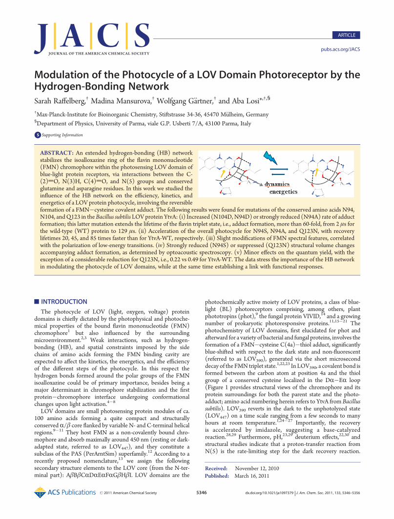
Interestingly, YtvA-Q123N shows a 3 nm blue-shift of the lowestenergy transition with respect to YtvA-WT, whereas its fluores-cence spectrum is virtually unaffected (Figure 3). A similarfeature was recently described for mutations of glutamate 46 inthe photoactive yellow protein and was predicted to arise from abroadening of the excited-state energy surface, although in ourcase the effects are very small.70
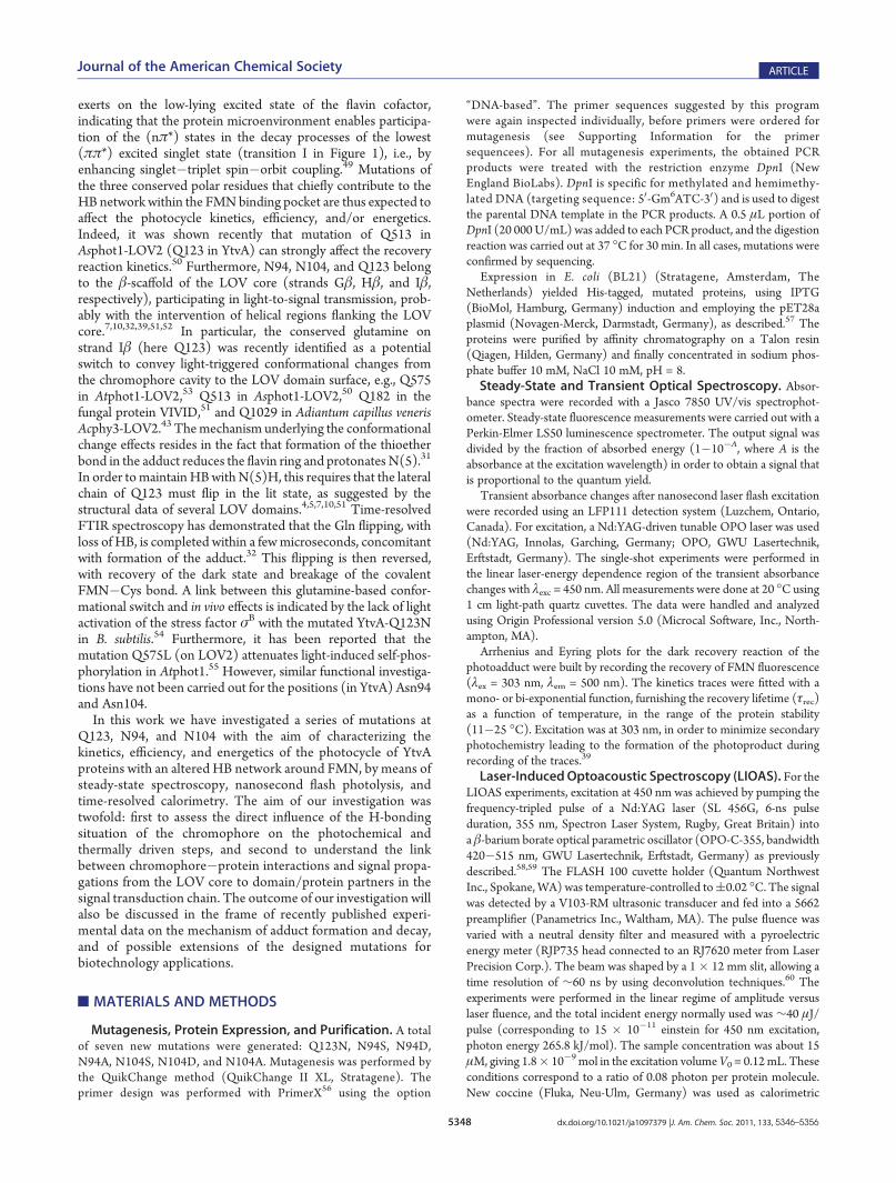
Photochemical Events: Triplet Decay and Formation ofthePhotoadduct.Themost notable features of themicro-secondtime-resolved transient absorbance measurements (flash photolysis)are summarized in Table 2: (i) the faster formation of the adduct/decay of triplet state observed for YtvA-N94D, YtvA-N104D, and, toa lesser extent, also for YtvA-Q123N; (ii) the remarkable slowingdown of the triplet decay/photoproduct formation in YtvA-N94A (Figure 4); and (iii) the smaller Φ390 for YtvA-Q123N(about half that for YtvA-WT).The data curves could be readily interpolated by means of a
mono-exponential decay (Table 2), with the exception of YtvA-N104A, for which the curve was better described by a bi-exponential decay (i.e., the χ2 improved more than 10% withrespect to a mono-exponential fitting).Photocalorimetric (LIOAS) Experiments: Triplet Forma-
tion and Decay, Energetics, and Structural Changes. LIOASexperiments have been previously performed with YtvA-WT57,59
and other LOV proteins.38 Given the time scale and timeresolution of LIOAS, the production of FMN triplet state andits decay into the adduct are readily recorded, but the formerprocess cannot be time-resolved (τ1 < 20 ns), and we see only theresult of the sub-nanosecond reactions leading to triplet forma-tion. The formation of the adduct can instead be followed in realtime, provided that it occurs with a lifetime τ2 e 5 μs (Table 3).Themajority of themutated proteins behave similarly to YtvA-
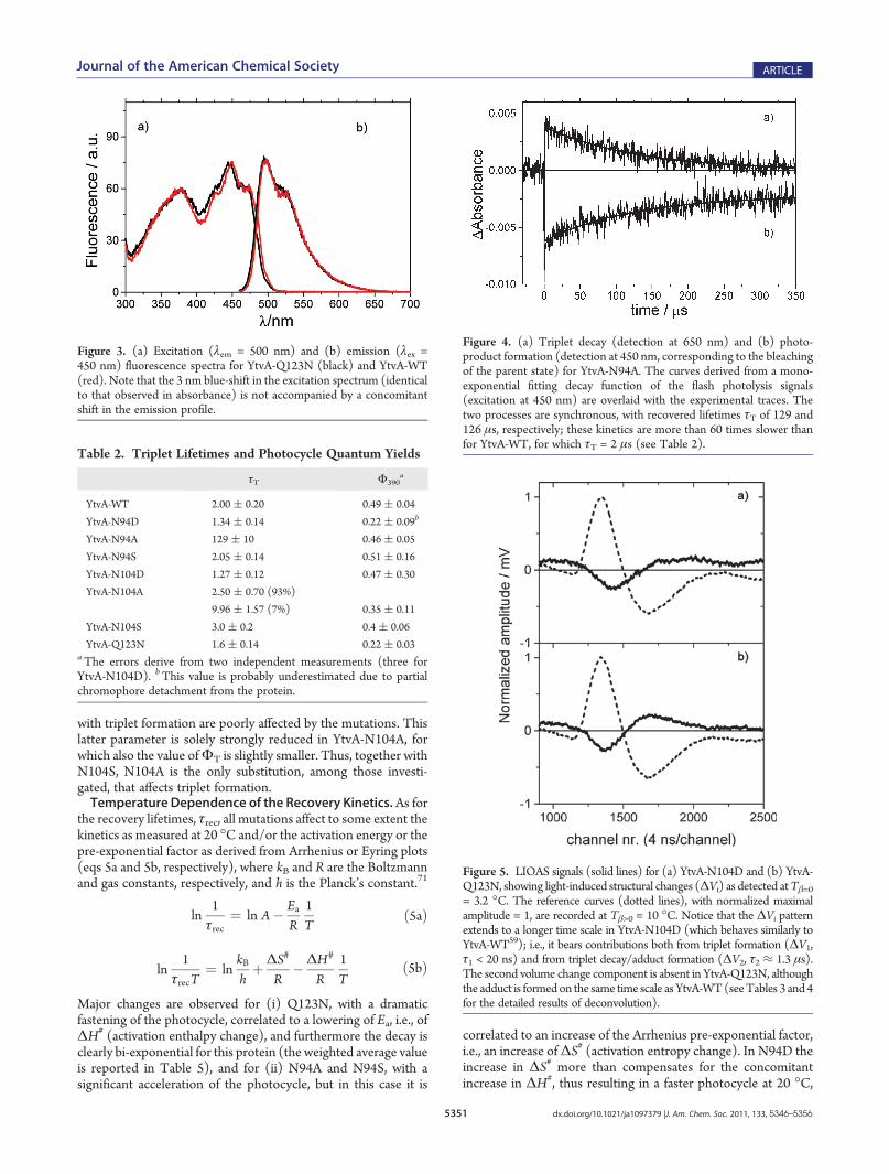
WT, with some notable exceptions: (i) YtvA-Q123N does notshow any structural volume change upon formation of theadduct, i.e., ΔV2 = 0, although the heat released in this step is
well detectable with the correct time constant (Table 3 andFigure 5); (ii) ΔV2 is strongly reduced in YtvA-N94S; and (iii)the formation of the adduct cannot be detected for YtvA-N94A,in agreement with the long lifetime of the triplet state (seeTable 2).The deconvolution of LIOAS waveforms thus provides the
fraction of heat released (Ri) in each step and the lifetime of thetriplet states; i.e., τ2 in Table 3 should be the same as τT asrecorded from transient absorption experiments (Table 2),provided that spectrally silent transitions do not occur on thesame time scale. By performing temperature-dependent experi-ments, it is also possible to obtain the structural volume changes(ΔVi, per mole of absorbed photons) that accompany each step(Table 3; eqs 2a, 2b, 3a, and 3b).Equation 3a allows us to determine the triplet formation
quantum yield,ΦT, which is very similar among all the proteins
investigated (Table 4), with the exception of YtvA-N104S, whichhas a higher value,ΦT = 0.79, underscored by the small fractionof released heat in the short nanosecond step (R1 in Table 3).The evaluation of E390 as the energy content of the photo-
adduct is negatively affected by the large errors associated insome cases with both Φ390 as determined by flash photolysis(Table 2) and the quantity E390Φ390, independently determinedby LIOAS. In at least two cases, YtvA-N104S and YtvA-Q123N,the energy stored in the signaling state appears larger with respectto YtvA-WT, i.e., close to the triplet energy level,ET≈ ca. 200 kJ/mol.38,57,66
The total contraction, ΔV390, that the system suffers uponlight activation (Table 4) is strongly reduced in YtvA-Q123N andYtvA-N94S. This is due to the fact that the volume changeassociated with the formation of the photoadduct is zero or closeto zero, respectively, whereas the structural changes associated
Table 1. Absorption Light�Dark (L-D) Maxima and Minima and Fluorescence Parameters of YtvA Proteins
YtvA-Q123N 273/350;373/447;473 396;300 496 0.27aTransitions in the UVB, UVA, and blue light spectral regions are separated by “/”; shoulders and vibrational bands in the same spectral region areseparated by “;”.
Figure 2. Light�dark difference absorption spectra for selected mutated YtvA proteins (black lines), compared to YtvA-WT (red line): (a) YtvA-N104D, (b) YtvA-N94D, (c) YtvA-Q123N, and (d) YtvA-N104S. See Table 1 for the maxima�minima patterns of all studied proteins.
with triplet formation are poorly affected by the mutations. Thislatter parameter is solely strongly reduced in YtvA-N104A, forwhich also the value ofΦT is slightly smaller. Thus, together withN104S, N104A is the only substitution, among those investi-gated, that affects triplet formation.Temperature Dependence of the Recovery Kinetics.As for
the recovery lifetimes, τrec, all mutations affect to some extent thekinetics as measured at 20 �C and/or the activation energy or thepre-exponential factor as derived from Arrhenius or Eyring plots(eqs 5a and 5b, respectively), where kB and R are the Boltzmannand gas constants, respectively, and h is the Planck’s constant.71
ln1τrec
¼ ln A� EaR
1T
ð5aÞ
ln1
τrecT¼ ln
kBhþΔS#
R�ΔH#
R1T
ð5bÞ
Major changes are observed for (i) Q123N, with a dramaticfastening of the photocycle, correlated to a lowering of Ea, i.e., ofΔH# (activation enthalpy change), and furthermore the decay isclearly bi-exponential for this protein (the weighted average valueis reported in Table 5), and for (ii) N94A and N94S, with asignificant acceleration of the photocycle, but in this case it is
correlated to an increase of the Arrhenius pre-exponential factor,i.e., an increase ofΔS# (activation entropy change). In N94D theincrease in ΔS# more than compensates for the concomitantincrease in ΔH#, thus resulting in a faster photocycle at 20 �C,
Table 2. Triplet Lifetimes and Photocycle Quantum Yields
τT Φ390a
YtvA-WT 2.00 ( 0.20 0.49 ( 0.04
YtvA-N94D 1.34 ( 0.14 0.22 ( 0.09b
YtvA-N94A 129 ( 10 0.46 ( 0.05
YtvA-N94S 2.05 ( 0.14 0.51 ( 0.16
YtvA-N104D 1.27 ( 0.12 0.47 ( 0.30
YtvA-N104A 2.50 ( 0.70 (93%)
9.96 ( 1.57 (7%) 0.35 ( 0.11
YtvA-N104S 3.0 ( 0.2 0.4 ( 0.06
YtvA-Q123N 1.6 ( 0.14 0.22 ( 0.03aThe errors derive from two independent measurements (three forYtvA-N104D). bThis value is probably underestimated due to partialchromophore detachment from the protein.
Figure 4. (a) Triplet decay (detection at 650 nm) and (b) photo-product formation (detection at 450 nm, corresponding to the bleachingof the parent state) for YtvA-N94A. The curves derived from a mono-exponential fitting decay function of the flash photolysis signals(excitation at 450 nm) are overlaid with the experimental traces. Thetwo processes are synchronous, with recovered lifetimes τT of 129 and126 μs, respectively; these kinetics are more than 60 times slower thanfor YtvA-WT, for which τT = 2 μs (see Table 2).
Figure 3. (a) Excitation (λem = 500 nm) and (b) emission (λex =450 nm) fluorescence spectra for YtvA-Q123N (black) and YtvA-WT(red). Note that the 3 nm blue-shift in the excitation spectrum (identicalto that observed in absorbance) is not accompanied by a concomitantshift in the emission profile.
Figure 5. LIOAS signals (solid lines) for (a) YtvA-N104D and (b) YtvA-Q123N, showing light-induced structural changes (ΔVi) as detected atTβ=0= 3.2 �C. The reference curves (dotted lines), with normalized maximalamplitude = 1, are recorded at Tβ>0 = 10 �C. Notice that the ΔVi patternextends to a longer time scale in YtvA-N104D (which behaves similarly toYtvA-WT59); i.e., it bears contributions both from triplet formation (ΔV1,τ1 < 20 ns) and from triplet decay/adduct formation (ΔV2, τ2 ≈ 1.3 μs).The second volume change component is absent in YtvA-Q123N, althoughthe adduct is formedon the same time scale asYtvA-WT(seeTables 3 and 4for the detailed results of deconvolution).
albeit not so dramatic as in N94A and N94S. Mutations of N104(see Figure 1 for the HB network centered on this residue) resultin less profound effects on the overall kinetics of the photocycle,but they still considerably lower the activation energy (N/A andN/S changes).
’DISCUSSION
Switching Glutamine on Strand Iβ and N(5) of the FlavinRing.The strong acceleration of the photocycle in YtvA-Q123N,which recovers with a τrec about 85 times faster than YtvA-WT(at 20 �C), parallels the 1.8 times acceleration observed for thecorresponding Q513N mutation in Asphot1-LOV2,50 althoughfor YtvA the effect is significantly more pronounced. We cantherefore safely state that this substitution destabilizes thecovalent adduct. This is reflected in the high energy content of
the photoadduct for YtvA-Q123N (Table 4) and in the loweringof the activation energy for the recovery reaction (Table 5,Figure 6), accounting for the fast photocycle at room tempera-ture. The decay of the adduct, clearly bi-exponential in thismutated protein, also points to a conformational heterogeneity inthe light-activated state. Interestingly, Alexandre and co-workersdetected a mixed one-/two-HB arrangement at C(4)dO in thedark state of Asphot-LOV2, indicative of a conformationalflexibility in this protein region.8 The asparagine substitutioncould enhance this flexibility, which becomes clearly evident inthe double-exponential decay. This mutation also affects theforward photochemistry, although to a minor extent, by accel-erating the triplet decay and lowering the quantum yield of thephotocycle (Table 2). These latter effects, together with thesubtle shifts observed in absorption and fluorescence, point to alimited but well detectable role of the direct HB partner of N(5)
Table 4. Triplet Yield, Energy Storage, and Structural Changes in Mutated YtvA Proteins
YtvA-Q123N 0.50 ( 0.03 48 ( 4 224 ( 30 �3.40 ( 0.20 �3.40 ( 0.20a Equation 3a. b Equation 3b. cUsingΦ390 values and the associated errors from Table 2. d Equation 4a, usingΦT from this table andΔV1 from Table 3,with associated errors. e Equation 4b, using Φ390 from Table 2 and ΔV2 from Table 3, with associated errors. fND = not determined. Theunderestimation ofΦ390 in Table 2, due to protein instability, would give for E390 a value of 345 kJ/mol; this value is larger than the excitation photonenergy of 265.8 kJ/mol.
Table 5. Recovery Kinetics (20 �C) and Arrhenius and Eyring Parameters
in the light state in determining the efficiency of the photo-chemical step in LOV domains. In addition, the much moresubstantial role that this residue plays in the later steps of thephotocycle and during the molecular events underlying signalpropagation has to be kept in mind. In Asphot1-LOV2, themolecular event crucial for activation of the C-terminal kinasedomain is the light-triggered unfolding of the JR helical linker,otherwise organized underneath the LOV2 β-scaffold in the darkstate.13,72 In these investigations, LOV2-Q513N is conforma-tionally locked in a pseudo-lit state, i.e., the linker is not helical inthe dark, whereas Q513L is locked in a pseudo-dark state, i.e.,there is no light-triggered unfolding of the linker.50 Theseobservations are fully compatible with the general proposed rolefor this conserved glutamine to act as a conformational switch inLOV domains.32,51,53 This suggestion is well supported by ourresults that reveal the lack of structural changes in Q123N duringthe formation of the adduct (Tables 3 and 4). Only in few caseshas the link between reduced or suppressed conformationalchanges and functional effects been established, e.g., in Atphot1,where the Q575L mutation on LOV2 diminishes light-drivenconformational changes53 and attenuates light-induced self-phosphorylation.55 Most important for our work is the findingof the physiological effect for this YtvA mutation (Q123N),which suppresses the light-dependent upregulation of the alter-native transcription factor σB,54 thus complementing ourphotophysical data.Hydrogen-Bond Network Builder Asparagines N94 and
N104: Tuning the Photocycle. Besides their obvious role instabilizing the polar part of the isoalloxazine ring via the forma-tion of hydrogen bonds to C(2)dO, N(3)H, and C(4)dOwithin the protein cavity, the two conserved asparagines seem toplay a major role in determining the kinetics of the photocycle,including the formation of the adduct, by building an optimal HBnetwork. Mutation of these polar residues into aspartates inprinciple is not innocent, in that we most probably introduce anegative charge within the protein cavity that requires a rearran-gement of the HB network shown in Figure 1. We note thatN94D induces a red-shift in the UVA absorption band, a
phenomenon that, for free flavins, has been correlated with thepresence of proton-donating solvents.47 The red-shift of transi-tion II, observed only for N94D and not for N104D, confirmsthat the transition dipole is oriented toward C(2)dO, aspreviously proposed (see Figure 2 for the transition dipoleorientation of this band).41,42 The data could indicate that theaspartate in position 94 is protonated or shares its proton suchthat the hydroxy group interacts with C(2)dO as a donor in theHB. However, we note that a similar red-shift of the UVA band isobserved for Asphot1-LOV2 with respect to LOV1,69 for whichNMR analysis has suggested a weaker HB to C(2)dO.45
Transition I undergoes a slight blue-shift for both mutations,larger for N104D (4 nm) than for N94D (2 nm). This could bewell explained by a geometric effect such that the HB formed bythe lateral chain of aspartic acid with the C(4)dO/N(3)Hgroups on FMN is extended in length or even is lost. Yet, residueN94 might also be partly involved in these effects in supportingthe ring geometry to be re-adjusted to the introduced carboxylatemoiety (N104D). A slight hypsochromic shift is observed for thisband in LOV2with respect to LOV1,69 for whichHB at C(4)dOand C(2)dO is stronger.45 Furthermore, Asphot1-LOV2 existsin an equilibrium of singly and doubly H-bonded C(4)dOconformers in the dark state, in which the stronger (double-bonded) HB induces a red-shift of this band.8 Therefore, we cansuggest that, in YtvA-Q123N, YtvA-N104D, and, to a lesserextent, YtvA-N94D, the HB at C(4)dO is weaker than in YtvA-WT, and this feature induces a moderate acceleration of tripletdecay/adduct formation. Nevertheless, the effects of aspartic acidmight be due instead to the presence of a negative charge withinthe cavity, due to the low pKa (ca. 4) of the lateral chain in thisresidue. Due to instability of the protein at low pH, we were notable to determine the pKa of D94 and D104, but we note that, asfor the energy of the electronic transitions, the introduction ofaspartic acid in position 94 or 104 is more perturbing thanintroducing residues such as serine or alanine that conceivablysuppress local HB with the chromophore (Table 1). The lattermutations have instead a more pronounced effect on the photo-chemical reactions, ranging from an increase of triplet quantumyield in YtvA-N104S to a slowing-down of triplet decay intothe adduct, which becomes dramatic in YtvA-N94A. In thelatter mutant, photoadduct formation is 65-fold slower than forYtvA-WT (see Table 2), counteracted by a 44-fold fasterrecovery kinetics. This large effect on the forward kineticsis not accompanied by a concomitant decrease of the photo-cycle quantum yield, indicating that possible competitiveprocesses, e.g., quenching from molecular oxygen, have anegligible yield.Recovery Reaction and the Link between Chromophore�
Protein Interactions and Signal Propagation. From Table 5,the increase in the rate of the recovery reaction due to thesubstitution of the “flipping” glutamine Q1237 appears to berelated to a more favorable activation enthalpy, i.e., a value lowerthan for YtvA-WT. Concomitantly, the LIOAS data indicate ahigh energy level for the photoadduct in YtvA-Q123N, whichmight be responsible for lowering the energy barrier for therecovery reaction. These results, together with the lack ofstructural changes concomitant with this step (Tables 3 and 4),suggest that the light-induced flipping does not occur in thismutated protein and that this missing “switch” could be respon-sible for the observed in vivo effects, where YtvA-Q123N does notshow any light-induced activity.54 A similar effect on the activationparameters is observed for N104A/S mutations that conceivably
Figure 6. Recovery kinetics of fast-cycling mutated YtvA proteins at20 �C, monitored via the growing fluorescence of FMN at 500 nm(excitation at 303 nm). Interpolated lines, as obtained by exponentialfitting, are superimposed on the experimental curves. (a) YtvA-Q123N(showing biexponential kinetics), τrec1= 44 s (51%) and τrec2 = 102 s(49%); (b) YtvA-N94A, τrec= 140 s; (c) YtvA-N94S, τrec= 300 s(Table 5).
disrupt the HB network centered on C(4)dO, thus also influen-cing the effect of Q123 (Figure 1). We note that N104 is adjacentto E105, a residue functionally essential for inter-domain signaltransmission in YtvA52 and for its in vivo light-induced effects.54
Therefore, this chromophore region, with its HB partneramino acids, could represent the link between light activation,photocycle duration, and signal propagation from the FMNbinding pocket to the surface of the LOV core, thereby con-firming and extending the proposed key role of the “switchingglutamine”.50,51,53
In contrast, suppression of the extended HB network centeredat N94 (N94S and N94A) and involving C(2)dO/N(3)H stillresults in an accelerated photocycle at 20 �C, but in this case theactivation parameters point to a more favorable entropic termthat is larger than for YtvA-WT. This indicates that the rearran-gement of this HB network is one of the rate-limiting steps on theway back to the dark state, besides being a major determinant fortriplet formation and decay (vide supra). We are instead notaware of any link between the protein region containing N94,localized on strand Gβ, and signal propagation. Strand Gβresidues are not part of the LOV�LOV interface in the crystalstructure7 and in the structural model52 of YtvA�LOV. Oneputative mechanism could rely on the adjacent conserved saltbridge formed by E56 (onDR) andK97 (Gβ-Hβ loop), linked tothe inner FMN cavity via a conserved arrangement of severalamino acids.73 Molecular dynamics (MD) simulations havesuggested that light-induced strengthening of the E�K saltbridge is a characteristics of LOV1 which thereby becomes lessmobile in the light state.74 In LOV2, instead, the E�K salt bridgeshould be stable in both dark and light states, with conforma-tional changes occurring mainly within the Hβ�Iβ loop and theadjacent regions of the central β-sheet that becomes moremobile.74 Freddolino et al. have suggested that these changesare initiated in LOV1 by the asparagines corresponding to N104in YtvA, thus solely indirectly involving N94, whereas LOV2activation should be mediated by Q123.74 Accordingly, mutationof the E�K in LOV2 does not affect light-driven self-phosphor-ylation of phot1.55 There is, however, up to now no experimentalevidence in favor of the MD simulations of the activationmechanism of LOV1, given that we still are missing a readoutresponse for LOV1 functional activity. In any case, these twoproposed different activation mechanisms between LOV1 andLOV2 should both be triggered by modifications of the HBaround FMN.Relevance for the Mechanism of Adduct Formation and
Decay.High-resolution FTIR techniques have recently providedevidence that the triplet state of FMN bound to LOV domains isunprotonated,31,32 thus disfavoring a previously proposed ionicmechanism.75 According to the latter, the increase in pKa of N(5)in the triplet state should trigger its protonation, leaving a singleHN(5)�C(4a) bond that can be easily attacked by the thiolate ofthe cysteine.75 The radical pair mechanism predicts the forma-tion of a triplet-state FMNH•�H2CS
• biradical, where a rapidtriplet�singlet conversion is induced by the sulfur atom, fol-lowed by radical-pair recombination between H2CS
• and theunpaired electron on C(4a).76,77 Recently it was suggested thatthe first FMN radical species formed might be the negativelycharged semiquinone FMN•�.33 During the revision of thismanuscript, the first direct evidence of an intermediate speciesbetween the triplet state and the adduct, bymeans of nanosecondflash photolysis with CCD camera detection and a special flowcell design, was reported.78 The new transient species is
spectrally similar to a neutral flavin radical, and it is suggestedto decay fast into the adduct.78
The time resolution and the molecular details provided by thetechniques employed in this work are certainly not suitable todetect the putative radical intermediates, but we can observe thatthe introduction of probably negatively charged aspartates in thecavity has no dramatic effect on the dynamics and energetics ofadduct formation (Tables 2 and 4). This would probably be thecase if transiently charged species (e.g., in the ionic mechanism)were present. Our data seem thus to agree with the occurrence ofa neutral flavin radical.78 The extremely slow decay of the flavintriplet in N94A suggests that polarity/polarizability effects mustbe operating during formation of the adduct, indeed pointing to astrong electronic rearrangement, which is not in contrast with theradical-pair hypothesis. High-resolution FTIR techniques ap-plied to this mutated protein could provide useful informationon the mechanism of adduct formation.Modulation of the photocycle by altering the HB network at
the flavin ring complements previous data suggesting that themicroenvironment around N(5) is a major determinant for thedark recovery reaction, in particular suggested by pH effects, basecatalysis, and mutations at position Q123.28,29,50 Other muta-tions are able to affect the photocycle kinetics by altering stericrestrictions or the HB network that stabilizes the ribitylchain.2,3,27,29,34,38,50 To our knowledge, we have here, for thefirst time, highlighted the role played by the asparagine residuesdirectly H-bonded to C(4)dO and C(2)dO, namely at polarsites on the isoalloxazine ring that determine chromophorespectral properties and photoreactivity.Hydrogen-Bond Network as a Tool To Modulate the
Photocycle: Relevance for Optogenetics Applications. BLreceptors of the LOV and BLUF families are increasinglyemployed as photofunctional proteins in the growing field ofoptogenetics, a research field prompted by the possibility ofregulating cellular machinery with optimal spatial and temporalcontrol.11,12,34,35,37,79,82 The lifetime of the photoactivated stateis of primary importance, in that it determines the duration of thecellular response of interest and has to be tuned to meet theparticular metabolic characteristics of a given cell.37 Mutations ofamino acids in the vicinity of the flavin chromophore, such asthose we have presented here, can be readily exploited to achievea given photocycle kinetics. The only condition required is theintactness of the light-to-signal transduction, namely the fact thatthe protein must remain functionally active, a condition thatmight not be met in certain instances, e.g., upon mutation of theflipping glutamine (vide supra).Hydrogen-Bond Network as a Possible Redox Potential
Regulator?While this article was under review, novel data on themidpoint potential of FAD bound to a BLUF domain werereported, suggesting that the HB network around the flavinchromophore might modulate both the absorption maximumand the midpoint potential of BL photoreceptors.83 Thoseauthors determined the values of both parameters for WTand mutated BLUF domains of the protein AppA (referred toas AppA1�125), and for two LOV proteins. The values are asfollows (absorption maximum/midpoint potential): AppA1�125,448 nm/�255 mV; AppA1�125Q63N, 446 nm/�260 mV;AppA1�125Y21F, 444 nm/�260 mV; AppA1�125W104A,444 nm/�260 mV; AppA1�125Q63H, 441 nm/�230 mVAppA1�125Y21FW104F, 442 nm/�217 mV; YtvA, 449 nm/�308mV; Asphot1-LOV2, 448 nm/�307 mV.83 The correlationbetween absorption maxima and midpoint potential is rather
vague and certainly needs to be better substantiated within theLOV series, in view of a potential integration of light and redoxsignals within the cell. Such integration has been shown to bepossible for the LOV kinase (LOVK) from C. crescentus, forwhich the midpoint potential of�258 mV has been determined,a value very similar to AppA, and in the same range as theproposed redox potential of a bacterial cell (�260 to�280 mV).84
The absorption maximum of LOVK lies indeed at 446 nm,as for AppA.83,84 Interestingly, the midpoint potential changesto �303 mV if the LOV core alone of LOVK (aa 1�138) isinvestigated; this truncation does not alter the absorption max-imum, indicating that structural aspects seem also to be impor-tant in determining the midpoint potential.84
This subject is certainly intriguing, and some of the mutatedYtvA proteins discussed here, in particular those able to shift theabsorption maximum, are good candidates to test the possibleinterrelation between transition energy and redox potential inLOV proteins.
’CONCLUSIONS
Modifications of the conserved HB network that stabilizes theisoalloxazine ring within a LOV domain, affects the photocyclekinetics, both for formation and decay of the signaling state. Amajor contribution is provided by the glutamine residue inter-acting with N(5) in the light-activated state, a position thatbecomes protonated, thus causing a flipping of the amino acidside chain and being identified as a major event during signaltransmission to the LOV domain surface. A second key elementis the extended HB network centered on N94: suppression of thenetwork by the Asn/Ala mutation dramatically slows down thephotochemical reaction and accelerates the decay of the photo-adduct. The intactness of the HB network thus appears to beessential for the optimization of the photocycle, most probablyby tuning it to the velocity and efficiency of signal-transductionevents and, ultimately, of functional and in vivo responses.
’ASSOCIATED CONTENT
bS Supporting Information. Details of primer design andsequences used in this work for site-directed mutagenesis. Thismaterial is available free of charge via the Internet at http://pubs.acs.org.
This work has been partially supported by the DeutscheForschungsgemeinschaft (FOR526). We thank Sarah Klassenfor her valuable help in the laboratory. The LIOAS equipmentand the laser system employed in this work were kindly donatedby the Max-Planck-Institute for Bioinorganic Chemistry(Muelhim and der Ruhr, Germany) to the University of Parma.
’REFERENCES
(1) Losi, A. Photochem. Photobiol. 2007, 83, 1283–1300.(2) Christie, J. M.; Corchnoy, S. B.; Swartz, T. E.; Hokenson, M.;
Han, I. S.; Briggs, W. R.; Bogomolni, R. A. Biochemistry 2007,46, 9310–9319.
(3) Brosi, R.; Illarionov, B.; Mathes, T.; Fischer, M.; Joshi, M.;Bacher, A.; Hegemann, P.; Bittl, R.; Weber, S.; Schleicher, E. J. Am.Chem. Soc. 2010, 132, 8935–8944.
Fuhrmann, M.; Hegemann, P. Biophys. J. 2003, 84, 2492–2501.(6) Iwata, T.; Nozaki, D.; Tokutomi, S.; Kagawa, T.; Wada, M.;
Kandori, H. Biochemistry 2003, 42, 8183–8191.(7) M€oglich, A.; Moffat, K. J. Mol. Biol. 2007, 373, 112–126.(8) Alexandre, M. T. A.; van Grondelle, R.; Hellingwerf, K. J.;
Kennis, J. T. M. Biophys. J. 2009, 97, 238–247.(9) Christie, J. M. Annu. Rev. Plant Biol. 2007, 58, 21–45.(10) Halavaty, A. S.; Moffat, K. Biochemistry 2007, 46, 14001–14009.(11) M€oglich, A.; Yang, X. J.; Ayers, R. A.; Moffat, K. Annu. Rev.
Plant. Phys. 2010, 21–47.(12) M€oglich, A.; Ayers, R. A.; Moffat, K. Structure 2009,
17, 1282–1294.(13) Harper, S. M.; Neil, L. C.; Gardner, K. H. Science 2003,
301, 1541–1544.(14) Schwerdtfeger, C.; Linden, H. EMBO J. 2003, 22, 4846–4855.(15) Briggs, W. R.; Tseng, T. S.; Cho, H. Y.; Swartz, T. E.; Sullivan,
S.; Bogomolni, R. A.; Kaiserli, E.; Christie, J. M. J. Integr. Plant Biol. 2007,49, 4–10.
(16) Losi, A.; G€artner, W. Photochem. Photobiol. Sci. 2008,7, 1168–1178.
(24) Kasahara, M.; Swartz, T. E.; Olney, M. A.; Onodera, A.;Mochizuki, N.; Fukuzawa, H.; Asamizu, E.; Tabata, S.; Kanegae, H.;Takano, M.; Christie, J. M.; Nagatani, A.; Briggs, W. R. Plant Physiol.2002, 129, 762–773.
(25) Nakasako, M.; Matsuoka, D.; Zikihara, K.; Tokutomi, S. FEBSLett. 2005, 579, 1067–1071.
(26) Kikuchi, S.; Unno, M.; Zikihara, K.; Tokutomi, S.; Yamauchi, S.J. Phys. Chem. B 2009, 113, 2913–2921.
(28) Alexandre, M. T. A.; Arents, J. C.; van Grondelle, R.; Hellingwerf,K. J.; Kennis, J. T. M. Biochemistry 2007, 46, 3129–3137.
(29) Zoltowski, B. D.; Vaccaro, B.; Crane, B. R. Nat. Chem. Biol.2009, 5, 827–834.
(30) Corchnoy, S. B.; Swartz, T. E.; Lewis, J. W.; Szundi, I.; Briggs,W. R.; Bogomolni, R. A. J. Biol. Chem. 2003, 278, 724–731.
(31) Alexandre, M. T. A.; Domratcheva, T.; Bonetti, C.; vanWilderen, L. J. G. W.; van Grondelle, R.; Groot, M. L.; Hellingwerf,K. J.; Kennis, J. T. M. Biophys. J. 2009, 97, 227–237.
(32) Pfeifer, A.; Majerus, T.; Zikihara, K.; Matsuoka, D.; Tokutomi,S.; Heberle, J.; Kottke, T. Biophys. J. 2009, 96, 1462–1470.
(33) Lanzl, K.; Sanden-Flohe, M. V.; Kutta, R. J.; Dick, B. Phys.Chem. Chem. Phys. 2010, 12, 6594–6604.
(34) M€oglich, A.; Moffat, K. Photochem. Photobiol. Sci. 2010,9, 1286–1300.
(35) Zoltowski, B. D.; Gardner, K. H. Biochemistry 2011, 50, 4–16.(36) Ryu, M. H.; Moskvin, O. V.; Siltberg-Liberles, J.; Gomelsky, M.
Tokutomi, S.; Kandori, H. Biochemistry 2006, 45, 15384–15391.(45) Eisenreich, W.; Joshi, M.; Illarionov, B.; Richter, G.; R€omisch-
Margl, W.; M€uller, F.; Bacher, A.; Fischer, M. FEBS J. 2007,274, 5876–5890.(46) Alexandre, M. T. A.; Purcell, E. B.; van Grondelle, R.; Robert,
B.; Kennis, J. T. M.; Crosson, S. Biochemistry 2010, 49, 4752–4759.(47) Kotaki, A.; Naoi, M.; Yagi, K. J. Biochem. 1970, 68, 287–292.(48) Yagi, K.; Ohishi, N.; Nishimoto, K.; Choi, J. D.; Song, P. S.
Biochemistry 1980, 19, 1553–1557.(49) Salzmann, S.; Silva-Junior, M. R.; Thiel, W.; Marian, C. M.
J. Phys. Chem. B 2009, 113, 15610–15618.(50) Nash, A. I.; Ko, W. H.; Harper, S. M.; Gardner, K. H.
Biochemistry 2008, 47, 13842–13849.(51) Zoltowski, B. D.; Schwerdtfeger, C.; Widom, J.; Loros, J. J.;
Bilwes, A. M.; Dunlap, J. C.; Crane, B. R. Science 2007, 316, 1054–1057.(52) Buttani, V.; Losi, A.; Eggert, T.; Krauss, U.; Jaeger, K.-E.; Cao,
Bilmes, G. M. Chem. Phys. Lett. 1999, 304, 167–172.(62) Braslavsky, S. E.; Heibel, G. E.Chem. Rev. 1992, 92, 1381–1410.(63) Rudzki-Small, J.; Libertini, L. J.; Small, E. W. Biophys. Chem.
1992, 41, 29–48.(64) Malkin, S.; Churio, M. S.; Shochat, S.; Braslavsky, S. E.
J. Photochem. Photobiol. B: Biol. 1994, 23, 79–85.(65) Losi, A.; Braslavsky, S. E. Phys. Chem. Chem. Phys. 2003,
5, 2739–2750.(66) Gauden,M.; Crosson, S.; van Stokkum, I. H.M.; vanGrondelle,
R.; Moffat, K.; Kennis, J. T. M. . In Femtosecond Laser Applications inBiology; Avrilleir, S., Tualle, J. M., Eds.; SPIE: Bellingham, WA, 2004; pp97�104.(67) Van den Berg, P. W.; Widengren, J.; Hink, M. A.; Rigler, R.;
Visser, A. G. Spectrochim. Acta A 2001, 57, 2135–2144.(68) Zirak, P.; Penzkofer, A.; Mathes, T.; Hegemann, P. Chem. Phys.
2009, 358, 111–122.(69) Salomon, M.; Christie, J. M.; Knieb, E.; Lempert, U.; Briggs,
W. R. Biochemistry 2000, 39, 9401–9410.
(70) Philip, A. F.; Nome, R. A.; Papadantonakis, G. A.; Scherer,N. F.; Hoff, W. D. Proc. Natl. Acad. Sci. U.S.A. 2010, 107, 5821–5826.
(71) Winzor, D. J.; Jackson, C. M. J. Mol. Recognit. 2006,19, 389–407.
(72) Harper, S. M.; Christie, J. M.; Gardner, K. H. Biochemistry 2004,43, 16184–16192.
(73) Crosson, S.; Rajagopal, S.; Moffat, K. Biochemistry 2003,42, 2–10.
(74) Freddolino, P. L.; Dittrich, M.; Schulten, K. Biophys. J. 2006,91, 3630–3639.
(75) Corchnoy, S. B.; Swartz, T. E.; Szundi, I.; Lewis, J. W.; Briggs,W. R.; Bogomolni, R. A. Biophys. J. 2003, 84, 398A.
(76) Schleicher, E.; Kowalczyk, R.M.; Kay, C.W.M.; Hegemann, P.;Bacher, A.; Fischer, M.; Bittl, R.; Richter, G.; Weber, S. J. Am. Chem. Soc.2004, 126, 11067–11076.
(77) Bonetti, C.; Stierl, M.; Mathes, T.; van Stokkum, I. H. M.;Mullen, K. M.; Cohen-Stuart, T. A.; van Grondelle, R.; Hegemann, P.;Kennis, J. T. M. Biochemistry 2009, 48, 11458–11469.
(78) Bauer, C.; Rabl, C. R.; Heberle, J.; Kottke, T. Photochem.Photobiol. 2011No. DOI: 10.1111/j.1751-1097.2011.00901.x.