Module 1 (Molecular Spectroscopy) Lecture 1 Date: Energy levels in a molecule A molecule has 4 types of energy levels 1. Electronic Energy levels. The energy associated with distribution of electrons in a molecule (bonding and antibonding orbitals). It is quantized, means it can have only certain discrete values. The energy gap between these levels falls in UV-visible region. 2. Vibrational Energy levels. The bonds in a molecule always vibrate (stretch or bend). Here during vibration the centre of gravity remains constant while the position of atoms changes. The energy associated with vibrational motion is vibrational energy. It is also quantized. The energy gap between two adjacent vibrational energy levels falls in IR region. 3. Rotational Energy levels. Gaseous molecule will always rotate in space about an axis passing through the centre of mass. The energy associated with rotation is called rotational energy levels. This is also quantized. Therefore it can be represented as levels. The energy gap between rotational energy levels corresponds to microwave region. 4. Translational energy levels. Energy associated with translational motion. During the translational motion the position of centre of mass changes. The difference between the energy levels is too small (almost zero). Therefore it appears as continuous. Therefore the spectroscopic transitions possible in a molecule are rotational, vibrational and electronic. Absorption Spectrum: When a continuous range of radiation is directed to a molecule, certain frequencies are absorbed by the molecule. The spectrum obtained is called absorption spectrum. Example (UV/Visible (electronic)spectroscopy, Vibrational (IR)spectroscopy) Emission Spectrum: The molecules get excited by irradiating with electromagnetic radiation. At a later time the molecules will relax back to the initial low energy state. During this process light is emitted. The spectrum obtained is called emission spectrum. Fluorescence spectroscopy, Phosphorescence spectroscopy
Transcript
Module 1 (Molecular Spectroscopy) Lecture 1 Date:
Energy levels in a molecule
A molecule has 4 types of energy levels
1. Electronic Energy levels. The energy associated with distribution of electrons in a molecule
(bonding and antibonding orbitals). It is quantized, means it can have only certain discrete
values. The energy gap between these levels falls in UV-visible region.
2. Vibrational Energy levels. The bonds in a molecule always vibrate (stretch or bend). Here
during vibration the centre of gravity remains constant while the position of atoms changes.
The energy associated with vibrational motion is vibrational energy. It is also quantized. The
energy gap between two adjacent vibrational energy levels falls in IR region.
3. Rotational Energy levels. Gaseous molecule will always rotate in space about an axis passing
through the centre of mass. The energy associated with rotation is called rotational energy
levels. This is also quantized. Therefore it can be represented as levels. The energy gap
between rotational energy levels corresponds to microwave region.
4. Translational energy levels. Energy associated with translational motion. During the
translational motion the position of centre of mass changes. The difference between the
energy levels is too small (almost zero). Therefore it appears as continuous.
Therefore the spectroscopic transitions possible in a molecule are rotational, vibrational and
electronic.
Absorption Spectrum:
When a continuous range of radiation is directed to a molecule, certain frequencies are absorbed by
the molecule. The spectrum obtained is called absorption spectrum. Example (UV/Visible
The molecules get excited by irradiating with electromagnetic radiation. At a later time the
molecules will relax back to the initial low energy state. During this process light is emitted. The
spectrum obtained is called emission spectrum. Fluorescence spectroscopy, Phosphorescence
spectroscopy
Beer Lamberts Law
When a monochromatic light is passed through a dye solution, the absorbance is proportional to
product of concentration and thickness of solution. A= c t [ is molar absorptivity, c is
concentration and t is thickness of solution.
Derivation:
Consider a parallel beam of monochromatic electromagnetic radiation of intensity Io, is
passed through an absorbing solution of thickness x and concentration c .
The rate of decrease in intensity -d I of radiation with thickness of solution dx is
proportional to intensity of incident light I at that point , concentration of solution and dx.
k constant
On rearranging
On integrating ∫
∫
is molar absorption coefficient (k/2.303) {unit = M-1cm-1}
Facts about Beer-Lamberts law
According to beer lamberts law absorbance increases when one increases concentrations
and/or path length (solution thickness).
In order to verify Beer-lamberts law, one has to measure absorbance (a) of a particular dye
solution at different concentrations (c). All measurement should be made in the same sample holder
(ie path length is constant). A plot of A vs c is a straight line passing through the origin. Colorimetric
analysis of quantity of chemicals is possible with Beer_lambers law.
If a non-linear plot is obtained it means there is deviation from Beer-lamberts law.
Beer-lamberts law is applicable only to dilute solution, hence it is a limiting law.
Few cases of deviation of Beer-lamberts law
At higher concentrations the average distance between absorbing species diminishes. Here
each absorbing species perturbs the charge distribution of nearby absorbing species. This interaction
alters their ability of to absorb a given wavelength of radiation.
According to Kortum and Seiller Molar extinction coefficient depends on refractive index of
absorbing solution. At low concentration refractive index is practically constant, hence Beer-
lamberts law is obeyed. At high concentration refractive index may vary appreciably, and system
deviates from beer-lamberts law.
Electronic spectroscopy or UV/Visible spectroscopy (Module 1 Lecture 2 N 3) It is an absorption spectroscopy. A molecule has quantized electronic energy levels. Energy gap between these levels corresponds to the energy of UV/Visible Photons. Therefore one can use UV/visible light to study electronic spectroscopy. In electronic spectroscopy, transition between electronic energy levels takes place. Absorption of UV/visible radiations results in the transition between electronic energy levels (electronic excitation). That is promotion of an electron from lower energy level to higher energy level. Thus molecular energy increases.
Plotting the Spectra
Instrumentation (Schematic representation of double beam UV-Visible spectroscope
Types of electronic transitions Three types of electrons are involved in organic molecules
a) σ - electrons: Electrons forming sigma bond. b) π – electrons: Electrons responsible for double and triple bond. c) n-electrons: These are unshared or non-bonded electrons.
Four types of transitions are possible in an organic molecule.
n-π* transitions : These types of transitions are related to the promotion of an electron from a non-bonding orbital to π* antibonding orbital. These transitions are shown by unsaturated molecules which contain hetero atoms like N, O, Cl, Br, S etc. These transitions are weak. Example, in aliphatic aldehydes and ketones n-π* transitions generally occurs in the wavelength range 270 to 300 nm. π-π* transitions : These types of transitions are related to the promotion of an electron from a π-bonding orbital to π* antibonding orbital. These are allowed transitions and therefore are very intense. The unsaturated organic molecules like alkenes have π-bonding orbital as HOMO and π* antibonding orbital as LUMO shows this type of transitions. For example ethylene molecule gives an absorption maximum at 169nm (which is not scanned by commercial spectrometer). Extending the conjugation, further red-shift the absorption maximum.
(169 nm)
n-* transitions : These types of transitions are related to the promotion of an electron from a non-bonding orbital to σ* antibonding orbital. These are forbidden transitions and therefore are weak intense. The saturated organic molecules with hetero atoms like N, O, S, Cl etc shows this type of transitions. Example ethers, chloroform etc. Usually absorption takes place below 200nm, therefore commercial spectrometers does not scan this. σ-σ* transitions : These types of transitions are related to the promotion of an electron from a σ-bonding orbital to σ* antibonding orbital. This type of transitions is common for saturated organic molecules without hetero-atoms. These are allowed transitions. Usually this transition occurs below 150 nm. These transitions cannot be observed in commercial spectrometers (200 nm – 750 nm).
Reason for Broadening of spectra (instead of a single line) Each electronic energy levels have vibrational sub levels and each vibrational levels have rotational sub-levels. Each peak represents group of transition from a particular combination of vibrational and rotational level of ground state to the corresponding one in the excited state.
Applications of UV-Visible spectroscopy 1). Qualitative analysis: For characterizing aromatic compound, conjugated dienes and dyes by comparing the spectra (Hartleys Rule). 2). Detection of impurities: It is one of the best method for detecting impurities in organic solvents. Example benzene is the most common impurity in cyclohexane. Benzene can be detected by its absorption band at 255 nm. 3). Quantitative analysis: Determination of unknown concentrations. Basis is Beer-Lamberts law.
4). Study kinetics of chemical reaction: Fix the wavelength of ether reactant or product and measure
absorbance at different time intervals. Plot absorbance vs time. Slope contains information about
rate constant.
Reading Assignments
Correlation of Molecular Structure and Spectra Conjugation
π to π * transitions, when occurring in isolated groups in a molecule, give rise to absorptions of fairly
low intensity. However, conjugation of unsaturated groups in a molecule produces a remarkable
effect upon the absorption spectrum. The wavelength of maximum absorption moves to a longer
wavelength and the absorption intensity may often increase.
It is useful to remember that 0%T = ∞ A 0.1% = 3.0A 1.0%T = 2.0A 10%T = 1.0A 100% = 0A
Home Work Date:
Q1. What are the commonly encountered electronic transitions in an organic molecule?
Q2. Arrange the following molecules in the increasing order of max.
Q3. Less energy is only required for a π → π* of 1,3-butadiene than similar transition in ethene.
Comment on the statement.
Q4. Predict the electronic transitions involved in the following compounds. (a) Methyl chloride (b)
Methyl alcohol (c) triethylamine.
Q5. Predict the electronic transitions involved in the following compounds. (a) Alkenes (b)
Q6. Name a compound which contains sigma, pi and n-electrons.
Q7. Predict the electronic transitions observable for a saturated aldehydes and ketones.
Q8. List the electronic transitions possible when UV/Visible light is absorbed by the following
molecule. (a) CH4 (b) CH3Cl and (c) HCHO
Q9. The UV spectrum of acetone shows two peaks at max = 189 nm and max = 273. Identify the
electronic transitions for each peak.
Q10. Which molecule absorbs at the longest wavelength, Cyclo-1,3-hexadiene or
Cyclo-1,4-hexadiene?
Vibrational spectroscopy / IR spectroscopy (Module 1 Lecture 4 and 5) IR spectroscopy or vibrational spectroscopy is an absorption spectroscopy (Absorption of IR radiations) Molecules have quantized vibrational energy levels. The energy gap between vibrational levels is in IR region. Therefore absorption of IR photons results in vibrational excitations.
Absorption of IR radiations (wave number 500 cm-1 to 4000 cm-1) by molecule results in transitions between two vibrational energy levels.
Plotting the spectra:
Modes of vibrations (in a molecule)
A linear molecule with n number of atoms will have 3n-5 types of vibrations are possible. For non-linear molecules 3n-6 types of vibrations are possible.
Mechanism of interaction of IR light with oscillating dipole moment
During vibration, if there occurs a change in dipole moment. It will lead to the generation of an oscillating electric field. Consider the asymmetric stretching vibration of carbondioxide.
Oscillating dipole is called electric field.
When a photon of frequency comes in resonance with the frequency of vibration of molecule, absorption of a photon takes place and molecule will be vibrating in next higher vibrational level
For a molecular vibrations to be IR active a change in dipole moment is must during vibrations.
Vibrational energy states:
The energies of Quantum mechanical simple harmonic oscillator are quantized and the permitted energy levels are
(
)
is the fundamental frequency of vibration.
√
μ is the reduced mass = m1.m2/(m1+m2)
Selection rule: Gross selection rule: A change in dipole moment is must during vibrations. Specific selection rule: Δ (A transition to next higher is only possible during absorption)
Reason for broadening of vibrational spectrum
Since vibrational levels are quantized and equally spaced, we expect the spectrum to be a
discrete line instead we obtain a narrow peak. This is due to anharmonicity effect. In reality the
molecule deviates from parabolic expressions and spacing between vibrational levels decreases as
vibrational quantum number increases.
Prove that frequency of absorbed IR radiation () is equal to fundamental frequency of molecular
vibrations ( ).
Vibrational energy level of a molecule is,
(
) (vibrational energy levels)
is the fundamental frequency of vibration. Consider a molecule undergoing a vibrational transition from lower vibrational level v to
upper vibrational level v’ by absorbing an IR radiation energy hv = E v’-v = 1 Selection rule: Δ
(
)
(
)
= v =
Thus v = (frequency of absorbed IR radiation () is equal to fundamental frequency
of molecular vibrations ( ))
Applications of IR spectroscopy:
1). Determination of force constant of diatomic molecule.
√
Kf is the force constant: μ is the reduced mass =
m1.m2/(m1+m2) : is the fundamental frequency of vibration = frequency of IR radiation
absorbed.
2. Identification of functional group. The presence of functional group can readily detect from their stretching vibrations.
3. Identification of unknown compound. (Qualitative analysis)
This is generally done by comparison of IR spectrum of unknown compound with
that of known compound. This comparison is especially made at the finger print region
(700 to 1500 cm-1). The spectrum in the finger print region is unique for each molecule.
4. To distinguish between intermolecular and intramolecular hydrogen bonding.
The OH (Hydrogen bonding) stretching frequency does not change on dilution in
the case of intramolecular hydrogen bonding. While the intermolecular H-bonding
weakens on dilution and there is change in absorption frequency.
5. To study the progress of a chemical reaction (Functional group inter conversion).
As the reaction proceeds the OH stretching band of secondary alcohols slowly
disappears and the carbonyl stretching band appear as ketone is formed.
Q. Sketch the normal modes of vibrations of a) H2O and CO2 and determine which of them
are IR-active/IR-inactive and why?
NMR Spectroscopy " Concept of Chemical Shift" Module 1 Lecture8
Chemical Shift When a molecule is placed in a magnetic field, the electronic orbital angular momentum can give rise to an additional magnetic field. This secondary field may oppose or reinforce the applied field.
Shielding: If the additional magnetic field oppose the applied field, the net field felt by an NMR active nuclei (proton 1H) in a molecule will be less than the applied field, and the NMR active nuclei (proton 1H) is said to be shielded. The more shielded a proton is greater must be the strength of applied field in order to achieve resonance with the RF to produce an absorption signal. Ie a more shielded proton absorbs RF radiation at higher field strength- up field.
Deshielding: If the additional magnetic field due to electrons reinforce the applied field, the net field felt by an NMR active nuclei (proton 1H) in a molecule will be greater than the applied field, and the NMR active nuclei (proton 1H) is said to be deshielded. In order to bring resonance for deshieleded proton one has to decrease the applied field strength. A more deshielded proton absorbs RF radiation at lower field strength – down field.
Due to shielding and deshielding chemically different protons in a molecule give NMR signal at different field strength. The position of NMR signals depends on the local electronic arrangement. This positional dependence is called chemical shift. Chemical shift is usually measured relative to a reference compound. For proton (1H)-NMR the standard reference is tetramethyl silane (TMS) – Si(CH3)4 . TMS is chemically inert and has a low boiling point. In TMS all the 12 protons are identical therefore gives one sharp signal. Si is more electropositive than C. Hence the protons in TMS are more shielded than most of the organic compounds (upfield). Therefore the 1H-NMR signals of most of the organic compounds appear down field to TMS. One of the scale for representing chemical shift is δ scale. For TMS δ value is assigned zero.
( )
Factors that affect Chemical Shift 1). Electro-negativity:
Electro-negative elements pulls the electron density away from the NMR active nuclei (proton) . Removal of electron density away from the NMR active nuclei reduces the magnitude of shielding effect (ie deshielded). Therefore the signals will appear more and more down field depending on electronegativity.
The deshielding effect of electronegative atom is cumulative.
The deshielding effect of electronegative atom decreases as the distance is increased from
electronegative atom.
2). Magnetic Anisotropy:
The aromatic protons are deshielded due to magnetic anisotropy.
Hence signals appear at down field.
Acetylenic protons are shielded due to magnetic anisotropy. Hence signals appear at up field.
Interpretation of Chemical shift NMR spectrum (low resolution NMR
spectrum)
>> Number of signals = Number of kinds (chemically different) of hydrogens.
>> Position of signals = relative shielding and de-shielding (local electronic arrangements)
>> Intensity ratio of signals = ratio of number of protons (1H) of each kind.
Q3.How many kinds of protons are there in (i) CH3-CH3. (ii) CH3-CH2-CH3, (iii)
C6H5-CH3 (iv) (CH3)2CHCH2CH3 (v) ClCH2CH2Cl
Ans:
(i) Only one kind of proton (ii) Two kinds of protons
Consider the chemical shift low resolution spectrum of Ethanol (CH3-CH2-OH) In ethanol we have three kinds of chemically different protons (3 Ha type, 2 Hb type and 1 Hc type. Therefore we will get three NMR absorption signals at three different field strengths. Hc protons are directly connected to electronegative O atom therefore the signal for Hc proton will be relatively de-shielded with respect to signal for Ha and Hb. Ha protons are far away from electronegative O atom therefore signal for Ha will be relatively shielded. ( n (Hc) : n(Hb) : n(Hc) = 1 : 2 : 3) Is the relative ratio of heights
Interpretation of Chemical shift NMR spectrum (low resolution NMR spectrum)
Consider the chemical shift low resolution spectrum of Methanol (CH3-OH) In methanol we have two different Kinds of Hydrogens (Chemically different), 3 Ha protons and 1 Hb proton. Therefore we will get two NMR absorption signals at two different field strengths. Hb protons are directly connected to electronegative atom whereas Ha protons are not. Therefore signal for Hb protons will be relatively de-shielded with respect to Ha. The Intensity (height) of signal for Ha is greater than the intensity (height) of signal for Hb. The ratio of intensity of heights is the relative ratio of number of protons of each kind (n(Hb): n(Ha) = 1 : 3).
Information from a low resolution chemical shift NMR spectrum
>> Number of signals (Peaks) = Number of kinds (chemically different) of hydrogens it has. For example, If an NMR spectrum contains 3 signals means it has 3 chemically different kinds of protons.
>> Position of signals = relative shielding and de-shielding (local electronic arrangements close to
each kind of proton)
>> Intensity ratio of signals = ratio of number of protons (1H) of each kind (How many protons of
each type are present)
High Resolution NMR spectrum (Spin-Spin splitting)
Consider the H-NMR spectrum of 1,1,2-trichloroethane. There are two types of protons (1Ha and 2Hb). Therefore you get two signals in NMR spectrum. Ha is connected to a carbon atom that contains 2 electronegative Cl atoms whereas Hb is connected to a carbon atom that contains only one Cl atom. Therefore the NMR absorption signal for Ha will be relatively downfield with respect to signal for Hb. Intensity of heights n(Ha) : n(Hb) = 1 : 2
Consider the chemical shift low resolution spectrum of Ethyl Benzene(C6H6-CH2-CH3) Ethyl-benzene has 3 kinds of protons. 5 aromatic Ha hydrogens, 2 Hb and 3Ha types. Therefore 3 NMR absorptions signals at 3 different fields.
Aromatic protons are highly de-shielded due to magnetic anisotropy ( 7-8)
n(Ha) : n(Hb) : n (Hc) = 5 : 2 : 3
Low resolution NMR spectra High Resolution NMR Spectra In the high resolution spectra, the signal for Ha is split into triplet (three peaks) whereas signal for Hb is split into a doublet (two peaks). This phenomenon of splitting of signals into multiplets is called spin-spin splitting. The signal for Hb is split into a doublet means magnetic resonance for Ha protons happens at two different field strengths that vary slightly. We know that, hydrogen nuclei produces a magnetic field due to its spin. Some of the Hydrogen nuclei in the neighbouring Carbon atom will be in +½ orientation and others will be –½ orientation. Therefore the one Hydrogen nuclei in the neighbouring Carbon produces two different magnetic field. Hb protons have one neighbouring vicinal (Separated by 3 bonds) proton (Ha). Ha can orient up-spin or down-spin. Then the signal for Hb proton will split into a doublet. Ha protons have two neighbouring vicinal (Separated by 3 bonds) proton (Hb). Two Hb can orient in 2+1 = 3 ways. Then the signal for Ha proton will split into a triplet.
This is a result of interaction between spins of hydrogen atoms of interest with the spins of neighbouring chemically different vicinal hydrogen atoms (separated by three bonds). Spins of Ha and Hb protons are coupled. 1 Ha proton will split the signal for Hb proton into 1 + 1 (doublet peak) 2 Hb protons will split the signal for Ha proton into 2+1 (triplet peak) In general the multiplicity (number of peaks) of an NMR signal is given by N+1 rule N+1 RULE The (n+1) Rule, For a particular set chemically identical Hydrogens have N number of Vicinal non-equivalent vicinal neighbouring Hydrogens then the multiplicity of the peak is N+1. Pascal’s Triangle: It predicts the ratio of heights of signals in multiplets.
Coupling Constant, J: The spacing between the lines of a doublet, triplet or quartet (Multiplet) is called the coupling constant.
Q. Predict the High resolution NMR spectrum of 1,1-dichloroethane CH3-CHCl2 (High resolution means spin-spin splitting).
. Predict the High resolution NMR spectrum of CH3-CH2-CHCl2 (High resolution means spin-spin splitting).
Applications of NMR
1. Structural elucidation of organic molecules. {Mapping C-H frame work} [1H-NMR and 13C-NMR]
2. 1H-NMR is basic principle of MRI of soft tissues. 3. 31P-NMR can be used for imaging bones. 4. To study dynamics of biomacromolecules (like protein folding). 5. Structure of inorganic molecules
Schematic diagram of NMR spectrometer:
1. A strong magnet to provide the main magnetic field. 2. A sweep circuit consisting of Helmholtz coil super imposing the main magnet to provide
additional field required to bring resonance condition. 3. A transmitter to supply the desired RF frequency. 4. A detector and amplifier circuit to pick up and amplify resonance signal. 5. A sample holder between two magnetic poles. 6. A device to receive and record signal.
MRI technique
MRI is magnetic resonance imaging. It is also called nuclear magnetic resonance imaging (NMRI) or magnetic resonance tomography (MRT). It is an imaging technique used in medical settings to produce images of inside of human body. It is based on the principle of nuclear magnetic resonance. Biological tissues consist predominantly of water and fat. Both contain NMR active hydrogen nuclei. (Huge concentration of hydrogen.) The Hydrogen nuclei has a spin I = ½ . It has two possible orientations (2I +1) of equal energy. +½ and -½ When the patient is placed in a magnetic field (0.3 T to 1.5 T), these energy levels splits. The difference in energy between the two spin states increases as the magnetic field strength increases.
19F is NMR active. 19F-NMR spectra of of bromo pentafluoride consists of two peaks with intensity ratio 4:1. More intense peak is split into doublet and less intense peak is split into quintet (5 peaks). Hint, structure of bromo pentafluoride contains 4 identical fluorine and the fifth fluorine is chemically different in nature. The possible structure is given above.
According to Boltzman distribution more nuclei will be in lower energy state than higher energy state. By using RF pulse excite the nuclei in lower energy state to higher energy state. At a later time after excitation, spins slowly return to the initial state of equilibrium. The energy is reradiated. This reradiated energy is observed as an MRI signal (called Free Induction Decay FID signal) and is processed to construct the image with the help of computer.
MRI advantages: 1. MRI is suited for examining soft tissues. – Tendon injuries, ligament injuries, spinal cord
injuries, brain tumour. Not suited for imaging of chest and lungs, bone injuries.
2. RF pulse used in MRI does not cause Ionization. No biological health hazards have been reported with the use of MRI.
3. Unlike techniques that examine small part of body (ultra sound scan and mammography) MRI can cover large portion of body. It also image at any angle.
Compare UV/Visible (electronic); IR (Vibrational) and NMR spectroscopy.
>> All the three methods are non-destructive detecting methods. >> All the three are absorption spectroscopy. >> In UV/Visible spectroscopy electronic excitation takes place. In IR spectroscopy, vibrational excitation takes place In NMR spectroscopy, nuclear excitation takes (Nuclear Spin Flips) >> The radiation absorbed in Electronic spectroscopy is UV/Visible light. The radiation absorbed in vibrational spectroscopy is IR light For NMR its RF light. >> For taking NMR we have to keep the sample in a strong magnetic field, where as to take electronic and vibrational spectrum, no need to keep the molecule in a magnetic field. >> Uv/Visible spectrum can be used to characterize pi-conjugated systems, aromatics and coloured compounds.
IR spectroscopy is used to identify the functional group present in a molecule. NMR spectroscopy can be used to map the C-H frameworks in a molecule. >> In Vibrational and electronic spectroscopy a continuous beam of electromagnetic radiation is irradiated to the sample, where as in NMR monochromatic RF light is used (usually in NMR we vary the magnetic field)
1
TUTORIAL -1 Date :
Q. Find the absorbance of the dye solution whose transmittance at 600 nm is 20%. Soln: Absorbance A = log(Io/I) Io is the intensity of incident light. I is the intensity of transmitted light.
% Transmittance %T = I/Io 100 = 20 then Io/I = 5 A = log (5) = 0.6989
Q. The percentage transmittance of a 0.01 M dye solution in ethanol is 20 % in a 2cm cell for light of wavelength 5000Ao. Find the absorbance A and molar absorption coefficient? Soln: Absorbance A = log(Io/I) Io is the intensity of incident light. I is the intensity of transmitted light.
% Transmittance %T = I/Io 100 = 20 then Io/I = 5 A = log (5) = 0.6989
Beer Lambertz law A = c t molar absorption coefficient c concentration (0.01 M) t solution thickness (2 cm)
= A/(c t) = 0.6989/(0.01 M 2 cm) = 34.9 M-1 cm-1
Q. The absorbance of a 0.01 M dye solution in ethanol is 0.6989 in a 2cm cell for light of wavelength 5000Ao. Find the molar absorption coefficient and percentage transmittance? Soln: Absorbance A = log(Io/I) Io is the intensity of incident light. I is the intensity of transmitted light. log(Io/I) = 0.6989 Io/I = antilog (0.6989) = 5
% Transmittance %T = I/Io 100 =
100 = 20%
Beer Lambertz law A = c t molar absorption coefficient c concentration (0.01 M) t solution thickness (2 cm) 2 cm = 0.2 dm and 0.01 M = 0.01 mol/dm3
= A/(c t) = 0.6989/(0.01 M 0.2 dm) = 349 dm2mol-1
Q. A dye solution of concentration 0.04 M shows absorbance of 0.045 at 530 nm; while a solution of same dye shows absorbance of 0.022 under same conditions. Find the concentration of the test solution?
Soln: A = c t Here and t are constants, therefore
c2 = 0.0195 M
2
Q. A 50 ppm std solution of Fe3+ after developing red colour with excess ammonium thiocyanate shows a transmittance of 0.2 at 620 nm. While an unknown solution of Fe3+ after developing red colour with same amount of ammonium thiocyanate gives a transmittance 0f 0.4. Find the concentration of Fe3+ solution?
Soln: - log T = c t Here and t are constants, therefore
c2 = 28.4 ppm
Q. The absorbance of a 0.01 M dye solution in ethanol is 0.62 in a 2 cm cell for light of wave length 5000 Å. If the path length of light through the sample is doubled and the concentration is made half, what will be the value of absorbance? Soln
= = = 0.62
--------------------------------------------------------------------------------------------------------- Q. A solution shows a transmittance of 20 % when taken in a cell of 2.5 cm thickness. Calculate its concentration if the molar absorption coefficient is 12000 dm2mol-1. Soln Absorbance A = log(Io/I) Io is the intensity of incident light. I is the intensity of transmitted light.
% Transmittance %T = I/Io 100 = 20 then Io/I = 5 A = log (5) = 0.6989
Beer Lambertz law A = c t molar absorption coefficient c concentration (0.01 M) t solution thickness (2 cm) 2.5 cm = 0.25 dm
c = A/( t) = 0.6989 / (12000 dm2mol-1 x 0.25 dm) = 2.32 x 10-4.mol / dm3
= 2.32 x 10-4.M
--------------------------------------------------------------------------------------------------------- Q. A dye solution of concentration of 0.05 M shows an absorbance of 0.055 at 540 nm, while a test solution of the same dye has an absorbance of 0.25 under same conditions. Calculate the concentration of test solution. Soln
Q1. Give two examples of molecules that will give n → * transition.
Hint. For a molecule to give n → * transition the molecule should contain at least a
heteroatom like O,S,N,Cl etc and a bond. Ans:
Q2. Give two examples of molecules that will give → * transition.
Hint. For a molecule to give → * transition the molecule should contain a bond.
Ans:
Q3. Which of the following molecules will give uv/visible absorption spectrum in a commonly used commercial spectrophotometer (Scanning range 200 to 750 nm). 1. Hexane 2. Benzene 3. Propanol 4. Butadiene 5. Formaldehyde 6. Ethene
Hint: → * and n→ * transition will fall below 200 nm therefore one cannot see these
transitions in a spectrophotometer. n → * and → * transitions are can only be
recorded.
Ethene has → * but it wavelength of maximum absorption is only 169 nm.
Ans: Benzene ( → * )
Butadiene ( → *)
Formaldehyde (n → *)
Q4. What are the type of electronic transitions possible for, a. Benzene b. Cyclohexane c. Acetone d. CCl4
Hint: Look for the type of electrons in the molecule . Ans:
a) Benzene has -electrons and -electrons (does not have non bonding electrons)
So the electronic transitions possible are * and * transitions.
b) Cyclohexane has only -electrons. Therefore * is only possible.
c) Acetone has -electrons, -electrons and non-bonding electrons. So four types of transitions are possible here, *, *, n * and n *.
d) CCl4 has -electrons and non-bonding electrons ( no electrons). The transitions
possible are * and n *.
4
Q5. Suggest a simple spectroscopic method to differentiate the isomeric dienes: 2,4-hexadiene and 2,5-hexadiene . Hint: Look for conjugation. Conjugated system will give a absorption signal greater than 200 nm.
Non-conjugated bond, wavelength of maximum absorption of → * is only 169 nm.
Ans:
By using UV-Visible spectroscopy one can differentiate the isomeric dienes, 2,4-hexadiene and 2,5-hexadiene.
2,4-hexadiene has a conjugated bond therefore will give an → * absorption signal
(>200 nm)
2,5-hexadiene is non-conjugated system therefore the → * absorption signal will fall
below 200nm. It cannot be observed in a commercial spectrophotometer.
Q5. Is it possible to differentiate - unsaturated aldehydes and ketones with - isomers using the electronic spectroscopy. If yes, explain.
Unsaturated aldehydes and ketones are conjugated systems therefore the wavelength
of maximum electronic absorption (max) will greater (red shifted) than non-conjugated
isomer.
Q6. Cyclohexane is an important solvent used in organic chemistry. Benzene is a common impurity present in cyclohexane. How can you identify the presence of benzene in cyclohexane using electronic spectroscopy? Ans
The electronic transition possible for cyclohexane is transition. max value of transition is less than 200 nm. Therefore in ordinary electronic spectrophotometer there will not be any UV-Visible absorption signal for cyclohexane.
But for benzene there is transition at 269 nm (max). When you take electronic spectrum of cyclohexane solvent if there is an absorption signal at 269 nm, then the solvent is contaminated with benzene.
5
Q7. How 1,3-pentadiene and 1,4-pentadiene can be distinguished by UV-Visible spectroscopy? Ans:
Q8. List the electronic transitions possible when UV light is absorbed by the following molecules. i) CH4 ii) CH3Cl iii) HCHO Ans
i) CH4 *transition ii) CH3Cl and n- transition
ii) HCHO n- , * and n-* transition Q9. List all the electronic transitions possible for CH3Cl and HCHO. (Answer Refer Q8)
Q10. Which among the following molecules will give n→π* transition. C2H6, CH3CHO, C6H5CONH2, C2H5OH, C2H4. Rationalize your answer. Ans C2H6 (CnH2n+2) therefore alkane. No heteroatom (non-bonding electrons)
CH3CHO Aldehyde – Have heteroatom(non-bonding electrons) + -bond (C=O)
C6H5CONH2 Amide - Have heteroatom(non-bonding electrons) + -bond (C=O)
C2H5OH Alcohol - Have heteroatom(non-bonding electrons) but no -bond. C2H4 (CnH2n) Therefore an alkene or cycloalkane (but with two carbon atom there is no
possibility for cyclic system). No heteroatom only -bond.
n-transition is possible for CH3CHO and C6H5CONH2.
6
Q11. Discuss the possible electronic transitions in acetaldehyde.
Ans
Has and n orbitals therefore all four n- , * and n-* transitions are possible for acetaldehyde.
Q12. Predict the kind of electronic transitions in a) Cl2 and CO molecule.
Ans
and n- transitions .
Has and n orbitals therefore all four n- , * and n-* transitions are possible.
Q13. Which of the following molecules show UV visible Spectrum. Ethane, Ethene, Butadiene,
compounds and coloured compounds give UV-Visible spectrum)
Ans Butadiene, Benzene, Phenol, Acetamide Acetic acid, Ethyl acetate, aniline, Azo dye. Can give UV-Visible spectrum
AQ1. Schematically represent and discuss the types of electronic transitions possible for a molecule and arrange them in the increasing order of energy.
AQ2. Draw the block diagram of a typical double beam UV-Visible Spectrophotometer instrument. Briefly describe the various components of the instrument.
After selecting the monochromatic light the light beam is split into two equal intensity beams, one is directed through the sample and the other beam through the reference.
Recorder will give the spectrum, a plot of molar absorptivity Vs wave length.
AQ3. Write the mathematical representation of the law governing the light absorption by molecules in solution.
Hint Beer-Lamberts law.
AQ4. Most absorption bands in the electronic spectra are very broad. Give reason.
AQ5. Write two important applications of electronic spectroscopy.
AQ6. Derive Beer Lamberts law.
AQ7. What is a spectrometer? Write the principle components of UV-visible spectrometer.
7
AQ8. What are the various transitions possible in a molecule? Why does electronic spectrum appear broad?
AQ7. Give a neat labelled sketch of instrumentation of UV-visible spectrometer.
AQ8. Discuss the effect of conjugation in electronic (UV-Visible) spectrum.
AQ9. What are the different kinds of electronic transitions possible in organic molecules? Give two applications of UV-Visible spectroscopy.
AQ10. Explain why the electronic absorption spectra are observed in UV-Visible region?
AQ11. Discuss the factor which determine the broadening of electronic spectrum.
MCQ1. Which of the following statements is consistent with an electronic absorption being
broad?
a> An electronic absorption includes vibrational and rotational structure. b> The absorption of a photon is slower than the timescale of molecular vibrations. c> Hydrogen bonding causes an electronic absorption to be broad. d> Electronic transitions are always localized on a single atomic centre.
Limitations of the Beer-Lambert law
deviations in absorptivity coefficients at high concentrations (>0.01M) due to electrostatic interactions between molecules in close proximity
scattering of light due to particulates in the sample fluoresecence or phosphorescence of the sample
8
TUTORIAL 3 (IR/Vibrational Spectroscopy)
DATE…………………………….. BATCH…………………….
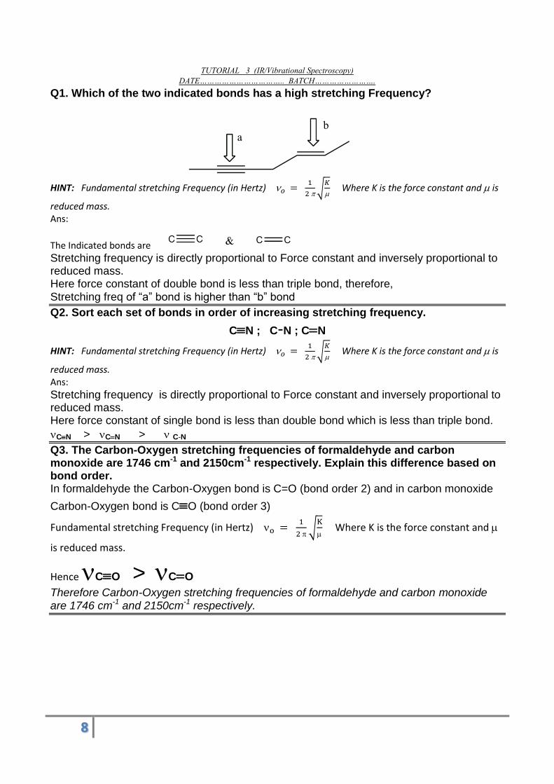
Q1. Which of the two indicated bonds has a high stretching Frequency?
HINT: Fundamental stretching Frequency (in Hertz)
√
Where K is the force constant and is
reduced mass. Ans:
The Indicated bonds are
Stretching frequency is directly proportional to Force constant and inversely proportional to reduced mass. Here force constant of double bond is less than triple bond, therefore, Stretching freq of ―a‖ bond is higher than ―b‖ bond
Q2. Sort each set of bonds in order of increasing stretching frequency.
CN ; C-N ; CN
HINT: Fundamental stretching Frequency (in Hertz)
√
Where K is the force constant and is
reduced mass. Ans:
Stretching frequency is directly proportional to Force constant and inversely proportional to reduced mass. Here force constant of single bond is less than double bond which is less than triple bond.
CN > CN > C-N
Q3. The Carbon-Oxygen stretching frequencies of formaldehyde and carbon monoxide are 1746 cm-1 and 2150cm-1 respectively. Explain this difference based on bond order. In formaldehyde the Carbon-Oxygen bond is C=O (bond order 2) and in carbon monoxide
Carbon-Oxygen bond is CO (bond order 3)
Fundamental stretching Frequency (in Hertz)
√
Where K is the force constant and
is reduced mass.
Hence CO > CO
Therefore Carbon-Oxygen stretching frequencies of formaldehyde and carbon monoxide are 1746 cm-1 and 2150cm-1 respectively.
9
Q4. Assign these stretching frequencies to the corresponding bonds in methanol.1030 cm-1 ; 2945 cm-1 and 3340 cm-1 Ans
The Stretching frequencies possible for methanol are O-H; C-O and C-H
Fundamental stretching Frequency (in Hertz)
√
Where K is the force constant and
is reduced mass. Stretching frequency is directly proportional to Force constant and inversely proportional to reduced mass. Reduced mass is very low for O-H and C-H than for C-O, therefore the stretching frequency at 1030 cm-1.
Usually O-H > C-H
Theirfore 2945 cm-1 corresponds to C-H stretching and 3340 cm-1corresponds to O-H
stretching.
Q5. Which of the following molecules are IR active?
H-Br, H-Cl, H-CN, H2, O2, O3, CO2, H2O, Benzene
Ans, >> A molecule is IR active if any of its vibration accompany with a change in dipole moment. >> A molecule with more than two atoms will have some asymmetric vibrations which generally changes the dipole moment. All molecules with atom number >2 will be IR active. >> Diatomic molecules have only one type of vibration. If it is homo-nuclear diatomic molecule the symmetric stretching will not change the dipole moment, but hetero-nuclear diatomic shows an oscillating dipole moment during asymmetric stretching.
IR active molecules are H-Br, H-Cl, H-CN, O3, CO2, H2O, Benzene.
IR inactive molecules are H2, O2
Q6. How can you distinguish between intermolecular and intramolecular Hydrogen bonding on the basis of IR spectrum? Ans: Intermolecular hydrogen bonding weakens on dilution where as intra-molecular hydrogen bonding is not affected. A change in stretching frequency of hydrogen bond can be observed upon dilution for intermolecular hydrogen bonded molecules.
10
Q7. How will you distinguish the following pair of compounds by IR spectroscopy? Trans-butene and cis-butene Ans: For the Trans-2-pentene, there was no C=C reading, at about 1600 cm-1 because during symmetric stretching there is no change in dipolemoment. For the cis-2-pentene, there is a C=C stretching IR Spectrum reading, here the stretching changes the dipole moment.
Q8. How will you distinguish the following pair of molecules using IR spectroscopy?
Ans
Q9. Sketch the normal modes of vibrations of HCN and which of them are IR active.
11
Q10. How many number of modes of vibrations are possible for acetylene, benzene and cyclohexane. Ans Acetylene is linear molecule. Therefore number of modes of vibrations 3N-5 (and N=4) = 12-5 = 7 nos. Benzene C6H6 is non-linear molecule. Therefore number of modes of vibrations 3N-6 (and N=12) = 36-6 = 30 nos. Cyclohexane C6H12 is non-linear molecule. Therefore number of modes of vibrations 3N-6 (and N=18) = 54-6 = 48 nos.
Q11. Define IR spectrum. Why HCl is IR active, but hydrogen molecule is not. Write the reason for the statement. Ans. In order for a vibrational mode in a sample to be "IR active", it must be associated with
changes in the dipole moment during vibrations. A permanent dipole is not necessary, as the rule requires only a change in dipole moment. A molecule can vibrate in many ways, and each way is called a vibrational mode Hydrogen and hydrogen chloride molecule are both diatomic linear molecules. Therefore 3N-5 = 6-5 = 1 vibration (N=2) is only possible (stretching).
Q12 Compare IR (or vibrational) spectroscopy and UV-Visible (or electronic) spectroscopy. Ans
a. Both are absorption spectroscopy. b. In vibrational spectroscopy vibrational excitation takes place by absorbing IR
light, but in electronic spectroscopy electronic excitation takes place by absorbing UV/Visible light.
c. Vibrational spectroscopy is useful for determining the functional group
present in a molecule, where as electronic spectroscopy is useful for finding -conjugations.
d. Plotting spectra: In electronic spectroscopy Absorbance A (or molar
absorptivity ) vs wave length is plotted, whereas in vibrational spectroscopy
percentage transmittance %T vs wave number is plotted. e. Both are non-destructible detecting systems. f. Both are non-destructible detecting devices.
12
Q13 Explain why some characteristic peaks are usually strong or weak and why some may be absent in an IR spectrum. Ans http://epgp.inflibnet.ac.in/epgpdata/uploads/epgp_content/S000005CH/P000667/M009109/ET/1459227637CHE_P12_M8_e-Text.pdf Q14 Define and give an example of each term in the contest of IR spectroscopy (a) Fingerprint region (b) an IR active vibration Ans https://scilearn.sydney.edu.au/organicspectroscopy/?type=Infrared&page=Fingerprint%20Region The absorption bands in the 4000 – 1500 cm-1 region allows identification of functional groups ; this region therefore is also termed the functional group region of the IR spectrum. The lower energy portion of the mid-IR region (1500 – 400 cm-1) usually contains a very complicated set of peaks arising due to complex vibrations involving several atoms. This region is unique to a particular compound and therefore is known as the fingerprint region of the IR spectrum. Though it is difficult to assign the vibrational modes to these peaks, these are useful to identify a compound if the spectrum of the compound is already known. (https://nptel.ac.in/courses/102103044/module2/lec10/6.html)
Q15 Discuss the strength and limitations of IR spectroscopy Ans The most useful aspect of IR spectroscopy is its ability to identify the functional groups, but IR does not provide much information about the carbon-hydrogen frame work of a molecule.
Q16 Which of the bonds shown in red are expected to have IR active stretching frequency.
Q17. Which of the following compound has a vibration that is IR inactive a) 1-butyne b) 2-butyne c) H2 d) H2O e) Cl2 f) ethene. Hint: Symmetric molecules symmetric stretching is IR inactive.
Ans:
Q18. How could you use IR spectroscopy to determine whether the following reaction has occurred?
Ans: If the reaction has occurred the intensity of IR absorption band at 1700 cm-1 (carbonyl stretching) of the reactant would have decreased.
Q19. Assume that the force constant is approximately same for C-C, C-N and C-O bonds. Predict the relative positions of their stretching vibrations in an IR spectrum. Ans: If the forces constant are approximately the same, the lighter atom will absorb at higher frequency.
C-C > C-N > C-O
14
Q20. For each of the following pairs of compounds, name one absorption band that could be used to distinguish between them.
A)
B)
C)
Q21. Why do CO2 and SO2 have a different number of degrees of vibrational freedom? Ans, Ans, CO2 is linear molecule, 3N-5 = 4, Vibrations possible and SO2 is a non-linear molecule 3N-6 = 3, vibrations possible.
15
TUTORIAL 4 (IR/Vibrational Spectroscopy)
DATE…………………………….. BATCH…………………….
Q1. Calculate the reduced mass of 1H—35.5Cl?
Soln: Reduced mass =
(M1 mass of first atom in Kg, M2 Mass of second atom
in Kg). M1 = 1 amu = 1 x 1.66 x 10-27 Kg M2 = 35.5 amu = 35.5 x 1.66 x 10-27 Kg
= (1 x 1.66 x 10-27 Kg) . (35.5 x 1.66 x 10-27 Kg) (1+35.5) 1.66 x 10⁻²⁷ Kg = 1.61 x 10⁻²⁷ Kg
Q2. 1H—35.5Cl has a force constant (k) value of 480 Nm-1. Calculate the fundamental frequency in terms of wave numbers?
Soln: Fundamental Frequency (in Hertz)
√
K is the force constant.
Reduced mass =
(M1 mass of first atom in Kg, M2 Mass of second atom in Kg).
M1 = 1 amu = 1 x 1.66 x 10-27 Kg
M2 = 35.5 amu = 35.5 x 1.66 x 10-27 Kg
= (1 x 1.66 x 10-27 Kg) . (35.5 x 1.66 x 10-27 Kg)
(1+35.5) 1.66 x 10⁻²⁷ Kg
= 1.61 x 10⁻²⁷ Kg
K = 480 Nm-1.
√
= 8.69 x 10¹³ Hz
=
= 2.89 105 m-1
= 2.89 103 cm-1
Q3. The fundamental frequency of 1H—35.5Cl is 8.69 x 10¹³ Hz. Calculate the force constant?
Soln: Fundamental Frequency (in Hertz)
√
K is the force constant. On
re-arranging K = 4 2
Reduced mass =
(M1 mass of first atom in Kg, M2 Mass of second atom in Kg).
M1 = 1 amu = 1 x 1.66 x 10-27 Kg
M2 = 35.5 amu = 35.5 x 1.66 x 10-27 Kg
= (1 x 1.66 x 10-27 Kg) . (35.5 x 1.66 x 10-27 Kg)
(1+35.5) 1.66 x 10⁻²⁷ Kg
= 1.61 x 10⁻²⁷ Kg
K = 4 3.142 ( )2 1.61 10-27 Kg
= 479.4 Nm-1
𝑜 𝑐
16
Q4. The IR stretching frequency of 1H—35.5Cl is 2890 cm-1. Calculate the force constant?
Soln: Fundamental Frequency (in Hertz)
√
K is the force constant. On
re-arranging K = 4 2 = 4 2 c2 2
Reduced mass =
(M1 mass of first atom in Kg, M2 Mass of second atom in Kg).
M1 = 1 amu = 1 x 1.66 x 10-27 Kg M2 = 35.5 amu = 35.5 x 1.66 x 10-27 Kg
= (1 x 1.66 x 10-27 Kg) . (35.5 x 1.66 x 10-27 Kg) (1+35.5) 1.66 x 10⁻²⁷ Kg = 1.61 x 10⁻²⁷ Kg
C = 3 * 108 m/s
= 2890 cm-1 = 289000 m-1
K = 4 3.142 ( )2 ( )2 1.61 10-27 Kg
= 477 Nm-1
Q5. Calculate the force constant of the CO molecule, if its fundamental vibrational frequency is 2140 cm-1.
Fundamental Frequency (in Hertz)
√
K is the force constant. On re-
arranging
K = 4 2 = 4 2 c2 2 [ fundamental frequency is given in
wavenumber unit]
Reduced mass =
(M1 mass of first atom in Kg, M2 Mass of second atom in Kg).
M1 = 12 amu = 12 x 1.66 x 10-27 Kg M2 = 16 amu = 16 x 1.66 x 10-27 Kg
= (12 x 1.66 x 10-27 Kg) . (16 x 1.66 x 10-27 Kg) (12+16) 1.66 x 10⁻²⁷ Kg = 11.4 x 10⁻²⁷ Kg
c = 3 * 108 m/s
= 2890 cm-1 = 214000 m-1 (Converting wave number in cm-1 to SI unit m-
1) K = 4 3.142 ( )2 ( )2 11.4 10-27 Kg
= 1853 Nm-1
17
Q6. Calculate the fundamental vibrational frequency HCl molecule, if the value of force constant of the molecule is 483 Nm-1. The atomic masses are 1H = 1.673 x 10⁻²⁷ kg and 35Cl = 58.06 x 10⁻²⁷ kg. Ans
Fundamental Frequency (in Hertz)
√
K is the force constant and is
reduced mass.
=
(M1 mass of first atom in Kg, M2 Mass of second atom in Kg).
= (1.673 x 10⁻²⁷ Kg) (58.06x 10⁻²⁷ Kg) (1.673 x 10⁻²⁷ Kg + 58.06x 10⁻²⁷ Kg) = 1.63 x 10⁻²⁷ Kg
Fundamental Frequency (in Hertz)
√
=
√
= 8.66 x 10¹³ Hz.
Fundamental Frequency (in wave number cm-1) =
=
= 2.89 X 10⁵ m-1
= 2.89 X 103 cm-1. Q7. The fundamental vibrational frequency of CO is 2140cm-1. Calculate force constant of the bonds if reduced mass of CO is 1.14 x 10⁻²⁶ Kg. Ans
Fundamental Frequency (in Hertz)
√
K is the force constant and is
reduced mass On re-arranging
k = 4 2 = 4 2 c2 2 [ fundamental frequency is given in
wavenumber unit] 2140 cm-1 = 214000 m-1. k = 4 X 3.14 X 3.14 X 3 x 108 m/s X 3 x 10⁸ m/s X 214000 m-1 X 214000 m-1 X 1.14 x
10-26Kg = 1853 Nm-1.
Q8. The fundamental vibrational frequency of carbon monoxide (12C16O) is 2140 cm-1.
Without calculating force constant, find the fundamental frequency of 13C17O in cm-1. Ans Assume that the force constant remains same for isotope substitution.
√
√
Squaring on both sides (
)
Therefore [ ]
[ ]
Then = √
[ ]
= 7.37 amu
= 6.86
amu
= √
= 2064 cm-1
18
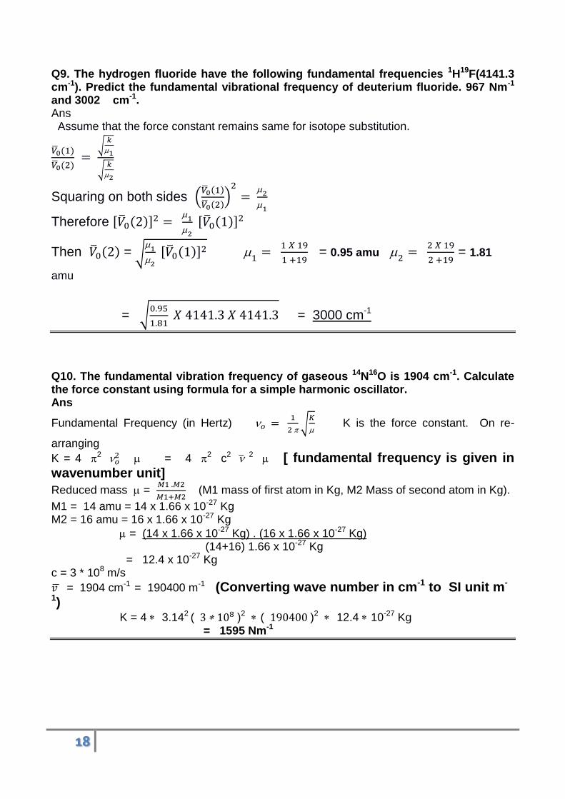
Q9. The hydrogen fluoride have the following fundamental frequencies 1H19F(4141.3 cm-1). Predict the fundamental vibrational frequency of deuterium fluoride. 967 Nm-1 and 3002 cm-1. Ans Assume that the force constant remains same for isotope substitution.
√
√
Squaring on both sides (
)
Therefore [ ]
[ ]
Then = √
[ ]
= 0.95 amu
= 1.81
amu
= √
= 3000 cm-1
Q10. The fundamental vibration frequency of gaseous 14N16O is 1904 cm-1. Calculate the force constant using formula for a simple harmonic oscillator. Ans
Fundamental Frequency (in Hertz)
√
K is the force constant. On re-
arranging
K = 4 2 = 4 2 c2 2 [ fundamental frequency is given in
wavenumber unit]
Reduced mass =
(M1 mass of first atom in Kg, M2 Mass of second atom in Kg).
M1 = 14 amu = 14 x 1.66 x 10-27 Kg M2 = 16 amu = 16 x 1.66 x 10-27 Kg
= (14 x 1.66 x 10-27 Kg) . (16 x 1.66 x 10-27 Kg) (14+16) 1.66 x 10⁻²⁷ Kg = 12.4 x 10⁻²⁷ Kg
c = 3 * 108 m/s
= 1904 cm-1 = 190400 m-1 (Converting wave number in cm-1 to SI unit m-
1) K = 4 3.142 ( )2 ( )2 12.4 10-27 Kg
= 1595 Nm-1
19
Q11. The fundamental vibration frequency of gaseous 14N16O is 1904 cm-1.Calculate the fundamental vibration frequency of 15N16O. Ans Assume that the force constant remains same for isotope substitution.
√
√
Squaring on both sides (
)
Therefore [ ]
[ ]
Then = √
[ ]
= 7.47 amu
= 7.74
amu
= √
= 1870 cm-1
Q12. A carbon deuterium C-D bond is electronically much like a C-H bond and it has similar stiffness, measured by spring constant, k. The deuterium isotope has twice the mass of hydrogen atom. Calculate the IR absorption frequency of a typical C-D bond. Given that IR absorption frequency of C-H is 3000 cm-1. ****
Ans: √
=
=
.
=
√
√
=
√
√
= √
= 0.734
Therefore
= 2202
20
Q13. The molecule 12C16O has a bond force constant of 1860 Nm-1. Calculate the vibrational zero point energy of this molecule. Reduced mass of CO is 1.14 x 10⁻²⁶ Kg. Ans
Fundamental Frequency (in Hertz)
√
K is the force constant and is
reduced mass.
= 1.14 x 10⁻²⁶ Kg
Fundamental Frequency (in Hertz)
√
=
√
= 6.43 x 10¹³ Hz.
Zero point energy
= ½ x 6.626 x 10
-36 x 6.43 x 1013 = 21.30 x 10-23
=
21
Tutorial 5 NMR realted
Q1. Which of the following nuclei shows NMR activity? H1, H2, He4, C12, C13, O18
Ans A Nuclei is NMR active when the nuclei have non-zero spin I. Rules to determine I If a nuclei has Proton number and Neutron number are both even, then I will be 0.NMR inactive. If a nuclei has Proton number and Neutron number are both odd, then I will be 1,2,3....(NMR active) If a nuclei has odd Proton number and even Neutron number or even Proton number and odd Neutron number (that is mass number is odd) then I will be 1/2 , 3/2 ,5/2 ... All Nuclei with odd mass number are NMR active Hydrogen has 1 Proton then H1, H2 are both NMR active Carbon has 6 Proton then C12 is NMR inactive C13 is NMR active Oxygen has 8 protons and 18-8 = 10 neutrons, both are even so NMR inactive.
Q2. Which of the following nuclei do not show nuclear magnetic resonance? 1H, 2H, 12C, 13C, 14N, 15N, 16O, 19F, 31P, 32S. Ans
Nuclei with a non-zero I value are NMR active. All nuclei with odd mass number are NMR active. also
Nuclei with even mass number having odd number of protons and neutrons are also NMR active.
Nuclei having even mass number with even number of protons as well as even number of neutrons are
NMR inactive.
12C, 16O and 32S are NMR inactive
Q3.How many kinds of protons are there in (i) CH3-CH3. (ii) CH3-CH2-CH3, (iii) C6H5-
CH3 (iv) (CH3)2CHCH2CH3 (v) ClCH2CH2Cl
Ans:
(i) Only one kind of proton (ii) Two kinds of protons
(iii) (iv) Four kinds of proton.
22
(v) Only one kind of proton.
Q4. Determine which of the following molecules will show spin-spin coupling in their
NMR spectra. If splitting is observed, give the multiplicity of each kind of proton.
(a) Same kind of protons cannot give spin-spin splitting
(b) Will give spin-spin splitting. Signal for Ha protons are split into triplet by 2 Hb
neighbours and signal for Hb is also a triplet because of 2 Ha neighbours.
(c) Will give spin spin splitting. Signal for Ha protons are split into doublet by 1 Hb
neighbour and signal for Hb will be a quartet due to 3 Ha neighbours.
(d) No vicinal hydrogens. No spin-spin splitting
(e) No vicinal hydrogens. Therefore no spin-spin splitting
23
Q5. Predict the low resolution NMR spctrum and high resolution NMR spectrum of
acetone.
Ans:
All six hydrogens are chemically
equivalent. Therefore we get a singlet in low resolution spectrum. Also there are no vicinal non-equivalent
neighbouring protons. Therefore high resolution spectrum and low resolution spectrum both look same.
Q6. Predict the low resolution NMR spctrum and high resolution NMR spectrum of
propanal.
Ans
Q7. An organic compound C3H6O contains a carbonyl group. How will its NMR
spectrum decide whether it is an aldehyde or a ketone.
CH3CO CH3 CH3-CH2-CHO
Ans: The propanone would only give one peak in its NMR spectrum because both CH3 groups are in an identical
environment - both are attached to -COCH3. The propanal would give three peaks with the areas underneath in the ratio
3:2:1.
24
Q8 Predict the NMR spectrum of the compound CH3CO CH2CΞCCH3.
25
Q9. How can the NMR spectrum disinguish between the isomers para-Xylene and
ethyl-benzene.
Q10. Predict the proton NMR spectrum of H2, CH4, C2H6 and C6H6.
a) H2 Only one kind of Hydrogen. Which do not show spin-spin interaction, hence a
singlet.
b) CH4 All four Hydrogens are identical—Singlet
c) C2H6 All 6 Hydrogens are chemically equivalent. Therefore no splitting. (Singlet)
26
d) C6H6 all six hydrogens are identical. Therefore no spin-spin splitting.
Q11. Below are given the molecular formula of some organic compounds that have only
one NMR signal. Draw the structural formula for each of them.
a) C5H12
Double bond Index or ring = C – H/2 + 1 = 5 – (12/2) + 1 = 5-6+1 =0
The compound is alkane.
b) C3H6 Double bond Index or ring = C – H/2 + 1 = 3 – (6/2) + 1 = 1 (a ring or 1 DB)
c) C3H4 Double bond Index or ring = C – H/2 + 1 = 3 – (4/2) + 1 = 2 (2DB or 1
ring+1DB)
d) C4H6 Double bond Index or ring = C – H/2 + 1 = 4 – (6/2) + 1 = 2 (2DB or 1
ring+1DB)
. All six hydrogens are equivalent, hence a singlet.
e) C8H18. Double bond Index or ring = C – H/2 + 1 = 8 – (18/2) + 1 = 0
27
f) C2H6O Double bond Index or ring = C – H/2 + 1 = 2 – (6/2) + 1 = 0
g) C2H4Br2 Double bond Index or ring = C – H/2 + 1 = 2 – (6/2) + 1 = 0
Q12. Below are given the molecular formula of some organic componds that give 2 singlets in their proton NMR spectra: Draw the structural formula for each of them.
h) C3H6O2 Double bond Index or ring = C – H/2 + 1 = 3 – (6/2) + 1 = 1
CH3 fragment + another CH3 fragment . Remaining COO ( )
i) C3H8O2 Double bond Index or ring = C – H/2 + 1 = 3 – (8/2) + 1 = 0
CH3 fragment + another CH3 fragment + a CH2 fragment . Remaining 2 O.
(j) C2H5OCl Double bond Index or ring = C – H/2 + 1 = 2 – (6/2) + 1 = 0
CH3 fragment + a CH2 fragment . Remaining O and Cl
(k) C3H5Cl3 Double bond Index or ring = C – H/2 + 1 = 3 – (8/2) + 1 = 0 CH3 fragment + a CH2 fragment + 3 Cl and a C
28
(l) C5H10Cl2 Double bond Index or ring = C – H/2 + 1 = 5 – (12/2) + 1 = 0
2 CH3 fragment + 2 CH2 fragment . Remaining a C and 2 Cl
Q13. Predict the number of signals, their relative positions and their multiplicities for
ethyl acetate.
There are three types of protons marked a, b and c ―a‖ protons is attached to electron withdrawing acetate group ―b‖ protons is attached to electronegative (electron withdrawing) O atom. ―c‖ protons does not have an electron withdrawing group attached. Electron withdrawing ability of O > -COO- therefore ―b‖ protons are more deshielded. ―a‖ protons have no neighbouring Hydrogens. Therefore the signal will be a singlet. ―b‖ protons have 3 neighbouring Hydrogens. Therefore the NMR signal will split into (3+1 = 4 ) quartet. ―c‖ protons have 2 neighbouring Hydrogens. Therefore the NMR signal will split into (2+1 = 3) triplet
29
Q14. Predict the number of signals, their relative positions and their multiplicities for 2-bromopropane.
There are two types of protons marked a and b
- ―b‖ protons is attached to electronegative (electron withdrawing) Br atom. - ―a‖ protons does not have an electron withdrawing group attached.
Signal for ―b‖ protons are more deshielded (left side).
- ―a‖ protons have one neighbouring Hydrogen. Therefore the signal will be a doublet. - ―b‖ protons have 3+3 = 6 neighbouring equivalent Hydrogens . Therefore the NMR
signal will split into (6+1 = 7) septet.
30
Q15. Predict the number of signals, their relative positions and their multiplicities for
ultra-pure Ethanol.
31
Q16. Predict the High resolution NMR spectrum of CH3-CH2-CHCl2 (High resolution means spin-spin splitting).
Q17. What is meant by spin active nuclei? Give 4 examples of spin active nuclei.
Ans: Spin active nuclei is nuclei having non-zero Spin value. They will show NMR phenomenon. Examples are 1H, 2H, 13C and 31P
Q18. Explain the term spin-spin splitting. Why does a peak for a particular set of proton split into a multiplet?
Ans.
Q19. “NMR spectroscopy is the most important tool compared to UV-Visible and IR spectroscopy for the structural determination of an unknown organic compound” Justify the statements.
Ans:
Number of signals in NMR spectrum gives the number of chemically different types of hydrogens present.
The intensity ratio is the relative ratio of number of hydrogens. Position of signals give an indication about its local electronic arrangement. Spin-Spin splitting gives information on number of hydrogen atoms attached to
neighbouring atoms.
Ultimately one can logically deduct the structure of molecule.
By UV visible spectroscopy one can find only the conjugation. By IR spectroscopy one can find only the functional groups present.
Q20. Why TMS is used a standard reference in proton NMR spectroscopy?
Ans: TMS Si(CH3)4. All 12 Hydrogens are equivalent therefore one NMR signal. Since Si is electropositive than C, Cl, F, O. The Hydrogens of TMS are highly shielded. The NMR signals of Hydrogens in all organic compounds will be deshielded with respect to TMS hydrogens.
32
Q21. Give the mechanism of absorption of RF pulse by NMR active nuclei kept in a magnetic field.
……………………………………………………………………………………..
Q22. What are the factors affecting Chemical shift?
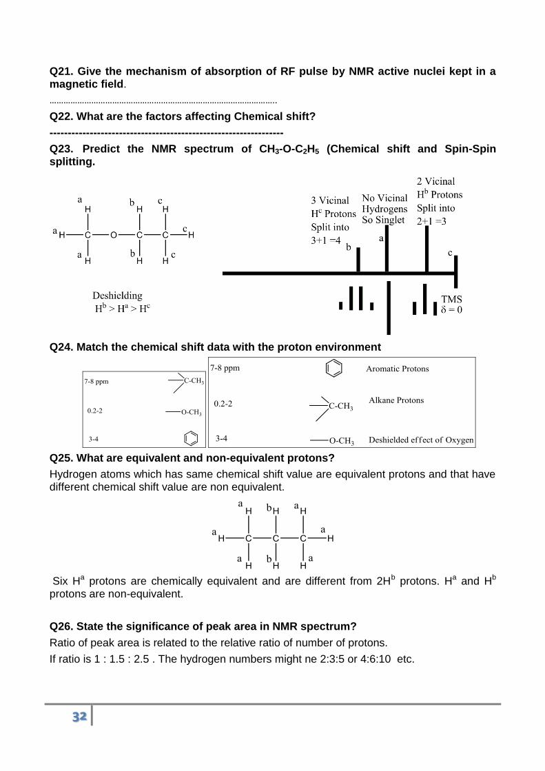
Q23. Predict the NMR spectrum of CH3-O-C2H5 (Chemical shift and Spin-Spin splitting.
Q24. Match the chemical shift data with the proton environment
Q25. What are equivalent and non-equivalent protons?
Hydrogen atoms which has same chemical shift value are equivalent protons and that have different chemical shift value are non equivalent.
Six Ha protons are chemically equivalent and are different from 2Hb protons. Ha and Hb protons are non-equivalent.
Q26. State the significance of peak area in NMR spectrum?
Ratio of peak area is related to the relative ratio of number of protons.
If ratio is 1 : 1.5 : 2.5 . The hydrogen numbers might ne 2:3:5 or 4:6:10 etc.
33
Q27. In which of the following systems energy level separation is largest? (a) a proton in 600 MHz instrument, (b) proton in 300 MHz instrument.
Splitting of energy level is proportional to magnetic field. Therefore splitting will be maximum in 600 MHz instrument.
Q28. Give the applications of NMR spectroscopy.
1. Structural elucidation of organic molecules. 2. 1H-NMR is basic principle of MRI of soft tissues. 3. 31P-NMR can be used for imaging bones. 4. To study dynamics of biomacromolecules (like protein folding).
Q29. What is the use of N+1 rule, Pascal triangle in NMR spectral analysis?
N+1 RULE: The (n+1) Rule, an empirical rule used to predict the multiplicity of each H-
NMR signal. It states that if a 1H nucleus has n number of vicinal non-equivalent
1H, then
the multiplicity of the peak is n+1.
Pascal’s Triangle: It predicts the ratio of heights of signals in a split NMR peak.
Coupling Constant, J: The spacing between the lines of a doublet, triplet or
quartet (Multiplet) is called the coupling constant.
Home Work Questions
HWQ1. Predict the NMR spectrum
a. CH3-CH3 b. CH3-CH2-CH3 c. CH3-O-CH3 d. CH3-CO-CH3 e. CH3-COO-CH3 f. Propanaldehyde g. 1-chloropropane
34
Tutorial 6 NMR related Numericals
Q1. Calculate the frequency of operation of NMR instrument if a particular proton of
value 4.2 shows a difference in frequency 1260 Hz from TMS?
Soln: Chemical Shift =
=
=
= 300MHz
Q2. When measured using 300 MHz instrument the signal for proton in CHCl3
appears 1260 Hz downfield from TMS signal. Calculate chemical shift.
Soln: Chemical Shift =
=
= 4.2 ppm
Q3. A 300-MHz spectrometer records a proton that absorbs at a frequency 2130 H z downfield from TMS. (a) Determine its chemical shift. (b) Predict this proton's chemical shift at 60 MHz. In a 60-MHz spectrometer, how far downfield (in hertz) from TMS would this proton absorb? Soln.
(a) Chemical Shift = =
= 7.10 ppm
(b) The chemical shift is unchanged 7.10 ppm when we change the operating frequency. Frequency Shift = Chemical Shift x Spectrometer Frequency = 7.1 x 60 = 426 Hz
Q4. A signal has been reported to occur at 600Hz downfield from TMS in an NMR spectrometer with a 300-MhZ operating frequency.
(a) What is the chemical shift of the signal? (b) What would its chemical shift be in an instrument operating at 500 MHz? (c) How many hertz downfield from TMS would the signal be in a 500 MHz NMR
spectrometer?
35
Q5. If two signals differ by 1.5 ppm in a 300 MHZ spectrometer, by how much do they differ in a 100 MHZ spectrometer. Ans The chemical shift is independent of the operating frequency. Therefore if the two signals differ by 1.5 ppm in a 300 MHZ instrument, they will still differ by 1.5 ppm in a 100 MHZ instrument.
Q6. If two signals differ by 90 Hz in a 300 MHZ spectrometer, by how much do they differ in a 100 MHZ Instrument.
Q6. If gyromagnetic ratio of 13C is ¼th of that of 1H. What is the frequency that must
be irradiated to take 13C NMR spectrum if the same instrument take 1H-NMR spectra
at 300 MHZ.
B is constant in both cases
We can write
36
That is
= 300/4 = 75MHz
Q1. How many kinds of proton are there in, a) Prop-1-ene b) (Z)-1,2-dibromoethene c) (E)-1-bromo-2-
chloroethene d) 1-chloro-1-iodoethene.
Hint
Ans
37
Q1. Determine which of the following molecules will show spin-spin coupling in their proton-NMR spectra.
If the coupling or splitting is observed give the multiplicity of the signal, a) Prop-1-ene b) (Z)-1,2-
dibromoethene c) (E)-1-bromo-2-chloroethene d) 1-chloro-1-iodoethene.
Ans
a)
b)
Both protons are identical, therefore spin-spin splitting is not observed.
c)
The two protons are not equivalent and each is split into a doublet.
d)
The two protons are not equivalent (one cis to Cl and other is trans to Cl) so that each is split into doublet.