+ All Categories
Home > Documents > MOHAMED EL-SAKHAWY , MAMDOUHEL …ijbpas.com/pdf/2016/January/1453466207MS IJBPAS 2015 3502.pdf ·...

MOHAMED EL-SAKHAWY , MAMDOUHEL …ijbpas.com/pdf/2016/January/1453466207MS IJBPAS 2015 3502.pdf ·...

Date post: 05-Aug-2018
Category:
Upload: vuongquynh
View: 213 times
Download: 0 times
Share this document with a friend
14
IJBPAS, January, 2016, 5(1): 362-375 ISSN: 2277–4998 362 IJBPAS, January 2016, 5(1) SEASONAL HISTOMORPHOMETRIC STUDIES ON THE EPIDIDYMAL DUCT OF THE ONE-HUMPED CAMEL (CAMELUS DROMEDARIUS) MOHAMED EL-SAKHAWY 1 , MAMDOUHEL-SHAMMAA 2 , SHAYMAA HUSSEIN 3* , ABDEL-ALEEMEL-SABAA, A 4 AND YAHYA AHMED 5 1, 2, 3, 4: Department of Cytology and Histology, Faculty of Veterinary Medicine, Cairo. University, Egypt 5: Department of Veterinary Anatomy, Mosul University, Iraq 5 E Mail: [email protected] ABSTRACT This investigation was carried on right and left epididymis of 76 sexually mature (6-7 years) apparently healthy, one humped camel ( Sudanese breed ), which collected directly after slaughtering from Kerdasa slaughter house, Giza , Egypt all over the year from December 2011 till November 2012. The epididymis was composed of the efferent ductules and the epididymal duct. The epididymal duct was divided into initial, middle and terminal segments. The middle segment was subdivided into proximal, intermediate and distal parts. The terminal segment was further subdivided into proximal and distal parts. Samples from different regions were processed histologically. Paraffin sections 5-6 μmthick were stained with H&E, Van Gieson's, Masson's trichromeand PASstains were used for histomorphometrical study. The epididymal duct showed significant seasonal changes in the total diameter, epithelial height, length of stereocilia, luminal diameter, thickness of peritubular muscular coat and the cellular distribution. The values of these elements were increase during spring season in comparison with other seasons. The results were analyzed statistically.The epididymal duct was lined by a pseudostratified columnar epithelium. The epididymal epithelium was composed of five cell types: principal, basal, apical, dark and halo cells. Abundant apical like protrusions, numerous cytoplasmic vacuoles and PAS positive granules characterizing the principal cells in spring season. In conclusion the spring season is the breeding season in camel in Egypt. Keywords: Seasonal changes, epididymis, camel, histomorphometry
Transcript

IJBPAS, January, 2016, 5(1): 362-375 ISSN: 2277–4998

362

IJBPAS, January 2016, 5(1)

SEASONAL HISTOMORPHOMETRIC STUDIES ON THE EPIDIDYMAL DUCT

OF THE ONE-HUMPED CAMEL (CAMELUS DROMEDARIUS)

MOHAMED EL-SAKHAWY1, MAMDOUHEL-SHAMMAA2, SHAYMAA HUSSEIN3*,

ABDEL-ALEEMEL-SABAA, A4AND YAHYA AHMED5

1, 2, 3, 4: Department of Cytology and Histology, Faculty of Veterinary Medicine, Cairo. University, Egypt

5: Department of Veterinary Anatomy, Mosul University, Iraq5 E Mail: [email protected]

ABSTRACT

This investigation was carried on right and left epididymis of 76 sexually mature (6-7 years)

apparently healthy, one humped camel ( Sudanese breed ), which collected directly after

slaughtering from Kerdasa slaughter house, Giza , Egypt all over the year from December

2011 till November 2012. The epididymis was composed of the efferent ductules and the

epididymal duct. The epididymal duct was divided into initial, middle and terminal segments.

The middle segment was subdivided into proximal, intermediate and distal parts. The

terminal segment was further subdivided into proximal and distal parts. Samples from

different regions were processed histologically. Paraffin sections 5-6 µmthick were stained

with H&E, Van Gieson's, Masson's trichromeand PASstains were used for

histomorphometrical study. The epididymal duct showed significant seasonal changes in the

total diameter, epithelial height, length of stereocilia, luminal diameter, thickness of

peritubular muscular coat and the cellular distribution. The values of these elements were

increase during spring season in comparison with other seasons. The results were analyzed

statistically.The epididymal duct was lined by a pseudostratified columnar epithelium. The

epididymal epithelium was composed of five cell types: principal, basal, apical, dark and halo

cells. Abundant apical like protrusions, numerous cytoplasmic vacuoles and PAS positive

granules characterizing the principal cells in spring season. In conclusion the spring season is

the breeding season in camel in Egypt.

Keywords: Seasonal changes, epididymis, camel, histomorphometry

Mohamed El-Sakhawy et al Research Article

363

IJBPAS, January 2016, 5(1)

INTRODUCTION

Camel is described as a seasonal breeder

(Al Eknah, 2000 and Maraiet al.,2009) or it

may maintain their reproductive capacity

throughout the year (Zayedet al., 1995).

The seasonal breeding behavior of the male

camel is referred to as rut (Khanet

al.,2003).The onset of breading season of

male camel is markedly divergent in Egypt.

The rutting season has been reported to

occur at December uptill March (Ebada,

1994), while according to Abd El-maksoud

(2010) the rutting season occurs during

spring. The excurrent duct system of the

testis includes the efferent ductules, the

ductus epididymidis and the ductus

deferens (Roberts, 2010). The epididymis

consists of highly convoluted duct that

links the efferent ductules to the vas

deferens (Turner et al.,2003).Like other

economically valuable animals, studying

the morphology of the camel reproductive

organs is essential to improving and

exploiting their reproductive activity.

Detailed description of the structure of the

epididymal duct and their morphometry in

the dromedary during breeding and non-

breeding seasons has been quite few (Abd

El-maksoud, 2010). In addition, data on the

stage of seasonal changes in camel are

quite divergent. In view of this, the aim of

this study is to elucidate more light and

some details on the histomorphometrical

features of the epididymal duct of the one

humped camel during different seasons.

MATERIAL AND METHODS

Right and left epididymis of 76 sexually

mature (6-7 years) apparently healthy, one

humped camel ( Sudanese breed ) were

collected directly after slaughtering from

Kerdasa slaughter house, Giza , Egypt all

over the year from December 2011 till

November 2012.

Anatomically, the epididymal duct was

consisted of three regions; head (caput),

body (corpus) and tail (cauda).

Histologically and histomorphometrically,

the epididymis in the present investigation

was subdivided into 7 parts. The head

(caput) was subdivided into two parts,

ascending first part of the head and initial

part of epididymal duct. The middle

segment (corpus) was further subdivided

into proximal, intermediate and distal parts

and finally the terminal segment (cauda)

was composed of proximal and distal parts

(Fig.1).

The present study included the following

lines:

1- General histological study:

The obtained segments were fixed in

buffered 10 % formalin of (pH 7.0) and

Bouin′s fluid for 24 hours. Fixed specimens

were dehydrated, cleared and embedded in

paraffin wax. Step serial sections of 5-6 µm

Mohamed El-Sakhawy et al Research Article

364

IJBPAS, January 2016, 5(1)

thick were obtained and stained with:Harris

hematoxyline and eosin (H&E), Van

Gieson's stain, Masson's trichrome

stain,Periodic acid Schiff technique (PAS).

The aformentioned methods were used as

outlined by Drury and Wallingtion (1980).

2– Histomorphometrical and statistical

analysis:

Sections stained with H&E, Van Gieson's,

Masson's trichromeand PAS stains were

used for histomorphometrical study.

Approximately 10 tubular cross sections

from each epididymal parts were measured

by high power lens (X 40). A computerized

microscopic image analyzer attached for

full HD microscopic camera (Leica

Microsystems, Germany) was used to

determine the histomorphometric

parametes. The data were tested firstly for

normal distribution using Normality test.

Normal distributed data were compared by

ANOVA (One-Way Analysis of Variance).

Significant differences were determined by

Duncan’s Multiple Range Test. The data

sets, that had not fulfilled the assumption of

normal distribution, were analyzed using

Kruskal- Wallis One Way Analysis of

Variance on Ranks. Significant differences

were determined by Dunn’s Test. All

statistical analyses were performed by

SigmaStat (Jandel scientific software

V3.1).

Table 1: Total diameter (Mean±SE) of different segments of the camel epididymal duct in different seasons

Segments Total diameter (µm)

Winter (January) Spring (may)

Summer (July )

Autumn (October)

Initial segment (IS) 156.851±4.102 a,b

163.498±4.649 a

139.31±4.183 b

150.219±5.054 a,b

Middle segment (MS)

Proximal part (P)

97.075±6.639 a

152.66±5.062 b

121.859±6.966 c

135.39±4.02 b,c

Intermediate part (I)

115.19±9.238 a

133.078±6.387 a

131.654±1.147 a

125.669±5.959 a

Distal part (D)

111.383±3.601 a

169.428±5.981 b

153.516±5.772 b,c

143.443±4.335 c

Terminal segment

(TS)

Proximal part (P)

168.33±3.263 a,c

208.316±9.284

187.385±18.487 c

163.104±7.396 a

Distal part (D)

182.421±8.001 a

246.311±13.871 b

217.951±5.759 c

232.325±6.216 b,c

a-c Values with a different superscript within rows were significantly different (P<0.05)

Table 2: Epithelial height (Mean±SE) of different segments of the camel epididymal duct in different seasons Segments Epithelial height (µm)

Winter (January) Spring (may) Summer (July ) Autumn (October) Initial segment (IS) 32.582±0.942

b 42.634±1.558

a 34.569±1.663

b 32.094±1.206

b Middle segment

(M) Proximal

part (P) 12.569±1.201

b 27.208±1.526

a 25.743±3.16

a 24.427±2.043

a Intermediate part

(I) 20.047±2.502

b 29.267±0.637

a 22.374±0.841

b 21.056±1.872

b Distal

part (D) 17.673±1.502

b 26.809±1.068

a 18.143±0.411

b 19.149±1.861

b Terminal segment

(TS)

Proximal part (P)

11.286±0.897 c

31.11±1.306 a

24.501±1.17 b

22.218±1.659 b

Distal part (D)

11.268±0.457 b

19.115±1.045 a

17.143±0.322 a,c

13.418±0.947 b,c

a-c Values with a different superscript within rows were significantly different (P<0.05)

Mohamed El-Sakhawy et al Research Article

365

IJBPAS, January 2016, 5(1)

Table 3: Length of stereocilia (Mean±SE) of different segments of the camel epididymal duct

Segments Length of stereocilia (µm)

Winter (January) Spring (may)

Summer (July )

Autumn (October)

Initial segment (IS) 3.329±0.334 A

4.16±0.503 A

3.346±0.598 a

3.972±0.532 a

Middle segment

(MS)

Proximal Part (P)

2.437±0.687 A

6.591±0.189 B

3.609±0.289 c

5.149±0.453 d

Intermediate part (I)

2.51±0.124 b

3.774±0.585 a

3.767±0.319 a

1.609±0.168 b

Distal part (D)

1.579±0.216 a

2.14±0.0374 a

2.049±0.285 a

1.151±0.0236 a

Terminal segment

(TS)

Proximal part (P)

1.803±0.141 a

1.915±0.129 a

1.242±0.232 a

1.888±0.174 a

Distal part (D)

1.325±0.0851 a

1.964±0.248 a

1.406±0.178 a

0.923±0.0494 a

a-d Values with a different superscript within rows were significantly different (P<0.05).

Table 4: Luminal diameter (Mean±SE) of different segments of the camel epididymal duct in different seasons

Segments Luminal diameter (µm)

Winter (January)

Spring (may)

Summer (July )

Autumn (October)

Initial segment (IS) 76.528±6.347 b

98.832±4.949 a

83.774±3.657 a,b

80.629±9.321 a,b

Middle segment

(MS)

Proximal part (P)

71.251±8.015 b,c

98.466±4.92 a

71.026±2.984 b,c

84.668±4.145 a,c

Intermediate part (I)

76.481±4.729 a

88.82±5.369 a

75.273±1.558 a

84.79±9.212 a

Distal part (D)

75.83±3.922 a

120.507±5.936 b

117.568±6.552 b

104.939±1.7 b

Terminal segment

(TS)

Proximal part (P)

145.91±3.035 a,c

164.503±10.372 a

139.586±20.754 b,c

100.901±6.057 b

Distal part (D)

162.501±7.978 a

206.266±7.312 b

184.759±6.255 a,b

201.229±8.58 b

a-c Values with a different superscript within rows were significantly different (P<0.05)

Table 5: Thickness of muscular coat (Mean±SE) of different segments of the camel epididymal duct in different seasons

Segments

Thickness of muscular coat (µm) Winter (January) Spring

(may) Summer (July )

Autumn (October)

Initial segment (IS) 8.616±1.151 a

12.174±1.315 b

10.826±0.524 a,b

10.303±0.754 a,b

Middle segment (MS)

Proximal part (P)

7.087±1.131 b,c

10.581±1.245 a

7.853±0.602 b,c

8.386±0.954 a,c

Intermediate part (I)

7.168±0.508 b

10.668±0.764 a

7.268±0.523 b

8.846±0.301 b

Distal part (D)

6.788±0.653 b,c

9.747±0.378 a

6.682±0.784 b,c

8.167±0.259 a,c

Terminal segment (TS)

Proximal part (P)

15.162±0.921 b

21.638±0.732 a

14.336±1.381 b

20.285±0.734 a

Distal part (D)

16.446±0.809 b

24.19±1.897 a

22.244±0.234 a

18.403±0.367 b

a-c Values with a different superscript within rows were significantly different (P<0.05).

Mohamed El-Sakhawy et al Research Article

366

IJBPAS, January 2016, 5(1)

Fig. (1). Schematic drawing of the right testis and epididymis of camel. The head, body and tail had been dissected from the upper

pole of the testis and stretched to show the different segment of the epididymis from which the specimens were obtained. 1-Ascending first region of the head, 2- Initial segment ( IS), 3-Proximal part of the middle segment (MS-P) ,4-Intermediate part of the middle segment (MS-I) ,5-Distal part of the middle segment (MS-D), 6-Proximal part of the terminal segment (TS-P), 7-Distal

part of the terminal segment (TS-D).

Fig. (2). A photomicrograph of a section at the level of the initial segment of camel epididymal duct during spring showing:

Principal cell (P) and its PAS positive supra and infranuclear cytoplasmis granules (arrow heads), Halo cell (H), apical cell (A) and PAS positive stereocilia (arrow) PAS X 1000

Fig. (3). A photomicrograph of a section at the level of the distal part of terminal segment of camel epididymal duct during spring showing: Principal cell (P) with its oval nuclei in the lower third, numerous vacuoles and PAS positive granules (star) in the apical

part, basal cell with its large PAS positive globule (arrow) and intense PAS positive stereocilia (arrow head). PAS X 1000

3

1

2

Mohamed El-Sakhawy et al Research Article

367

IJBPAS, January 2016, 5(1)

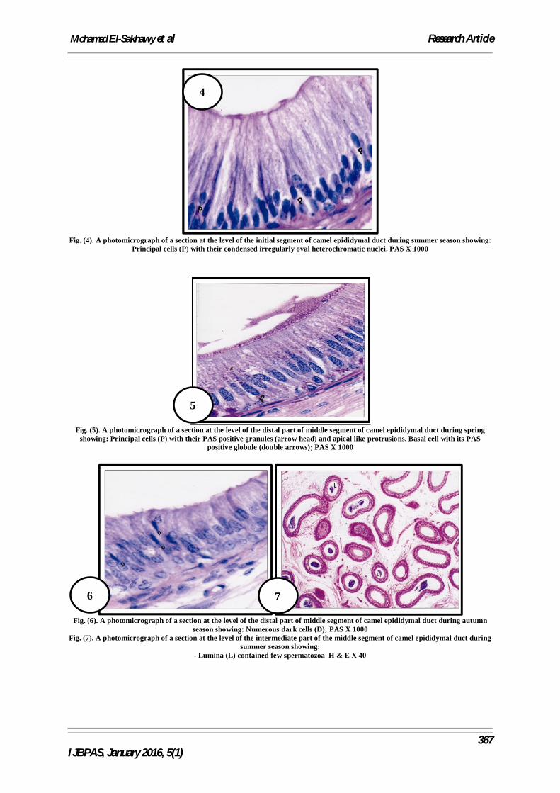
Fig. (4). A photomicrograph of a section at the level of the initial segment of camel epididymal duct during summer season showing:

Principal cells (P) with their condensed irregularly oval heterochromatic nuclei. PAS X 1000

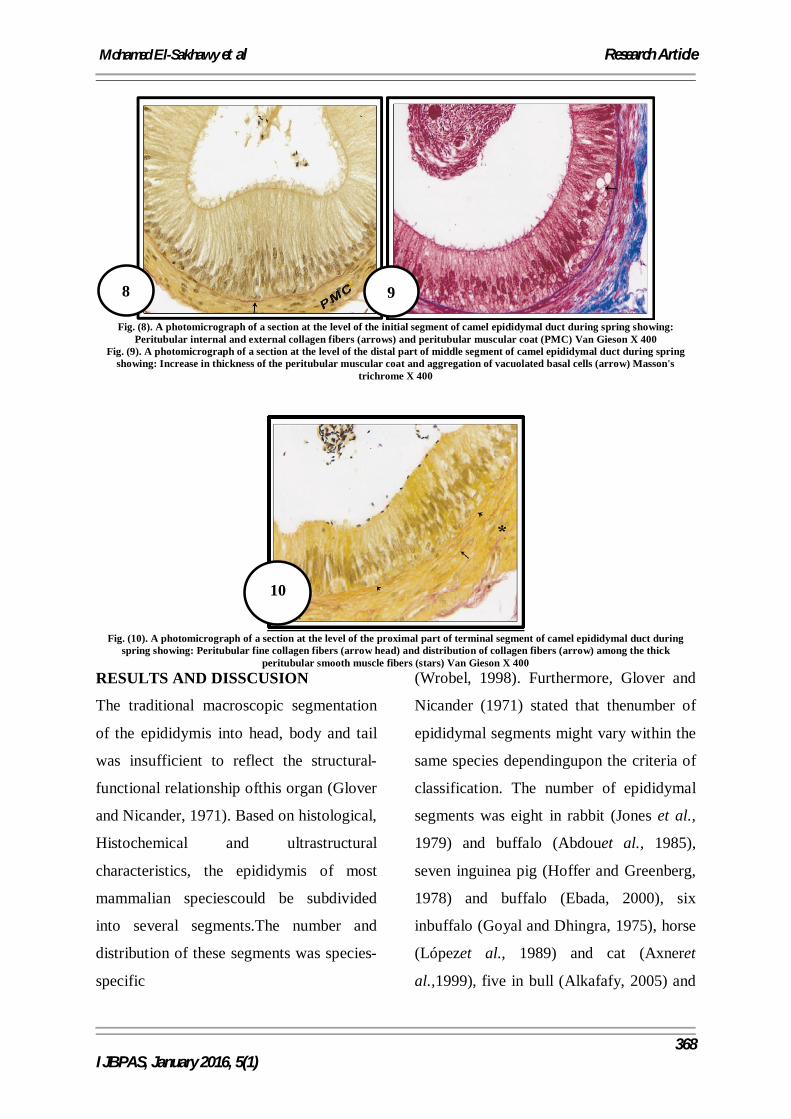
Fig. (5). A photomicrograph of a section at the level of the distal part of middle segment of camel epididymal duct during spring

showing: Principal cells (P) with their PAS positive granules (arrow head) and apical like protrusions. Basal cell with its PAS positive globule (double arrows); PAS X 1000

Fig. (6). A photomicrograph of a section at the level of the distal part of middle segment of camel epididymal duct during autumn

season showing: Numerous dark cells (D); PAS X 1000 Fig. (7). A photomicrograph of a section at the level of the intermediate part of the middle segment of camel epididymal duct during

summer season showing: - Lumina (L) contained few spermatozoa H & E X 40

4

5

6 7

Mohamed El-Sakhawy et al Research Article

368

IJBPAS, January 2016, 5(1)

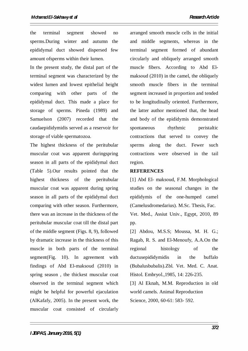
Fig. (8). A photomicrograph of a section at the level of the initial segment of camel epididymal duct during spring showing:

Peritubular internal and external collagen fibers (arrows) and peritubular muscular coat (PMC) Van Gieson X 400 Fig. (9). A photomicrograph of a section at the level of the distal part of middle segment of camel epididymal duct during spring

showing: Increase in thickness of the peritubular muscular coat and aggregation of vacuolated basal cells (arrow) Masson's trichrome X 400

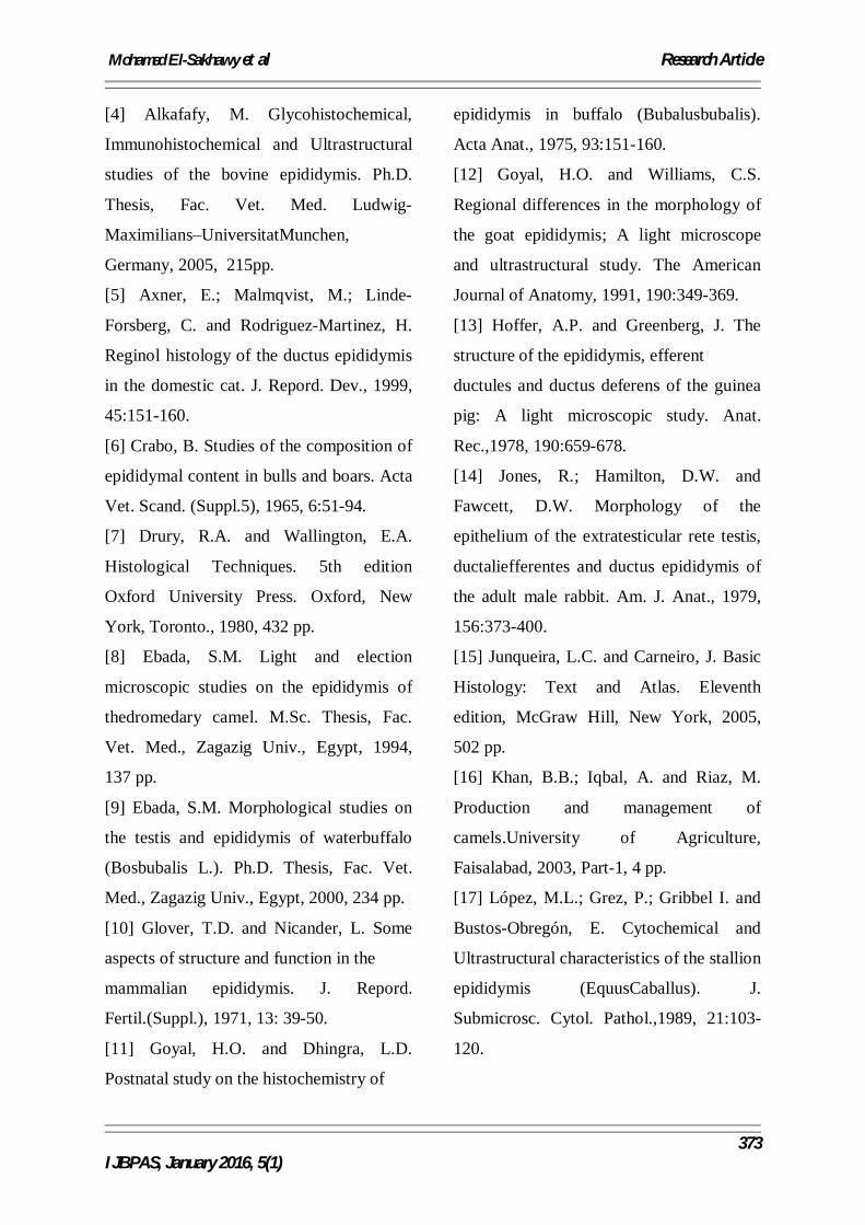
Fig. (10). A photomicrograph of a section at the level of the proximal part of terminal segment of camel epididymal duct during

spring showing: Peritubular fine collagen fibers (arrow head) and distribution of collagen fibers (arrow) among the thick peritubular smooth muscle fibers (stars) Van Gieson X 400

RESULTS AND DISSCUSION

The traditional macroscopic segmentation

of the epididymis into head, body and tail

was insufficient to reflect the structural-

functional relationship ofthis organ (Glover

and Nicander, 1971). Based on histological,

Histochemical and ultrastructural

characteristics, the epididymis of most

mammalian speciescould be subdivided

into several segments.The number and

distribution of these segments was species-

specific

(Wrobel, 1998). Furthermore, Glover and

Nicander (1971) stated that thenumber of

epididymal segments might vary within the

same species dependingupon the criteria of

classification. The number of epididymal

segments was eight in rabbit (Jones et al.,

1979) and buffalo (Abdouet al., 1985),

seven inguinea pig (Hoffer and Greenberg,

1978) and buffalo (Ebada, 2000), six

inbuffalo (Goyal and Dhingra, 1975), horse

(Lópezet al., 1989) and cat (Axneret

al.,1999), five in bull (Alkafafy, 2005) and

8 9

10

Mohamed El-Sakhawy et al Research Article

369

IJBPAS, January 2016, 5(1)

goat (Goyal and Williams,1991), and four

in cat (Sanchez et al., 1998).

In camel and in consistence with Tingari

and Moniem (1979), Ebada(1994) and Abd

El-maksoud (2010) whom stated that, the

epididymis wassubdivided into initial,

middle and terminal segments, of which the

middlesegment was further subdivided into

proximal, intermediate and distal parts.The

current study did not support the previous

findings in camel, since theepididymis was

divided into initial, middle and terminal

segments. The middlesegment was further

subdivided into proximal, intermediate and

distal parts andthe terminal segment was

also subdivided into proximal and distal

parts. For these reasons, the epididymis of

camel in the present work was subdivided

into seven segments (with addition of

efferent ductules) (Fig. 1).

The epididymal duct was lined by a

pseudostratified columnar epithelium. The

epididymal epithelium was composed of

five cell types: principal, basal, apical,

darkand halo cells. These findings

simulated with former observations in the

camel (Tingari, 1989; Ebada, 1994 and Abd

El-maksoud, 2010).

The principal and basal cells were the most

frequent cell types throughout the whole

length of the epididymal duct. The

occurrence of the apical, dark and halo cells

was less numerous in comparisons with

those of principal and basal cells.

Macrophages and lymphocytes were found

scattered throughout the epididymal duct

and also present within the lumen.It was

possible that the macrophages along with

the lymphocytes might participate in the

induction of immune tolerance and thus

prevent the body from mounting immune

reaction against spermatozoa

(Marchlewicz, 2001). Also, the presence of

macrophages might be of some importance

in the process of sperm resorption.

Similar to findings of Abd El-maksoud

(2010) in camel, the present study showed

significant seasonal differences of the

epididymal duct expressed by total

diameter, epithelial height, length of

stereocilia, luminal diameter, thickness of

peritubular muscular coat and the cellular

distribution.

The total diameter showed significant

changes among different segments of the

epididymal duct in different seasons. It

decreased gradually from initial segment to

the intermediate part of the middle segment

then increased gradually in the distal part of

the middle segment until reaching the

maximum diameter in the distal part of the

terminal segment (Table 1). The above was

observed in all seasons.

On the other hand, the height of the

epithelium decreased toward the distal part

of the middle segment, and then the

Mohamed El-Sakhawy et al Research Article

370

IJBPAS, January 2016, 5(1)

epithelial height increased in the proximal

part of the terminal segment followed by a

marked decrease in the distal part of such

segment (Table 2 and Figs. 2, 3).This

gradual decrease in the epithelial height

distal wards the epididymal duct might

mechanically facilitate passage of the

sperms toward the terminal segment (Abd

El-maksoud, 2010). Our observation

revealed that, the high epithelium in the

initial segment might however, indicated a

more absorptive power and secretory

activity of the epithelium in this segment.

In this respect, Crabo (1965) in bull had

been reported that, over 90% of the fluids

entering the epididymal duct was absorbed

in the head of the epididymis.

In the current study, the highest epididymal

epithelium was demonstrated in spring

season (Fig. 2) followed by summer (Fig.

4), autumn and winter. Singh and

Bharadwaj (1980) had been revealed that,

the epididymal epithelium increased in

height at December to March and Abd El-

maksoud (2010) recorded that, the slightly

low lining epithelium was identified in

spring season. The latter author illustrated

that the decreasing epithelial height in

spring season to higher activity, whereas

the high epithelium in other seasons was an

indication to low activity. This was due to

accumulation of secretion within the cells

of high epithelium.

The present investigation revealed that the

distribution and morphology of various

cells constituting the epididymal epithelium

showed significant seasonal changes. In all

seasons, the principle cells formed the

predominant cells throughout the whole

epididymal duct and displayed important

morphological changes from season to

another. In spring season, the nuclei of the

principal cells were vesicular oval with few

chromatin condensations mainly adherent

to the inner surface of the nuclear

membrane. These nuclei were displaced at

different levels in the cells (Fig. 2). The

nuclei of the principal cells were elongated

oval in winter season and condensed

irregular oval in summer (Fig. 4) and

autumn. The nuclei in the latter seasons

possessed numerous heterochromatin

patches and they were mostly arranged in

the basal parts of the cells. Similar to

findings of Abd El-maksoud (2010) in the

camel, the lightly stained oval nuclei in the

spring season indicating increased activity

of the principal cells comparing to the

darkly stained nuclei in other seasons; a

sign of the decreased activity.

In the same manner, our observations

showed that the abundant apical like

protrusions, the numerous cytoplasmic

vacuoles and PAS positive granules

characterizing the principal cells in spring

season (Fig. 5) were indication to increase

Mohamed El-Sakhawy et al Research Article

371

IJBPAS, January 2016, 5(1)

absorptive and secretory activities

comparing with other seasons.In this

respect, López et al. (1989) had mentioned

that the epididymal epithelium synthesized

and secreted glycoproteins into the luminal

fluid, which was the carrier medium for

spermatozoa during the epididymal passage

and played a role in sperm maturation,

capacitation and fertilization activities.

Furthermore, Mandalet al.(2006) had found

protein factor termed as forward motility

stimulating protein (FMSF-1). This factor

was glycoprotein in nature and possessed a

physiological activator of sperm motility.

The principal cells function was secretion

and absorption. In addition to

glycoproteins, the principal cells secreted

proteins and enzymes such as phosphatases

and glycosidases which were essential in

the process of fertilization (Robaireet

al.,2006 and Samuelson, 2007).

In the present work, the basal cells did not

exhibited significant changes among the

different seasons. The dark cells in the

current study were never found in the distal

part of the terminal segment in spring

season, whereas in the other season

particularly in autumn, they were increased

in number distalwards of the epididymal

duct (Fig. 6). Abd El-maksoud (2010) in

camel mentioned that, the dark cells

increased in summer indicating decreased

activity after an activeperiod in spring.

The longest stereociliawere noticed in the

proximal part of the middlesegment, and

then decreased gradually distalwards till

they reached the lowest length in

theterminal segment (Table 3). These

findings were in accordance with the

precedingstudies in camel (Tingari and

Moniem, 1979) and contradict other studies

in the same animal (Ebada, 1994 and Abd

El-maksoud, 2010). The principal cells of

the equine caput and corpus epididymidis

were equipped with long stereocilia and

displayed morphological hallmarks of

absorption and digestive activities (López

et al., 1989). The stereocilia increased the

cell surface area facilitating the movement

of molecules into and out of the cell

(Junqueira and Carneiro, 2005).

In spring, the luminal diameter decreased

gradually toward the intermediatepart of

the middle segment, then increased

distalwards greatly. In spring, themaximum

luminal diameter was noticed in the distal

part of the terminal segment.Also,

maximum luminal diameter was reached in

all parts in spring season followed

byautumn, summer and winter (Table 4). In

spring, the lumina of the middleand

terminal segments were densely crowded

with sperms.

In summer, the middle segment of the

epididymal duct revealed few sperms

intheir lumen (Fig.7), while the lumen of

Mohamed El-Sakhawy et al Research Article

372

IJBPAS, January 2016, 5(1)

the terminal segment showed no

sperms.During winter and autumn the

epididymal duct showed dispersed few

amount ofsperms within their lumen.

In the present study, the distal part of the

terminal segment was characterized by the

widest lumen and lowest epithelial height

comparing with other parts of the

epididymal duct. This made a place for

storage of sperms. Pineda (1989) and

Samuelson (2007) recorded that the

caudaepididymidis served as a reservoir for

storage of viable spermatozoa.

The highest thickness of the peritubular

muscular coat was apparent duringspring

season in all parts of the epididymal duct

(Table 5).Our results pointed that the

highest thickness of the peritubular

muscular coat was apparent during spring

season in all parts of the epididymal duct

comparing with other season. Furthermore,

there was an increase in the thickness of the

peritubular muscular coat till the distal part

of the middle segment (Figs. 8, 9), followed

by dramatic increase in the thickness of this

muscle in both parts of the terminal

segment(Fig. 10). In agreement with

findings of Abd El-maksoud (2010) in

spring season , the thickest muscular coat

observed in the terminal segment which

might be helpful for powerful ejaculation

(AlKafafy, 2005). In the present work, the

muscular coat consisted of circularly

arranged smooth muscle cells in the initial

and middle segments, whereas in the

terminal segment formed of abundant

circularly and obliquely arranged smooth

muscle fibers. According to Abd El-

maksoud (2010) in the camel, the obliquely

smooth muscle fibers in the terminal

segment increased in proportion and tended

to be longitudinally oriented. Furthermore,

the latter author mentioned that, the head

and body of the epididymis demonstrated

spontaneous rhythmic peristaltic

contractions that served to convey the

sperms along the duct. Fewer such

contractions were observed in the tail

region.

REFERENCES

[1] Abd El- maksoud, F.M. Morphological

studies on the seasonal changes in the

epididymis of the one-humped camel

(Camelusdromedarius). M.Sc. Thesis, Fac.

Vet. Med., Assiut Univ., Egypt, 2010, 89

pp.

[2] Abdou, M.S.S; Moussa, M. H. G.;

Ragab, R. S. and El-Menoufy, A.A.On the

regional histology of the

ductusepididymidis in the buffalo

(Bubalusbubalis).Zbl. Vet. Med. C. Anat.

Histol. Embryol.,1985, 14: 226-235.

[3] Al Eknah, M.M. Reproduction in old

world camels. Animal Reproduction

Science, 2000, 60-61: 583- 592.

Mohamed El-Sakhawy et al Research Article

373

IJBPAS, January 2016, 5(1)

[4] Alkafafy, M. Glycohistochemical,

Immunohistochemical and Ultrastructural

studies of the bovine epididymis. Ph.D.

Thesis, Fac. Vet. Med. Ludwig-

Maximilians–UniversitatMunchen,

Germany, 2005, 215pp.

[5] Axner, E.; Malmqvist, M.; Linde-

Forsberg, C. and Rodriguez-Martinez, H.

Reginol histology of the ductus epididymis

in the domestic cat. J. Repord. Dev., 1999,

45:151-160.

[6] Crabo, B. Studies of the composition of

epididymal content in bulls and boars. Acta

Vet. Scand. (Suppl.5), 1965, 6:51-94.

[7] Drury, R.A. and Wallington, E.A.

Histological Techniques. 5th edition

Oxford University Press. Oxford, New

York, Toronto., 1980, 432 pp.

[8] Ebada, S.M. Light and election

microscopic studies on the epididymis of

thedromedary camel. M.Sc. Thesis, Fac.

Vet. Med., Zagazig Univ., Egypt, 1994,

137 pp.

[9] Ebada, S.M. Morphological studies on

the testis and epididymis of waterbuffalo

(Bosbubalis L.). Ph.D. Thesis, Fac. Vet.

Med., Zagazig Univ., Egypt, 2000, 234 pp.

[10] Glover, T.D. and Nicander, L. Some

aspects of structure and function in the

mammalian epididymis. J. Repord.

Fertil.(Suppl.), 1971, 13: 39-50.

[11] Goyal, H.O. and Dhingra, L.D.

Postnatal study on the histochemistry of

epididymis in buffalo (Bubalusbubalis).

Acta Anat., 1975, 93:151-160.

[12] Goyal, H.O. and Williams, C.S.

Regional differences in the morphology of

the goat epididymis; A light microscope

and ultrastructural study. The American

Journal of Anatomy, 1991, 190:349-369.

[13] Hoffer, A.P. and Greenberg, J. The

structure of the epididymis, efferent

ductules and ductus deferens of the guinea

pig: A light microscopic study. Anat.

Rec.,1978, 190:659-678.

[14] Jones, R.; Hamilton, D.W. and

Fawcett, D.W. Morphology of the

epithelium of the extratesticular rete testis,

ductaliefferentes and ductus epididymis of

the adult male rabbit. Am. J. Anat., 1979,

156:373-400.

[15] Junqueira, L.C. and Carneiro, J. Basic

Histology: Text and Atlas. Eleventh

edition, McGraw Hill, New York, 2005,

502 pp.

[16] Khan, B.B.; Iqbal, A. and Riaz, M.

Production and management of

camels.University of Agriculture,

Faisalabad, 2003, Part-1, 4 pp.

[17] López, M.L.; Grez, P.; Gribbel I. and

Bustos-Obregón, E. Cytochemical and

Ultrastructural characteristics of the stallion

epididymis (EquusCaballus). J.

Submicrosc. Cytol. Pathol.,1989, 21:103-

120.

Mohamed El-Sakhawy et al Research Article

374

IJBPAS, January 2016, 5(1)

[18] Mandal, M.; Saha, S.; Ghosh, A.K.

and Majumder, G.C. Identification and

characterization of a sperm motility

promoting glycoprotein from buffalo blood

serum. Journal of Cellular Physiology,

2006, 209:353-362.

[19] Marai, I. F.M.; Zeidan, A.F.B.; Abdel-

Samee, A.M.; Abisaid A. and Fadiel, A .

Camels reproductive and physiological

performance traits as affected by

environmental conditions. Tropical and

Subtropical Agroecosystems, 2009, 10:129-

149.

[20] Marchlewicz, M. Localization of

immunocompetent cells in the human

epididymis. Folia Histochemica El

Cytobiologica, 2001, 39:173-174.

[21] Pineda, M.H. Male reproductive. In:

“Veterinary Endocrinology and

Reproduction”. 4th Edition (Eds. LE

McDonald and MH Pineda). Lea and

Febiger Philadelphia, London, 1989, pp

261-302.

[22] Robaire, B.; Hinton, B. and Orgebin-

Crist, M. The epididymis. In: “The

physiology of reproduction”. Neil, J. (Eds,

Knobile and J. Neil). Elsevier, New York,

2006, p.1071-1148.

[23] Roberts, KP. What are the components

of male reproductive system? In: “Hand

book of Andrology” (Eds, Robaire B, Chan

P) Lawrence, KS: Allen Press, Inc, 2010,

pp 1-5.

[24] Samuelson, D.A. Text book of

Veterinary Histology, Saunders an imprint

of Elsevier Inc., 2007,pp 546.

[25] Sanchez, B.; Flores, J.M.; Pizarro, M.

and Garcia, P. Histological and

immunohistochemical study of the cat

epididymis. Anat. Histol. Embryol., 1998,

27: 135-140.

[26] Singh, U.B. and Bharadwaj, M.B.

Histological studies on the testicular

seminal pathway and changes in the

epididymis of the camel

(Camelusdromedarius). Acta Anat., 1980,

108:481-489.

[27] Tingari, M.D. The fine structure of the

epithelial lining of the epididymis of the

camel (Camelusdromedarius) with special

reference to regional differences. J. Anat.,

1989, 165: 201-214.

[28] Tingari, M.D. and Moniem, K.A. On

the regional histology and histochemistry

of the epididymis of camel

(Camelusdromedarius).Reprod.

Fertil.,1979, 57: 11-20.

[29] Turner, T.T.; Bomgardner, D.; Jacobs,

J.P. and Nguyen, Q.A. Association of

segmentation of the epididymal interstitium

with segmented tubule function in rats and

mice. Reprod.,2003, 125: 871-878.

[30] Wrobel, K.H. Male Reproductive

system: In: “Textbook of Veterinary

Mohamed El-Sakhawy et al Research Article

375

IJBPAS, January 2016, 5(1)

Histology”. (Eds. H.D. Dellmann and J.

Eurell) Williams and Williams, 1998, p.

226- 246.

[31] Zayed, A.E.; Hifny, A.; Abou-Elmagd,

A. and Wrobel, K.H. Seasonal changes in

the intertubular tissue of the camel testis

(Camelusdromedarius). Ann. Anat., 1995,

177: 199-212.


Recommended