+ All Categories
Home > Documents > Molecular Analysis of Bacterial Community DNA in Sludge ... · Autothermal thermophilic aerobic...

Molecular Analysis of Bacterial Community DNA in Sludge ... · Autothermal thermophilic aerobic...

Date post: 15-Apr-2018
Category:
Upload: doanhuong
View: 219 times
Download: 0 times
Share this document with a friend
22
Diversity 2010, 2, 505-526; doi:10.3390/d2040505 diversity ISSN 1424-2818 www.mdpi.com/journal/diversity Article Molecular Analysis of Bacterial Community DNA in Sludge Undergoing Autothermal Thermophilic Aerobic Digestion (ATAD): Pitfalls and Improved Methodology to Enhance Diversity Recovery Anna V. Piterina 1 , John Bartlett 2 and J. Tony Pembroke 1, * 1 Department of Chemical and Environmental Sciences, Material and Surface Science Institute, University of Limerick, Limerick, Ireland; E-Mail: [email protected] 2 Centre for Sustainability, Institute of Technology Sligo, Sligo, Ireland; E-Mail: [email protected] * Author to whom correspondence should be addressed. E-Mail: [email protected]; Tel.: +353- 61-202-491; Fax: +353-61-202-568. Received: 13 February 2010; in revised form: 21 March 2010 / Accepted: 24 March 2010 / Published: 31 March 2010 Abstract: Molecular analysis of the bacterial community structure associated with sludge processed by autothermal thermophilic aerobic digestion (ATAD), was performed using a number of extraction and amplification procedures which differed in yield, integrity, ability to amplify extracted templates and specificity in recovering species present. Interference to PCR and qPCR amplification was observed due to chelation, nuclease activity and the presence of thermolabile components derived from the ATAD sludge. Addition of selected adjuvant restored the ability to amplify community DNA, derived from the thermophilic sludge, via a number of primer sets of ecological importance and various DNA polymerases. Resolution of community profiles by molecular techniques was also influenced by the ATAD sludge extraction procedure as demonstrated by PCR-DGGE profiling and comparison of taxonomic affiliations of the most predominant members within 16S rRNA gene libraries constructed from ATAD DNA extracted by different methods. Several modifications have been shown to be necessary to optimize the molecular analysis of the ATAD thermal niche which may have general applicability to diversity recovery from similar environments. OPEN ACCESS
Transcript

Diversity 2010, 2, 505-526; doi:10.3390/d2040505

diversity ISSN 1424-2818

www.mdpi.com/journal/diversity

Article

Molecular Analysis of Bacterial Community DNA in Sludge Undergoing Autothermal Thermophilic Aerobic Digestion (ATAD): Pitfalls and Improved Methodology to Enhance Diversity Recovery

Anna V. Piterina 1, John Bartlett 2 and J. Tony Pembroke 1,*

1 Department of Chemical and Environmental Sciences, Material and Surface Science Institute,

University of Limerick, Limerick, Ireland; E-Mail: [email protected] 2 Centre for Sustainability, Institute of Technology Sligo, Sligo, Ireland;

E-Mail: [email protected]

* Author to whom correspondence should be addressed. E-Mail: [email protected];

Tel.: +353- 61-202-491; Fax: +353-61-202-568.

Received: 13 February 2010; in revised form: 21 March 2010 / Accepted: 24 March 2010 /

Published: 31 March 2010

Abstract: Molecular analysis of the bacterial community structure associated with sludge

processed by autothermal thermophilic aerobic digestion (ATAD), was performed using a

number of extraction and amplification procedures which differed in yield, integrity, ability

to amplify extracted templates and specificity in recovering species present. Interference to

PCR and qPCR amplification was observed due to chelation, nuclease activity and the

presence of thermolabile components derived from the ATAD sludge. Addition of selected

adjuvant restored the ability to amplify community DNA, derived from the thermophilic

sludge, via a number of primer sets of ecological importance and various DNA polymerases.

Resolution of community profiles by molecular techniques was also influenced by the

ATAD sludge extraction procedure as demonstrated by PCR-DGGE profiling and

comparison of taxonomic affiliations of the most predominant members within 16S rRNA

gene libraries constructed from ATAD DNA extracted by different methods. Several

modifications have been shown to be necessary to optimize the molecular analysis of the

ATAD thermal niche which may have general applicability to diversity recovery from

similar environments.

OPEN ACCESS

Diversity 2010, 2

506

Keywords: ATAD; thermophilic sludge; DNA extraction; PCR optimisation; PCR

inhibition; microbial community analysis; DGGE; clone library

1. Introduction

The development of DNA-based techniques has revolutionized the ability to characterise and

identify the diversity and taxonomy of environmental organisms in a wide variety of niches [1,2], such

as food [3], soil [4], water [5] and the human body [6]. A major advantage of this approach is that it

allows monitoring, detection and analysis of the genetic targets of interest directly from environmental

samples, without the additional steps of cultivation and recovery [7,8], which are known to be

inefficient in recovering symbiotic, facultative, stationary, slow growing, pH sensitive and various

other fastidious microorganisms [9,10]. In spite of its attractiveness, many molecular studies applied to

soil and water have indicated that the choice of processing method and the design of extraction

protocols may affect the degree of lysis of the microorganisms present in the sample (and hence the

recovery of their template DNAs) [11], the integrity and size of DNA obtained [12] and the extent of

co-extraction of both organic and inorganic impurities which may interfere with PCR

amplification [13]. These factors may also affect the usefulness and applicability of the DNA for

further molecular analysis [14] and drastically effect the recovery of molecular diversity, leading to

mistakes in the interpretation of the true diversity and taxa present [15,16].

It is thus essential, when examining any new ecological niche to carefully apply methodology to

evaluate the efficiency of sample extraction and determine the heterogeneity of the amplified DNA

target obtained [17]. Among the various methods applied, denaturing gradient gel electrophoresis

(DGGE) has been shown to be effective in rapid screening of diversity within the pool of amplified

PCR fragments from taxonomic targets like 16S rRNA [18-20]. To provide precise identification and

quantification of the phylotypes present in different DNA samples, amplification of a near full-length

taxonomic marker, such as 16S rRNA, is necessary, followed by clone library generation and

sequencing [21,22]. However, a key initial factor of both methodologies, is successful template

amplification and here multiple strategies have been used in an attempt to overcome inhibition of

amplification of problematic DNA samples. These strategies include serial dilution of the DNA [23]

and titration with MgCl2 [24]. However this can reduce the resolution power of DNA based techniques

and lead to loss of amplification of rare genetic targets (particularly by dilution) while giving rise to

low reproducibility and reduced diversity [25,26]. A range of chemical additives (adjuvants) have been

shown to be beneficial to Taq-polymerase performance in PCR reactions but their applicability and

effectiveness depends on the nature and origin of the DNA samples, as well as the types of inhibitor

potentially present in the samples [27-29]. The choice of adjuvant can be made by empirically

screening numerous compounds in the PCR reaction until the desired effects are achieved, which in

this case would be successful amplification of diverse targets. Thus for each new environmental niche

being analysed, a number of suitable adjuvants could be screened based on previous experience with

particular samples or based on the nature of impurities expected or found associated with that niche.

Such an analysis should be a key component in the optimisation of molecular examination for any

Diversity 2010, 2

507

environmental niche. A range of commercial extraction kits are now available and many are attractive

in terms of short operation times, ease of performance, lack of refrigeration necessary in the field, and

the minimal set of chemicals and equipment required. Indeed there have been numerous reports of

their successful application in a variety of niches [30,31]. However the composition of ingredients is

often proprietary, a full understanding of the protocol and what is being achieved is often absent and

most importantly the extent to which the full biodiversity of the niche under examination is being

explored is rarely determined or examined.

Bioengineered ecosystems are artificially created habitats, designed to utilise the metabolic and

genetic potential of living organisms [32] and such systems often vary in design, size, operational

parameters and purpose [33,34]. Autothermal thermophilic aerobic digestion (ATAD) is such a

system, which utilises the metabolic activity of predominately aerobic microorganisms to generate a

thermophilic degradative environment for sludge treatment, producing a stabilized class A biosolids

product capable of land spread without site restriction. Such an engineered environment can be

expected to select novel communities of microorganisms adapted to the physicochemical environment

pertaining within and adapting to changes in conditions of feed, oxygen, temperature, pH and products

formed. The community structure operating within ATAD systems treating domestic sludge has been

little studied. There has been limited culture based examination of ATAD systems [35,36], and limited

DNA-based examination of ATAD systems treating swine-waste [37], pharmaceutical waste [38] and

secondary sludge [39]. In all cases DNA extraction tools were used with no comparison or

optimisation of the techniques used to examine the diversity obtained. Here we have examined an

ATAD system treating domestic sludge operating up to 65 oC to determine optimal strategies to

maximise recovery of molecular targets of taxonomical, biotechnological and ecological interest.

2. Experimental Section

2.1. Sampling, ATAD Location, Feed Characteristics and Sampling Procedure

Sludges were collected from the Killarney Sewage Treatment Works, which processes the majority

of the domestic wastewater for a population of 20,000–51,000 people (mean solids production of 500

tones per annum). The ATAD feed consists of a combination of primary and secondary treated

sludges, thickened to 4–6 % TS on a belt filter and is processed in a semi-batch process via two reactor

systems with aeration, one operating at mesophilic temperatures (Reactor 1A) and the other at

thermophilic temperatures (Reactor 2A), details of the process have been described [34,39]. Reactors

1A and 2A (110 m3) are operated in series, with partially digested sludge being fed from the first

mesophilic ATAD reactor (operational temperature range 35–49 °C) to the second thermophilic ATAD

reactor (operation temperature range 58–65 °C). Temperatures were monitored via in line

thermocouples during each ATAD cycle and while sampling, process variations have been described

[34,39,40]. Samples were taken aseptically from ATAD reactors during or following thermal

treatment. For sampling of reactor 2A, samples of sludge were taken after 23 hours of digestion

following the sludge feeding procedure at the later stages of processing.

Diversity 2010, 2

508

2.2. DNA Extraction

2.2.1. Solvent-based method

One mL of ATAD sludge was centrifuged at 30,000 x g to maximise collection of microcolloidal

particles, flocs and bacterial cells. Extracellular DNA was removed by washing pelleted sludge twice

with TE buffer (10 mM Tris–HCl, 1 mM EDTA, pH 8.0) and collected by centrifugation at 16000 x g

at 16 °C for 20 min and the supernatant discarded. The pellet was resuspended in 100 µL of

pre-treatment buffer I (TE buffer + 20 mg/mL of lysozyme), and 0.03 g of 0.1 mm, 0.5 mm or 1.2 mm

acid-washed beads (G8772, G1145, G152, Sigma-Aldrich) were added to the mixture [41]. After

incubation on ice for 15 min and at 37 °C for 10 min three short pulses (5 s each) of high speed

vortexing was performed to physically disintegrate sludge flocs, microparticles and cells. Following

this step, 0.5 L of lysis solution II was added, containing 0.05 M Tris–HCl pH 7.6, 0.1 M NaCl,

0.05 M EDTA, 2% SDS (to dissolve hydrophobic material or cell membranes), 0.2% PVP (facilitating

penetration of extracelluar materials and removal of phenolics) and 0.1% β-mercaptoethanol (to

inactivate proteases and nucleases) [42]. The mixture was incubated at room temperature for

15–30 min, followed by incubation at 65 °C for 30 min after which the cell debris was pelleted by

centrifugation at 4,000 x g for 5 min at room temperature. Removal of protein was performed by the

addition of 0.4 mL of Tris-buffered phenol (pH 8.0). The phases were mixed again gently for 5 min at

room temperature, 0.3 mL of chloroform was then added, the phases mixed and then separated by

centrifugation at 13,000 x g for 10 min. The aqueous phase was re-extracted with a phenol: chloroform

mixture (1:1 ratio) [43]. Traces of phenol were removed via a single chloroform extraction (0.8 mL).

Total nucleic acid was precipitated by adding 0.6 volumes of isopropanol for 30 min and incubated at

room temperature to minimise co-precipitation of inorganic, organic or humic components, which can

occur during incubation at lower temperatures and in the presence of ethanol. The resulting precipitate

was collected by centrifugation at 13,000 x g, at room temperature for 30 min and washed twice with

70% ethanol to remove traces of isopropanol. DNA pellets were resuspended in 100 µL of 10 mM

Tris-HCl (pH 8.0) [43] for immediate analysis. For long term storage, DNA pellets were kept in 80%

ethanol, and stored at −80 °C until further analysis. Before further analysis, DNA sample were

defrosted on ice and then the DNA pellets were air dried and dissolved in 100 µl of 10 mM Tris

(pH 8.0) [43].

2.2.2. Extraction of genomic DNA via the MoBIO commercial kit

Total genomic DNA was isolated from ATAD sludge using the Power Soil DNA isolation kit

(MoBIO, UK) according to the manufacturer’s instructions. DNA was eluted in 100 µl of 10 mM

Tris-HCl (pH 8.0) [43] for immediate analysis. For long term storage, DNA was precipitated by

addition of 3 volumes of 96 % ethanol, 0.3M sodium acetate (pH 5.5) and 20 ng of glycogen (Roche,

UK) [43] or stored as in 2.2.1.

Diversity 2010, 2

509

2.3. Analysis of Integrity and Size of Extracted Genomic DNA

Agarose gels (0.8% w/v) were prepared in 1xTAE buffer and 1 µL of each DNA extract was

electrophoresed at 80 V in 1xTAE buffer and post-stained in 1x TAE buffer containing 0.1% Ethidium

Bromide [43].

2.4. Amplification of Extracted DNA and Detection of Co-Extracted Contaminants

2.4.1. PCR conditions

A list of oligonucleotides used is presented in Table 1. Conditions for amplification of the 16S

rRNA, rpoB and integrase genes were applied as described elsewhere [44-49].

Table 1. Oligonucleotides used in this study.

a -GC clamp added to the 5' end of the primer 338F, 5'-CGCCCGCCG CGCGCGGCGGGCGGGGCGGGGGCACGGGGGG-3'.

2.4.2. Addition of additives to improve PCR amplification and relieve inhibition of ATAD sludge

derived components

A number of adjuvants, shown to be effective in relieving the inhibitory effect of various inhibitory

compounds derived from soil, meat, blood, organic polluted samples and stool samples [50-60], were

added to the reaction mix to evaluate their ability to facilitate amplification, reverse or prevent

inhibitory effect. The additives used were 50 ng non-acetylated Bovine Serum Albumin (naBSA),

0.1% (w/v) PVPP, 10% (v/v) glycerol and DMSO.

Primer name

Nucleotide sequence (5’–3’) Target sequences Techniques Reference

27f AGAGTTTGATCCTGGCTCAG V1, 16S rRNA gene PCR [44]

1492r GGTTACCTTGTTACGACTT V 9, 16S rRNA gene PCR [44]

518r ATTACCGCGGCTGCTGG V5, 16S rRNA gene PCR, DGGE, [45] 338fGC a ACTCCTACGGGAGGCAGCAG V3, 16S rRNA gene DGGE [45]

968f AACGCGGAAGAACCTTAC V6, 16S rRNA gene PCR [46]

1401r CGGTGTGTACAAGAAGACCC V8, 16S rRNA gene PCR [44]

1698f AACATCGGTTTGATCAAC rpoB PCR [47]

2041r CGTTGCATGTTGGTACCCAT rpoB PCR [47]

Int R391f AACTAGGGCTGGGCTTATAACA

TGGCC

Integrase, R391 PCR [48,49]

Int R391r AAAGATGGCAGCTTGCCGCAA

CCTC

Integrase, R391 PCR [48,49]

Kanf TATCGATTGTATGGGAAGCC aph, R391 [46] PCR, cloning, qRT-PCR

[This study]

Kanr CAGCGCATCAACAATATTTTCA aph, R391 [46] PCR, cloning, qRT-PCR

[This study]

T7f ATTTAGGTGACACTATAG pGEM vector sequencing Promega, UK SP6r TAATACGACTCACTATAGGG pGEM vector sequencing Promega, UK

Diversity 2010, 2

510

2.5. The Effect of Inhibiting Co-Contaminants on the Activity of Taq-Polymerases of Different Origin

Nine different DNA polymerases, differing in origin, specificity, polymerisation and proof-reading

capacity, were analyzed for their ability to amplify ATAD DNA samples obtained following

solvent-based extraction to determine the sensitivity of the DNA polymerases to inhibitory compounds

and unknown impurities which may be present in the thermophilic sludge. Primer set 27f and 1492r

was used for PCR amplification of DNA obtained by the two extraction methods following serial

dilution of the extracts with and without addition of adjuvants. The following DNA polymerases were

analysed: AmpliTaq Gold DNA polymerase (Thermus flavus), Expand High Fidelity (Expand HF)

PCR system (a mixture of two DNA polymerases, Taq (Thermus aquaticus) and Pwo (Pyrococcus

woese)), Pwo DNA polymerases (Pyrococcus woesei), rTth DNA polymerase (Thermus thermophilus),

Taq DNA polymerase (Thermus aquaticus) (Bio-Line), Tfl DNA polymerase (Thermus flavus)

(Promega,UK), RedTaq Genomic DNA polymerase (Sigma) and Long-target genomic DNA Taq DNA

polymerase (TaKaRa), HotTub DNA polymerase (Thermus ubiquatous). Conditions were as

recommended by

the manufacturer.

2.6. Development of an Internal Standard for the qRT-PCR Assay and Inhibition Assessment

The presence of PCR inhibitory substances in ATAD DNA extracts obtained by the solvent-based

method, was analysed by the method described by Kleiboeker [61]. PCR performance, target

enumeration, and degree of relief of inhibition due to addition of additives were determined by

monitoring the change in detection of the arbitrary fluorescence threshold Ct value ( Ct) for a number

of target molecules added to the Real-Time PCR reaction (exogeneous genomic target) with and

without the addition of various amounts of ATAD DNA extract.

Primer set, Kanf and Kanr MWG Biotech (Germany) (Table 1), annealing to the aminoglycoside

phosphotransferase gene [orf 20 of the Providencia rettgeri ICE R391 (NCBI accession number

AY090559 [48])], were designed for this study to amplify a 250 bp DNA fragment as a model target.

DNA was extracted from Escherichia coli R391 and PCR performed using primers Kanr and Kanf

(Table 1) with an initial denaturation at 95 °C for 5 min, followed by 35 cycles of 95 °C for 20 s, 53 °C

for 10 s, and 72 °C for 15 s, followed by a final extension at 72 °C for 9 min. The resulting PCR

product was purified using a Wizard SV gel and PCR clean-up system (Promega), ligated into the

pGEM-TA vector and trasformed into Escherichia coli JM109 by electroporation [43]. Sequence

identity was confirmed by sequencing and the construct termed pGEM-TA Kan.

ATAD DNA samples extracted by the solvent-based protocol were diluted 10 and 100 fold and

samples without pGEM-TA-Kan were assessed by qPCR for the presence of the kanamycin resistance

determinant and shown to be negative. To ‘spike’ the ATAD DNA, 103 copies of the target gene (1 µL

pGEM-TA-Kan plasmid construct) were mixed with diluted and non-diluted ATAD DNA extracts and

examined by qRT-PCR to determine potential inhibition and interference of potential co-contaminants.

Real-time fluorescence measurements were recorded via a Light Cycler II instrument (Roche). Cycle

thresholds (Ct) values were recorded for plasmid standards, and plasmid standards spiked with ATAD

DNA extracts at various dilutions for three independent experiments. The ΔCt values were calculated

Diversity 2010, 2

511

by subtracting the mean control Ct value for the uninhibited control reactions from the sample

containing plasmid control Ct.

2.7. Analysis of the Total Bacterial Community Profile and Predominant and Rare Species as a

Function of Extraction Procedures

ATAD DNA extracts were amplified with primer pair 338f GC and 518r [45] annealing to the

V3-V5 region of 16S rRNA gene. 3−5 µg of PCR product was analysed via a DGGE gel system

(Shaw, USA). DGGE gels (dimension 1.5 mm/22 cm/22 cm) contained 4% (w/v) PAAG, 0.5 × TAE

buffer) Gradients were formed between 30% and 80% denaturant. Electrophoresis was performed at

constant temperature of 60 °C, and 25 V (15 min) followed by 75 V for 18 hours. Gels were stained

with EtBr, digitally imaged to allow band detection and DGGE profile comparison.

PCR amplification for each ATAD DNA extract was also carried out using universal bacterial

1492r and 27f primers (Table 1) via touchdown PCR, and low cycle number [62-64] to avoid PCR

biases. PCR products were ligated into the pGEM-TA vector (Promega), transformed into

electrocomptent JM109 cells and positive clones with vector insert of correct size confirmed by

amplification with vector specific primers T7f and SP6r (Table 1). These PCR amplicons were

subjected for ARDRA analysis [65] with CfoI, RsaI, MspI and AluI restriction enzymes. Plasmids

which were shown to posess a unique ARDRA pattern were used as templates for amplification of the

V3-V5 region of the 16S rRNA gene followed by DGGE analysis. Clones with unique ARDRA and

DGGE patterns were sequenced using the SequiTherm Excel II DNA sequencing KiT-LC (Epicentre

Technologies, Madison, Wisconsin, USA), with fluorescence DNA primers (MWG-Biotech,

Germany), labelled at the 5’end with the dye IRD-800 (Li-COR Inc.,USA) and analysed on a LI-COR

Inc (Lincoln, Nebraska, USA) LONGREADER 4200 DNA sequencer as recommended by the

manufacturer. The newly obtained sequencing reads were aligned and assembled to a reference

sequence within CLC Biosoftware [66]. Sequence trimming and removal of vector contamination was

performed using the on-line bioinformatic tool VecScreen at the NCBI [67]. The directionality of all

sequences was synchronised using “Sequence manipulation suite” [68]. “CHECK_CHIMERA” from

the Ribosomal Database Project (RDP) [69] was applied to all sequences to examine possible chimeric

artifacts. Nucleotide sequences were compared with GeneBank entries using BlastN [70] and

SEQUENCE_MATCH from the RDP [71].

The 16S rRNA gene sequences obtained in this study have been deposited in the GenBank database

with accession numbers GU320654, GU320658, GU320663, GU325832, GU325838, GU320661,

GU320662, GU325830, GU325829 and GU325836.

3. Results and Discussion

The suitability of genomic DNA extracted from ATAD sludge for further molecular analysis can

be determined by three important characteristics, its molecular weight, the absence of PCR

amplification inhibitors and extracts that recover all possible genomes present. Two DNA extraction

protocols were compared for their ability to produce ATAD derived genomic DNA which fulfills these

requirements.

Diversity 2010, 2

512

3.1. Integrity, Size and Stability of the Extracted Nucleic Acid

The two methods applied were the commercial MoBIO Power DNA extraction kit and a modified

laboratory method based on a solvent-extraction technique (solvent-based method). The color of the

DNA extracts varied from a slightly-yellowish (MoBIO kit) to a slightly-brown color (the solvent

method) which is reflective of humic substances [72,73], known to co-purify because of their chemical

characteristics. Such substances can be the major source of inhibitors of molecular manipulations with

microbial DNA [74] with as little [75] as 1 μL of undiluted humic-acid-like substance completely

inhibiting DNA polymerases activity.

The MoBIO commercial kit produced DNA of greater than 15 kb (Figure 1a) while DNA extracted

by the solvent-based method, incorporating a glass beating technique, resulted in some shearing.

(Figure 1b) possibly as a consequence of the physical disruption step. DNA yield was shown to

decrease as a function of ATAD operation temperature and may reflect a decrease in sludge biomass.

Typical yields of 34 2.3 μg of DNA from 1 ml of the sludge from ATAD Reactor 1A and

11.6 1.9 μg of DNA from 1 ml of the sludge from ATAD Reactor 2A were obtained. Replicate

extractions of DNA from ATAD sludge obtained early from the thermophilic reactor (Figure 1b, Lanes

1−3) and sludge at a later stage (Figure 1b, Lanes 4−6) indicated that the procedure was reproducible

and yielded DNA suitable for further analysis.

Figure 1. Integrity and molecular size of extracted ATAD DNA by two extraction methods

as visualized by agarose gel electrophoresis: (a) commercial MoBIO kit from ATAD

Reactor 1A (a, Lanes 1−2) and ATAD Reactor 2A (a, Lanes, 3−4) and (b) by solvent-based

method. ATAD Reactor 1A (b, Lanes 1−3) and ATAD Reactor 2A (b, lanes 4−6). (a) M,

λDNA/Hind III molecular weight marker (Invitrogen) (a) and (b) M, 1 kb DNA Ladder

(Invitrogen) were used as recommended by manufacturer.

Diversity 2010, 2

513

A near-full length 16S rRNA gene fragment (1450 bp) was successfully amplified from samples of

ATAD DNA extracted by the MoBIO extraction kit (Figure 2, Lane 1−3), whereas amplification failed

on ATAD DNA extracted by the solvent-based protocol (Figure 2, Lane 4–6) showing inhibition with

no visible products, excess primers or primer-dimers. Replicate DNA extracts from thermophilic

sludge in Reactor 2A at 10 different sampling points all showed signs of PCR inhibition and

amplification with 35 cycles was unsuccessful.

Figure 2. The amplifiability of ATAD DNA extracts from Reactor 2A with two extraction

protocols using primers 27f−1491r (Table 1) (with expected amplicon size of 1450 bp)

analysed via 1% agarose TBE gel electrophoresis, visualised by EtBr. Lane M, 1kb DNA

Marker (Invitrogen) (5 μL per lane), Lanes 1−3, MoBIO extracted ATAD DNA. Lanes

4−6, Solvent extracted ATAD DNA.

To evaluate the nature of this inhibition five different primers sets for various DNA target

sequences (R391 integrase gene, the rpoB gene and the V3-V5, V6-V8 and V1-V9 regions of the

bacterial 16S rRNA gene) and nine different DNA polymerases were used for PCR amplification [76].

No visible PCR product was detected for any set of primers and the activity of seven of the nine

polymerases tested were inhibited by co-extracted impurities from the ATAD DNA samples obtained

by the solvent-based method. Such inhibition of PCR amplification could compromise attempts to

apply DNA-based molecular analysis of ATAD DNA samples. When the ATAD DNA extract was

added to the control plasmid, used as an internal control, the Ct value of the qRT-PCR reaction

lowered to a non-detectable level after 45 cycles of amplification. This data confirmed that poor

amplification results from exogenous inhibitory components which lower the detection sensitivity and

may thus lead to underestimation of the amount of genetic target under investigation.

Diversity 2010, 2

514

Figure 3. Nuclease activity in ATAD DNA extracted from ATAD sludge following

solvent extraction and the ability of various chemical and physical factors to inhibit these

co-extracted nucleases. M, GeneRuler 1kb DNA ladder (Fermentas) (5 μL per lane). Lane

C, Genomic bacterial DNA (200 ng) and pGEM-TA plasmid DNA (100 ng) were run alone

untreated. Lane 1, 1 μL of ATAD DNA (20 ng) were added to substrate mixture of

genomic bacterial DNA (200 ng) and pGEM-TA plasmid DNA (100 ng) and incubated at

37 ºC. Lane 2, 1 μL of ATAD DNA (20 ng) were added to substrate mixture of genomic

bacterial DNA (200 ng) and pGEM-TA plasmid DNA (100ng) and incubated at 65 ºC.

Lane 3, 1 μL of ATAD DNA (20ng) was added to a substrate mixture of genomic bacterial

DNA (200 ng) and pGEM-TA plasmid DNA (100 ng) with addition of 5 mM EDTA, and

incubated at 55 ºC for 30 min. Lane 4, 1 μL of ATAD DNA heat treated 95 ºC for 20 min

(20 ng) with substrate mixture of genomic bacterial DNA (200 ng) and pGEM-TA plasmid

DNA (100 ng) with incubation at 55 ºC for 30 min. Lane 5, 1 μL of ATAD DNA (20 ng)

was added to a substrate mixture of genomic bacterial DNA (200 ng) and pGEM-TA

plasmid DNA (100 ng) with addition of 1% formamide and incubated at 55 ºC for 30 min.

Lane 6, Control sample without addition of ATAD DNA, incubated at 55 ºC for 30 min.

After incubation all samples were extracted with phenol/chloroform solution and 10 μL

separated by agarose gel electrophoresis and visualized by EtBr.

Degradation of target DNA or short oligonucleotides (primers) by nuclease action may be a cause

of poor amplification and may be a particular issue with ATAD sludge. These nucleases result from

thermal lysis, may be thermostable and survive the thermal cycling reactions during PCR. ATAD

DNA samples occasionally showed evidence of degradation particularly following storage and hence

the presence of nucleases was investigated by addition of exogenous plasmid or genomic DNA to

ATAD extracts. These added DNA substrates were found to be rapidly degraded within 30 min after

addition of ATAD DNA extracts at 37 ºC (Figure 3, Lane 1) and 65 ºC (Figure 3, Lane 2). The loss of integrity of the substrates supports the hypothesis, that active nucleases are present within the ATAD

DNA extracts in spite of using EDTA as a nuclease inhibitor. Approaches to inhibit nuclease activity

Diversity 2010, 2

515

included the addition of formamide, EDTA, and temperature treatment followed by assessment of

changes in added substrate integrity during subsequent incubation with ATAD DNA. Addition of up to

5mM EDTA (Figure 3, Lane 3) to the incubation mix, did not inhibit nuclease activity and heating up

to 95 ºC for 20 min before addition to the substrate mixture had only a slight effect on nuclease

activity (Figure 3, Lane 4). These data may only be explained by the presence of thermostable

nucleases originating from thermophilic microorganisms in the ATAD sludge. The addition of

formamide however (Figure 3, Lane 5) was found to be a major aid in reducing nuclease action and

maintaining the DNA extracts on ice during manipulation appeared to be a key factor in stabilising

ATAD DNA integrity. Nuclease action was only manifest at higher incubation temperatures as might

be expected for thermostable nucleases with high optimum operating temperatures. Given the reaction

conditions operating during PCR there activity at elevated temperatures would be a major cause of

reduced diversity recovery not only from ATAD sludge but potentially for any thermal source being

investigated. Due to these observations DNA extraction from ATAD sludge was carried out

immediately on sampling, longer-term storage of ATAD DNA samples was achieved as a pellet in

80% ethanol while addition of 1% formamide with storage at -80 ºC was found to be an effective

method of maintaining ATAD DNA integrity.

Other factors associated with the high organics content of ATAD sludge may also contribute to

poor efficiency of genetic target recovery by PCR amplification through binding to DNA or inhibiting

polymerase action. Apparently inert components such as cellulose and cellulose derivatives, which are

present in the ATAD sludge matrix samples [39] are known to adsorb to DNA molecules and may be highly inhibitory. Many DNA purification protocols applied to ATAD sludge extracts such as

precipitation of DNA with 5% polyethylene glycol 8000 (PEG)/0.6 M NaCl [77] and filtration with a

combined Sepharose 4B/polyvinyl polypyrrolidone (PVPP) spin column [78] were shown to have

little effect on amplification but did reduce DNA yields. Thus, while it may not be possible to remove

all impurities from ATAD DNA samples, a more satisfactory approach to resolve problem of PCR

inhibition would be to relieve interference of co-extracted compounds, rather than attempt to remove

all offending substances.

3.3. Other Strategies to Overcome Inhibition of PCR Amplification

Mg2+ ions are a vital cofactor for DNA polymerase and their concentration will affect the success

and specificity of amplification [79] while the sequestration of Mg2+ ions by various compounds and

interference by Ca2+ ions may inhibit amplification [80]. Addition of Mg2+ in the range of 2.5–8 mM in

PCR mixture had a slight effect on the amplification efficacy indicating that the presence of chelating

agents in the non-diluted ATAD DNA extracts may also contribute to the PCR inhibition. Dilution of

DNA extracts was examined as a means of alleviating inhibition using 10- and 100-fold dilutions of

ATAD DNA samples obtained by the solvent–based method. Partial relief of inhibition occurred on

dilution suggesting that other factors which potentially inhibit DNA-polymerase action may also be

present. Different primer sets were effected, the least effected primer set 25f–1497r, amplified near-full

length 16S rRNA gene amplicons (Figure 4b, Lane 2). Given that dilution should equally dilute

co-extracted materials this observation may indicate that the affinity of this primer set may be greater

for this DNA target than some of the other primer sets used. The efficiency of PCR amplification did

Diversity 2010, 2

516

not correlate with the length of amplicon, and primers sets for the V3-V5 and V1-V9 regions of 16S

rRNA gene appeared to be more resistant to the inhibitory effects of co-extracted compounds (data not

shown). Although the dilution scheme (1/10 and 1/100) was not sufficient to totally eliminate

inhibition of amplification of the internal standard ( Ct = 6.5 and 5.5), the effect of inhibiting

compounds were shown to be lowered by this approach.

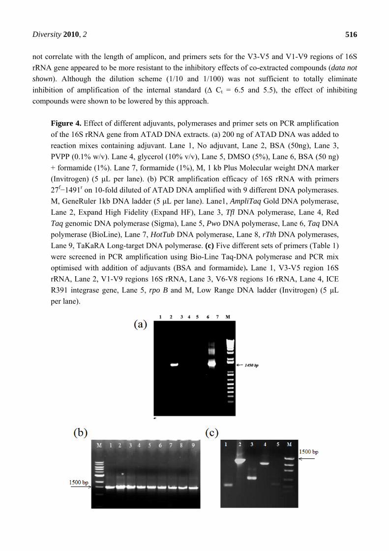
Figure 4. Effect of different adjuvants, polymerases and primer sets on PCR amplification

of the 16S rRNA gene from ATAD DNA extracts. (a) 200 ng of ATAD DNA was added to

reaction mixes containing adjuvant. Lane 1, No adjuvant, Lane 2, BSA (50ng), Lane 3,

PVPP (0.1% w/v). Lane 4, glycerol (10% v/v), Lane 5, DMSO (5%), Lane 6, BSA (50 ng)

+ formamide (1%). Lane 7, formamide (1%), M, 1 kb Plus Molecular weight DNA marker

(Invitrogen) (5 μL per lane). (b) PCR amplification efficacy of 16S rRNA with primers

27f−1491r on 10-fold diluted of ATAD DNA amplified with 9 different DNA polymerases.

M, GeneRuler 1kb DNA ladder (5 μL per lane). Lane1, AmpliTaq Gold DNA polymerase,

Lane 2, Expand High Fidelity (Expand HF), Lane 3, Tfl DNA polymerase, Lane 4, Red

Taq genomic DNA polymerase (Sigma), Lane 5, Pwo DNA polymerase, Lane 6, Taq DNA

polymerase (BioLine), Lane 7, HotTub DNA polymerase, Lane 8, rTth DNA polymerases,

Lane 9, TaKaRA Long-target DNA polymerase. (c) Five different sets of primers (Table 1)

were screened in PCR amplification using Bio-Line Taq-DNA polymerase and PCR mix

optimised with addition of adjuvants (BSA and formamide). Lane 1, V3-V5 region 16S

rRNA, Lane 2, V1-V9 regions 16S rRNA, Lane 3, V6-V8 regions 16 rRNA, Lane 4, ICE

R391 integrase gene, Lane 5, rpo B and M, Low Range DNA ladder (Invitrogen) (5 μL

per lane).

Diversity 2010, 2

517

Since many ATAD sludge factors such as nucleases, carbohydrates, humic substances, and

synthetic fiber material may result in inhibition of PCR amplification, several adjuvants were added in

an attempt to improve amplification and recovery of the 16S rRNA gene by various DNA polymerases

(Figure 4b). With the exception of BSA and BSA with formamide (Figure 4a, Lane 2 and 9), none of

the other adjuvants had positive effects. Synergistic or additive effects were observed when BSA

(Figure 4a, Lane 2) was combined with formamide (Figure 4a, Lane 2) and resulted in PCR products

suitable for further molecular biological analysis. Enhanced efficiency of PCR was observed for

polymerase used with the addition of adjuvant (Figure 4a) and was shown to enhance detection

sensitivity and recovery of amplicons from multiple primers targeting different genetic loci (Figure

4c). These targets are the most commonly utilised for investigation of molecular diversity and its

dynamics. The addition of adjuvant worked well to eliminate inhibition and enhanced the efficiency of

PCR in the spiked internal standard assay and restored Ct ( Ct = 0.4) compared to mixtures without

adjuvants ( Ct =7.5) clearly demonstrating the positive effect of adjuvants and indeed modification of

different amplification parameters in optimizing recovery from ATAD sludges.

Optimising the PCR mixture allowed amplification of the 16S rRNA gene with as little as 2 pg of

ATAD DNA obtained by the solvent-based method and the sensitivity was comparable with titration

assays obtained on ATAD DNA extracts prepared by the commercial MoBIO kit (data not shown).

The use of touchdown PCR in conjunction with optimisation of PCR mix composition did not lead to

any non specific products in this study. The ability of the PCR reaction to recover rare genetic targets

within ATAD samples is extremely important for diversity studies, for pathogen detection and for

detection of rare species and it is clear that adjuvant addition may play a key role in this regard. BSA is

thought to bind polyphenolic substances, humic substances, and anions by virtue of its high lysine

content and bind lipids via hydrophobic interactions [81], all of which are present in ATAD

sludge [40]. In addition BSA may provide an alternate substrate for exogenous proteases and hence

protect the DNA polymerase somewhat from proteolysis. BSA in the presence of formamide was more

effective (Figure 4a, Lane 6) than BSA on it own (Figure 4a, Lane 2). Formamide is known to affect

the efficacy of PCR reactions via weakening of the hydrogen bonds between nucleotides, reducing the

formation of complexes and secondary structures in the targeted DNA molecule [82]. It enhances

specificity by lowering the effective annealing temperature of the primers and simultaneously provides

effective denaturation of the DNA template during amplification [83]. Formamide has also been shown

to possess DNase inhibitory activity on DNA templates isolated from human and animal saliva [84]

and pathogenic biofilms [85,86] and this may be of key importance in the case of ATAD sludge.

3.4. Capacity of DNA Extraction Methods to Reproduce a Total Community Profile

The effect of the extraction procedure on the detection of predominant and rare members of

bacterial community was assessed by examining amplified pools of the 16S rRNA gene from ATAD

DNA extracts The migration behavior of recovered V3-V5 region amplicons of the 16S rRNA gene via

DGGE and detailed taxonomical analysis based on clone library construction and nucleotide

sequencing were applied. DGGE fingerprinting of the ATAD bacterial community DNA, amplified by

primer pair 338fGC−518r (Table 1) was used to assess the ability of different methods to recover

community DNA from predominant and rare bacterial members. DGGE profiles differed for the two

Diversity 2010, 2

518

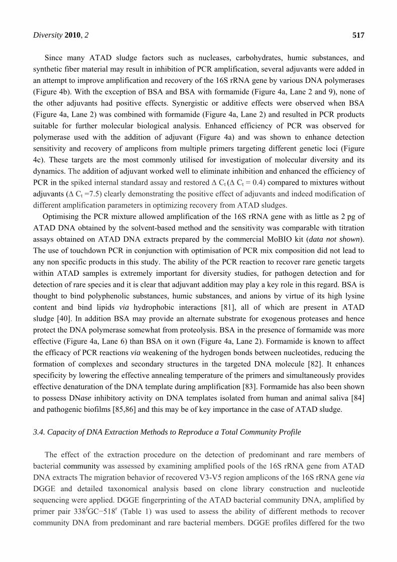
extraction methods used (Figure 5b), which were stable and reproducible in replicates (Figure 5b,

Lanes 1 and 3, Lane 2 and 4). Indexes of bacterial diversity were calculated from DGGE profiles, such

as total band number (richness of diversity) and the number of unique bands within each profile. The

MoBIO extraction method recovered a higher number of bands (N = 21) (Figure 5b, Lane 2 and 4) and

had a higher mean diversity compared to the DGGE profile obtained from the DNA extracted by the

solvent-based method (19 band) (Figure 5b, Lanes 1 and 3). Comparison of the two profiles revealed

that each profile contained 3 unique bands, those originating from the solvent extraction method had a

longer migration distance corresponding to a higher denaturant percentage within the gel suggesting a

higher GC content, often associated with thermophilic organisms [87]. The two DGGE profiles shared

16 common bands, with different migration behavior and nucleotide sequences which may originate

from 16 different bacterial species or indeed from a lower number of species with multiple copies of

the 16S rRNA gene [88]. Taxonomical identification of dominant and rare sequences in a constructed

clone library of near full length 16S rRNA gene sequences (~1450 bp) obtained by amplification of

ATAD DNA was performed. The resolution power and the distribution of the predominant and rare

bacterial members for each DNA extraction method is presented (Table 2).

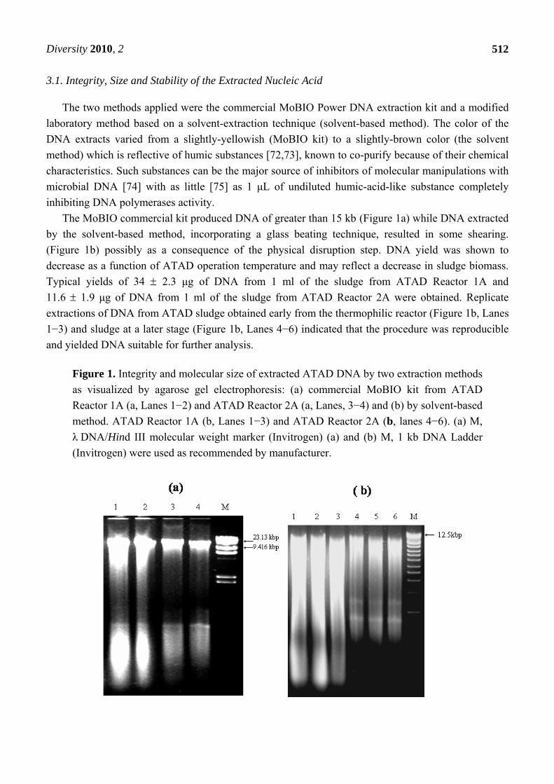
Figure 5. DGGE analysis of ATAD bacterial community DNA extracted by the two

extraction procedures with amplification of the V3-V5 hypervariable region of 16S rRNA

gene. (a) Non-denaturing gel electrophoresis of PCR products of the V3-V5 region

amplified from ATAD DNA extracted by MoBIO commercial kit Lane (1) and solvent-

based method Lane (2). M, 100 bp DNA ladder (5 μl per lane). (b) DGGE (10%

acrylamide, 30 to 80% denaturant) profile of the V3-V5 region amplified from MoBIO

extracts (Lane 1 and 3), solvent based DNA extract (Lane 2 and 4), M, pBR 322/Alu I

(1 μg per lane).

Diversity 2010, 2

519

Table 2. Taxonomic affiliation of predominant and unique clones within the clone libraries

constructed with ATAD DNA extracts obtained by two different DNA extraction

protocols.

Both libraries shared most of the sequence types, but the identity of the predominant members and

the presence of unique sequences differed within each library. The predominant members of the clone

library (Table 2) obtained from DNA extracted by the MoBIO method were bacterial species whose

nearest taxonomic identity were Clostridium ultunense (NCBI accession number EF174267), isolate

Clostridial sp. PO (NCBI AN AJ002593) and Clostridium sp. PML3-1 (NCBI accession number

EF165015). The presence of anaerobes in an aerobic treatment process may be reflective of poor

oxygen solubility, poor mixing, insulating effects of flocs, and incomplete aeration in large scale

ATAD systems. Analysis of predominant members in the clone library obtained via the solvent-based

method revealed a predominance of the species with identity to Ureibacillus thermosphaericus

(AB101594), Bacillus thermocloaceae (Z26939) and Symbiobacterium thermophilum (AP006840),

which are known to be aerobic thermophilic bacteria with high metabolic and enzymatic activity. The

presence of several clones, for example, ER 9 (closest match Moorella glycerini SQL, GQ872425), ER

17 (closest match Anoxybacillus toebii NS1-1, AY466700), ER 59 (closest match Sphingosinicella

microcystinivorans, EU337119) (Table 2), were unique to the clone library constructed with DNA

extracted by the solvent-based method and provides information on the presence of a more

OTU

from

ATAD

library

Accesssion

number Closest affiliation %

Closest affiliation

to cultivated strains %

% of total clones in the library

Library type

MoBIO

kit

Solvent- based method

CK5 GU320654 Uncultured bacterium, clone H1-814, EF174267

99 Clostridium ultunense Z69293 94 17.5 8

CK17 GU320658 Uncultured bacterium, clone BSA1B-10, AB175364

95

Mesophilic clostridial digester isolate PO AJ002593

95

13 3

CK29 GU320663 Uncultured bacterium E91, AM500794

95 Clostridia sp.PM13-1, EF165015

95 9.5 2

ER78 GU325838 Uncultured compost bacterium, clone 4B18, DQ346582

99 Bacillus sp. MSP06G, AB084065 Bacillus thermocloaceae; DSM 5250, Z26939

98 98

0 14.5

CK27 GU320661 Uncultured bacterium, clone SMQ30, AM930327

99 Bacillus sp. MSP06G ; AB084065 Bacillus thermocloaceae; DSM 5250; Z26939

98 97.5

4 17.5

CK28 GU320662 Uncultured bacterium clone F24; AM500822

99 Bacillus sp. 50LAy-1, AB375754 Ureibacillus thermosphaericus; S7, AF403019

99 99

3 9.5

ER32 GU325832

Symbiobacterium sp. KY38, AB361629

99 Symbiobacterium thermophilum IAM 14863 DNA AP006840

97

1 30.5

ER 9 GU325829

Uncultured compost bacterium clone 1B07, DQ346486

98 Moorella glycerini SQL, GQ872425

97 0 2.5

ER 17 GU325830 Uncultured bacterium, clone SMQ48; AM930332

98 Anoxybacillus toebii NS1-1, AY466700

98 0 3.5

ER 59 GU325836

UnculturedSphingomonadaceae clone 113-Cadma, AB478689

96 Sphingosinicella microcystinivorans, EU337119

94 0 2

Diversity 2010, 2

520

physiologically diverse group. It also indicates for the first time the presence of symbionts and

ammonia reducing organisms within the ATAD sludge matrix.

The application of two DNA extraction methods to molecular analysis of ATAD sludge offers

useful insights and attributes. The MoBIO method is rapid, easy and affordable, resulting in the

recovery of high molecular weight ATAD DNA with little inhibition of PCR. The solvent-based

method recovered other species not detected in the MoBIO extracts which leads to the conclusion that

combining extracts from several procedures could aid maximal recovery of diversity. Extraction of

DNA from tertiary sludge and its subsequent molecular analysis is problematic and thus far no method

gives a perfect picture of the bacterial diversity. With optimisation, sample modification, use and

pooling of extracts generated by different procedures, richness and diversity recovery can be

improved. ATAD sludge contributes significant nuclease activity which must be addressed while other

co-extracted ATAD materials influence primer specificity, PCR performance and yield. Specific

modifications to extraction methods, addition of adjuvant and use and pooling of multiple extraction

methodologies have been shown to be useful in diversity recovery from ATAD samples but may also

have more widespread application in the analysis of other thermal environmental niches.

Acknowledgements

This work was financially supported by a collaborative research grant from the Higher Education

Authority, PRTLI 4, Ireland. Authors acknowledge the assistance of staff at the Killarney WasteWater

treatment facilities during sampling at the ATAD plant.

References and Notes

1. Nocker, A.; Burr, M.; Camper, A.K. Genotypic microbial community profiling: a critical

technical review. Microb. Ecol. 2007, 54, 276-893.

2. Holben, W.E.; Harris, D. DNA-based monitoring of total bacterial community structure in

environmental samples. Mol. Ecol. 1995, 4, 627-631.

3. Giraffa, G.; Neviani, E. DNA-based, culture-independent strategies for evaluating microbial

communities in food-associated ecosystems. Int. J. Food Microbiol. 2001, 20, 19-34.

4. Ranjard, L.; Poly, F.; Nazaret, S. Monitoring complex bacterial communities using

culture-independent molecular techniques: application to soil environment. Res. Microbiol. 2000,

151, 167-177.

5. Kawai, M.; Matsutera, E. 16S ribosomal DNA-based analysis of bacterial diversity in purified

water used in pharmaceutical manufacturing processes by PCR and denaturing gradient gel

electrophoresis. Appl. Environ. Microbiol. 2000, 68, 699-704.

6. Zhou, X.; Bent, S.J.; Schneider, M.G.; Davis, C.C.; Islam, M.R.; Forney, L.J. Characterizationof

vaginal microbial communities in adult healthy women using cultivation-independent methods.

Microbiology 2004, 150, 2565-2573.

7. Amann, R.I.; Ludwig, W.; Schleifer, K.H. Phylogenetic identification and in situ detection of

individual microbial cells without cultivation. Microbiol. Rev. 1995, 59, 143-146.

8. Dahllöf, I. Molecular community analysis of microbial diversity. Curr. Opin. Microbiol. 2002, 13,

213-217.

Diversity 2010, 2

521

9. Colwell, R.; Grimes.J. Semantics and strategies. In Non-culturable Microorganisms in the

Environment; Colwell, R., Grimes, J., Eds.; ASM: Washington. DC, USA, 2000; pp. 1-5.

10. Kazuhiro, K. Nonculturable bacterial populations that control environmental processes. Biosci.

Ind. 1999, 57, 731-736.

11. Frostegard, A.; Courtois, S.; Ramisse, V.; Clerc, S.; Bernillon, D.; Le Gall, F.; Jeannin, P.;

Nesme, X.; Simonet, P. Quantification of bias related to the extraction of DNA directly from

soils. Appl. Environ. Microbiol. 1999, 65, 5409-5420.

12. Wu, L.; Li, F.; Deng, C.; Xu, D.; Jiang, S; Xiong, Y. A method for obtaining DNA from compost.

Appl. Biochem. Biotechnol. 2009, 84, 389-395.

13. Al-Soud, W.A.; Radstrom, P. Capacity of nine thermostable DNA polymerases to mediate DNA

amplification in the presence of PCR-inhibiting samples. Appl. Environ. Microbiol. 1998, 64,

3748-3753.

14. Weiss, A.; Jerome, V.; Freitag, R. Comparison of strategies for the isolation of PCR-compatible,

genomic DNA from a municipal biogas plants. J. Chromatogr. B Analyt. Technol. Biomed. Life

Sci. 2007, 15, 190-197.

15. Stach, J.E.; Bathe, S.; Clapp, J.P.; Burns, R.G. PCR-SSCP comparison of 16S rDNA sequence

diversity in soil DNA obtained using different isolation and purification methods. FEMS

Microbiol. Ecol. 2001, 36, 139-151.

16. McIlroy, S.J.; Porter, K.; Seviour, R.J; Tillett, D. Extracting nucleic acids from activated sludge

which reflect community population diversity. Antonie van Leeuwenhoek 2009, 96, 593-605.

17. McOrist, A.L.; Jackson, M.; Bird, A.R. A comparison of five methods for extraction of bacterial

DNA from human faecal samples. J. Microbiol. Meth. 2002, 50, 131-139.

18. Muyzer, G.; de Waal, E.C.; Uitterlinden, A.G. Profiling of Complex Microbial populations by

Denaturing Gradient Gel Electrophoresis Analysis of Polymerase Chain Reaction-Amplified

Genes Coding for 16S rRNA. Appl. Environ. Microbiol. 1993, 59, 695-700.

19. Fischer, S.G.; Lerman, L.S. DNA fragments differing by single base-pair substitutions are

separated in denaturing gradient gels: correspondence with melting theory. Proc. Natl. Acad. Sci.

USA 1983, 80, 1579-1583.

20. Durtschi, J.D.; Voelkerding, K.V.; Wittwer, C.T. Seipp, M.T. Multiplex amplicon genotyping by

high-resolution melting. J. Biomol.Tech. 2009, 20, 160-164.

21. Relman, A. Universal bacterial 16S rDNA amplification and sequencing. In: Diagnostic

Molecular Microbiology, Principles and Applications; Persing, D.H., Smith, T.F., Tenover F.C.,

White, T.J., Eds.; ASM: Washington, DC, USA, 1993; pp. 489-495.

22. Petti, C.A.; Polage, C.R.; Schreckenberger, P. The role of 16S rRNA gene sequencing in

identification of microorganisms misidentified by conventional methods J. Clin. Microbiol. 2005,

43, 6123-6125.

23. Alvarez, A.J.; Buttner, M.P.; Stetzenbach, L.D. PCR for bioaerosol monitoring: sensitivity and

environmental interference. Appl. Environ. Microbiol. 1995, 61, 3639-3644.

24. Lantz, P.G.; Abu Al-Soud, W.A.; Knutsson, R.; Hahn-Hägerdal, B.; Radstrom, P. Biotechnical

use of polymerase chain reaction for microbiological analysis of biological samples. Biotechnol.

Annu. Rev. 2000, 5, 87-130.

Diversity 2010, 2

522

25. Heuer, H.; Smalla, K. Application of denaturing gradient gel electrophoresis and temperature

gradient gel electrophoresis for studying soil microbial communities. In Modern Soil

Microbiology; van Elsas, J.D., Trevors, J.T., Wellington, E.M.H., Eds.; Marcel Dekker: New

York, NY, USA, 1997; pp. 353-373.

26. Chandler, D.P.; Frederickson, J.K.; Brockman, J. Effect of PCR template concentration on the

composition and distribution of total community 16S rDNA clone libraries. Mol. Ecol. 1997, 6,

475-482.

27. Pomp, D.; Medrano, J.F. Organic solvents as facilitators of polymerase chain reaction.

BioTechniques 1991, 10, 58-59.

28. Sidhu, M.K.; Liao, M.J.; Rashidbaigi, A. Dimethyl sulfoxide improves RNA amplification.

BioTechniques 1996, 21, 44-47.

29. Nol, P.; Williamson, J.L.; Rocke, T.E.; Yuill, T.M. Detection of Clostridium botulinum type C

cells in the gastrointestinal tracts of Mozambique Tilapia (Oreochromis mossambicus) by

polymerase chain reaction. J. Wildl Dis. 2004, 40, 749-753.

30. Klammer, S.; Mondini, C., Insam, H. Microbial community fingerprint of compost stored under

different condition. Ann. Microbiol. 2005, 55, 299-305.

31. Maarit-Niemi, R.; Heiskanen, I.; Wallenius, K.; Lindstrom, K. Extraction and purification of

DNA in rhizosphere soil samples for PCR-DGGE analysis of bacterial consortia. J. Microbiol.

Meth. 2001, 45, 155-165.

32. Bramucci, M.G.; Nagarajan, V. Industrial wastewater bioreactors: sources of novel

microorganisms for biotechnology. Trends Biotechnol. 2000, 18, 501-505.

33. Sen, R; Chakrabarti, S. Biotechnology—applications to environmental remediation in resource

exploitation. Curr. Sci. 2009, 97, 768-775.

34. Layden, N.; Mavinic, D.; Kelly, H.; Moles, R.; Bartlett, J. Autothermal thermophilic aerobic

digestion (ATAD)–Part II: Review of research and full-scale operating experiences. J. Environ.

Eng. Sci. 2007, 6, 679-690.

35. Sonnleitner, B.; Fiechter, A. Bacterial diversity in thermophilic aerobic sewage sludge II. Types

of organisms and their capacities. Eur. J. Appl. Microbiol. Biotechnol. 1983, 18, 174-180.

36. Sonnleitner, B.; Fiechter, A. Microbial flora studies in thermophilic aerobic sludge treatment.

Conserv. Recycl. 1985, 8, 303-313.

37. Juteau, P.; Tremblay, D.; Villemur, R.; Bisaillon, J.G.; Beaudet, R. Analysis of the bacterial

community inhabiting an aerobic thermophilic sequencing batch reactor (AT-SBR) treating swine

waste. Appl. Microbiol. Biotechnol. 2004, 66, 115-122.

38. LaPara, T.M.; Nakatsu, C.H.; Pantea, L.; Alleman, J.E. Phylogenetic analysis of bacterial

communities in mesophilic and thermophilic bioreactors treating pharmaceutical wastewater.

Appl. Environ. Microbiol. 2000, 66, 3951-3959.

39. Piterina, A.V.; MacCausland, C.; Bartlett, J.; Pembroke, J.T. Microbial ecology of autothermal

aerobic digestion (ATAD): diversity, dynamics and activity of bacterial communities involved in

treatment of municipal wastewater. In Modern Multidisciplinary Applied Microbiology,

Exploiting Microbes and Their Interactions; Mendez-Vilas, A., Ed.; Wiley-VCH: Weinheim,

Germany, 2006; pp. 526-535.

Diversity 2010, 2

523

40. Piterina, A.V.; Barlett, J.; Pembroke, T.J. 13C-NMR assessment of the pattern of organic matter

transformation during domestic wastewater treatment by Autothermal Aerobic Digestion

(ATAD). Int. J. Environ. Res. Public Health 2009, 6, 2288-2306.

41. Miller, D.N.; Bryant, J.E.; Madsen, E.L.; Ghiorse, W.C. Evaluation and optimization of DNA

extraction and purification procedures for soil and sediment samples. Appl. Environ. Microbiol.

1999, 65, 4715-4724.

42. Martin-Laurent, F.; Philippot, L.; Hallet, S.; Chaussod, R.; Germon, J.C.; Soulas, G.; Catroux G.

DNA extraction from soils: old bias for new microbial diversity analysis method, Appl. Environ.

Microbiol. 2001, 67, 2354-2359.

43. Sambrook, J. Molecular Cloning: A Laboratory Manual, 3rd ed.; Cold Spring Harbor Laboratory

Press: Cold Spring Harbor, NY, USA, 2001.

44. Lane, D.J. 16S/23S rRNA Sequencing. In Nucleic Acid Techniques in Bacterial Systematics;

Stackebrandt, E., Goodfellow, M., Eds.; John Wiley & Sons: New York, NY, USA, 1991;

pp. 115-175.

45. Muyzer, G.; Hottentrager, S.; Teske, A.; Wawer, C. Denaturing gradient gel electrophoresis of

PCR-amplified 16S rDNA. A new molecular approach to analyse the genetic diversity of mixed

microbial communities. In Molecular Microbial Ecology Manual; Akkermans, A., Ed.; Kluwer

Academic Publ.: Norwell, MA, USA, 1996; p. 23.

46. Nubel, U.; Engelen, B.; Felske, A.; Snaidr, J.; Wieshuber, A.; Amann, R.I.; Ludwig, W.;

Backhaus, H. Sequence heterogeneities of genes encoding 16S rRNAs in Paenibacillus polymyxa

detected by temperature gradient gel electrophoresis. J. Bacteriol. 1996, 178, 5636-5643.

47. Dahllof, I.; Baillie, H.; Kjelleberg, S. rpoB-based microbial community analysis avoids

limitations inherent in 16S rRNA gene intraspecies heterogeneity. Appl. Environ. Microbiol.

2000, 66,

3376-3380.

48. Böltner, D.; MacMahon, C.; Pembroke, J.T.; Strike, P.; Osborn, A.M. R391: a conjugative

integrating mosaic comprised of phage, plasmid and transposon elements. J. Bacteriol. 2002, 184,

5158-5169.

49. McGrath, B.M; O’Halloran, J.A; Piterina, A.V.; Pembroke, J.T. Molecular tools to detect the IncJ

elements: a family of integrating, antibiotic resistant mobile genetic elements. J. Microbiol.

Methods 2006, 66, 32-42.

50. Wilson, I.G. Inhibition and facilitation of nucleic acid amplification. Appl. Environ. Microbiol.

1997, 63, 3741-3751.

51. Demeke, T.; Adams, R.P. The effects of plant polysaccharides and buffer additives on PCR.

Biotechniques 1992, 12, 332-334.

52. Furrer, B.; Candrian, U.; Wieland, P.; Luthy, J. Improving PCR efficiency. Nature 1990, 26, 324.

53. Kreader, C.A. Relief of amplification inhibition in PCR with bovine serum albumin or T4 gene 32

protein. Appl. Environ. Microbiol. 1996, 62, 1102-1106.

54. Sarkar, G.; Kapelner, S.; Sommer, S.S. Formamide can dramatically improve the specificity of

PCR. Nucl. Acids Res. 1990, 25, 7465.

Diversity 2010, 2

524

55. Rossen, L.; Norskov, P.; Holmstrom, K.; Rasmussen, O.F. Inhibition of PCR by components of

food samples, microbial diagnostic assays and DNA-extraction solutions. Int. J. Food Microbiol.

1992, 17, 37-45.

56. Akane, A.; Matsubara, K.; Nakamura, H.; Takahashi, S.; Kimura, K. Identification of the heme

compound copurified with deoxyribonucleic acid (DNA) from bloodstains, a major inhibitor of

polymerase chain reaction (PCR) amplification. J. Forensic Sci. 1994, 39, 362-372.

57. Suarez, D. Molecular diagnostic techniques: can we identify influenza viruses, differentiate

subtypes and determine pathogenicity potential of viruses by RT-PCR? Avian Dis. 1997, 47,

318-325.

58. Sarkar, G.; Kapelner, S.; Sommer, S.S. Formamide can dramatically improve the specificity of

PCR. Nucl. Acids Res. 1990, 18, 7465.

59. Abu Al-Soud, W.A.; Radstrom, P. Effects of amplification facilitators on diagnostic PCR in the

presence of blood, feces, and meat. J. Clin. Microbiol. 2000, 38, 4463-4470.

60. Chou, S.W. Optimizing polymerase chain reaction technology for clinical diagnosis. Clin. Chem.

1991, 37, 1893-1894.

61. Kleiboeker, S.B. Applications of competitor RNA in diagnostic reverse transcription-PCR. J.

Clin. Microbiol. 2003, 41, 2055-2061.

62. Sipos, R.; Székely, A.; Révész, S.; Márialigeti, K. Addressing PCR biases in environmental

microbiology studies. Methods Mol. Bio 2010, 599, 37-58.

63. Don, R.H.; Cox P.T.; Wainwright, B.J.; Baker, K.; Mattick, J.S. “Touchdown” PCR to circumvent

spurious priming during gene amplification. Nucl. Acids Res. 1991, 19, 4008.

64. Hongoh, Y.; Yuzawa, H.; Ohkuma, M.; Kudo, T. Evaluation of primers and PCR conditions for

the analysis of 16S rRNA genes from a natural environment. FEMS Microbiol. Lett. 2003, 25,

299-304.

65. Massol-Deya, A.A.; Odelson, D.A.; Hickey, R.F.; Tiedje, J.M. Bacterial Community

Fingerprinting of Amplified 16S and 16-23S Ribosomal DNA Gene Sequences and Restriction

Endonuclease Analysis (ARDRA). In: Molecular Microbial Ecology Manual. Akkermans,

A.D.L., Ed.; Kluwer Academic Publishers: Dordrecht, The Netherlands, 1995; pp. 1-8.

66. CLC bio’s Workbenches (http://www.clcbio.com).

67. VecScreen (http://www.ncbi.nlm.nih.gov/VecScreen/VecScreen.html).

68. Sequence manipulation suite (http://www.gobozzy.com/sequence-analysis-tools).

69. Maidak, B.L; Cole, J.R.; Lilburn, T.G.; Parker, C.T., Jr; Saxman, P.R.; Farris, R.J.; Garrity, G.M.;

Olsen, G.J.; Schmidt, T.M.; Tiedje, J.M. The RDP-II (Ribosomal Database Project). Nucl. Acids

Res. 2001, 29, 173-174.

70. Altschul, S.F.; Gish, W.; Miller, W.; Myers, E.W.; Lipman, D.J. Basic local alignment search

tool. J. Mol. Biol. 1990, 215, 403-410.

71. Wang, G.C.T.; Wang, Y. The frequency of chimeric molecules as a consequence of PCR

co-amplification of 16S rRNA genes from different bacterial species. Microbiology 1996, 142,

1107-1114.

72. Kuske, C.R.; Banton, K.L.; Adorada, D.L.; Stark, P.C.; Jackson P.J. Small-scale DNA sample

preparation method for field PCR detection of microbial cells and spores in soil. Appl. Environ.

Microbiol. 1998, 64, 2463-2472.

Diversity 2010, 2

525

73. DellAnno, A.; Fabiano, M.; Duineveld, G.C.A.; Kok, A.; Danovaro, R. Nucleic acid (DNA, RNA)

quantification and RNA/DNA ratio determination in marine sediments: comparison of

spectrophotometric, fluorometric and High Performance Liquid Chromatography methods and

estimation of detrital DNA. Appl. Environ. Microbiol. 1998, 64, 3238-3245.

74. Jackson, C.R.; Harper, J.P.; Willoughby, D.; Roden, E.E.; Churchill, P.F. A simple, efficient

method for the separation of humic substances and DNA from environmental samples. Appl.

Environ. Microbiol. 1997, 63, 4993-4995.

75. Radstrom, P.; Knutsson, R.; Wolffs, P.; Lövenklev, M.; Löfström, C. Pre-PCR processing:

Strategies to generate PCR-compatible samples. Mol. Biotechnol. 2004, 26, 133-146.

76. Abu Al-Soud, W.; Râdström, P. Capacity of nine thermostable DNA polymerases to mediate

DNA amplification in the presence of PCR-inhibiting samples. Appl. Environ. Microbiol. 1998,

64, 3748-3753.

77. LaMontagne, M.G.; Michel, F.C., Jr.; Holden, P.A.; Reddy, C.A. Evaluation of extraction and

purification methods for obtaining PCR-amplifiable DNA from compost for microbial community

analysis. J. Microbiol. Meth. 2002, 49, 255-264.

78. Arbeli, Z.; Fuentes, C.L. Improved purification and PCR amplification of DNA from

environmental samples. FEMS Microbiol. Lett. 2007, 272, 269-275.

79. Blanchard, M.M.; Tailon-Miller, P.; Nowotny, P.; Nowotny, V. PCR buffer optimization with a

uniform temperature regimen to facicilate automation. PCR Meth. Applic. 1993, 2, 234-240.

80. Innis, M.A.; Gelfand, D.H. Optimization of PCRs. In PCR protocols. A guide to methods and

applications; Innis, M.A., Gelfand, D.H., Sninsky, J.J., White, T.J., Eds.; Academic Press Inc.:

San Diego, CA, USA, 1990; p. 12.

81. Loomis, W.D. Overcoming problems of phenolics and quinones in the isolation of plant enzymes

and organelles. Meth. Enzymol. 1974, 31, 528-545.

82. Zhang, W.; Hu, G.Y.; Deisseroth, A. Improvement of PCR sequencing by formamide. Nucleic

Acids Res. 1991, 19, 6649.

83. Loeffelholz, M.;Deng, H. PCR and Its Variations. In Advanced techniques in Diagnostic

Microbiology; Tang, Y.W., Stratton, C.W., Eds.; Springer: New York, NY, USA, 2006;

Volume 1, pp. 166-183.

84. Wiedbrauk, D.L.; Werner, J.C.; Drevon, A.M. Inhibition of PCR by aqueous and vitreous fluids.

J. Clin. Microbiol. 1995, 33, 2643-2646.

85. Wilson, I.G.; Cooper, J.E.; Gilmour A. Some factors inhibiting amplification of the

Staphylococcus aureus enterotoxin C1 (sec1) by PCR. Int. J. Food Microbiol. 1994, 22, 55-62.

86. Gibson, J.R.; Sutherland, K.; Owen, R.J. Inhibition of DNAse activity in PFGE analysis of DNA

from Campylobacter jejuni. Lett. Appl. Microbiol. 1994, 19, 357-358.

87. Singer, G.A.; Hickey, D.A. Thermophilic prokaryotes have characteristic patterns of codon usage,

amino acid composition and nucleotide content. Gene 2003, 23, 39-47.

88. Klappenbach, J.A.; Saxman, P.R.; Cole, J.R.; Schmidt, T.M. rrndb: the Ribosomal RNA Operon

Copy Number Database. Nucleic Acids Res. 2001, 29, 181-184.

Diversity 2010, 2

526

© 2010 by the authors; licensee Molecular Diversity Preservation International, Basel, Switzerland.

This article is an open-access article distributed under the terms and conditions of the Creative

Commons Attribution license (http://creativecommons.org/licenses/by/3.0/).


Recommended