Molecular Analysis of Bacterial Microbiota Associated with Oysters (Crassostrea gigas and Crassostrea corteziensis) in Different Growth Phases at Two Cultivation Sites Natalia Trabal & José M. Mazón-Suástegui & Ricardo Vázquez-Juárez & Felipe Asencio-Valle & Enrique Morales-Bojórquez & Jaime Romero Received: 6 December 2011 / Accepted: 6 March 2012 / Published online: 27 March 2012 # Springer Science+Business Media, LLC 2012 Abstract Microbiota presumably plays an essential role in inhibiting pathogen colonization and in the maintenance of health in oysters, but limited data exist concerning their dif- ferent growth phases and conditions. We analyzed the bacte- rial microbiota composition of two commercial oysters: Crassostrea gigas and Crassostrea corteziensis. Differences in microbiota were assayed in three growth phases: post- larvae at the hatchery, juvenile, and adult at two grow-out cultivation sites. Variations in the microbiota were assessed by PCR analysis of the 16S rRNA gene in DNA extracted from depurated oysters. Restriction fragment length polymorphism (RFLP) profiles were studied using Dice’ s similarity coeffi- cient (Cs) and statistical principal component analysis (PCA). The microbiota composition was determined by sequencing temperature gradient gel electrophoresis (TGGE) bands. The RFLP analysis of post-larvae revealed homology in the micro- biota of both oyster species (Cs>88 %). Dice and PCA analyses of C. corteziensis but not C. gigas showed differ- ences in the microbiota according to the cultivation sites. The sequencing analysis revealed low bacterial diversity (primar- ily β-Proteobacteria, Firmicutes, and Spirochaetes), with Burkholderia cepacia being the most abundant bacteria in both oyster species. This study provides the first description of the microbiota in C. corteziensis, which was shown to be influenced by cultivation site conditions. During early growth, we observed that B. cepacia colonized and remained strongly associated with the two oysters, probably in a symbiotic host– bacteria relationship. This association was maintained in the three growth phases and was not altered by environmental conditions or the management of the oysters at the grow-out site. Introduction Oysters are the most produced and most valuable bivalve mollusks [23]. However, one of the main problems in the aquaculture of oysters is the repetitive episodes of mortality, which seriously reduces production. These outbreaks of disease affect the larval and post-larval stages in hatcheries and also affect juveniles and adults cultured in the natural environment. In some cases, studies have demonstrated dis- eases of bacterial etiology, including vibrioses nocardosis and infections by Chlamydia-, Mycoplasma-, and Rickett- sia-like organisms [7, 39, 59, 65, 68, 75]. Because oysters may concentrate pathogenic microorganisms, the microbiota of oysters have been examined mainly from the public health perspective, and most of the studies have focused on fecal contamination, enteric pathogens, and pathogenic species of Vibrio [17, 34, 39, 54, 67, 75–77]. The association between aquatic invertebrates and gut microbes is typically attributed to the ingestion of bacteria [31, 62]. Research with aquatic organisms has suggested a variety of beneficial roles for microbiota, including the development of the host gastrointestinal tract, nutrition N. Trabal : J. M. Mazón-Suástegui : R. Vázquez-Juárez (*) : F. Asencio-Valle : E. Morales-Bojórquez Centro de Investigaciones Biológicas del Noroeste, (CIBNOR), Mar Bermejo 195, Col. Playa Palo de Santa Rita, La Paz, Baja California Sur 23095, Mexico e-mail: [email protected]N. Trabal e-mail: [email protected]J. Romero Laboratorio de Biotecnología, Instituto de Nutrición y Tecnología de los Alimentos, Universidad de Chile, El Líbano 5524, Macul, Santiago, Chile Microb Ecol (2012) 64:555–569 DOI 10.1007/s00248-012-0039-5
Transcript
Molecular Analysis of Bacterial Microbiota Associatedwith Oysters (Crassostrea gigas and Crassostrea corteziensis)in Different Growth Phases at Two Cultivation Sites
Natalia Trabal & José M. Mazón-Suástegui &Ricardo Vázquez-Juárez & Felipe Asencio-Valle &
Enrique Morales-Bojórquez & Jaime Romero
Received: 6 December 2011 /Accepted: 6 March 2012 /Published online: 27 March 2012# Springer Science+Business Media, LLC 2012
Abstract Microbiota presumably plays an essential role ininhibiting pathogen colonization and in the maintenance ofhealth in oysters, but limited data exist concerning their dif-ferent growth phases and conditions. We analyzed the bacte-rial microbiota composition of two commercial oysters:Crassostrea gigas and Crassostrea corteziensis. Differencesin microbiota were assayed in three growth phases: post-larvae at the hatchery, juvenile, and adult at two grow-outcultivation sites. Variations in the microbiota were assessed byPCR analysis of the 16S rRNA gene in DNA extracted fromdepurated oysters. Restriction fragment length polymorphism(RFLP) profiles were studied using Dice’s similarity coeffi-cient (Cs) and statistical principal component analysis (PCA).The microbiota composition was determined by sequencingtemperature gradient gel electrophoresis (TGGE) bands. TheRFLP analysis of post-larvae revealed homology in the micro-biota of both oyster species (Cs>88 %). Dice and PCAanalyses of C. corteziensis but not C. gigas showed differ-ences in the microbiota according to the cultivation sites. Thesequencing analysis revealed low bacterial diversity (primar-ily β-Proteobacteria, Firmicutes, and Spirochaetes), with
Burkholderia cepacia being the most abundant bacteria inboth oyster species. This study provides the first descriptionof the microbiota in C. corteziensis, which was shown to beinfluenced by cultivation site conditions. During early growth,we observed that B. cepacia colonized and remained stronglyassociated with the two oysters, probably in a symbiotic host–bacteria relationship. This association was maintained in thethree growth phases and was not altered by environmentalconditions or the management of the oysters at the grow-outsite.
Introduction
Oysters are the most produced and most valuable bivalvemollusks [23]. However, one of the main problems in theaquaculture of oysters is the repetitive episodes of mortality,which seriously reduces production. These outbreaks ofdisease affect the larval and post-larval stages in hatcheriesand also affect juveniles and adults cultured in the naturalenvironment. In some cases, studies have demonstrated dis-eases of bacterial etiology, including vibrioses nocardosisand infections by Chlamydia-, Mycoplasma-, and Rickett-sia-like organisms [7, 39, 59, 65, 68, 75]. Because oystersmay concentrate pathogenic microorganisms, the microbiotaof oysters have been examined mainly from the publichealth perspective, and most of the studies have focusedon fecal contamination, enteric pathogens, and pathogenicspecies of Vibrio [17, 34, 39, 54, 67, 75–77].
The association between aquatic invertebrates and gutmicrobes is typically attributed to the ingestion of bacteria[31, 62]. Research with aquatic organisms has suggested avariety of beneficial roles for microbiota, including thedevelopment of the host gastrointestinal tract, nutrition
N. Trabal : J. M. Mazón-Suástegui :R. Vázquez-Juárez (*) :F. Asencio-Valle : E. Morales-BojórquezCentro de Investigaciones Biológicas del Noroeste, (CIBNOR),Mar Bermejo 195, Col. Playa Palo de Santa Rita,La Paz, Baja California Sur 23095, Mexicoe-mail: [email protected]
(providing vitamins, enzymes, and essential fatty acids forthe host), immune responses, and disease resistance [3, 19,26, 31, 46, 56, 62, 64]. An increased susceptibility to infec-tions may be related to the lack of the barrier provided bymicrobiota, which compete with pathogenic microorgan-isms for nutrients and space in the intestinal tract [5, 46,51, 52, 61] or produce substances that inhibit pathogens [26,28, 61].
In recent years, the natural bacterial microbiota of differ-ent oysters (Crassostrea gigas [33], Crassostrea virginica[40], Saccostrea glomerata [29], Ostrea chilensis [69], andCrassostrea iredalei [54]) have been described. However,these studies do not focus on the changes in microbiota thatmay occur during the growth of the oysters. With theexception of the study by Romero et al. [69], these reportsdo not distinguish between resident and transient bacteria,which lead to an overestimation of bacterial diversity withinthe microbiota. Because oysters are filter feeders, their gillsare covered with mucus and vibrating cilia that facilitate gasexchange for respiration and simultaneously trap suspendedparticles, including bacteria and viruses [62]. In oysters, twocategories of normal bacterial microbiota can be described:(1) indigenous or resident bacteria, including relativelyfixed types of organisms that remain relatively stable overtime and do not change with the particles ingested by thehost (even if the bacteria are disturbed, they tend to sponta-neously recover), and (2) transient or non-indigenous micro-biota, which are composed of potentially pathogenicmicroorganisms that are ingested with the food, survivepassage through the gut (possibly proliferating in the gut),and are voided with the feces [6, 31, 52]. During larvaldevelopment, transient microbiota rapidly become residentsof the oyster microbiota [8, 37, 51, 52, 62], although little isknown about the dynamics and stability of microbiota in thejuvenile and adult growth stages. It is, however, recognizedthat the physiology of the invertebrate marine organism caninfluence the gut microbiota [31, 62]. In bivalve mollusks,bacteria have been reported in the style, gland of Deshayes,esophagus, stomach, and intestine [31, 37, 51, 62]; however,many other studies on the microbiota were performed on thewhole-oyster homogenate [31, 40, 63, 69]. Colonization bybacteria in the oyster gastrointestinal tract has a particulardependence on the external environment because of the flowof water passing through the digestive tract [26, 31, 62].Considering that ambient conditions and seasonality havebeen shown to alter the gut bacteria community in someaquatic invertebrates, it is probable that the habitat can influ-ence the conditions in the gut and may therefore be an impor-tant factor in the composition of gut microbiota [26, 31, 62].However, studies of the changes in microbiota under differentenvironmental conditions are lacking [40, 62, 63].
One of the most critical steps in the cultivation of theseoysters is the planting of post-larvae (spat) from production
at hatcheries to the field, where large numbers die [7, 11,18]. Additionally, at high stocking densities in the field, theidentification of microbiota is important because the resi-dent microbiota can be disturbed, and transient microorgan-isms subsequently exploit a temporary advantage [51, 52].Productivity is related to larval transfer to the field andenvironmental conditions that can alter the bacterial com-munity associated with oysters.
During the past 20 years, oyster cultivation in Mexico hasimproved; commercially important mollusks, such as thePacific oyster C. gigas (Thunberg, 1793) and the Cortezoyster C. corteziensis (Hertlein, 1951), have been used. C.gigas is the most cultivated and widely studied oysterworldwide [30] and is the most prevalent species along thePacific coast; however, massive deaths have led to manystudies of the regional species C. corteziensis [11, 13,47–49]. Presently, the microbiota composition of Cortezoyster is unknown. In this study, using molecular techni-ques, we analyzed the composition of the bacterial micro-biota of C. gigas and C. corteziensis. Variations anddifferences in the bacterial microbiota composition wereassayed in three growth phases: post-larvae at the hatchery,juvenile, and adult at two grow-out cultivation sites in thefield. In particular, we studied the variation of the micro-biota composition when the post-larvae were planted atdifferent cultivation sites.
Methods
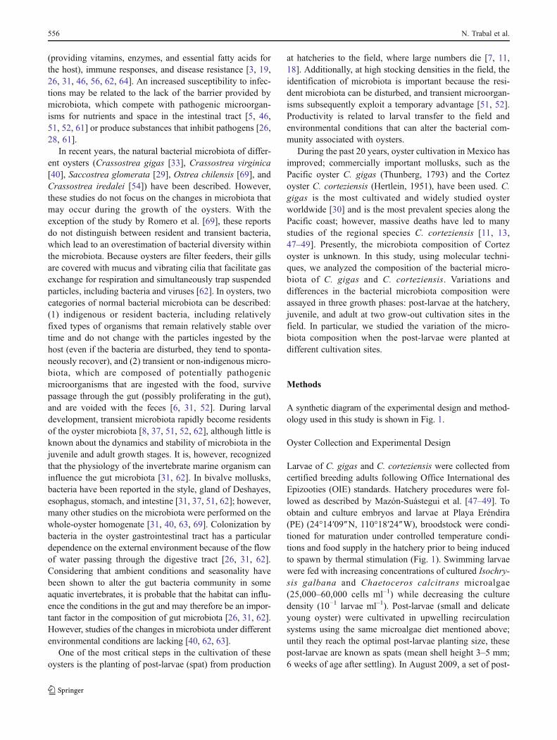
A synthetic diagram of the experimental design and method-ology used in this study is shown in Fig. 1.
Oyster Collection and Experimental Design
Larvae of C. gigas and C. corteziensis were collected fromcertified breeding adults following Office International desEpizooties (OIE) standards. Hatchery procedures were fol-lowed as described by Mazón-Suástegui et al. [47–49]. Toobtain and culture embryos and larvae at Playa Eréndira(PE) (24°14′09″N, 110°18′24″W), broodstock were condi-tioned for maturation under controlled temperature condi-tions and food supply in the hatchery prior to being inducedto spawn by thermal stimulation (Fig. 1). Swimming larvaewere fed with increasing concentrations of cultured Isochry-sis galbana and Chaetoceros calcitrans microalgae(25,000–60,000 cells ml–1) while decreasing the culturedensity (10−1 larvae ml–1). Post-larvae (small and delicateyoung oyster) were cultivated in upwelling recirculationsystems using the same microalgae diet mentioned above;until they reach the optimal post-larvae planting size, thesepost-larvae are known as spats (mean shell height 3–5 mm;6 weeks of age after settling). In August 2009, a set of post-
556 N. Trabal et al.
larvae was transferred (planting) to two grow-out cultivationsites (Fig. 1). An initial culture of spats from the oysters inthe cultured site was performed in NestierTM-type trayssuspended from a floating long-line. The juvenile oysters(3–5 cm in shell height; age 5 months after planting; Fig. 1)were cultured in cages suspended on rigid structures near thebottom. The adult oysters (6–12 cm in shell height; age6–10 months after planting, Fig. 1) were cultured exclusivelyat the bottom of the cages for improved stability and foodsupply.
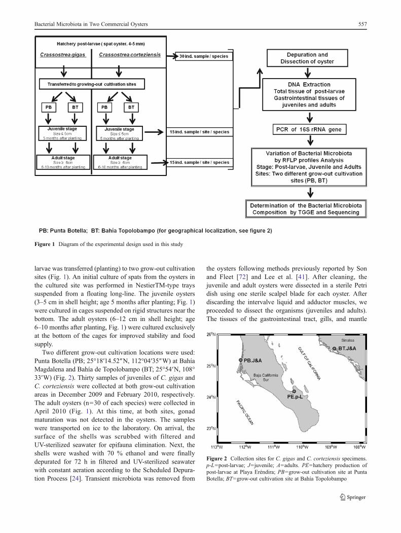
Two different grow-out cultivation locations were used:Punta Botella (PB; 25°18′14.52″N, 112°04′35″W) at BahíaMagdalena and Bahía de Topolobampo (BT; 25°54′N, 108°33′W) (Fig. 2). Thirty samples of juveniles of C. gigas andC. corteziensis were collected at both grow-out cultivationareas in December 2009 and February 2010, respectively.The adult oysters (n030 of each species) were collected inApril 2010 (Fig. 1). At this time, at both sites, gonadmaturation was not detected in the oysters. The sampleswere transported on ice to the laboratory. On arrival, thesurface of the shells was scrubbed with filtered andUV-sterilized seawater for epifauna elimination. Next, theshells were washed with 70 % ethanol and were finallydepurated for 72 h in filtered and UV-sterilized seawaterwith constant aeration according to the Scheduled Depura-tion Process [24]. Transient microbiota was removed from
the oysters following methods previously reported by Sonand Fleet [72] and Lee et al. [41]. After cleaning, thejuvenile and adult oysters were dissected in a sterile Petridish using one sterile scalpel blade for each oyster. Afterdiscarding the intervalve liquid and adductor muscles, weproceeded to dissect the organisms (juveniles and adults).The tissues of the gastrointestinal tract, gills, and mantle
Figure 1 Diagram of the experimental design used in this study
Figure 2 Collection sites for C. gigas and C. corteziensis specimens.p-L0post-larvae; J0juvenile; A0adults. PE0hatchery production ofpost-larvae at Playa Eréndira; PB0grow-out cultivation site at PuntaBotella; BT0grow-out cultivation site at Bahía Topolobampo
Bacterial Microbiota in Two Commercial Oysters 557
were separated, and the gastrointestinal tract was frozenat −20°C. Post-larval oysters (n030, for each species) werevigorously rinsed in sterile seawater, and after the intervalveliquid and adductor muscles were eliminated, the organismswere frozen at −20°C. In all cases, at the time of dissection,the oysters were alive, and the appearance of the bodies wasnormal with no evidence of infection.
Environmental Conditions of the Grow-Out CultivationArea
The Punta Botella grow-out cultivation site (PB) is locatedin Bahía Magdalena, which has one of the most diverseecosystems in the state of Baja California Sur. This locationhas low pollution and minor human activity, and it is influ-enced by oceanic currents of temperate seawater from fur-ther south. In Bahía Magdalena, the large lagoon has anirregularly shaped barrier island. There are numerous smallestuaries and shallow channels bordered by mangroves thatprovide organic matter deposited as sediment. Mangrovesare the common feeding habitat for communities of manyvertebrate and invertebrate species and possess commercialand ecological value [1, 12].
The Bahía Topolobampo grow-out site (BT) is a coastallagoon complex with relatively large mouths connecting tothe Gulf of California that is influenced by oceanic currents.In the last several decades, BT has been supporting shrimpand oyster aquaculture projects. However, the lagoon wateris contaminated by agricultural runoff and urban and indus-trial sewage effluents [57, 58].
The salinity at both sites ranged from 35 to 40 psu withan average of 37 psu.
The weekly sea surface temperature (SST) data for 2009–2010 were derived from the Moderate Resolution ImagingSpectroradiometer-Aqua (MODIS-Aqua) satellite. The SSTdata were downloaded from the National Aeronautics andSpace Administration web site (http://modis.gsfc.nasa.gov/data/). The monthly SST was estimated along a straight linecentered at each one of the study areas, and thecorresponding mean was calculated. The following datashow the SST during grow-out of juveniles and adults atboth sites: Punta Botella (August, 26.6°C; September, 28.0°C;October, 26.2°C; November, 25.5°C; December, 23.4°C;January, 21.7°C; February, 20.6°C; March, 20.4°C; and April,19.9°C) and Bahía Topolobampo (August, 27.9°C; September,29.1°C; October, 26.7°C; November, 25.9°C; December,24.6°C; January, 21.2°C; February, 19.7°C; March, 23.8°C;and April, 25.5°C). Temperate temperatures were found at thePB site, whereas the temperatures at the BT site weresubtropical. A temperature drop was also noted inOctober, which indicated the beginning of autumn atboth sites. C. gigas grew faster at PB, and C. corte-ziensis grew faster at BT. The difference is based on
temperate Pacific environmental conditions that aremore suitable for C. gigas, which is a temperate waterspecies, whereas the subtropical conditions in parts ofthe Gulf of California are more suitable for C. corte-ziensis, which is a subtropical species [13, 47, 48].
DNA Extraction and Purification
The total tissue of the post-larvae was homogenized by lysisusing a buffer containing Tris–EDTA–SDS (100 mM NaCl,50 mM Tris [pH 8], 100 mM EDTA [pH 8], sodium dodecylsulfate [SDS 1 %]) and 20 μl proteinase K (20 mg ml−1)(Sigma, St. Louis, MO) and was incubated for 1 h at 65°C.The DNA was subsequently extracted with phenol/chloro-form and precipitated with ethanol, as previously described[69].
The gastrointestinal tissues (cut into pieces) of juvenileand adult oysters were lysed with 50 ml of Tris–EDTA–SDSbuffer (as previously described), and the DNAwas extractedusing a QIAmp® DNA Mini kit (Qiagen, Valencia, CA)following the manufacturer's instructions. A final purifica-tion was performed using the Wizard® SV Gel and PCRClean-up System (Promega, Madison, WI). The concentra-tion and quality of the DNA were determined at A260 andA280 nm using an ND-1000 spectrophotometer (NanoDropTechnologies, Wilmington, DE).
PCR Amplification of the 16S rRNA Gene and Analysisof the Products
The total DNA was extracted from each oyster (total tissuepost-larvae and gastrointestinal tissues of juveniles andadults), diluted in nuclease-free water to obtain a concentra-tion of 50 ng μl−1, and used as a template in PCR. NestedPCR of the V3–V5 region of the 16S rRNA gene was usedto obtain profiles of the bacterial communities present inthree different growth stages in both oyster species and atthe two grow-out cultivation sites. Eubacterial 16S rRNAgenes were amplified with the following primers: EUB 27 F(5′-AGAGTTTGATCCTGGCTCAG-3′) and EUB1492R(5′-GTTACCTTGTTACGACTT-3′) [43] in the first ampli-fication and 341 F (5′-GCCTACGGGAGGCAGCAG-3′with GC clamps at the 5′ end for temperature gradient gelelectrophoresis analysis , TGGE) [53] and 939R(5′-CTTGTGCGGGCCCCCGTCAA TTC-3′) [70] for thesecond phase. The primers used in the specific amplificationof the eubacterial 16S rRNA gene variable region wereselected from a set of primers available in the literatureand aligned in silico using the Sequence Match softwarewith reference sequences (1,921,179 16S rRNAs) from theRibosomal Database Project II Database (RDP II). Thecriteria used to choose the best primers were as follows:greater alignment with eubacteria domain sequences,
especially with the Proteobacteria group; lack of alignmentwith Archaea domain sequences; and no cross-amplificationwith the oyster genome. PCR was performed with a reactionmixture (25 μl) containing 0.2 mM of each deoxynucleosidetriphosphate, 0.05 Uml−1 Platinum Taq DNA polymerase(Invitrogen, San Diego, CA), 1× polymerase reaction buffer,2 mM MgCl2, and 0.25 pmol/ml−1 of each primer. Thereaction mixtures were incubated in an Eppendorf Master-cycler (Eppendorf, Hamburg, Germany) with an initialdenaturation at 95°C for 10 min followed by 15 cycles at95°C for 1 min 30 s, 55°C for 1 min 30 s, 72°C for 1 min30 s, and a final elongation at 72°C for 10 min for the firstphase of the nested PCR. Subsequently, 1 μl of the ampli-fied DNA was re-amplified with an initial denaturation at94°C for 10 min followed by 20 cycles at 97°C for 1 min,54°C for 1 min, 72°C for 1 min 30 s, and a final elongationat 72°C for 10 min for second phase of the nested PCR. ThePCR products were analyzed using polyacrylamide gel elec-trophoresis and silver nitrate staining [20].
Restriction Fragment Length Polymorphism Analysisof the 16S rRNA Gene
By applying restriction fragment length polymorphism(RFLP) analysis to the PCR amplification products of the16S rRNA gene, we sought to observe variations of theresident microbiota in C. gigas and C. corteziensis in thethree growth stages at two different grow-out sites. Theproducts of the 16S rRNA gene amplification (10 μl) fromthe DNA of post-larval and gastrointestinal tissues of juve-niles and adults were digested for 2 h at 37°C with 1.5 U ofAlu1 restriction endonuclease (Invitrogen, San Diego, CA).The resulting fragments were subsequently analyzed bypolyacrylamide gel electrophoresis and silver nitrate stain-ing [20]. Each gel included BenchTop 100 bp DNA Laddermarkers (Promega) and standard bacterial RFLP controls(Vibrio harveyi ATCC 14126, Listonella pelagia ATCC25916) to validate the comparisons between the gels.
Analysis of the Variation of Microbiota Using RFLPPatterns
The RFLP profiles were analyzed using software (GelCom-par II® 5.2, Applied Maths, Austin, TX) and by applying theDice similarity index (Cs). The pairwise Cs is a similarityindex used to compare microbiotal variation in differentsamples based on the presence or absence of bands in eachlane in the gels [50]. Thus, two identical profiles create a Csvalue of 100 %, whereas completely different profiles resultin a Cs value of 0 %. Each lane in the gels (samples fromindividual oysters) can be compared with every other sam-ple; therefore, the mean percent similarities (Cs values) wereused to compare the similarity of microbiota profiles in
post-larvae, juveniles, and adults in the two grow-out sites[50]. Using this index, microbiota profiles derived fromdifferent species (C. gigas vs. C. corteziensis) and fromeach individual of the same species were compared. Thecluster analysis results were displayed as a dendrogram.UPGMAwas applied with GelComparII software. The reli-ability was tested by calculating the co-phenetic correlationsof the nodes with tools available in the GelCompar program.
Analysis of Microbiota Composition Using 16S rRNA GeneTemperature Gradient Gel Electrophoresis
Using TGGE analysis, we determined the composition ofthe microbiota associated with the oysters. We chose indi-vidual samples that showed different RFLP profiles andtriplicate samples of organisms having the same patternfrom each group analyzed (the two species of post-larvae,juveniles, and adults from Punta Botella and BahíaTopolobampo). Products obtained from the nested PCR ofthe 16S rRNA gene were separated by TGGE and analyzedusing the DCode System (Bio-Rad, Hercules, CA, USA) in0.8 mm gels composed of 6 % [w/v] acrylamide, 0.1 %bisacrylamide, 8 M urea, 20 % [v/v] formamide, and 2 %[v/v] glycerol with 1× Tris–acetate (Tris 0.04 M, acetate0.002 M, EDTA 0.001 M, pH 8.5) as the electrophoresisbuffer. The polymerization was catalyzed by 110 μl of N-N-N′-N′-tetramethylethylenediamine and 80 μl of 10 %ammonium persulfate solution added to 50 ml of the gelsolution. The gel was loaded with 6 μl of 341f-939r-GCclamp PCR products and 1 μl of dye solution and run for18 h 30 min at a fixed voltage of 65 V (±9 mA) with aparallel temperature gradient ranging from 66 to 70°C. Afterelectrophoresis, the gels were stained for 1 h by incubationwith SYBR Green at 25±2°C (room temperature). Each gelincluded standards containing PCR amplicons of knownbacterial sequences ( % GC) to validate comparisonsbetween gels [55]. The standard was composed of ampli-cons from the following bacteria: Pseudomona aeruginosaP1 (%GC: 51.8), V. harveyi ATCC 14126 (%GC: 53.1),Bacillus subtilus B4 (% GC: 54.6) and Citrobacter gillenii(%GC: 56).
Sequence Analysis of the 16S rRNA Gene
The dominant bands were identified by their correspondingintensity in each TGGE pattern. The bands were excisedfrom the gel and eluted for 12 h at 25°C in 50 μl MilliQwater. After elution, 1 μl of DNA was used forre-amplification, using the same conditions describedabove. To test for the presence of similar amplicons, bandsshowing the same migration in different lanes were checkedby RFLP using AluI (an enzyme that cuts frequently) [55].Next, 16S rDNA from the re-amplified bands or from the
Bacterial Microbiota in Two Commercial Oysters 559
bacterial isolates was purified using the Wizard® SV Geland PCR Clean-up System and sequenced (by Macrogen,Rockville, MD) with an automatic sequencer (Applied Bio-systems ABI3730XL, Carlsbad, CA).
The sequences were initially compared to the avail-able databases using the basic Local Alignment SearchTool network service [2] and aligned with referencesequences using Sequence Match software from theRibosomal Database Project II (RDP II) website [15]to determine the approximate phylogenetic affiliations.The sequences were deposited in GenBank (accessionnumbers JF522195–JF522232).
Statistical Analysis
We analyzed the differences in the bacterial communitiesassociated with C. gigas and C. corteziensis in three growthphases (post-larvae, juvenile, and adult) between two grow-out cultivation sites using orthogonal empirical functions.The differences were estimated using a multivariate analy-sis, namely, the principal component analysis (PCA). Thevectors computed from PCA are orthogonal and are uncor-related, which prevents the regression on residuals [36]. ThePCA permitted the ranking and simplification of the newvariables, called principal components, and determination ofthe total variation of the data Xj, thereby explaining thevariation with a few principal components (factor load-ings≥0.7). Only the principal component was consideredto be statistically significant for eigenvalues (l)>1.0 [36].PCA was performed from correlation matrices generatedfrom a binary matrix considering the presence/absence ofthe RFLP patterns bands, which were expressed as a valueof the Dice similarity coefficient [25]. The PCA was con-ducted using the Statistica 8.0 software package (StatSoftInc., Tulsa, OK).
Results
Similarity of Bacterial Microbiota in the Post-larvaeof C. gigas and C. corteziensis
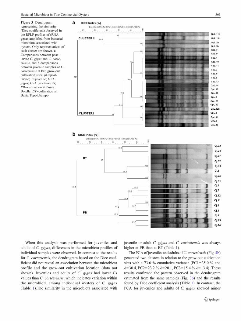
The similar RFLP profiles obtained from the cluster of post-larvae of C. corteziensis revealed a homology in the micro-biota of this oyster with Cs>92 % (Table 1, Fig. 3a). C.gigas, however, exhibited a lower similarity than did C.corteziensis with Cs>72 % (Fig. 3a, Table 1). A comparisonof the patterns obtained from the post-larvae of both speciesshowed uniformity in the composition of the microbiota andclustering with high similarity (cluster I, Cs>88 %). How-ever, cluster II was observed with high similarity (Cs>83 %), which partly corresponded to the post-larvae of C.gigas (Fig. 3a).
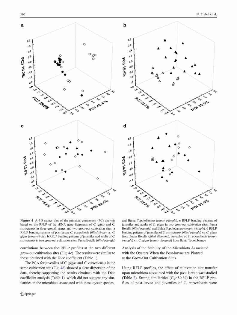
The PCA statistical method was used to compare thebacterial microbiota variation in the three growth phases(post-larvae, juvenile, and adult) of C. gigas and C. corte-ziensis at different grow-out cultivation sites, and the resultsare presented as a tridimensional scatter plot in Fig. 4. Theresults for the microbiota associated with the post-larvae inboth species of oyster are shown in Fig. 4a. The observedpost-larval cluster of C. gigas and C. corteziensis wasexplained by three principal components (PC1042.4 %and l036.9, PC2019.8 % and l017.1, PC3010.2 % andl08.8) with a 72.4 % cumulative variance. However, aseparated group of C. gigas post-larvae was also observed,which agrees with the results of the overall cluster analysisof RFLP patterns of post-larvae (Fig. 3a).
Variation in the Bacterial Microbiota of Juvenilesand Adults in Different Grow-Out Cultivation Sites
The microbiota composition in juveniles and adults of bothspecies at the two sites was studied by analyzing the patternsobtained by RFLP (Table 1). The profiles obtained fromjuvenileC. corteziensis indicated differences in the microbiotacomposition when these oysters were raised at different sites(Fig. 3b). Two groups were defined on the basis of the RFLPprofiles, which correlated with the C. corteziensis grow-outsite (Punta Botella cluster and Bahía Topolobampo cluster).For the population at Bahía Topolobampo, the Cs was >82 %,whereas at Punta Botella, the Cs was >87 %, which indicatesuniformity of the microbiota associated with each localitywith the exception of oyster JC6 (Fig. 3b, Table 1). For theC. corteziensis adults, the similarity between samples washigher at PB (Cs>75 %) than at BT (Cs>55 %) (Table 1).
Table 1 Summary of Dice similarity coefficients (percent) of theRFLP profiles of the bacterial microbiota associated with oysters,depending on the growth stage and two grow-out cultivation sites
Species Growth stage Site
H PB BT
C. corteziensis Post-larvae 92
Juvenile 87 81
Adult 75 55
C. gigas Post-larvae 72
Juvenile 53 57
Adult 56 68
Both speciesa Post-larvae 88
Juvenile 80 55
Adult 40 40
H hatchery production of post-larvae at Playa Eréndira, PB grow-outcultivation at Punta Botella, BT grow-out cultivation at BahíaTopolobampoa Comparison of RFLP profiles of C. gigas and C. corteziensis
560 N. Trabal et al.
When this analysis was performed for juveniles andadults of C. gigas, differences in the microbiota profiles ofindividual samples were observed. In contrast to the resultsfor C. corteziensis, the dendrogram based on the Dice coef-ficient did not reveal an association between the microbiotaprofile and the grow-out cultivation location (data notshown). Juveniles and adults of C. gigas had lower Csvalues than C. corteziensis, which indicates variation withinthe microbiota among individual oysters of C. gigas(Table 1).The similarity in the microbiota associated with
juvenile or adult C. gigas and C. corteziensis was alwayshigher at PB than at BT (Table 1).
The PCA of juveniles and adults ofC. corteziensis (Fig. 4b)generated two clusters in relation to the grow-out cultivationsites with a 73.6 % cumulative variance (PC1035.0 % andl030.4, PC2023.2 % l020.1, PC3015.4 % l013.4). Theseresults confirmed the pattern observed in the dendrogramestimated from the same samples (Fig. 3b) and the resultsfound by Dice coefficient analysis (Table 1). In contrast, thePCA for juveniles and adults of C. gigas showed minor
Figure 3 Dendrogramrepresenting the similarity(Dice coefficient) observed inthe RFLP profiles of rRNAgenes amplified from bacterialmicrobiota associated withoysters. Only representatives ofeach cluster are shown. aComparisons between post-larvae C. gigas and C. corte-ziensis, and b comparisonsbetween juvenile samples of C.corteziensis at two grow-outcultivation sites. pL0post-larvae; J0juvenile; G0C.gigas; C0C. corteziensis;PB0cultivation at PuntaBotella; BT0cultivation atBahía Topolobampo
Bacterial Microbiota in Two Commercial Oysters 561
correlations between the RFLP profiles at the two differentgrow-out cultivation sites (Fig. 4c). The results were similar tothose obtained with the Dice coefficient (Table 1).
The PCA for juveniles of C. gigas and C. corteziensis in thesame cultivation site (Fig. 4d) showed a clear dispersion of thedata, thereby supporting the results obtained with the Dicecoefficient analysis (Table 1), which did not suggest any sim-ilarities in the microbiota associated with these oyster species.
Analysis of the Stability of the Microbiota Associatedwith the Oysters When the Post-larvae are Plantedat the Grow-Out Cultivation Sites
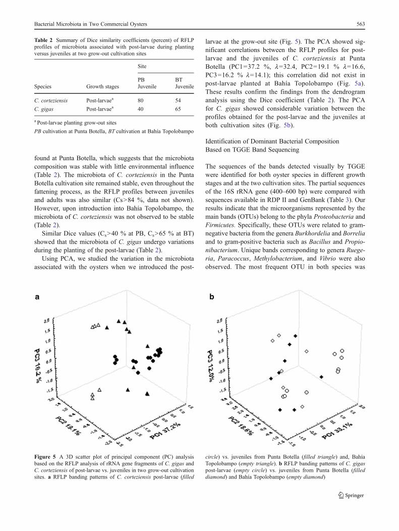
Using RFLP profiles, the effect of cultivation site transferupon microbiota associated with the post-larvae was studied(Table 2). Strong similarities (Cs>80 %) in the RFLP pro-files of post-larvae and juveniles of C. corteziensis were
Figure 4 A 3D scatter plot of the principal component (PC) analysisbased on the RFLP of the rRNA gene fragments of C. gigas and C.corteziensis in three growth stages and two grow-out cultivation sites. aRFLP banding patterns of post-larvae C. corteziensis (filled circle) vs. C.gigas (empty circle). bRFLP banding patterns of juveniles and adults of C.corteziensis in two grow-out cultivation sites. Punta Botella (filled triangle)
and Bahia Topolobampo (empty triangle). c RFLP banding patterns ofjuveniles and adults of C. gigas in two grow-out cultivation sites. PuntaBotella (filled triangle) and Bahia Topolobampo (empty triangle). d RFLPbanding patterns of juveniles of C. corteziensis (filled triangle) vs. C. gigasfrom Punta Botella (filled diamond), juveniles of C. corteziensis (emptytriangle) vs. C. gigas (empty diamond) from Bahia Topolobampo
562 N. Trabal et al.
found at Punta Botella, which suggests that the microbiotacomposition was stable with little environmental influence(Table 2). The microbiota of C. corteziensis in the PuntaBotella cultivation site remained stable, even throughout thefattening process, as the RFLP profiles between juvenilesand adults was also similar (Cs>84 %, data not shown).However, upon introduction into Bahía Topolobampo, themicrobiota of C. corteziensis was not observed to be stable(Table 2).
Similar Dice values (Cs>40 % at PB, Cs>65 % at BT)showed that the microbiota of C. gigas undergo variationsduring the planting of the post-larvae (Table 2).
Using PCA, we studied the variation in the microbiotaassociated with the oysters when we introduced the post-
larvae at the grow-out site (Fig. 5). The PCA showed sig-nificant correlations between the RFLP profiles for post-larvae and the juveniles of C. corteziensis at PuntaBotella (PC1037.2 %, l032.4, PC2019.1 % l016.6,PC3016.2 % l014.1); this correlation did not exist inpost-larvae planted at Bahía Topolobampo (Fig. 5a).These results confirm the findings from the dendrogramanalysis using the Dice coefficient (Table 2). The PCAfor C. gigas showed considerable variation between theprofiles obtained for the post-larvae and the juveniles atboth cultivation sites (Fig. 5b).
Identification of Dominant Bacterial CompositionBased on TGGE Band Sequencing
The sequences of the bands detected visually by TGGEwere identified for both oyster species in different growthstages and at the two cultivation sites. The partial sequencesof the 16S rRNA gene (400–600 bp) were compared withsequences available in RDP II and GenBank (Table 3). Ourresults indicate that the microorganisms represented by themain bands (OTUs) belong to the phyla Proteobacteria andFirmicutes. Specifically, these OTUs were related to gram-negative bacteria from the genera Burkhordelia and Borreliaand to gram-positive bacteria such as Bacillus and Propio-nibacterium. Unique bands corresponding to genera Ruege-ria, Paracoccus, Methylobacterium, and Vibrio were alsoobserved. The most frequent OTU in both species was
Table 2 Summary of Dice similarity coefficients (percent) of RFLPprofiles of microbiota associated with post-larvae during plantingversus juveniles at two grow-out cultivation sites
Site
PB BTSpecies Growth stages Juvenile Juvenile
C. corteziensis Post-larvaea 80 54
C. gigas Post-larvaea 40 65
a Post-larvae planting grow-out sites
PB cultivation at Punta Botella, BT cultivation at Bahía Topolobampo
Figure 5 A 3D scatter plot of principal component (PC) analysisbased on the RFLP analysis of rRNA gene fragments of C. gigas andC. corteziensis of post-larvae vs. juveniles in two grow-out cultivationsites. a RFLP banding patterns of C. corteziensis post-larvae (filled
circle) vs. juveniles from Punta Botella (filled triangle) and, BahíaTopolobampo (empty triangle). b RFLP banding patterns of C. gigaspost-larvae (empty circle) vs. juveniles from Punta Botella (filleddiamond) and Bahía Topolobampo (empty diamond)
Bacterial Microbiota in Two Commercial Oysters 563
Burkholderia cepacia, which was found during all growthstages and at both sites. A particular bacterium (probablyBacillus subtilis or another in the genus Bacillus) was acomponent ofC. gigas throughout its growth at Punta Botella.In most cases, the bacterial diversity was higher in C. gigasthan in C. corteziensis, especially in adults (Table y3).
Discussion
The composition of microbiota associated with oysters canbe influenced by numerous factors, including particularcharacteristics of the host, diet, and environmental condi-tions. In this study, bacterial microbiota of depurated C.
Table 3 Nearest-match identification of 16S rRNA gene sequences obtained by PCR-TGGE from known bacterial sequences in the RDP II andGenBank databases
Accession number OTU Class affiliation Identity (%) Closest relative Growth stage N/Total Sample Site
Crassostrea corteziensis
JF522209 B2 β-Proteobacteria 100 Burkholderia cepacia Post-larvae 8/8 H
N Number of oysters presenting the closest relative
OTU operational taxonomic unit/number total of oyster samples, H hatchery production of post-larvae at Playa Eréndira, PB cultivation at PuntaBotella, BT cultivation at Bahía Topolobampo
564 N. Trabal et al.
gigas and C. corteziensis oysters were compared using theRFLP molecular technique to analyze the microbiota varia-tion in three growth stages of the oysters and at two grow-out cultivation sites. In parallel, the microbiota compositionin these two species of oysters was also determined usingthe TGGE method.
At the post-larval stage, the microbiota presented a uni-form composition that was independent of the host (C. gigasor C. corteziensis) with similar RFLP profiles and formed agroup when analyzed by PCA. During the cultivation of thelarvae at the hatchery, the bacteria associated with the food,which influence the composition of the microbiota, werecontrolled in the laboratory [4, 5, 26, 31, 74]. All post-larvae were produced in the same hatchery, where they werefed a mixture of microalgae. Generally, the microalgaecultivated in shellfish hatcheries are not free of bacteriaand are a highly nutritive product, that is, notably useful asoyster food [38, 39]. Thus, when oysters are fed microalgae,the associated bacteria may colonize the gastrointestinaltract and become part of the microbiota of the oyster species.It has been reported that bivalve mollusks, which havesimilar diets, provide similar gastrointestinal environmentsfor bacterial colonization [4, 27, 31, 66]; this condition mayexplain the similarity found in the composition of the micro-biota in C. gigas and C. corteziensis.
There is evidence that certain aquatic invertebrates, par-ticularly oysters, maintain a permanent microbiota in thegastrointestinal tract [31, 33, 40, 62, 69, 74]. Several studieshave revealed that the bacterial populations isolated fromthe gut differ in species composition from those isolatedfrom the habitat [31, 40, 62, 63]. Other studies have indi-cated that ambient conditions significantly affect the micro-biota in marine invertebrates; oysters of the same speciescaught in the wild and laboratory-reared animals may havedifferent bacterial communities in the gastrointestinal tract[31, 40, 62, 75]. During the cultivation of marine organisms,changes in environmental parameters, such as salinity, tem-perature [17, 34], nutrients, availability of food, and opera-tional design in grow-out and management methods, mayinduce stress in the animals [38] and influence the compo-sition of the gastrointestinal microbiota. Our results showthat C. corteziensis microbiota composition was influencedby the conditions presented at the cultivation site. Theresults obtained by RFLP profile analysis and PCA showthat the associated microbiota vary depending on where C.corteziensis was fattened (differences were observed be-tween Punta Botella and Bahía Topolobampo). It is note-worthy that the microbiota composition of C. corteziensisremained stable from the post-larvae stage to the growth ofthe oyster when the oysters were planted at Punta Botella,which was not observed at Bahía Topolobampo. This resultwas somewhat surprising because the subtropical tempera-ture observed at Bahía Topolobampo promotes better
growth in C. corteziensis [13, 47, 48]. There is limited dataabout the environmental parameters of the field site used inthis study. Thus, we opted to report the temperature ranges,but these data are not sufficient to establish a relationshipwith the microbiota composition. It will be interesting toevaluate the impact of environmental parameters on micro-biota composition. The differences found in the stability ofthe microbiota in C. corteziensis at Punta Botella and BahíaTopolobampo may be the result of the characteristics of eachcultivation site. In particular, the environmental conditionsand grow-out management at Punta Botella reflect lowpollution and little anthropogenic activity, which supportthe stability of the associated microbiota. In contrast, BahíaTopolobampo is a highly impacted site, where the lagoonwater is contaminated by agricultural runoff and urban andindustrial sewage effluents [57, 58].
The difference in the filtering capacity of the two speciesis another factor that may explain the differences in micro-biota [31, 62]. C. gigas has a higher filtration capacity thannative oysters, and the capacity increases further when foodis abundant; thus these oysters are typically larger in size[11, 22]. A difference in filtration capacity might explain thegreater diversity of bacteria in C. gigas because this oysterhas a greater capacity to ingest bacteria during respirationand feeding.
Despite the differences in behavior among microbiotaassociated with oysters in different grow-out sites, we founda common bacterial component associated with C. corte-ziensis and C. gigas. This bacterium was a member of theBurkholderia genus, was acquired in the post-larval stage,and remained associated with the gastrointestinal tract injuvenile and adult oysters at both cultivation sites. Presum-ably, the changes during the different growth stages of theoysters result in changes in the environment of the micro-biota associated with the gastrointestinal tract [8, 31, 37,62]. Changes in the composition of the microbiota weremainly observed in C. gigas in the three growing phasesincluded in this study and at the two cultivation sites. Thesechanges are probably related to a particular feature of thehost [31]. However, our results show that at least part of themicrobiota remains strongly associated with the gastrointes-tinal tracts of C. corteziensis and C. gigas, presumablybecause of attributes of the bacteria (such as adhesion tothe gut wall) that prevent expulsion from the gut. In aprevious report, B. cepacia was found in the three growthstages and was not influenced by the conditions at thecultivation site, which could indicate a symbiotic associa-tion between this bacterium and the host (i.e., the two oysterspecies). It would be interesting to demonstrate positiverelationships between these host oysters and B. cepaciaand to determine whether the bacteria play an important rolein the physiological processes of C. gigas and C. cortezien-sis. The genus Burkholderia was originally classified with
Bacterial Microbiota in Two Commercial Oysters 565
Pseudomonas [44, 47, 78]. In the past two decades, Bur-kholderia has been found in a variety of ecological niches,including soil [21], plants [14, 44, 71], and freshwater andmarine habitats [16]. Recently, a strain was isolated from C.corteziensis and identified as B. cepacia. This strain hadbeen isolated from the digestive gland of native C. corte-ziensis from the State of Nayarit, Mexico [9, 10].
Another common bacterium in the post-larvae of C. gigaswas Bacillus sp., which was also present in juveniles andadults at Punta Botella. Bacillus spp. have been reported inoysters, especially in Saccrostrea glomerata [29] and Ostreaedulis [63]. Hernández and Olmos [33] reported findingssimilar to ours, and they explained that Bacillus spp. adheresto the gills of C. gigas at the cultivation site in the west coastof Mexico. It has been reported that some species of Bacil-lus may have potential as a probiotic in cultivated marineanimals because it can colonize marine organisms and havean antagonistic effect on pathogenic microorganisms. Inaddition, the spores of these bacteria can be easily intro-duced into the food supply of the oysters [26, 52].
Several OTUs were found in C. corteziensis sampled atthe Punta Botella cultivation site and were recognized asbacteria of the family Spirochaetaceae. The biodiversity ofspirochetes is high, and they are present in large numbers inthe gastrointestinal tract of humans, mammals, insects, andbivalves (especially oysters), as well as in aquatic and ma-rine environments [45, 60, 73]. These bacteria seem to belimited to the crystalline styles (mucoproteinaceous rod ofthe digestive gland) [45, 73], but the hypothesis that style-associated spirochetes play a major role in the extracellularenzymatic digestion processes of bivalves has not beentested [35]. Recently, Husmann et al. [35] investigated thephylogeny of spirochete groups present in crystalline stylesfrom select bivalves at different habitats and found thatspirochetes are not randomly distributed, which implies anassociation between oyster species and specific spirocheteclusters. However, the spirochetes were present in only aportion of the crystalline styles investigated; therefore, theauthors concluded that spirochetes are not essential sym-bionts for these bivalves. All of the identified spirocheteswere grouped into two families: Spirochaetaceae, whichincluded two genera (Cristispira and Spirochaeta), andBrachyspiraceae, with one genus (Brachyspira) [35]. Sim-ilarity analyses of the OTU found in this study using data-bases and GenBank RPDII revealed an 85 % sequencesimilarity with Borrelia spp. This result is similar to theresults of Green & Barnes [29] for microbiota in the diges-tive gland of the Sydney rock oyster. In this study, wedemonstrate that molecular biology techniques may clarifydifferences in bacterial species associated with an oysterspecies, as most spirochetes cannot be cultured.
The α-, β-, and γ-Proteobacteria, Firmicutes, Spiro-chaetes, and Actinobacteria species that we identified
among the microbiota associated with C. gigas or C. corte-ziensis have been reported in other oysters using culture-independent techniques [33, 40, 54, 63, 69]. However, ourresults showed low bacterial diversity compared with otherstudies. We used TGGE methodology to analyze the bacte-rial community composition in the oysters. To rule out thepossibility that the observed lack of diversity may be theresult of primer bias, the 16S rRNA primers were analyzedusing the informatics tool Probe Match in RDP II, whichcontains almost 2 million sequences that are distributed in33 phyla. Each primer matched with over 80 % of the totalsequences deposited in RDP II, recognizing all phyla. Morethan 70 % of the sequences assigned in each phylum wererecognized by these primers. Therefore, no specific prefer-ence during amplification should be expected, and the nar-row diversity is therefore not a consequence of primer bias.To reduce the nested PCR bias, the first amplification stepwas limited to 15 cycles. For TGGE analysis, we used apreviously established protocol [55]. This protocol includesthe use of known sequences (%GC content) as a control foreach run and checks the separation of bands with differentGC contents.
Regardless of the growth stage or the cultivation site ofoysters, we found low bacterial diversity and this may bedue to the use of depurated oysters. With the exception ofthe study by Romero et al. [69], the reports on microbiotaassociated with other oysters, do not distinguish betweenresident and transient bacteria. This could lead to an over-estimation of bacterial diversity within the microbiota.
The Vibrio and Pseudomonas species are commonly iso-lated from oyster tissues using traditional methods [31, 54,62, 63, 75, 77] and are recognized as symbionts of diversemarine invertebrates [32]. In general, the Vibrios specieshave been reported in sick oysters and have been detectedusing selective culturing [17, 59, 75, 76]. However, we didnot find Vibrios in this study (only one OTU in an adult C.gigas). It has been demonstrated that the distribution ofVibrios is typically seasonal with peaks of abundance thatdepend on environmental conditions, similar to mollusks[17, 32, 75, 76]. However, although the juveniles and adultsof C. corteziensis and C. gigas were collected in warmwater, which has a positive effect on the abundance ofVibrios, these bacteria were not found. In recent years, someauthors have suggested that Vibrio is not necessarily pre-dominant in the microbiota of healthy oysters [29, 42]. Weanalyzed samples from healthy and depurated oysters,which may explain why we did not find Vibrios to be acomponent of the microbiota of C. gigas and C. cortezien-sis. It would be interesting in a future study to use Vibrio-specific primers to verify our results.
This study provides the first description of the bacterialcommunity in Crassostrea corteziensis. Additionally, thisstudy is the first attempt to describe what happens to the
566 N. Trabal et al.
stability of the microbiota when the post-larvae are plantedat grow-out cultivation sites. Considering that one of themost critical steps in the cultivation of these oysters is theplanting of post-larvae (spat) at the cultivation site in thenatural environment in which large numbers of oyster die [7,11, 18], the studies described here are probably important.We observed that during the early growth stages, certainbacteria (especially B. cepacia) colonize oysters and remainstrongly associated with their host, probably in a symbiotichost–bacteria relationship. This association is maintainedduring the growth phases of the oysters and is not alteredby environmental conditions or the management of theorganisms at the grow-out site. This finding is importantbecause probiotics have been used in aquaculture to controlpathogenic microorganisms by competitive exclusion andmay help to reduce and prevent outbreaks in several marinespecies [26–28, 51, 61]. Resident bacteria with potentialprobiotic characteristics that strongly associate with theoysters seem to be stable in the host because the communitywas not affected by the conditions of the grow-out site,which suggests a competitive advantage for the sustainabledevelopment of aquaculture.
The main findings of the present study were (1) the bacte-rial communities associated with the oysters, especially C.corteziensis, at each grow-out site were similar, if not identi-cal, and were influenced by the environment. (2) The micro-biota associated with the oysters underwent certain changeswhen the post-larvae were planted at grow-out sites andduring their growth from juvenile to adult. (3) However, B.cepacia was established in the post-larval phase and remainedassociated with the oysters throughout their growth, regardlessof the grow-out site. (4) There were certain differences amongthe microbiota associated with C. gigas and C. corteziensis,even when grown at the same site. (5) C. gigas has moreintraspecific differences in its composition of microbiota.
Acknowledgments We thank Acuícola Robles and Acuícola Cuate-Machado for providing the oysters used in this research. We also thankVictoria Urzúa (INTA), Alejandra Givovich (INTA), Angel CarrilloGarcía (CIBNOR), and Hever Latisnere-Barragan of Marine Biotech-nology Laboratory (CIBNOR) for technical support. Funding wasprovided by Consejo Nacional de Ciencia y Tecnología of Mexico(SEP-CONACYT grants 129025 and 106887). N. A. is a recipient of aCONACYT doctoral fellowship and also an internship grant at theInstituto Nacional de Tecnología de los Alimentos (Universidad deChile).
References
1. Acosta-Velázquez J, Ruíz-Luna A (2007) Variación en la cober-tura, distribución y estructura de los manglares del complejo lagu-nar Bahía Magdalena-Bahía Almejas (1990–2005). In: Funes-Rodríguez R, Gómez-Gutiérrez J, Palomares-García R (eds) Estu-dios ecológicos en Bahía Magdalena, CICIMAR [Centro
Interdiciplinario de Ciencias Marinas]-IPN [Instituto PolitécnicoNacional], La Paz. B.C.S, Mexico, pp 127–141
2. Altschul SF, Madden TL, Schäffer AA, Zhang J, Zhang Z, MillerW, Lipman DJ (1997) Gapped BLAST and PSI-BLAST: a newgeneration of protein database search programs. Nucleic Acids Res17:3389–3402
3. Austin B (2006) The bacterial microflora of fish, revised. SciWorld J 6:931–945
4. Avendaño R, Riquelme C (1999) Establishment of mixed-cultureprobiotics and microalgae as food for bivalve larvae. Aquacul Res30:893–900
5. Avendaño R, Riquelme C, Escribano R, Reyes N (2001) Postlarvalsurvival and growth of Argopecten purpuratus (Lamarck, 1819) inBahia Inglesa, Chile: effects of origin, distribution in the bay andlarval bacterioflora. Rev Chilena Hist Nat 74:669–679
6. Berg R (1996) The indigenous gastrointestinal microflora. TrendsMicrobiol 4:430–435
7. Bower SM, McGladdery SE (2003) Disease interactionsbetween wild and cultured shellfish. In: Fisheries and OceansCanada (ed) A scientific review of the potential environmentaleffects of aquaculture in aquatic ecosystems. Can Tech RepFish Aquat Sci 2:1–5
8. Brown C (1973) The effects of some selected bacteria on embryosand larvae of the American oyster Crassostrea virginica. J Inver-tebr Pathol 21:215–233
9. Campa-Córdova AI, González-Ocampo H, Luna-González A,Mazón-Suástegui JM, Ascencio F (2009) Growth, survival, andsuperoxide dismutase activity in juvenile Crassostrea corteziensis(Hertlein, 1951) treated with probiotics. Hidrobiologia 19:151–157
10. Campa-Córdova AI, Luna-González A, Mazón-Suástegui JM,Aguirre-Guzmán G, Ascencio-Valle F, González-Ocampo HA(2011) Efecto de bacterias probióticas en el cultivo larvario delostión de placer Crassostrea corteziensis (Bivalvia: Ostreidae). IntJ Trop Biol 59:183–191
11. Castillo-Durán A, Chávez-Villalba J, Arreola-Lizárraga A,Barraza-Guardado R (2010) Comparative growth, condition, andsurvival of juvenile Crassostrea gigas and C. corteziensis oysterscultivated in summer and winter. Cienc Mar 36:29–39
12. Chávez S (2006) El papel de los manglares en la producción de lascomunidades acuáticas de Bahía Magdalena, BCS. In: Funes-Rodríguez R, Gómez-Gutiérrez J, Palomares-García R (eds) Estudiosecológicos en Bahía Magdalena, CICIMAR [Centro Interdiciplinariode Ciencias Marinas]-IPN [Instituto Politécnico Nacional], La Paz.B.C.S, Mexico, pp 127–141
13. Chávez-Villalba J, López-Tapia M, Mazón-Suástegui JM, Robles-Mungaray M (2005) Growth of the oyster Crassostrea corteziensis(Hertlein, 1951) in Sonora, Mexico. Aquacul Res 36:1337–1344
14. Coenye T, Vandamme P (2003) Diversity and significance ofBurkholderia species occupying diverse ecological niches. Envi-ron Microbiol 5:719–729
15. Cole J, Chai B, Farris R, Wang Q, Kulam S, McGarrell D, GarrityG, Tiedje J (2005) The Ribosomal Database Project (RDP-II):sequences and tools for high-throughput rRNA analysis. NucleicAcids Res 33:294–296
16. Compant S, Nowak J, Coenye T, Clément C, Barka EA (2008)Diversity and occurrence of Burkholderia spp. in the natural envi-ronment. FEMS Microbiol Rev 32:607–626
17. DePaola A, Nordstrom JL, Bowers JC, Wells JG, Cook DW (2003)Seasonal abundance of total and pathogenic Vibrio parahaemoly-ticus in Alabama oysters. Appl Environ Microbiol 69:1521–1526
18. Eckmaye W (1983) Growth and survival of hatchery-reared Amer-ican oysters set on three types of cultch and in Bon Secour Bay,Alabama. North Amer J Fish Manage 3:171–175
19. Erasmus JH, Cook PA, Coyne VE (1997) The role of bacteria inthe digestion of seaweed by the abalone Haliotis midae. Aquacul-ture 155:377–386
Bacterial Microbiota in Two Commercial Oysters 567
20. Espejo RT, Escanilla D (1993) Detection of HIV1 DNA by asimple procedure of polymerase chain reaction, using “primer-dimer” formation as an internal control of amplification. Res Virol144:243–246
21. Estrada-de Los Santos P, Bustillos-Cristales R, Caballero-MelladoJ (2001) Burkholderia, a genus rich in plant-associated nitrogenfixers with wide environmental and geographic distribution. ApplEnviron Microbiol 67:2790–2798
22. Farías A (2008) Nutrición y alimentación en moluscos bivalvos.In: Lovatelli A, Farías, Uriarte I (eds). Estado actual del cultivo ymanejo de moluscos bivalvos y su proyección futura: factores queafectan su sustentabilidad en América Latina, FAO Actas de Pescay Acuicultura, No. 12., Rome, pp 297–308
23. FAO (2009) El estado mundial de la pesca y la acuicultura 2008.Food and Agriculture Organization, Rome, pp 36–39
24. Food and Drug Administration (1992) Scheduled Depuration Pro-cess. National Shellfish Sanitation Program.
25. Fromin N, Hamelin J, Tarnawski S, Roesti D, Jourdain-Miserez K,Forestier N, Teyssier-Cuvelle S, Gillet F, Aragno M, Rossi P(2002) Statistical analysis of denaturing gel electrophoresis(DGE) fingerprinting patterns. Environ Microbiol 4:634–643
26. Gatesoupe FJ (1999) The use of probiotics in aquaculture. Aqua-culture 180:147–165
27. Gibson LF, Woodworth J, George AM (1998) Probiotic activity ofAeromonas media on the Pacific oyster, Crassostrea gigas, whenchallenged with Vibrio tubiashii. Aquaculture 169:111–120
28. Gómez-Gil B, Roque A, Turnbull JF (2000) The use and selectionof probiotic bacteria in the larval culture of aquatic organisms.Aquaculture 191:259–270
29. Green TJ, Barnes AC (2010) Bacterial diversity of the digestivegland of Sydney rock oysters, Saccostrea glomerata, infected withthe paramyxean parasite, Marteilia Sydney. J Appl Microbiol109:613–622
30. Guo X, Yongping W, Lingling W, Lee JH (2008) Genome map-ping and genomics in oyster. In: Kole C, Kocher TD (eds) Fishesand Aquatic Animals. Springer, Berlin, pp 23–36
31. Harris JM (1993) The presence nature, and role of gut microflorain aquatic invertebrates: a synthesis. Microb Ecol 25:195–231
32. Hazen TH, Pan L, Gu JD, Sobecky PA (2010) The contribution ofmobile genetic elements to the evolution and ecology of Vibrios.FEMS Microbiol Ecol 74:485–499
33. Hernández-Zárate G, Olmos-Soto J (2006) Identification of bacte-rial diversity in the oyster Crassostrea gigas by fluorescent in situhybridization and polymerase chain reaction. J Appl Microbiol100:664–667
34. Hofmann E, Ford S, Powell E, Klinck J (2001) Modeling studiesof the effect of climate variability on MSX disease in eastern oyster(Crassostrea virginica) populations. Hydrobiologia 460:195–212
35. Husmann G, Gerdts G, Wichels A (2010) Spirochetes in crystallinestyles of marine bivalves: group-specific PCR detection and 16 srRNA sequence analysis. J Shellfish Res 29:1069–1075
36. Krzanowski WJ (2000) Principles of multivariate analysis. A user'sperspective. Oxford University Press, Oxford
37. Kueh C, Chan K (1985) Bacteria in bivalve shellfish with specialreference to the oysters. J Appl Bacteriol 59:41–47
38. Lacoste A, Jalabert F, Malham SK, Cueff A, Poulet S (2001) Stressand stress-induced neuroendocrine changes increase the suscepti-bility of juvenile oysters (Crassostrea gigas) to Vibrio splendidus.Appl Environ Microbiol 67:2304–2309
39. Lafferty KD, Porter JW, Ford SE (2004) Are diseases increasing inthe ocean? Annu Rev Ecol Evol Syst 35:31–54
40. LaValley KJ, Steve Jones LA, Gómez-Chiarri M, Dealteris J, RiceM (2009) Bacterial community profiling of the eastern oyster(Crassostrea virginica): comparison of culture-dependent andculture-independent outcomes. J Shellfish Res 28:827–835
41. Lee R, Lovatelli T, Ababouch A (2008). Bivalve depuration:fundamental and practical aspects. FAO, Fisheries Technical Pa-per, Food and Agriculture Organization, Rome, No 551: pp 11–39
42. Li Q, Zhang Y, Juck D, Fortin N, Greer CW, Tang Q (2010)Phylogenetic analysis of bacterial communities in the shrimp andsea cucumber aquaculture environment in northern China by cul-turing and PCR–DGGE. Aquacult Int 18:977–990
43. Liesack W, Weyland H, Stackebrandt E (1991) Potential risks ofgene amplification by PCR as determined by 16S rDNA analysisof a mixed culture of strict barophilic bacteria. Microb Ecol21:191–198
44. Mahenthiralingam E, Baldwin A, Dowson CG (2008) Burkholde-ria cepacia complex bacteria: opportunistic pathogens with impor-tant natural biology. J Appl Microbiol 104:1539–1551
45. Margulis L, Nault L, Sieburth J (1991) Cristispira from oysterstyles: complex morphology of large symbiotic spirochetes. Sym-biosis 11:1–19
46. Marques A, Ollevier F, Verstraete W, Sorgeloos P, Bossier P (2006)Gnotobiotically grown aquatic animals: opportunities to investi-gate host-microbe interactions. J Appl Microbiol 100:903–918
47. Mazón-Suástegui JM, Parres-Haro MA, Ruíz-Ruíz KM, Rodríguez-Jaramillo MC, Saucedo PE (2009) Influence of hatchery diets onearly grow-out of the Cortez oyster Crassostrea corteziensis inGuasave, Sinaloa, Mexico. Aqua Res 40:1908–1914
48. Mazón-Suástegui JM, Ruíz-GarcíaMC, Chávez-Villalba J, Rodríguez-Jaramillo C, Saucedo PE (2011) Analysis of growth and firstreproduction of hatchery-reared juvenile Cortez oyster (Crassostreacorteziensis) in northwestern Mexico: proposal of a minimal fishingsize. Aqua Res 42:1–11
49. Mazón-Suástegui JM, Ruiz-Ruiz KM, Parres-Haro A, Saucedo P(2008) Combined effects of diet and stocking density on growthand biochemical composition of seed of the Cortez oyster Cras-sostrea corteziensis at the hatchery. Aquaculture 284:98–105
50. McCracken VJ, Simpson JM, Mackle RI, Gaskins HR (2001)Molecular ecological analysis of dietary and antibiotic-inducedalterations of the mouse intestinal microbiota. J Nutrition131:1862–1870
51. Moriarty DJW (1990) Interactions of microorganisms and aquaticanimals, particularly the nutritional role of the gut flora. In: Lésel R(ed) Microbiology in Poecilotherms. Elsevier, Paris
52. Moriarty DJW (1997) The role of microorganisms in aquacultureponds. Aquaculture 151:333–349
53. Muyzer G, Dewaal EC, Uitterlinden AG (1993) Profiling of com-plex microbial populations by denaturing gradient gel electropho-resis analysis of polymerase chain reaction-amplified genes codingfor rRNA. Appl Environ Microbiol 59:695–700
54. Najiah M, Nadirah M, Lee KL, Lee SW, Wendy W, Ruhil HH,Nurul FA (2008) Bacteria flora and heavy metals in cultivatedoysters Crassostrea iredalei of Setiu Wetland, East Coast Penin-sular Malaysia. Vet Res Commun 32:377–381
55. Navarrete P, Espejo RT, Romero J (2009) Molecular analysis ofmicrobiota along the digestive tract of juvenile Atlantic salmon(Salmon salar L.). Microb Ecol 57:550–561
56. Nayak SK (2010) Role of gastrointestinal microbiota in fish.Aquac Res 41:1553–1573
57. Osuna Flores I, Riva MC (2002) Organochlorine pesticide residueconcentrations in shrimps, sediments, and surface water from Bayof Ohuira, Topolobampo, Sinaloa, Mexico. Bull Environ ContamToxicol 68:532–539
58. Páez-Osuna F, Ruiz-Fernández JI, Botello AV, Ponce-Velez G,Osuna-López JI, Frías-Espirucueta MG, López-López G,Zazueta-Padilla HM (2002) Concentrations of selected trace met-als (Cu, Pb, Zn), organochlorines (PCBs, HCB) and total PAHs inmangrove oysters from the Pacific coast of Mexico: an overview.Mar Pollut Bull 44:1303–1308
568 N. Trabal et al.
59. Paillard C, Le Roux F, Borrego JJ (2004) Bacterial disease inmarine bivalves, a review of recent studies: trends and evolution.Aquat Living Resour 17:477–498
60. Paster BJ, Dewhirst FE (2000) Phylogenetic foundation of spiro-chetes. J Mol Microbiol Biotechnol 4:341–344
62. Prieur D, Mvel G, Nicolas J-L, Plusquellec A, Vigneulle M (1990)Interactions between bivalve molluscs and bacteria in the marineenvironment. Oceanogr Mar Biol Annu Rev 28:277–352
63. Pujalte MJ, Ortigosa M, Macián MC, Garay E (1999) Aerobic andfacultative anaerobic heterotrophic bacteria associated to Mediter-ranean oysters and seawater. Int Microbiol 2:259–266
64. Rawls JF, Samuel BS, Gordon JI (2004) Gnotobiotic zebrafishreveal evolutionarily conserved responses to the gut microbiota.Proc Natl Acad Sci U S A 13:4596–4601
65. Renault T, Cochennec N (1995) Chlamydia-like organisms inctenidia and mantle cells of the Japanese oyster Crassostrea gigasfrom the French Atlantic coast. Dis Aquat Org 23:153–159
66. Riquelme CE, Avendaño RE (2003) Microalgae and bacteria in-teraction in the aquatic environment and their potential use inaquaculture. Rev Chil Hist Nat 76:725–736
67. Rippey SR (1994) Infectious diseases associated with molluscanshellfish consumption. Clin Microbiol Rev 7:419–425
68. Romalde JL, Barja JL (2010) Bacteria in molluscs: good and badguys. In: Current Research, Technology and education topics inApplied Microbiology and Microbial Biotechnology. Mendez –Vila ed, Spain. pp:136-147
69. Romero J, Garcia-Varela M, Laclette JP, Espejo RT (2002) Bacterial16S rRNA gene analysis revealed that bacteria related to Arcobacterspp. constitute an abundant and common component of the oystermicrobiota (Tiostrea chilensis). Microb Ecol 44:365–371
70. Rudi K, Skulberg OM, Larsen F, Jacoksen KS (1997) Strainclassification of oxyphotobacteria in clone cultures on the basisof 16S rRNA sequences from variable regions V6, V7 and V8.Appl Environ Microbiol 63:2593–99
71. Salles JF, Van Veen JA, Van Elsas JD (2004) Multivariate analysesof Burkholderia species in soil: effect of crop and land use history.Appl Environ Microbiol 70:4012–4020
72. Son TH, Fleet GH (1980) Behavior of pathogenic bacteria in theoyster, Crassostrea commercialis, during depuration, re-laying,and storage. Appl Environ Microbiol 40:994–1002
73. Tall BD, Nauman RK (1981) Scanning electron microscopy ofCristispira species in Chesapeake Bay oysters. Appl EnvironMicrobiol 42:336–343
74. Tanaka R, Otsubob M, Sawabec T, Ezurac Y, Tajimac K (2004)Biodiversity and in situ abundance of gut microflora of abalone(Haliotis bulmes) determined by culture-independent techniques.Aquaculture 241:453–463
75. Thompson JR, Marcelino LA, Polz MF (2005) Diversity, sources,and detection of human bacterial pathogens in the marine environ-ment. In: Belkin C (ed) Oceans and health: pathogens in the marineenvironment. Springer, New York, pp 29–68
76. Thompson JR, Randa MA, Marcelino LA, Tomita-Mitchell A, LimE, Polz MF (2004) Diversity and dynamics of a north atlanticcoastal Vibrio community. Appl Environ Microbiol 70:4103–4110
77. Thompson FL, Tetsuya I, Swings J (2004) Biodiversity of Vibrios.Microbiol Mol Biol Rev 68:403–431
78. Yabuuchi E, Kosako Y, Oyaizu H, Yano I, Hotta H, Hashimoto Y,Ezaki T, Arakawa M (1992) Proposal of Burkholderia gen. nov.and transfer of seven species of the genus Pseudomonas homologygroup II to the new genus, with the type species Burkholderiacepacia (Palleroni and Holmes 1981) comb. nov. MicrobiolImmuno 36:1251–1275
Bacterial Microbiota in Two Commercial Oysters 569