30
Molecular and Integrative Toxicology Tryptophan Metabolism: Implications for Biological Processes, Health and Disease Atilla Engin Ayse Basak Engin Editors

Molecular and Integrative Toxicology
Tryptophan Metabolism: Implications for Biological Processes, Health and Disease
Atilla EnginAyse Basak Engin Editors

Molecular and Integrative Toxicology
Series editor Rodney R. Dietert , Department of Microbiology & Immunology , Cornell University College of Veterinary Medicine , Ithaca , New York , USA

Molecular and Integrative Toxicology presents state-of-the-art toxicology in a useful context. Volumes emphasize the presentation of cellular and molecular information aimed toward the protection of human or animal health or the sustainability of environmental systems.
More information about this series at http://www.springer.com/series/8792

Atilla Engin • Ayse Basak Engin Editors
Tryptophan Metabolism: Implications for Biological Processes, Health and Disease

ISSN 2168-4219 ISSN 2168-4235 (electronic) Molecular and Integrative Toxicology ISBN 978-3-319-15629-3 ISBN 978-3-319-15630-9 (eBook) DOI 10.1007/978-3-319-15630-9
Library of Congress Control Number: 2015937768
Springer Cham Heidelberg New York Dordrecht London © Springer International Publishing Switzerland 2015 This work is subject to copyright. All rights are reserved by the Publisher, whether the whole or part of the material is concerned, specifi cally the rights of translation, reprinting, reuse of illustrations, recitation, broadcasting, reproduction on microfi lms or in any other physical way, and transmission or information storage and retrieval, electronic adaptation, computer software, or by similar or dissimilar methodology now known or hereafter developed. The use of general descriptive names, registered names, trademarks, service marks, etc. in this publication does not imply, even in the absence of a specifi c statement, that such names are exempt from the relevant protective laws and regulations and therefore free for general use. The publisher, the authors and the editors are safe to assume that the advice and information in this book are believed to be true and accurate at the date of publication. Neither the publisher nor the authors or the editors give a warranty, express or implied, with respect to the material contained herein or for any errors or omissions that may have been made.
Printed on acid-free paper
Humana Press is a brand of Springer Springer International Publishing AG Switzerland is part of Springer Science+Business Media (www.springer.com)
Editors Atilla Engin Faculty of Medicine,
Department of General Surgery Gazi University Besevler, Ankara , Turkey
Ayse Basak Engin Faculty of Pharmacy,
Department of Toxicology Gazi University Hipodrom, Ankara , Turkey

v
Prefa ce
Tryptophan Metabolism: Implications for Biological Processes, Health and Disease
In many organs and tissues, the major route for the metabolism of tryptophan is the kynurenine pathway. One of the initial enzymes for this pathway is indoleamine- 2,3-dioxygenase, present in most organs and tissues except the liver. The second enzyme, tryptophan-2,3-dioxygenase, is almost exclusively found in the mammalian liver and is responsible for tryptophan catabolism. A small portion of tryptophan is used for the synthesis of serotonin. Serotonin is a key neurotransmitter that modulates a wide variety of functions in both peripheral organs and the central nervous system. In response to signals from the circadian clock, N-acetylserotonin is converted to melatonin, which is synthesized not only in the pineal gland but also in many other parts of the body. Melatonin shows a strong antitumor activity by decreasing tumor cell viability and reactive oxygen species generation.
Most of the endogenous metabolites of tryptophan particularly derived from kynurenine pathway are implicated in cell damage in a wide range of psychiatric, neurological, and systemic disorders such as osteoporosis, neurodegenerative diseases, allergic and infectious diseases, brain injury, ischemic stroke injury, depression, immune response modulation, and immune tolerance. Additionally disrupted circadian rhythm, sleep restriction, and sleep deprivation-associated metabolic disorders are the subject of current research; however, extremely limited data has been obtained concerning the immune modulation, immune escape mechanisms, spontaneous immune tolerance, and the biosynthesis of quorum-sensing molecules.
Extensive screening of the tryptophan degradation pathway components aimed to clarify and update the selected topics within the scope of recent opinions. However, reappraisal of conceptualized defi nitions of tryptophan-related disorders within the current perspectives surprisingly revealed that several details of trypto-phan metabolism still remain unknown. Last of all, complementary investigations

vi
are required to comprehend the complex interaction between tryptophan-derived metabolites among themselves and within the central nervous system and in the periphery. Overall this publication focuses on the critical and controversial points of tryptophan metabolism. We believe that the reassessment of tryptophan metabolism may lead to new perceptions.
Ankara, Turkey Atilla Engin Ayse Basak Engin
Preface

vii
Contents
1 Tryptophan-Related Signaling Molecules: Targets and Functions............................................................................. 1Atilla Engin
2 Tryptophan and Cell Death .................................................................... 31Atilla Engin and Ayse Basak Engin
3 Tryptophan and Nitric Oxide in Allergy ............................................... 55Kathrin Becker, Giorgio Ciprandi, Johanna Gostner, Heinz Kofl er, and Dietmar Fuchs
4 Tryptophan Metabolites: A Microbial Perspective .............................. 75Evren Doruk Engin
5 The Role of L-Tryptophan Kynurenine Pathway Metabolism in Various Infectious Diseases: Focus on Indoleamine 2,3-Dioxygenase 1 ............................................. 95Yuki Murakami, Hiroyasu Ito, and Kuniaki Saito
6 Evaluation of Tryptophan Metabolism in Chronic Immune Activation .................................................................................. 121Ayse Basak Engin
7 Diabetes and Tryptophan Metabolism .................................................. 147Ugur Unluturk and Tomris Erbas
8 3-Hydroxykynurenic Acid and Type 2 Diabetes: Implications for Aging, Obesity, Depression, Parkinson’s Disease, and Schizophrenia .................................................................... 173Gregory Oxenkrug

viii
9 Therapeutical Implications of Melatonin in Alzheimer’s and Parkinson’s Diseases ........................................................................ 197Daniel P. Cardinali, Daniel E. Vigo, Natividad Olivar, María F. Vidal, and Luis I. Brusco
10 Tryptophan Metabolism and Sleep ....................................................... 239Oguz Kokturk and Asiye Kanbay
11 Tryptophan in Molecular Hematopoiesis ............................................. 253Ibrahim C. Haznedaroglu
12 Night Shifts and Melatonin: Relevance to Age and Breast Cancer ....................................................................... 269Atilla Engin and Ayse Basak Engin
13 Chemotherapeutic Agents in Cancer Treatment and Tryptophan Metabolism .................................................................. 291S. Altug Kesikli and Nilufer Guler
14 Indoleamine 2,3-Dioxygenase-Competent Regulatory Dendritic Cells and Their Role in Alloimmune Regulation and Transplant Immune Tolerance ....................................................... 335Atilla Engin and Ayse Basak Engin
15 Wine Flavor and Tryptophan ................................................................. 361Atilla Engin
Index ................................................................................................................ 379
Contents

ix
Contributors
Kathrin Becker , Ph.D. Division of Biological Chemistry , Biocenter, Innsbruck Medical University , Innsbruck , Austria
Luis I. Brusco , M.D., Ph.D. Centro de Neuropsiquiatría y Neurología de la Conducta, Hospital de Clínicas “José de San Martín”, Facultad de Medicina , Universidad de Buenos Aires , Buenos Aires , Argentina
Daniel P. Cardinali , M.D., Ph.D. Departamento de Docencia e Investigación, Facultad de Ciencias Médicas , Pontifi cia Universidad Católica Argentina , Buenos Aires , Argentina
Giorgio Ciprandi , M.D. IRCCS-Azienda Ospedaliera Universitaria San Martino , Genoa , Italy
Atilla Engin , M.D., Ph.D. Professor Emeritus, Faculty of Medicine, Department of General Surgery , Gazi University , Besevler, Ankara , Turkey
Mustafa Kemal Mah. 2137. Sok. 8/14 , Cankaya , Ankara , Turkey
Ayse Basak Engin , Ph.D. Faculty of Pharmacy, Department of Toxicology , Gazi University , Hipodrom, Ankara , Turkey
Evren Doruk Engin , M.D., Ph.D. Biotechnology Institute , Ankara University , Ankara , Turkey
Tomris Erbas , M.D. Department of Endocrinology and Metabolism , Hacettepe University School of Medicine , Sihhiye, Ankara , Turkey
Dietmar Fuchs , M.D. Division of Biological Chemistry , Biocenter, Innsbruck Medical University, Innsbruck , Austria
Johanna Gostner , Ph.D. Division of Medical Biochemistry , Biocenter, Innsbruck Medical University , Innsbruck , Austria

x
Nilufer Guler , M.D. Professor Emeritus, Department of Medical Oncology , Hacettepe Univesity Institute of Oncology , Ankara , Turkey
Ibrahim C. Haznedaroglu , M.D. Department of Hematology , Hacettepe University, Medical School , Ankara , Turkey
Hiroyasu Ito , M.D., Ph.D. Department of Informative Clinical Medicine , Gifu University Graduate School of Medicine , Gifu City , Japan
Asiye Kanbay , M.D. Faculty of Medicine, Department of Pulmonary Medicine , Istanbul Medeniyet University , Kadıköy, Istanbul , Turkey
S. Altug Kesikli , M.D., Ph.D. Department of Basic Oncology, Hacettepe University Cancer Institute, Altindag, Ankara, Turkey
Heinz Kofl er , M.D. Allergy Ambulance , Hall , Austria
Oguz Kokturk , M.D. Faculty of Medicine, Department of Pulmonary Medicine, Sleep Disorders Center , Gazi University , Beşevler, Ankara , Turkey
Yuki Murakami , Ph.D. Human Health Sciences, Graduate School of Medicine and Faculty of Medicine , Kyoto University , Sakyo-Ku , Kyoto , Japan
Natividad Olivar , M.D. Centro de Neuropsiquiatría y Neurología de la Conducta, Hospital de Clínicas “José de San Martín”, Facultad de Medicina , Universidad de Buenos Aires , Buenos Aires , Argentina
Gregory Oxenkrug , M.D., Ph.D. Neuroinfl ammation Program, Department of Psychiatry , Tufts Medical Center and Tufts University School of Medicine , Boston , MA , USA
Kuniaki Saito , Ph.D. Human Health Sciences, Graduate School of Medicine and Faculty of Medicine , Kyoto University , Sakyo-Ku , Kyoto , Japan
Ugur Unluturk, M.D. Department of Endocrinology and Metabolism , Kirklareli State Hospital , Kirklareli , Turkey
María F. Vidal , M.D. Departamento de Docencia e Investigación, Facultad de Ciencias Médicas , Pontifi cia Universidad Católica Argentina , Buenos Aires , Argentina
Daniel E. Vigo , M.D., Ph.D. Departamento de Docencia e Investigación, Facultad de Ciencias Médicas , Pontifi cia Universidad Católica Argentina , Buenos Aires , Argentina
Contributors

xi
About the Editors
Atilla Engin, M.D., Ph.D., was formerly Professor of General Surgery at Gazi University. He is currently professor emeritus. He earned Ph.D. in Biochemistry from Hacettepe University in 1971. His doctoral research led to the discovery of the relationship between the cancer growth rate and tissue glutathione concentrations, and these works have been cited for several times and won the “Science Award in Surgery.” He is a pioneer scientist in experimental surgery. His nationally and internationally funded research projects’ fi ndings have been cited in numerous international journals and books. While some of these outcomes won national and international awards, some of them were registered and included into scientifi c databases. His works are mainly focused on oxidative stress and antioxidants and endocrine and metabolic response to trauma.
Ayse Basak Engin, Ph.D., is Associate Professor of Toxicology and currently working in Gazi University, Faculty of Pharmacy, Department of Toxicology, in Ankara, Turkey. Dr. Engin has several publications related to pteridine and tryptophan metabolism, immunological alterations in biological systems, application of these biomarkers in cancer diagnosis and prognosis, oxidative stress, nanotoxicology-nanomedicine, and mycotoxins. She has participated as principal investigator or researcher, in many research projects funded by national and international organizations and her fi ndings have been cited in numerous international journals. While some of these were registered and included into scientifi c databases, some of them won national and international awards. She edited and wrote two chapters in a book related to endothelium. Dr. Engin is currently the Secretary General of Turkish Society of Toxicology.

1© Springer International Publishing Switzerland 2015 A. Engin, A.B. Engin (eds.), Tryptophan Metabolism: Implications for Biological Processes, Health and Disease, Molecular and Integrative Toxicology, DOI 10.1007/978-3-319-15630-9_1
Chapter 1 Tryptophan-Related Signaling Molecules: Targets and Functions
Atilla Engin
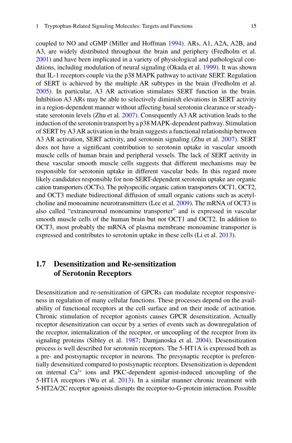
Abstract Most of the daily dietary tryptophan (Trp) is oxidatively degraded through the kynurenine (Kyn) pathway, and the remaining may be consumed either in serotonin synthesis or in conversion into melatonin through the methoxyindole pathway. Trp degradation products along the Kyn pathway include three neuroac-tive metabolites: the neuroinhibitory agent kynurenic acid (KA), the free radical generator 3-hydroxykynurenine (3HK), and the excitotoxin quinolinic acid (QA). Kyn is the major metabolite of Trp and is readily transported across the blood–brain barrier into the brain where it can be further metabolized in perivascular macro-phages, microglia, and astrocytes, also to generate neuroactive intermediates. In contrast to Kyn, QA, KA, and 3-hydroxyanthranilic acid (3HAA) penetrate through the blood–brain barrier only poorly due to its polar nature. Although the cytokines do not pass through the blood–brain barrier, their signals reach the brain through humoral, neural, and cellular pathways and stimulate Trp degradation by interacting with a cytokine network in the brain. The induction of Kyn pathway by indoleamine 2,3-dioxygenase (IDO) activity exhausts L -Trp in the medium and produces toxic metabolites. While Kyn to Trp ratio refl ects IDO activity, Kyn to KA ratio indicates the neurotoxic challenge. Alpha7 nicotinic acetylcholine receptor (alpha7nAChR) constitutes a crucial link between excessive KA formation and reduction in gluta-mate. KA-induced reduction in prefrontal glutamate levels emerges as a result of alpha7nAChR inhibition. Changes in the endogenous concentrations of KA, as a potent alpha7nAChR and N-methyl- D -aspartate (NMDA) receptor antagonist, affect extracellular dopamine levels in the brain. The entire monoaminergic neurotrans-mission involves functional interactions between serotonin, norepinephrine, and dopamine systems (Fig. 1.1 ). Serotonin transporter (SERT) reuptakes biogenic amine neurotransmitters following release in the nervous systems and terminates the action of serotonin. SERT can be regulated by a membrane-bound G-protein- coupled receptor, and this occurs via nitric oxide (NO) and cyclic guanosine monophosphate (cGMP). Desensitization and re-sensitization of G-protein-coupled
A. Engin , M.D., Ph.D. (*) Faculty of Medicine, Department of General Surgery , Gazi University , Besevler, Ankara , Turkey
Mustafa Kemal Mah. 2137. Sok. 8/14 , 06520 , Cankaya, Ankara , Turkey e-mail: [email protected]

2
receptors (GPCRs) can modulate receptor responsiveness in regulation of many cellular functions. Diet restriction-induced exaggerated feedback control over serotonin synthesis decreases serotonin neurotransmission at postsynaptic sites by reducing availability of Trp. Enterochromaffi n (EC) cells of the intestinal mucosa respond to chemical and mechanical stimuli by releasing serotonin. The enteric serotonin transporter plays a critical role in serotonergic neurotransmission and in the initiation of peristaltic and secretory refl exes.
Keywords Tryptophan • Kynurenine • Kynurenic acid • Quinolinic acid • Indoleamine 2,3-dioxygenase • N-Methyl- D -aspartate receptor • Serotonin • Serotonin transporter • Serotonin receptors
1.1 Introduction
Amino acids are not only regulators of gene expression and the protein phosphoryla-tion cascade but are also cell signaling molecules. Carbon skeletons of essential amino acids cannot be synthesized by animal cells and, therefore, must be provided from the diet (Wu 2010 ). The average daily nutritional requirement of L -tryptophan (Trp) as an essential amino acid is 5 mg/kg. In order to improve mood or sleep, many adults may consume Trp much more, up to 4–5 g/day (60–70 mg/kg) (Fernstrom 2012 ). Ninety-fi ve percent of dietary Trp is oxidatively degraded in the liver through the kynurenine (Kyn) pathway. Actually there are two rate-limiting enzymes of Kyn formation: fi rst, tryptophan 2,3-dioxygenase (TDO) and, the second, indoleamine 2,3-dioxygenase (IDO) (Marazziti et al. 2013 ). TDO reaction generates nicotinamide adenine dinucleotide [NAD + ] following Trp oxidation. A small amount of Trp degra-dation can also occur extrahepatically by the enzyme IDO. IDO is expressed by a large variety of cells and can be directly activated by proinfl ammatory cytokines such as interferon (IFN)-gamma and tumor necrosis factor (TNF)-alpha, whereas TDO is only located in the liver cells and is activated by stress hormones (Wirleitner et al. 2003 ). Degradation of Trp mainly occurs along the Kyn pathway. Eventually Kyn is metabolized along one of two catabolic branches, leading to the formation of either hydroxykynurenine (3HK) and quinolinic acid (QA) or kynurenic acid (KA). The cerebral Kyn pathway is driven mainly by blood-borne L -Kyn, which enters from the circulation to the brain using the large neutral amino acid transporter, whereas QA, KA, and 3-hydroxyanthranilic acid (3HAA) cannot pass the blood–brain barrier eas-ily (Fig. 1.1 ) (Fukui et al. 1991 ). In the brain, L -Kyn is then rapidly taken up by astrocytes and, presumably, by microglial cells. Almost all enzymes of the Kyn path-way are primarily contained in astrocytes and microglial cells (Schwarcz 2004 ). However, astrocytes do not contain kynurenine 3-hydroxylase and therefore favor KA synthesis, whereas microglial cells have very little kynurenine aminotransferase (KAT) activity which catalyzes the irreversible transamination of L -Kyn to KA and preferentially forms intermediates of the QA (Guillemin et al. 2001 ). KA can antago-nize the neuronal degeneration mediated by excessive stimulation of N-methyl- D -aspartate (NMDA) receptors in vivo (Lekieffre et al. 1990 ). During the stress response
A. Engin

3
100- to 1,000-fold elevations in 3HK and QA occur upon microglial cell activation or macrophage infi ltration to the brain (Schwarcz 2004 ). 3HK generates free radical species that can cause oxidative stress and lipid peroxidation. QA-induced excitation and neurotoxicity are mediated by N-methyl-D-aspartate receptor (NMDA) recep-tors. Because of the absence of effective removal mechanisms for extracellular QA (Foster et al. 1984 ), its ability to induce concentration-dependent increases in reac-tive oxidative species (ROS) formation (Santamaría et al. 2001 ), and its specifi c interaction with the NMDA receptor (De Carvalho et al. 1996 ), QA is particularly excitotoxin, whereas KA acts as a competitive blocker of the glycine co-agonist site of the NMDA receptor (Kessler et al. 1989 ) and as a noncompetitive inhibitor of the
5-HT1B
IDO
KYNURENINE
Kynurenine3-hydroxylase
Xanthurenic acid
Kynureninase
3-Hydroxyanthranilic acid
3-Hydroxyanthranilic acid oxygenase
Quinolinic acid
Quinolinic acidphosphoribosyltransferase
NAD+
3-Hydroxykynurenine
KynurenineAminotransferase
KynurenineAminotransferase
Anthranilic acid
Kynurenic acid
5-HydroxytryptophanhpTrpH1/hnTrpH2
BH4 qBH2Aromatic amino acid Decarboxylase (AAADC)
SEROTONIN
N-acetyl serotonin
AA-NAT
MELATONINHIOMTROS/RNS
NMDARalpha7nAChR
Glutamate
Dopamine
NMDAR ROS/RNS
5-HT2A 5-HT2C 5-HT1A
Norepinephrine Dopamine Gi Gi
Gs
cAMP
SSRISERTsIPSC
GABA
MT1MT2
TDO
TRYPTOPHAN
IFN-gamma/IFN-alpha/TNF-alpha
STAT1-alpha/IRF-1p38-MAPK/NFkappaB
SOCS3IDO-ITIM
IL-6
Fig. 1.1 Catabolic cascade of tryptophan metabolism. A simplifi ed version of the kynurenine, serotonin, and methoxyindole pathways demonstrating the major enzymes, intermediates, and receptors. TDO tryptophan 2,3-dioxygenase, IDO indoleamine 2,3-dioxygenase, SOCS suppressor of cytokine signaling, STAT1-alpha signal transducer and activator of transcription 1-alpha, IRF-1 interferon regulatory factor-1, NF-kappaB nuclear factor kappa B, p38-MAPK p38 mitogen- activated protein kinase, IDO-ITIM immunoreceptor tyrosine-based inhibitory motif for IDO, IFN-gamma interferon gamma, IFN-alpha , interferon alpha, TNF-alpha tumor necrosis factor alpha, IL-6 interleukin-6, ROS reactive oxygen species, RNS reactive nitrogen species, NMDAR N-methyl- D -aspartate receptor, NAD+ nicotinamide adenine dinucleotide, hpTrpH1 human periph-eral tryptophan hydroxylase1, hnTrpH2 human neural tryptophan hydroxylase2, BH4 tetrahy-drobiopterin, qBH2 quinonoid dihydrobiopterin, alpha7nAChR alpha7 nicotinic acetylcholine receptor, AA-NAT arylalkylamine-N-acetyltransferase, HIOMT hydroxyindole-O- methyltransferase, 5-HT2A, 5-HT2C, 5-HT1B, 5-HT1A serotonin receptors, Gi inhibitory G pro-tein, Gs stimulatory G protein, SSRI selective serotonin reuptake inhibitor, SERT serotonin transporter, sIPSC spontaneous inhibitory postsynaptic currents, GABA gamma-aminobutyric acid, cAMP cyclic adenosine monophosphate, MT1, MT2 membrane-bound melatonin receptors
1 Tryptophan-Related Signaling Molecules: Targets and Functions

4
alpha7 nicotinic acetylcholine receptor (alpha7nAChR) (Hilmas et al. 2001 ). Therefore, KA is considered to be neuroprotective.
In the second metabolic pathway of L -Trp degradation, a small amount of Trp is converted to 5-hydroxytryptophan by the tetrahydrobiopterin (BH4)-dependent tryptophan hydroxylase (TrpH). Subsequently aromatic amino acid decarboxylase (AAADC ) catalyzes the second step of serotonin synthesis (Chen and Miller 2012 ).
The third metabolic pathway of L -Trp degradation involves its conversion into melatonin through the methoxyindole pathway. Biosynthetic steps of melatonin comprise two major rate-limiting enzymes: arylalkylamine-N-acetyltransferase (AA-NAT) and hydroxyindole-O-methyltransferase (HIOMT). Although trans-forming of Trp into melatonin originally occurred in pinealocytes, it has been also detected in many other parts of the body, including the eyes, bone marrow, skin, lymphocytes, and enteroendocrine cells of the gastrointestinal tract (Konturek et al. 2007 ; Srinivasan et al. 2011 ). However, cytokine-driven Trp degradation pathways and how they infl uence each other under different physiologic and pathologic conditions are open to debate.
1.2 Cytokine-Mediated Signaling
Contrary to Kyn, cytokines are relatively large molecules that do not freely pass through the blood–brain barrier. Nevertheless, cytokine signals are able to reach the brain through humoral, neural, and cellular pathways and interact with a cytokine network in the brain consisting of neurons, microglia, and astrocytes (Capuron and Miller 2011 ). Considering the abovementioned issues, cytokine signals reach to the brain with fi ve different mechanisms: (1) passage of cytokines through the leaky regions of the blood–brain barrier, (2) active transport with cytokine-specifi c trans-port molecules on brain endothelium, (3) activation of endothelial cells, (4) trans-mission of cytokine signals via afferent nerve fi bers, and (5) entry into the brain parenchyma and involvement of microglia and astrocytes (Rivest et al. 2000 ; Konsman et al. 2002 ; Plotkin et al. 1996 ).
Cytokine overexpression in the brain due to infl ammation is an important factor in the pathogenesis of neurotoxic disorders. However, peripheral and central cytokine compartments appear to be integrated, and their effects might synergize or inhibit each other (Szelényi 2001 ). Although numerous cytokines and their receptors have been identifi ed in the brain, interleukin-1 (IL-1), IL-6, and TNF-alpha have been implicated in the central control of responses to neuroendocrine, immune, and behavioral alterations (Rothwell et al. 1996 ). Actually the innate and adaptive immune responses are triggered by microglia in the central nervous system including the release of proinfl ammatory mediators. In this case toll-like receptor (TLR)-induced activation of microglia and the release of proinfl ammatory molecules are responsible for neurotoxic processes (Lehnardt 2010 ). Following activation of the immune system pathways, a number of cytokines alone or in combination including IFN-alpha, IFN-gamma, and TNF-alpha through activation of a number of infl am-
A. Engin

5
matory signaling pathways such as signal transducer and activator of transcription 1-alpha (STAT1-alpha), interferon regulatory factor (IRF)-1, nuclear factor (NF) kappa B, and p38 mitogen-activated protein kinase (MAPK) stimulate IDO (Fig. 1.1 ) (Fujigaki et al. 2006 ). IDO breaks down Trp into Kyn. Kyn is preferen-tially converted to KA and QA in astrocytes and in microglia, respectively (Schwarcz and Pellicciari 2002 ). As mentioned above, activated microglia is a chronic source of multiple neurotoxic molecules, including TNF-alpha, nitric oxide (NO), IL-1beta, and ROS, which cause progressive neuron damage (Lull and Block 2010 ). Initially released cytokines, IL-1beta and TNF-alpha, signal neuroendocrine, autonomic, limbic, and cortical areas of the central nervous system to control neural activity, behaviors, hormone release, and autonomic functions (Lorton et al. 2006 ).
Acute activation of pattern-recognition receptors, TLR-4 and TLR-2, by expos-ing to bacterial lipopolysaccharide and peptidoglycan, respectively, also increases circulating levels of IFN-gamma and potently activates IDO in both the periphery and the brain (Lestage et al. 2002 ). Indeed glial cells and TLRs are vital components of immune response in the central nervous system. Intrauterine infection/infl amma-tion promotes infl ammatory processes in glial cells by upregulating cytokines and by activating signaling pathways and transcriptional factors (Yuan et al. 2010 ).
Response to cytokines seems to be related to the hypothalamic–pituitary–adrenal (HPA) axis activation. Thus IL-1 administration increases noradrenaline secretion and stimulates indoleamine metabolism and most prominently increases the metab-olism of serotonin (5-hydroxytryptamine, 5-HT). IL-6 also induces a short-lived activation of the HPA axis. Its effects on Trp and serotonin metabolism are similar to those of IL-1 (Dunn et al. 1999 ). Furthermore suppressors of cytokine signaling (SOCS) proteins are critical modulators of cytokine-mediated processes, and janus kinase 2 (JAK)–STAT–SOCS signaling modules can have diverse effects on infl am-matory diseases (O’Shea and Murray 2008 ). In the long term, IL-6-dependent upregulation of SOCS3 is responsible for inhibiting the IFN-gamma-driven transcriptional expression of IDO (Fig. 1.1 ) (Orabona et al. 2004 ). Hence, an inverse correlation between SOCS3 and IDO expression is evident. Immunomodulatory mechanisms extensively use negative regulators in the form of signaling proteins bearing one or more immunoreceptor tyrosine-based inhibitory motifs (ITIMs). IL-6 upregulates SOCS3 and promotes SOCS3 binding to ITIMs of IDO. This process causes shortening of the half-life and proteasome-mediated degradation of IDO (Orabona et al. 2008 ).
1.3 IDO-Mediated Signaling
The extrahepatic Trp degradation enzyme IDO is induced by IFN-gamma-mediated effects of the STAT1-alpha and IRF-1. The induction of IDO can also be mediated through an IFN-gamma-independent mechanism which may be related to the activity of the p38-MAPKinase pathway and NF-kappaB (Fujigaki et al. 2006 ). Actually the enzymatic activity of IDO is enhanced in conditions of acute or chronic
1 Tryptophan-Related Signaling Molecules: Targets and Functions

6
activation of the immune system, including immunotherapy, acquired immunodefi -ciency syndrome, atherosclerosis and coronary heart disease, rheumatoid arthritis, and obesity (Wirleitner et al. 2003 ). In particular secretion of IFN-gamma is signifi cantly higher in the obese than that of the control subjects. Initially this might be partly dependent on the action of leptin that shifts T-helper (Th) cells toward a Th1 phenotype. A shift to Th1-cytokine profi le is dominated by the production of IFN- gamma and is related to insulin resistance in obesity (Pacifi co et al. 2006 ). Hereby T cells and IFN-gamma participate in the regulation of the chronic infl am-matory response in obese individuals (Rocha et al. 2008 ). Chronic infl ammation might trigger and maintain the transcriptional induction of IDO-mediated Trp catabolism. Consequently chronic immune activation is the cause for reduced Trp plasma levels in morbidly obese patients (Brandacher et al. 2007 ). In case of obesity, activation of IDO simultaneously causes excessive synthesis of kynuren-ines (Brandacher et al. 2006 ). Furthermore, decrease in Trp levels and subsequent reduction in serotonin due to shift to Kyn pathway provoke satiety dysregulation and ultimately lead to increase in caloric intake and favor obesity (Brandacher et al. 2007 ). Even after weight reduction in morbidly obese patients, Trp depletion persists (Brandacher et al. 2006 ). The induction of the Kyn pathway by IDO activity and subsequent decrease in the Trp availability in the brain results in the IFN-alpha-induced depressive symptoms. While Kyn to Trp ratio refl ects IDO activity, the Kyn/KA indicates the neurotoxic challenge (Wichers et al. 2005 ). Higher IDO activity has also been implicated in immune tolerance because it can inhibit the immune response, either by exhausting L -Trp in the medium or producing toxic metabolites. Trp metabolites in the Kyn pathway, such as 3HAA and QA, induce the selective apoptosis in vitro of murine thymocytes and of Th1 but not Th2 cells (Fallarino et al. 2002 ). As stated above IDO activity is characterized best by the Kyn to Trp ratio, but considering the immune tolerance, it should be correlated with the concen-tration of immune activation marker such as neopterin (Schröcksnadel et al. 2006 ).
Until recently, the conversion of Trp to N-formylkynurenine was thought to be performed by either of two enzymes, TDO and IDO. However a third enzyme, indoleamine 2,3-dioxygenase-2 (IDO2) [indoleamine 2,3-dioxygenase-like protein (INDOL1) or proto-indoleamine 2,3-dioxygenase (proto-IDO)], with the Trp degra-dation activity has been described (Ball et al. 2009 ). Although IDO2 is not as widely expressed as IDO (IDO1), it is also expressed in antigen-presenting dendritic cells where Trp catabolism drives immune tolerance. Like IDO, IDO2 catabolizes Trp and triggers phosphorylation of the translation initiation factor eIF2alpha. Trp restoration switches off this signaling pathway when activated by IDO, but not IDO2, arguing that IDO2 has a distinct signaling role (Metz et al. 2007 ). IDO2 has 43 % similarity to classical IDO protein and shares the same critical catalytic residues. Although IDO2 enzyme activity is weaker than IDO, it is less sensitive to dextro- methyl tryptophan inhibition than IDO. Thus a more recent study indicated that human CD4+ and CD8+ T-cell proliferation was inhibited by IDO2, but both levo-1-methyl tryptophan and dextro-methyl tryptophan which are the gold stan-
A. Engin
7
dard inhibitors of IDO enzyme activity could not reverse IDO2-mediated arrest of cell proliferation, even at high concentrations (Qian et al. 2012 ). In fact, IDO- dependent tolerogenic effects induced by transforming growth factor beta (TGF- beta) are abolished by IDO gene silencing, but not by the use of 1-meth-yltryptophan. TGF-beta/IDO/phosphotyrosine phosphatase SHP-1 axis activates the anti- infl ammatory NF-kappaB pathway by inhibiting the IL-1 receptor-associ-ated kinase-1 (Orabona et al. 2012 ).
1.4 Aryl Hydrocarbon Receptor Activation
Gene transcription in response to xenobiotics can be stimulated by aryl hydrocarbon receptor (AhR) which is one of the several ligand-dependent intracellular respon-sive elements (Denison and Nagy 2003 ). In this respect Trp photoproducts modulate light-dependent regulation of circadian rhythm through triggering of AhR signal-ing. Thus these by-products, including 6-formylindolo(3,2-b)carbazole, have high affi nity for AhR (Mukai and Tischkau 2007 ). Ligand activation provokes the AhR to migrate from cytosol to the nucleus and form a complex with the aryl hydrocar-bon nuclear translocator (ARNT) that can bind dioxin-responsive elements in the promoter regions of xenobiotic-metabolizing cytochrome P450 (CYP1A) enzymes and 2,3,7,8-tetrachlorodibenzo-p-dioxin-inducible poly (ADP-ribose) polymerase (PARP7) (TiPARP) (Diani-Moore et al. 2010 ). TiPARP is an AhR target gene that can mediate a 2,3,7,8-tetrachlorodibenzo-p-dioxin (TCDD) toxicity. TCDD suppresses glucose metabolism-related pathways such as hepatic glucose produc-tion, expression of key gluconeogenic genes, phosphoenolpyruvate carboxykinase, and glucose-6-phosphatase activities, and NAD + levels. Nicotinamide, a known pre-cursor of NAD + , is an AhR antagonist. There is a link between signaling path-ways for AhR toxicity and nutrient homeostasis NAD + /peroxisome proliferator-activated receptor gamma coactivator 1 alpha (PGC1alpha), regulator of mitochondrial biogenesis, and function/silent mating type information regulation 2 homolog 1 [(SIRT1), NAD-dependent deacetylase sirtuin-1] via the AhR target gene TiPARP (Diani-Moore et al. 2010 ). Consequently the effects of TCDD are mediated through its binding to the AhR, as a ligand-activated transcription factor. Subsequent to binding AhR, TCDD inhibits CD4+ T-cell differentiation into T helper (Th)1, Th2, and Th17 effector cells while inducing forkhead transcription factor (Foxp3)-negative and/or preserving Foxp3+ regulatory T cells (Tregs) (Marshall and Kerkvliet 2010 ). The AhR is a key transcriptional regulator of Th17- cell differentiation. Th17 cells express kynurenine 3-monooxygenase, which is an enzyme involved in catabolism of the Trp metabolite Kyn (Stephens et al. 2013 ). On the other hand, activation of AhR induces IDO and IDO2 expression of dendritic cells. Hence AhR activation is an important signaling pathway for IDO expression and displays a critical role in the mechanism leading to the generation of Tregs. Eventually induction of Tregs mediates the immune suppression through the activation of AhRs (Vogel et al. 2008 ).
1 Tryptophan-Related Signaling Molecules: Targets and Functions

8
1.5 Glutamate Neurotransmission
Infl ammatory cytokines and their signaling pathways have signifi cant effects on the synthesis, release, and reuptake of serotonin, dopamine, and glutamate (Miller et al. 2013 ). In this context, higher glutamate receptor, mGluR1alpha, and lower guanine nucleotide-binding protein (G-protein)-coupled receptor, regulator of G-protein signaling 4 (RGS4) mRNA levels, play an important role in regulating gamma- aminobutyric acid (GABA) and glutamate neurotransmission in the brain cortex by initiating intracellular signaling cascade (Fig. 1.1 ). Suppression of GABA release in GABA neurons of the prefrontal cortex and diminished glutamate neurotransmis-sion due to NMDA receptor hypofunction are evident in certain cognitive defi cits (Volk et al. 2010 ). Activation of serotonin 5-HT1A receptors or dopamine D (4) receptors downregulates the function of NMDA receptor channel in pyramidal neurons of the prefrontal cortex. Blocking RGS4 function signifi cantly potentiates the 5-HT1A regulation of NMDA receptor. Conversely, overexpression of RGS4 couples RGS4 to serotonin signaling in cortical neurons and attenuates the 5-HT1A effect (Gu et al. 2007 ). Furthermore elevated levels of KA in the prefrontal cortex may contribute to the abnormal glutamatergic and nicotinic functions in cognitive defi cits (Schwarcz et al. 2001 ; Erhardt et al. 2009 ). This concept is partly based on the fi nding that endogenous KA is an astrocyte-derived metabolite of Trp degradation via Kyn pathway (Kiss et al. 2003 ). As already mentioned above, Trp degradation products along the Kyn pathway include three neuroactive metabolites: the neuroin-hibitory agent KA, the free radical generator 3HK, and the excitotoxin QA. Inhibition of kynurenine 3-hydroxylase shifts Kyn pathway metabolism from 3HK formation toward enhanced KA formation in the mature brain. Therefore acute kynurenine 3-hydroxylase inhibition effectively increases KA formation (Ceresoli- Borroni et al. 2007 ). Following the systemic administration of Kyn, a signifi cant reduction in prefrontal glutamate occurs. Alpha7nAChRs constitutes a crucial link between excessive KA formation and reduction in glutamate. Subsequent to peripheral administration, Kyn penetrates the blood–brain barrier and dose dependently raises extracellular KA levels in the prefrontal cortex. Actually systemic Kyn administra-tion duplicates the reduction in extracellular glutamate seen after a local perfusion of Kyn in the prefrontal cortex. Resultant KA-induced reduction in prefrontal gluta-mate levels emerges as a result of alpha7nAChRs inhibition (Konradsson-Geuken et al. 2010 ). The cognitive defi cits are likely related to abnormal glutamatergic and cholinergic neurotransmission in the prefrontal cortex. These defects may be secondary to increased levels of the astrocyte-derived KA, which inhibits alpha7A-ChR and may thereby reduce glutamate release. Fluctuations in endogenous KA formation bidirectionally infl uence cortical glutamate concentrations. Consequently selective attenuation of cerebral KA production by increasing glutamatergic tone might improve cognitive functions (Wu et al. 2010 ). Endogenous glutamate acts locally within the striatum via ionotropic receptors to control impulse-independent and transporter-mediated mode of dopamine release. When the KA inhibits the release of glutamate, low glutamate level may inhibit the secretion of dopamine
A. Engin
9
(Borland and Michael 2004 ). Modulation of glutamate release by the alpha7nAChRs on striatal glutamatergic terminals, in turn, activates presynaptic ionotropic gluta-mate receptors on striatal dopaminergic nerve terminals (Kaiser and Wonnacott 2000 ). Decrease in extracellular levels of striatal dopamine due to KA-induced blockade of alpha7nAChRs can be enhanced by stimulating the endogenous forma-tion of KA via kynurenine 3-hydroxylase inhibition (Rassoulpour et al. 2005 ). Blood-derived Kyn rapidly accesses to KAT II-containing astrocytes, and KA synthesis takes place in astrocytes (Guidetti et al. 2007 ). Fluctuations in KA indi-rectly regulate extracellular dopamine levels in the striatum. Acute inhibition of KAT II reduced the de novo synthesis of KA; thus, KAT II is a critical determinant of functionally relevant KA fl uctuations (Amori et al. 2009 ). On the other hand, cytokine-activated Kyn pathway not only depletes Trp but also generates neuroac-tive metabolites that can signifi cantly infl uence the regulation of dopamine and glutamate (Miller et al. 2013 ). Excitotoxic damage is a common pathologic event in a number of neurologic diseases occurring after accumulation of excess extracellu-lar glutamate in the central nervous system and subsequent overstimulation of glu-tamate receptors. High extracellular glutamate increases risk of glutamate excitotoxicity. However, astrocytes can take up synaptically released glutamate and maintain glutamate homeostasis (Pitt et al. 2003 ). Nevertheless astrocytes can release glutamate together with the various chemical transmitters which may medi-ate communication between neurons and astrocytes (Ida et al. 2008 ). There are complex cross talks between microglia and astrocytes during neuroinfl ammatory insults which would infl uence glutamate-dependent responses in astrocytes (Tilleux et al. 2007 ). Astrocytosis due to the destruction of neurons is accompanied by microglial activation. Actually in proinfl ammatory processes, activated microglia stimulates the increase in number of astrocytes and enhances mRNA expression of IL-6 (Röhl et al. 2007 ). Cytokine release from microglia also causes downregula-tion of mGluR5, mGluR5 protein, and mRNA expression in astrocytes (Tilleux et al. 2007 ). On the other hand, the inhibition of inducible nitric oxide synthase (iNOS) eliminates the cytokine-induced enhancement of glutamate release, whereas treatment with a NO donor, even in the absence of cytokines, increases glutamate release (Ida et al. 2008 ). Nonspecifi c NOS inhibitors decrease the homocysteine-induced lipid peroxidation more than does the selective neuronal nitric oxide syn-thase (nNOS) inhibitor. In this case homocysteine can induce oxidative injury to nerve terminals, and this effect involves the NMDA receptor stimulation, NOS activation, and associated free radical formation (Jara-Prado et al. 2003 ). Higher concentration of QA induces concentration-dependent increases in ROS formation in all synaptosomes, but the increase in the production of peroxidized lipids only emerges in the striatum and the hippocampus. These fi ndings suggest that the excitotoxic action of QA involves regional selectivity in the oxidative status of brain synaptosomes (Santamaría et al. 2001 ). However, NMDA receptor antagonists completely inhibit the increase of QA-induced lipid peroxidation (Santamaría and Ríos 1993 ).
1 Tryptophan-Related Signaling Molecules: Targets and Functions
10
1.6 Serotonin Neurotransmission
Trp is only available in the diet. It is therefore likely that excessive diet restriction and malnutrition decrease brain serotonin stores. Evidence shows that diet restriction- induced exaggerated feedback control over serotonin synthesis and the smaller availability of Trp decrease serotonin neurotransmission at postsynaptic sites, leading to hyperactivity, depression, and behavioral disorders (Haleem 2012 ). Conversely excessive L -Trp ingestion raises brain Trp levels and stimulates its conversion to serotonin in neurons. Adverse effects may be seen at higher doses (70–200 mg/kg) and include tremor, nausea, and dizziness. When Trp is taken alone or with a drug that enhances serotonergic effects, it may provoke side effects (Fernstrom 2012 ). In fact serotonin neurotransmission comprises multiple consecutive processes including synthesis, storage/release, signaling, reuptake, and metabo-lism, of which the fi rst step, synthesis, is a critical modulator of serotonin neuro-transmission (Chen and Miller 2012 ). Serotonin is synthesized by a two-step enzymatic reaction. Firstly, the essential amino acid L -Trp is hydroxylated into 5-hydroxy- L -tryptophan by the limiting enzyme TrpH. Two isoforms of TrpH enzyme, TrpH1 and TrpH2, have been characterized so far: TrpH1 is mainly expressed in the gastrointestinal tract and the pineal gland, whereas TrpH2 is primarily expressed in the central nervous system (Watts 2009 ). TrpH2 polymor-phisms directly infl uence serotonergic function and thus impact on mood disor-ders. TrpH2-defi cient mice display alterations in anxiety-like behavior which is accompanied by adaptational changes of 5-HT1A receptors and its associated sig-naling pathway (Waider et al. 2011 ). Genetic inactivation of TrpH2 function in mice led to the identifi cation of phenotypic changes, ranging from growth retarda-tion and late-onset obesity to enhanced conditioned fear response, increased aggression, and depression-like behavior (Lesch et al. 2012 ). In fact TrpH, a BH4-dependent amino acid hydroxylase, is the key regulator of serotonin biosynthesis (Carkaci-Salli et al. 2006 ). 5-Hydroxy- L - tryptophan is converted to serotonin by AAADC. Actually AAADC defi ciency is a severe genetic neuro-metabolic disor-der that is characterized with combined defi ciency of serotonin, dopamine, and catecholamines (Manegold et al. 2009 ). Furthermore endothelial AAADC plays an important role in cardiac synthesis of serotonin and possibly in serotonin-depen-dent regulation of NO generation. 5-Hydroxy- l -tryptophan administration in mice increased phosphorylation of aortic endothelial NOS (eNOS) at Ser-1177 as well as accumulation of nitrates in cardiac tissue (Rouzaud-Laborde et al. 2012 ). eNOS is known to be stimulated by serotonin via 5-HT1B receptor/eNOS pathway (McDuffi e et al. 1999 ). Phosphorylation of eNOS produces NO without requiring any changes in [Ca 2+ ]i (Boo et al. 2003 ). Actually 5-HT2B receptor stimulation plays a critical role in the phosphorylation of both extracellular signal-regulated kinase 1/2 (ERK1/2) and eNOS (Asada et al. 2009 ). In human endothelial cells, serotonin markedly stimulates eNOS expression and the phosphorylation of eNOS, Akt, and ERK1/2. Consequently serotonin induces angiogenesis through activation
A. Engin
11
of Akt in endothelial cells. Selective inhibition of 5-HT2A causes induction of the eNOS/Akt pathway via the endothelial 5-HT1B receptors and enhances vasodila-tion in diabetes mellitus (Iwabayashi et al. 2012 ).
Increased activity of the liver enzyme TDO is stimulated by an excess of circulat-ing corticosteroids. In hypercortisolemic conditions, metabolism of Trp turns to the Kyn pathway from serotonin synthesis. Upregulation of the Trp-Kyn pathway and diminished availability of Trp are the primary causes of serotonin defi ciency (Oxenkrug 2010 ). Hypercortisolism affects the gene encoding TrpH2 and the expression of TrpH2. Also chronic corticosterone intake disrupts the diurnal varia-tion of TrpH2 mRNA expression in the brain stem dorsal raphe nucleus and of plasma adrenocorticotropin and corticosterone levels in a dose-dependent manner (Donner et al. 2012 ). The hippocampus plays a central role in regulation of the HPA axis and release of endogenous glucocorticoids. Exposure to serotonin increases the glucocorticoid receptor mRNA levels in hippocampal neurons. Eventually synthetic and endogenous glucocorticoids, as well as serotonin, infl uence glucocorticoid receptor expression during hippocampal development (Erdeljan et al. 2005 ). Stress signifi cantly increases extracellular serotonin release in the basolateral amygdaloid nucleus and the prefrontal cortex (Kawahara et al. 1993 ). Indeed serotonin dramati-cally enhances frequency and amplitude of spontaneous inhibitory postsynaptic currents (sIPSCs) in the basolateral amygdala through 5-HT2A receptors. Because of the basolateral amygdaloid GABAergic inhibition is blocked by selective 5-HT2A receptor antagonists, the stress-induced effect appeared to be specifi c to 5-HT2A receptor downregulation (Jiang et al. 2009 ).
Monoaminergic neurotransmission involves functional interactions between serotonin, norepinephrine, and dopamine systems. First of all serotonin system exerts negative effect on norepinephrine system through 5-HT2A and on dopamine system through 5-HT2C receptor-mediated mechanisms. Positive and negative effect of norepinephrine system on serotonin neurotransmission is mediated through alpha1- and alpha2-adrenergic receptors, respectively (Hamon and Blier 2013 ). Actually BH4 is an essential cofactor in the synthesis of serotonin, dopamine, epinephrine, norepinephrine, and NO. BH4 availability infl uences many cells, including neurons. Following peripheral nerve damage, BH4 dramatically increases in sensory neurons and causes pain hypersensitivity (Latremoliere and Costigan 2011 ). Fatigue and impaired executive functions are commonly linked to disturbed cerebral dopaminergic and noradrenergic neurotransmission. Moreover selective serotonin reuptake inhibitors (SSRIs) contribute to fatigue, which is a common residual symptom associated with depression (Stenman and Lilja 2013 ). During the prolonged exercise, fatigue is attributed to the muscle glycogen depletion. “Central fatigue hypothesis” previously was based on the increase in the concentration of brain serotonin during exercise. However according to the revised central fatigue hypothesis, an increase in central ratio of serotonin to dopamine is associated with feelings of tiredness and lethargy (Meeusen and Piacentini 2003 ). Actually a complex interplay between the different neurotransmitter systems induces fatigue: dopamine and noradrenaline rather than serotonin alone (Roelands and Meeusen 2010 ).
1 Tryptophan-Related Signaling Molecules: Targets and Functions

12
Diet restriction-induced exaggerated feedback control over serotonin synthesis and the reduced availability of Trp decrease serotonin neurotransmission at postsyn-aptic sites. A compensatory upregulation of postsynaptic 5-HT1A receptors and hypophagic serotonin receptors may be involved in suppression of appetite (Haleem 2012 ). In this case although the levels of Trp in the plasma and of serotonin in the hypothalamus decrease, no effect is found on the levels of Trp in the hypothalamus. Diet restriction-induced decrease of serotonin is due to an increase in the respon-siveness of negative feedback control over serotonin, not due to smaller availability of Trp (Haleem 2009 ). Likewise 20–25 % reduction in body weight due to food restriction decreases serotonin concentration in the brain of male but not female rats (Haider and Haleem 2000 ). Conversely in sugar-diet-treated rats, when the cumula-tive food intakes increase, body weights decrease. Hyperphagic effects of selective 5-HT1A agonist are greater in normal diet than sugar-diet-treated rats. However serotonin and 5-hydroxyindole acetic acid levels are not changed. Desensitization of pre- as well as postsynaptic 5-HT1A receptors in rats treated with sugar diet causes the precipitation of obesity (Jabeen and Haleem 2008 ). Actually long-term consumption of sugar diet results in a decrease in the effectiveness of pre- as well as postsynaptic 5-HT1A receptor-dependent responses (Inam et al. 2006 ). Malnourished offspring have a signifi cant elevation of L -Trp, TrpH activity, and serotonin in the brain stem. Both isoforms of TrpH (TrpH1 and TrpH2) are expressed at birth in both groups; however, TrpH1 expression is signifi cantly higher in offspring with intrauterine malnutrition when compared to the controls. Malnourished offspring show reduced expression of TrpH2 compared to controls. Thus it has been confi rmed that intrauterine malnutrition produces an increase in serotonin in the brain stem and also shows increased expression of TrpH1 at birth, with decreased expression of TrpH2 (Manjarrez-Gutiérrez et al. 2012 ).
The dorsal raphe nucleus (DRN) is the largest serotonin-containing nucleus in the brain and has extensive ascending projections that innervate most forebrain struc-tures. Targets of DRN innervation receive input from both serotonergic and nonseroto-nergic cells. Selective serotonergic neurotoxins, including 5,7- dihydroxytryptamine (5,7-DHT), have been shown to disrupt axonal transport in serotonergic neurons (Callahan et al. 2001 ; Araneda et al. 1980 ). Human LIM homeobox transcription factor 1-beta (Lmx1b)-encoded gene is essential for the development of central serotonergic neurons. This gene is required for the normal biosynthesis of serotonin in the adult brain and for regulating normal functions of central serotonergic neurons. Lmx1b deletion in the adult brain leads to reduction in central serotonin levels. However the overall number of serotonergic neurons is not affected by delet-ing Lmx1b, and Pet1 promoter expression in the adult brain is independent of Lmx1b. Reduction in central serotonin levels seems to be the consequence of TrpH2 downregulation (Song et al. 2011 ). In fact Pet1 in the brain is necessary for terminal differentiation of serotonergic neuron phenotype during embryonic development (Hendricks et al. 2003 ). Considering the serotonergic signaling mechanisms, ETS domain transcription factor Pet1 is also required for maintaining the serotonergic neurotransmitter system during adult stages as well as for expression of the presyn-aptic 5-HT1B autoreceptor. Therefore adult central nervous system expression of
A. Engin

13
TrpH2 and serotonin transporter (SERT) is restricted to Pet1- expressing serotonin neurons and is rate limiting for the essential serotonergic functions of serotonin synthesis and reuptake (Liu et al. 2010 ). Pet1 RNA co-localizes with TrpH-positive neurons in raphe nuclei. Loss of Pet1 in the serotonergic neurons leads to a decrease of TrpH2 expression but no change in Lmx1b expression (Song et al. 2011 ). Virtually serotonergic and nonserotonergic axons innervate distinct but partially overlapping fi elds within vestibular nuclei (Halberstadt and Balaban 2007 ). Both local GABAergic and glutamatergic cells project onto DRN serotonergic neurons (Jolas and Aghajanian 1997 ). 5-HT1A receptors are present on nonserotonergic as well as serotonergic DRN neurons. While the majority of serotonin-immuno-positive cells are double-labeled for 5-HT1A receptor, small but signifi cant population of sero-tonin-immuno-negative cells express the 5-HT1A receptor (Kirby et al. 2003 ). Both 5-HT1A and alpha1b adrenergic mRNA are highly expressed throughout the DRN, and the vast majority of serotonergic neurons express both receptors. A smaller percentage of GABAergic neurons also express 5-HT1A or alpha1b adrenergic mRNA. A small amount of catecholaminergic cells express either 5-HT1A or alpha1b adrenergic mRNA (Day et al. 2004 ).
Hence, serotonin not only affects neuronal excitability through activating post-synaptic receptors (Guo and Rainnie 2010 ) but also affects presynaptic excitatory or inhibitory neurotransmission in the central nervous system, because of the serotonin activating 5-HT1A and/or 5-HT1B receptors located on the presynaptic terminals. Serotonin exerts signifi cant control over the synaptic inputs and the autonomous activity of subcortical pallidal neurons (Bouryi and Lewis 2003 ; Hashimoto and Kita 2008 ). The serotonin receptors have been divided into 7 subfamilies, 6 of which include 13 different genes for G-protein-coupled receptors (GPCR). Post- genomic modifi cations create 20 more G-protein-coupled serotonin receptors. Consequently there are at least 30 distinct serotonin receptors that signal through G proteins (Raymond et al. 2001 ). 5-HT1A and 5-HT1B receptors interface primarily with inhibitory G proteins (Gi) to decrease adenylyl cyclase activity. Subsequently the action of the SSRI is mediated through the 5-HT1A receptor (Blier and Ward 2003 ; Monaca et al. 2003 ). While SSRI inhibiting the SERT density and function, it maintains the normal fi ring rates and release of serotonin and immediately increases activation of postsynaptic serotonin receptors (Nemeroff and Owens 2003 ). All of the seven specifi c serotonin receptors mediate SSRI effects; however, the second- class receptors, 5-HT6 and 5-HT7, primarily interact with stimulatory G proteins (Gs) to increase adenylyl cyclase activity. In particular the 5-HT6 receptor is involved in neuronal serotonergic transmission and may have effects on anxiety and mood (Yoshioka et al. 1998 ). The 5-HT7 receptor is involved in hippocampal func-tion (Gill et al. 2002 ), and has been implicated in the regulation of the glucocorti-coid receptors (Laplante et al. 2002 ). SSRIs also affect the function of the 5-HT2C receptor (Bristow et al. 2000 ) with some adverse effects potentially mediated by 5-HT2C. Other than for the 5-HT3 receptor, most of the downstream effects of serotonin are mediated by G proteins (Raymond et al. 2001 ). Actually G proteins are a family of guanine nucleotide-binding regulatory components that couple neurotransmitter receptors to various types of intracellular effector systems. Gs/Gi
1 Tryptophan-Related Signaling Molecules: Targets and Functions

14
mediates stimulation/inhibition of adenylate cyclase system, which forms cyclic adenosine monophosphate (cyclic AMP) as a second messenger (Lesch and Lerer 1991 ). There are 16 genes for G-protein alpha subunits, 5 for beta, and 12 for gamma (Downes and Gautam 1999 ). SSRIs have been associated with increased transcrip-tion of adenylyl cyclase 1. 5-HT1A receptor mediates inhibition of basal and Gs-induced cAMP formation in the absence of adenylyl cyclase 2. 5-HT1A activation decreases activity of neuronal adenylyl cyclase 2 (Albert et al. 1999 ). Among serotonin receptors, the 5-HT3 receptor is a member of the Cys-loop family of ligand-gated ion channels and located in both the peripheral and central nervous systems. Chronic activation of 5-HT3 receptor produces signifi cant desensitization of 5-HT3 and postsynaptic 5-HT1A receptors without major changes in the expres-sion of SERT and TrpH-2 genes (Kondaurova et al. 2012 ).
Human SERT reuptakes biogenic amine neurotransmitters following release in the nervous systems and terminates the action of serotonin (Murphy et al. 2004 ).
SERTs are tightly controlled by multiple signaling pathways, including G-protein-coupled receptor-linked pathways (Blakely et al. 2005 ). Two protein kinase G (PKG)-dependent pathways have been proposed to support rapid SERT regulation by A3 adenosine receptors (ARs). The fi rst enhances SERT surface traffi cking to clear serotonin following vesicular release, and the second is a separate, p38 MAPK-dependent process which augments SERT intrinsic activity (Zhu et al. 2004 ). p38 MAPK activation downstream of PKG via SERT catalytic regulatory pathway in a traffi cking-independent mode is distinct from events controlling SERT surface den-sity. Protein phosphatase 2A is a critical component of the pathway responsible for p38 MAPK upregulation of SERT catalytic activity (Zhu et al. 2005 ). Thus A3 ARs activation stimulates serotonin uptake via PKG- and p38 MAPK-linked pathway (Zhu et al. 2004 ). MAP kinase kinase (MAPKK) superfamily molecules, MKK3, MKK3b, and MAPKK6, can act as a specifi c activator for p38. Furthermore as a major activator for p38, the MAPKK6/p38 kinase cascade is activated strongly by TNF-alpha and H 2 O 2 (Moriguchi et al. 1996 ). Eventually PKG-linked and p38 MAPK-linked pathways provide a rapid increase in SERT surface expression and function. In contrast, the activity of protein phosphatase 2A inhibitors attenuates MAPK or other signal transduction pathways and facilitates the stimulation of sero-tonin transport (Zhu et al. 2005 ), whereas activated protein kinase C (PKC) interacts with SERT and alters the subcellular localization of the transporter resulting in a reduction of serotonin transport. SERT proteins are rapidly phosphorylated in paral-lel with transporter redistribution and loss of functional uptake capacity. Indeed loss of surface SERT protein after PKC activation refl ects transporter redistribution rather than irreversible loss of transporter protein via degradation (Haase et al. 2001 ; Blakely et al. 1998 ). In brief, one of the well-known mechanisms in the ter-mination of the stimulation of monoamine neurotransmitters is the removal from the synapse by transporter molecules. Transporters are located within the plasma mem-brane of presynaptic cells and may be readily regulated by a variety of receptor-mediated intracellular signals.
SERT can be rapidly regulated by a membrane-bound G-protein-coupled receptor and this occurs via NO and cyclic guanosine monophosphate (cGMP). A3 AR is
A. Engin
15
coupled to NO and cGMP (Miller and Hoffman 1994 ). ARs, A1, A2A, A2B, and A3, are widely distributed throughout the brain and periphery (Fredholm et al. 2001 ) and have been implicated in a variety of physiological and pathological con-ditions, including modulation of neural signaling (Okada et al. 1999 ). It was shown that IL-1 receptors couple via the p38 MAPK pathway to activate SERT. Regulation of SERT is achieved by the multiple AR subtypes in the brain (Fredholm et al. 2005 ). In particular, A3 AR activation stimulates SERT function in the brain. Inhibition A3 ARs may be able to selectively diminish elevations in SERT activity in a region-dependent manner without affecting basal serotonin clearance or steady- state serotonin levels (Zhu et al. 2007 ). Consequently A3 AR activation leads to the induction of the serotonin transport by a p38 MAPK-dependent pathway. Stimulation of SERT by A3 AR activation in the brain suggests a functional relationship between A3 AR activation, SERT activity, and serotonin signaling (Zhu et al. 2007 ). SERT does not have a signifi cant contribution to serotonin uptake in vascular smooth muscle cells of human brain and peripheral vessels. The lack of SERT activity in these vascular smooth muscle cells suggests that different mechanisms may be responsible for serotonin uptake in different vascular beds. In this regard more likely candidates responsible for non-SERT-dependent serotonin uptake are organic cation transporters (OCTs). The polyspecifi c organic cation transporters OCT1, OCT2, and OCT3 mediate bidirectional diffusion of small organic cations such as acetyl-choline and monoamine neurotransmitters (Lee et al. 2009 ). The mRNA of OCT3 is also called “extraneuronal monoamine transporter” and is expressed in vascular smooth muscle cells of the human brain but not OCT1 and OCT2. In addition to OCT3, most probably the mRNA of plasma membrane monoamine transporter is expressed and contributes to serotonin uptake in these cells (Li et al. 2013 ).
1.7 Desensitization and Re-sensitization of Serotonin Receptors
Desensitization and re-sensitization of GPCRs can modulate receptor responsive-ness in regulation of many cellular functions. These processes depend on the avail-ability of functional receptors at the cell surface and on their mode of activation. Chronic stimulation of receptor agonists causes GPCR desensitization. Actually receptor desensitization can occur by a series of events such as downregulation of the receptor, internalization of the receptor, or uncoupling of the receptor from its signaling proteins (Sibley et al. 1987 ; Damjanoska et al. 2004 ). Desensitization process is well described for serotonin receptors. The 5-HT1A is expressed both as a pre- and postsynaptic receptor in neurons. The presynaptic receptor is preferen-tially desensitized compared to postsynaptic receptors. Desensitization is dependent on internal Ca 2+ ions and PKC-dependent agonist-induced uncoupling of the 5-HT1A receptors (Wu et al. 2013 ). In a similar manner chronic treatment with 5-HT2A/2C receptor agonists disrupts the receptor-to-G-protein interaction. Possible
1 Tryptophan-Related Signaling Molecules: Targets and Functions

16
mechanism underlying this desensitization process may be phosphorylation of the 5-HT2A receptor and/or G alpha q/11 proteins. The desensitization of 5-HT2A receptors is most likely due to posttranslational modifi cations of the 5-HT2A receptor and G alpha q/11 proteins altering the 5-HT2A receptor-to-G alpha q/11 protein interface (Damjanoska et al. 2004 ). Activation of 5-HT2A receptors stimulates activation of G alpha q/11, which in turn activates effector enzyme phospholipase C (PLC). Desensitization of 5-HT2A receptor-stimulated PLC activity is dependent on activation of the JAK–STAT pathway and is associated with increases in RGS7 protein levels. This increase in RGS7 protein plays a role in the desensitization of 5-HT2A receptor signaling by terminating the activated G alpha q/11 proteins (Singh et al. 2009 ). Recycled internalized receptors return to the cell surface and recover their ability to couple with G proteins that involve in the re- sensitization process (Bhattacharyya et al. 2002 ). On the other hand in the absence of serotonin, PKC-activated receptors also recycle to the cell surface. Even in the presence of 5-HT, blocking the activation of PKC prevents the receptor internal-ization. Therefore PKC activation is necessary for the internalization of serotonin receptors. In order to internalize the receptor, PKC-mediated phosphorylation occurs in the absence of serotonin or G-protein activation (Bhattacharyya et al. 2002 ). Eventually 5-HT2A receptors become available again at the cell surface after both serotonin- and PKC-mediated processes.
1.8 Enterochromaffi n Cell and Serotonergic Signaling
Actually one of the predominant sites of serotonin synthesis, storage, and release is the enterochromaffi n (EC) cells of the intestinal mucosa. Serotonin released from EC cells activates neural refl exes associated with intestinal secretion, motility, and sensation. In this respect 5-HT3 and 5-HT4 are the two important receptors for serotonin signaling in pathologic conditions (Costedio et al. 2007 ). Hence serotonin is not taken up by mucosal nerve fi bers (Gershon and Sherman 1982 ). EC cells activate both intrinsic and extrinsic primary afferent neurons through their release of serotonin. Upon stimulation of 5-HT1P receptors by serotonin, submucosal intrinsic primary afferent neurons trigger peristaltic and secretory refl exes. Serotonin also enhances the release of transmitters through 5-HT4 receptors in prokinetic refl ex pathways. However in infl ammatory conditions, serotonergic signaling is specifi cally diminished within the mucosa due to decrease of transcripts encoding tryptophan hydroxylase-1 and 5-HT reuptake transporter. Stimulation of serotonin secretion and desensitization of its receptor can account for the symptoms seen in diarrhea-predominant and constipation-predominant irritable bowel syndrome, respectively (Gershon 2004 ).
Th17 cells, a novel subtype of proinfl ammatory T-helper cell, seem to have an important role in the development of infl ammatory bowel diseases (Brand 2009 ). Increase in the plasma IL-17 and mRNA levels of the Th17-specifi c transcription factor, retinoic acid-related orphan receptor gammat (RORgammat), is an evident
A. Engin

17
fi nding in patients with active ulcerative colitis. The levels of p-STAT3 and p-STAT5 in peripheral blood mononuclear cells, as well as the ratio of p-STAT3/p-STAT5, are also elevated in these patients. Rising circulating Th17 and the aberrant activation of the STAT pathway may be effective in the progression of infl ammatory bowel diseases (Dong et al. 2013 ). Despite the importance of STAT3 signaling, it should be emphasized that this stimulus alone is not suffi cient to drive Th17 dif-ferentiation. STAT3 is necessary but not suffi cient for IL-17 expression (Chen et al. 2007 ). Thus IL-27 inhibits the development of proinfl ammatory Th17 cells by sup-pressing in a STAT1-dependent manner the expression of the Th17-specifi c tran-scription factor of RORgammat (Diveu et al. 2009 ). Stimulation of intestinal epithelial cells with IL-27 results in the activation of the MAPK signaling pathways p38 and ERK as well as of the phosphoinositol-3-kinase (PI3K)-Akt pathway. IL-27 also activates the transcription factors STAT1, STAT3, and STAT6. IL-27-mediated IDO1 enzymatic activity is also strongly dependent on STAT1 as determined by the IL-27-induced Kyn levels. While silencing of STAT3 has a weak positive effect on IDO1 mRNA and protein expression, silencing of STAT6 does not infl uence IL-27- activated IDO expression and enzymatic activity (Diegelmann et al. 2012 ). STAT1 DNA-binding site in the IDO promoter is identical to a described STAT1-binding site following IFN-gamma stimulation (Chon et al. 1995 ). The response of the IDO gene promoter region to IFN-gamma is dependent on two regulator elements IFN-gamma- activated site and the IFN-stimulated response element. The location of the IFN-gamma-activated site-related sequence is important in relation to the IFN- stimulated response element sequence for a response to IFN-gamma. A cooperative role of IFN-gamma-IRF1 and STAT1 is described in the induction of the IDO1 gene by IFN-gamma (Chon et al. 1996 ).
EC cells are the sensory transduction elements in the gastrointestinal mucosa and respond to chemical and mechanical stimuli by releasing serotonin. The uptake of serotonin by SERT-dependent mechanisms is a key factor in controlling serotonin availability in the gastrointestinal tract. EC cell numbers increase in the ileum of these rats (Bertrand et al. 2011 ). In obesity a signifi cant decrease in the total number of EC cells per crypt and a reduction in the levels of serotonin occur in western type of diet-fed rats compared with in chow-fed rats. SERT protein levels and SERT- dependent uptake of serotonin are constant. Although there is no change in trypto-phan hydroxylase 1 mRNA, SERT mRNA increases. Reduction of serotonin availability is associated with decreased intestinal motility in vivo (Bertrand et al. 2012 ). The enteric SERT is the only transporter expressed in the bowel with a high affi nity for serotonin. In SERT defi cient bowel expresses dopamine transporter (DAT) and OCT3 that transport serotonin, although they lack the selectivity and affi nity of SERT for 5-HT. DAT and the OCTs might thus compensate, at least partially, for the absence of SERT. Although there is an excessive increase in colonic motility and watery diarrhea in the majority of SERT-defi cient subjects, a striking decrease in colonic motility and constipation may be evident in a minority of these animals (Chen et al. 2001 ). No difference in OCT1 expression is detected between SERT defi cient and control animals. Upregulation of OCT3 expression and enhanced low-affi nity serotonin uptake may limit the adverse effects of elevated extracellular
1 Tryptophan-Related Signaling Molecules: Targets and Functions

18
serotonin in the absence of SERT (Schmitt et al. 2003 ). Consequently OCT3 contributes to serotonin clearance if the expression of the SERT is low or absent.
As mentioned already, OCTs and the plasma membrane monoamine transporters are capable of clearing biogenic amines from extracellular fl uid and may serve to buffer the effects of selective serotonin reuptake inhibitors (Daws 2009 ). Proliferation of intestinal mucosal cells is signifi cantly greater in mice with lack of the serotonin reuptake transporter and in mice given selective serotonin reuptake inhibitors. On the other hand serotonin promotes growth and turnover of the intestinal mucosal epithelium. These processes are mediated by neuronal rather than mucosal serotonin (Gross et al. 2012 ). Likewise, constitutive gastrointestinal motility depends on neuronal rather than on mucosal serotonin, and the development of dopaminergic, GABAergic, and calcitonin gene-related peptide (CGRP)-expressing enteric neurons requires neuronal serotonin (Li et al. 2011 ).
Since EC cells are sensitive to oxygen, alterations in oxygen levels differentially activate hypoxia-inducible factor 1alpha (HIF-1alpha) and TpH1, as well as NF-kappaB signaling. Changes in the amount of serotonin production and secretion determine the oxygen sensing role of EC cells. Decrease in oxygen concentration elevates serotonin secretion by 2–3.2-fold, as well as protein levels of HIF-1alpha by 1.7–3-fold. Whereas rising of the oxygen concentration to 100 % reduces serotonin secretion, inhibits hypoxia transcriptional response element (HRE)-mediated signaling, and signifi cantly lowers HIF-1alpha levels. NF-kappaB signaling is also elevated during hypoxia by 1.2–1.6-fold (Haugen et al. 2012 ).
1.9 Taste Receptor Signaling
GPCRs are key transmembrane recognition molecules for regulatory signals such as light, odors, taste hormones, and neurotransmitters. In addition to activating G proteins, GPCRs associate with a variety of GPCR-interacting proteins (GIPs) (Bockaert et al. 2010 ). GIPs infl uence the targeting, traffi cking, and signal trans-duction properties of serotonin receptors (Marin et al. 2012 ). Three currently recognized types of taste bud cells exhibit distinct morphological features and cel-lular functions: nucleoside triphosphate diphosphohydrolases (NTPDase)2 and glial glutamate/aspartate transporter (GLAST) co-localized type I cells (Bartel et al. 2006 ), the taste-specifi c G-protein α-gustducin expressed type II cells (Yang et al. 2000a ), and serotonin, neuron-specifi c enolase, ubiquitin carboxyl terminal hydrolase, and neural cell adhesion molecule expressed type III taste cells (Yee et al. 2001 ). Adenosine triphosphate (ATP) activated presynaptic (type III) cells release serotonin and norepinephrine following ATP secretion from receptor (type II) taste bud cells during taste stimulation. Subsequently, serotonin released from presynaptic (type III) cells provides a negative paracrine feedback onto receptor cells by activating 5-HT1A receptors. Finally, taste-evoked Ca 2+ mobilization is
A. Engin


