Research Signpost
37/661 (2), Fort P.O.
Trivandrum-695 023
Kerala, India
Molecular Aspects of Inflammation, 2013: 209-231 ISBN: 978-81-308-0528-3 Editors: Leonor Pérez-Martínez, Gustavo Pedraza-Alva and Eduardo Ferat Osorio
9. Therapeutic use of scorpion venom
Vera L. Petricevich1, Leticia Barbosa Navarro1 and Lourival D. Possani2 1Laboratorio de Inflamación y Toxicología, Facultad de Medicina de la Universidad Autónoma del Estado
de Morelos. Calle Leñeros esquina Iztaccihuatl s/n Col. Volcanes. Cuernavaca, Morelos, 62350 México; 2Departamento de Medicina Molecular y Bioprocessos, Institute of Biotechnology Universidad Nacional Autonoma de Mexico, Avenida Universidad, 2001 Colonia Chamilpa
Apartado Postal 510-3 Cuernavaca, Mexico. E.mail: [email protected]
Summary. Scorpion venoms contain a mixture of peptides, free amines,
nucleotides, lipids and many other bioactive compounds, that when
injected into humans cause a severe systemic inflammation. In some
species of scorpions the presence of toxic peptides capable of affecting
the normal function of excitable and non excitable cells can lead to a
high degree of morbility and mortality, especially among children. The
pathophysiology of envenomation involves a highly integrated response
that includes the activation of a number of cell types and inflammatory
mediators. Excessive immune response induced multiple organ
dysfunctions in envenomed patients and/or experimental animals to large
extent and contributes to strong inflammatory response. Scorpion
venoms contain peptides that block or modify ion-channel function and
could present some possible applications to control cell excitability, but
also contain proteins that can impair development of parasites or have a
potential application as antibiotics. The venom itself is used for
production of antibodies in experimental animals (horse and sheep) and
is normally used for neutralization of the venom deleterious effects to
humans. Specific literature is revised here, concerning the effects of
scorpion venom components on T-cell differentiation, autoimmunity, as
well as on cardiac, hematological, neoplasic and infectious diseases.
Correspondence/Reprint request: Dr. Vera L. Petricevich, Laboratorio de Inflamación y Toxicología, Facultad de
Medicina de la Universidad Autónoma del Estado de Morelos. Calle Leñeros esquina Iztaccihuatl s/n Col. Volcanes
Cuernavaca, Morelos, 62350, México. E-mail: [email protected]
Vera L. Petricevich et al. 210
Introduction
Envenomation of humans by scorpion stings constitutes a serious health
problem in certain regions of the word (1). The most important components,
responsible for severe intoxication are short- and long-chain peptides that
affect ion-channel (Na+, K
+, Ca
+2, Cl
-) function, either by blocking the
channels or modifying their gating mechanisms (2). The best known are those
specific for Na+
and K+ channels (3,4). They cause abnormal depolarization of
the cells and if not treated on time can lead to death. However many
components might be present: enzymes such as phospholipase A2, proteases,
hyaluronidase and other peptides with bradykinin-potentiating, antimicrobial,
hemolytic and immune-modulating activities (5). Anti-venoms have been
prepared by hyper-immunization of horses, and their immunoglobulins have
been purified and are currently used for control of envenomation (see review
by Espino-Solis et al., 2009 (6).
However, due to rich variety of components present in these venoms there
are some that have shown potential applications as therapeutic agents. The
advancements in biotechnology have made it possible to synthesize new
natural products such as components of venom purified with therapeutic
properties. The therapeutic effects of these agents are usually achieved by
mechanisms that are different from that of conventional therapeutic agents.
Scorpion and its organs have been used to cure epilepsy, rheumatism and male
impotency since medieval times. This review is focus at certain scorpion
venom components and their potential applications for the treatment of various
diseases including autoimmune, cardiovascular, infectious, inflammatory,
hematological and malignant.
1. Scorpions venoms
Scorpion body is divided into three parts: the head (cephalothorax), the
abdomen (mesosoma) and the tail (metasoma). Scorpions are venomous
arthropods, members of Arachnida class and order Scorpiones. These animals
are found in all continents except Antarctica, and are known to cause problems
in tropical and subtropical regions. The scorpion species that present medically
importance belonging to the family Buthidae are represented by the genera
Androctonus, Buthus, Mesobuthus, Buthotus, Parabuthus and Leiurus located
in North Africa, Asia, the Middle East and India. Centruroides spp are located
in Southwest of United States, Mexico and Central America, while Tityus spp
are found in Central and South America and Caribbean. In these different
regions of the world the scorpionism is considered a public heath problem,
with frequent statements that scorpion stings are dangerous. The signs of the
Therapeutic use of scorpion venom 211
scorpion envenomation are determined by the: a) scorpion species; b) venom
composition and c) the victim´s physiological reaction to the venom. The
symptoms start immediately with a few minutes after the sting and usually
progress to a maximum severity within 5 hours. At this period the massive
release of neurotransmitters results in sweating, nausea and vomiting (7,8).
The victims may exhibit signs and symptoms involving the central nervous
system, stimulation of the autonomic nervous system, and occasionally,
respiratory and heart failure, and even death. The victims of scorpion
envenoming that presented multi-system-organ failure characterized by
changes in hormonal environment with a massive release of counter-regulatory
hormones, such as catecholamine, glucagon, cortisol, angiotensin-II, and with
decreased levels of insulin and an increase blood glucose level. The grading of
these scorpions envenomation depend local signs and whether or not
neurological signs predominate. The local signs observed in victims can
present effects that can separate in a neurotoxic and cytotoxic local. Central
nervous system signs are: sympathetic, parasympathetic, somatic, cranial and
peripheral nervous system. The signs are also classified as non-neurological
(cardiovascular, respiratory, gastrointestinal, genitourinary, hematological, and
metabolic signs), and neurological signs (release of catecholamine from the
adrenal glands or the release of acetylcholine from postganglionic
parasympathetic neurons) (9). Scorpions use their venoms for killing or paralyzing their prey. The venom helps the capture and digestion of preys, but also can serve to defend them against predators. The venom is constituted by mucopolysaccharides, hyaluronidase, phospholipase, serotonin, histamine, enzyme inhibitors and proteins usually named neurotoxins (10,11). This reflects millions of years of evolution of specialized venom producing glands. Scorpions are among the oldest (400 million years) living groups of animals. They are represented by 1,500 distinct species and sub-species and their venoms are a mixture of components containing about 50 – 100 distinct polypeptides (12-17). Scorpion toxins are classified according to their structural properties,
mode of action and binding site on different channels or channel subtypes
(2,3,18). Several studies have shown the effect and the biochemistry of these
toxins (2,18-28). The long-chain toxins affecting sodium channels have been
subdivided primarily into two major sub-types, - and -toxins (19,20). While
the -toxins bind to receptor site 3 of the voltage-gated Na+ channels of
vertebrates in a membrane-dependent manner and induce a prolongation of the
action potential of muscles and nerves (21), the -toxins present in American
scorpions bind to receptor site 4 of vertebrate Na+ channels producing a shift to
a more negative membrane potential (29-38). The -scorpion toxin is believed
to bind, to only one of the four voltage sensors of the sodium channel
Vera L. Petricevich et al. 212
(30,32,38-40). In accordance to the classical models of sodium channel gating,
the voltage sensors of the sodium channel activate independently, and at least
three of them have to be in an activated position for the channel to open (41-
44). However, if one of them is activated by the -toxin, the threshold of
activation is unlikely to shift significantly since other voltage sensors remain
unaffected.
Sodium channels toxins (NaTx) are critical for generation and propagation
of action potentials initiation and propagation in excitable cells (21,29,45). The
sodium channel specific toxins are composed of 60-76 amino acid residues and
are usually stabilized by four disulfide bridges. They are targeted to various
receptors of different organisms, affecting in different manners distinct sub-
types of sodium channels through recognition of several receptor sites on the
pore-forming -subunit (30,31,45). Some toxins were shown to be species
specific, recognizing only certain types of tissues, such as those from
mammals, insects, crustaceans and others (29-31,46,47). Although sodium-
channel activators are typically toxic, the sub-type selective inhibitors might
have considerable therapeutic potential.
Potassium channels toxins (KTx) play an important role in a large variety
of biological processes and their therapeutic value are involved in an
increasing number of human pathologies specially autoimmune disorders,
inflammatory neuropathies and cancer (14,48). The scorpion toxin that target
K+
channels (KTx) are composed by circa 31-39 amino acid residues. The
potassium channels specific toxins are authentic blockers of the channels; they
bind to the extracellular face of the channel and impede the flow of ions
through the biological membrane. The -KTx family is constitutes by more
than 50 different -KTx. They have been reported and listed in more than 18
families (3,49-52). Various studies describe the three-dimensional structure of
these KTx toxins. In case of T. serrulatus venom the neurotoxin -KTx 12.1
initially named as TsTX-IV is constituted by four disulfide-bridged (3,50,53-
56). The voltage gated potassium channel has been shown to play a role in
decreasing of T cell activation and delayed type hypersensitivity (57). In
venoms of three Brazilian scorpions T. serrulatus, T. bahiensis and
T. stigmurus, the butantoxin has shown to block reversibly the potassium
channels and inhibit the proliferation of T cells and IL-2 production (54).
More recently two inmunomodulatory peptides (Vm23 and Vm24) were
purified from the venom of the Mexican scorpion Vaejovis mexicanus smithi,
and described to block with high affinity (picomolar concentration) and high
specificity the Kv1.3 channels of human lymphocytes (58). These peptides are
supposed to be potential therapeuthic agents for the control of inmunological
diseases (59).
Therapeutic use of scorpion venom 213
Calcium and chloride channel toxins play important roles in regulating a
variety of cellular functions such as second messenger-coupling-receptor to
active many cellular processes that including cellular excitability,
neurotransmitter release, intracellular metabolism and gene expression (14,21).
Chlorotoxin specific for Cl- channels, has only 36 amino acid residues and
stabilized by four disulfide bridges.
As earlier mentioned scorpion venom consists of numerous peptides that
may interfere with the activity of ion channels and modulate their functional
properties. Various studies have been shown that scorpion toxins have
different physiological and pharmacological activities with potential
therapeutic uses.
Cysteine-free peptides with and without antimicrobial activity
Scorpion venoms have been reported to contain peptides such as:
a) Cysteine-free antimicrobial peptides (AMP) capable of self-
integrating into mammalian and bacterial membranes to form
transmembrane pores, that make the membranes leaky (60,61).
b) Cysteine-free non-antimicrobial peptides (NAMPs) which might show
the ability of potentiating bradykinin activity (62-64).
Venoms and toxins have found a niche in the pharmaceutical market. Several
isolated toxins with known mode of action have practical applications as
pharmaceutical agents, diagnostic reagents or preparative tools.
2. Effect of scorpion venom on T-cell differentiation
T lymphocyte response to antigenic challenges is called the immune
response. T lymphocytes can be categorized and functionally divided into
CD4+ (T helper lymphocytes) cells and CD8+ (cytotoxic T lymphocytes) by
the type of antigen receptors and small number of accessory markers on their
cell surface. Naive T cells can differentiate into at least two different types of
T helpers Th1 and Th2 cells (65).
The presence of IL-12 T-cells undergoing an immune response show
varying patterns of cytokine production. The patterns are represented in both
T helper and T cytolytic populations, and have been named type 1 and type 2
(65). The original patterns were identified by analysis of murine CD4+ T
helper cell clones. Th1/Th2 concept rests largely on a dichotomy of cytokine;
however, as with other immune cells, the array of cytokines produced by the
Th1 and Th2 cells varies greatly and is influenced by a larger number of
experimental variables. Both the Th1 and Th2 cells are produced from a
Vera L. Petricevich et al. 214
non-committed population of precursor T cells. The differentiation proceeds
within a few days of direct contact with naive cells by APC (65,66). This
process is called polarization. The naive T cells may pass through a transient,
pre-activation state (T0) on their way to becoming Th1 or Th2 cells. Both
subsets contain effector cells that do the immediate work, and memory cells
that retain the experience for future action as necessary. The polarization
already begins with those cells having the primary contact with antigens,
including the DC, monocytes and macrophages, and other APCs. These APC
likely polarize into type 1 and type 2 in response to the type of antigen, then
subsequently bias the polarization of the T-helper population functionally. The
polarization process is driven mainly by cytokines. The Th1 cells
differentiation is promoting by IL-12; IFN-α and IL-18; while IL-4 and to
extent IL-13 are the cytokines that determine Th2 differentiation.
3. Therapeutic use of scorpion venom
Autoimmune diseases
Immunoregulatory abnormalities have been shown to exist in a wide
variety of autoimmune and chronic inflammatory diseases including systemic
lupus erythematosis, chronic rheumatoid arthritis, diabetes mellitus types I and
II, inflammatory bowel disease, cirrhosis biliar, uveitis, multiple sclerosis and
other disorders such as Crohn´s disease, ulcerative colitis, psoriasis, ichthyosis
and Graves ophthalmopathy. Although the underlying pathogenesis of each of
these conditions may be quite different they have in common the appearance of
a variety of autoantibodies and self-reactive lymphocytes. Such self-reactivity
may be due, in part to a loss of the homeostatic controls under which the
normal immune system operates. The end result of an autoimmune or a
rejection process is tissue destruction caused by inflammatory cells and the
mediators they release. Anti-inflammatory agents act principally by blocking
the effect or secretion of these mediators without modifying the immunologic
basis of the disease. The process of inhibition of potassium channels has been
described by immunosuppressive response. Potassium channels can modulate a
number of cellular events such as muscle contraction, neuro-endocrine
secretion, frequency and duration of action potentials, electrolyte homeostasis,
and resting membrane potential. The process of inhibition of potassium
channels has been described by immunosuppressive response. Scorpion
venoms have been recognized as a source of peptidyl inhibitors of various
types of potassium-channels. Some of these peptides are capable of
depolarizing human T cells, and preventing inflammatory and proliferative
responses, and thus might play a potent treatment of autoimmune diseases,
in the prevention of rejection of foreign organ transplants and/or related
Therapeutic use of scorpion venom 215
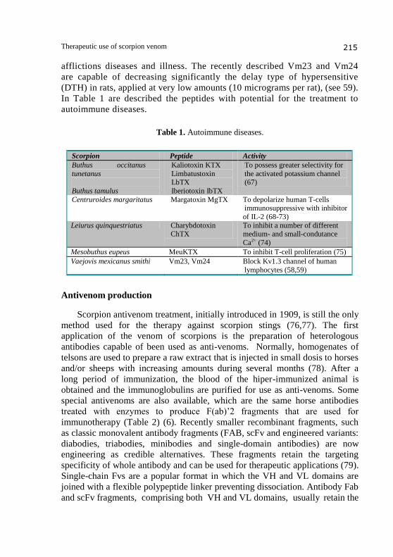
afflictions diseases and illness. The recently described Vm23 and Vm24
are capable of decreasing significantly the delay type of hypersensitive
(DTH) in rats, applied at very low amounts (10 micrograms per rat), (see 59).
In Table 1 are described the peptides with potential for the treatment to
autoimmune diseases.
Table 1. Autoimmune diseases.
Scorpion Peptide Activity
Buthus occitanus
tunetanus
Buthus tamulus
Kaliotoxin KTX
Limbatustoxin LbTX
Iberiotoxin IbTX
To possess greater selectivity for
the activated potassium channel (67)
Centruroides margaritatus Margatoxin MgTX To depolarize human T-cells immunosuppressive with inhibitor
of IL-2 (68-73)
Leiurus quinquestriatus Charybdotoxin ChTX
To inhibit a number of different medium- and small-condutance
Ca2+ (74)
Mesobuthus eupeus MeuKTX To inhibit T-cell proliferation (75)
Vaejovis mexicanus smithi Vm23, Vm24 Block Kv1.3 channel of human
lymphocytes (58,59)
Antivenom production
Scorpion antivenom treatment, initially introduced in 1909, is still the only
method used for the therapy against scorpion stings (76,77). The first
application of the venom of scorpions is the preparation of heterologous
antibodies capable of been used as anti-venoms. Normally, homogenates of
telsons are used to prepare a raw extract that is injected in small dosis to horses
and/or sheeps with increasing amounts during several months (78). After a
long period of immunization, the blood of the hiper-immunized animal is
obtained and the immunoglobulins are purified for use as anti-venoms. Some
special antivenoms are also available, which are the same horse antibodies
treated with enzymes to produce F(ab)’2 fragments that are used for
immunotherapy (Table 2) (6). Recently smaller recombinant fragments, such
as classic monovalent antibody fragments (FAB, scFv and engineered variants:
diabodies, triabodies, minibodies and single-domain antibodies) are now
engineering as credible alternatives. These fragments retain the targeting
specificity of whole antibody and can be used for therapeutic applications (79).
Single-chain Fvs are a popular format in which the VH and VL domains are
joined with a flexible polypeptide linker preventing dissociation. Antibody Fab
and scFv fragments, comprising both VH and VL domains, usually retain the
Vera L. Petricevich et al. 216
Tabla 2. Antivenoms.
Antivenom Scorpion Neutralization
Alacramyn C. limpidus, C. noxius, C. suffusus
C. limpidus, C. noxius, C. suffusus
Antiscorpion Tityus serrulatus Tityus spp.
Polyvalent scorpion antinvenoms
Leiurus quinquetriatus
Androctonus
crassicauda
A. amoreuxi, A. crassicauda, A. australis: B. arenicola, B. mimax,
B. occitanus, L.quinquestriatus
hebreus, Scorpiomarus palmatus
specific, monovalent, antigen binding affinity of the parent IgG, while showing
improved pharmacokinetics for tissue penetration (79). In this context, recently
single chain antibodies of human origin were developed and shown to be
effective for neutralization of scorpion toxin envenomation (80,81,82)
Cardiac diseases
Cardiac diseases are constituted by coronary heart and cerebro-vascular
diseases. Peptides from animal venoms are active as bradykinin-potentiating
factors are of particular interest because of their strong effect as hypotensive
agent. These factors have been found in Leiurus quinquestriatus, Tityus
serrulatus, Buthus martensii and B. occitanus scorpions. Pharmacologically,
these peptides obtained from scorpions venoms act as bradykinin-potentiating
peptides and can be used as hypotensive agents in the treatment of
hypertension. Moraes et al., 2011 (83) described that sodium channel gating
from Tityus bahiensis scorpion venom modified present different effects on
sodium channel isoforms.
Hematological diseases
The scorpion venom exerts its lethal action by interference with blood
coagulation, either by accelerating the process or inhibits the coagulation
processes. A peptide with anti-thrombotic action was described to be present in
the venom from the scorpion Buthus martensii Karsch (84). This same peptide
is related to the resistance against platelet aggregation and causes increment of
the concentration of prostanglandin I2 in plasma (84). Tityus discrepans
scorpion venom modifies clotting times in humans. Brazon et al., 2008 (85)
described the effect of T. discrepans venom on a partial thromboplastin time
prothrombin time and its direct clotting activity. This venom contains
anticoagulant components which prolong prothrombin time and partial
thromboplastic time.
Therapeutic use of scorpion venom 217
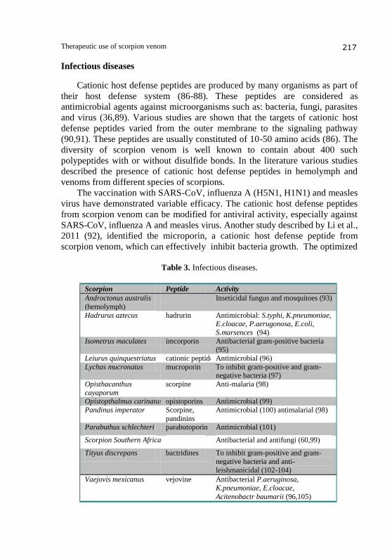
Infectious diseases
Cationic host defense peptides are produced by many organisms as part of
their host defense system (86-88). These peptides are considered as
antimicrobial agents against microorganisms such as: bacteria, fungi, parasites
and virus (36,89). Various studies are shown that the targets of cationic host
defense peptides varied from the outer membrane to the signaling pathway
(90,91). These peptides are usually constituted of 10-50 amino acids (86). The
diversity of scorpion venom is well known to contain about 400 such
polypeptides with or without disulfide bonds. In the literature various studies
described the presence of cationic host defense peptides in hemolymph and
venoms from different species of scorpions.
The vaccination with SARS-CoV, influenza A (H5N1, H1N1) and measles
virus have demonstrated variable efficacy. The cationic host defense peptides
from scorpion venom can be modified for antiviral activity, especially against
SARS-CoV, influenza A and measles virus. Another study described by Li et al.,
2011 (92), identified the microporin, a cationic host defense peptide from
scorpion venom, which can effectively inhibit bacteria growth. The optimized
Table 3. Infectious diseases.
Scorpion Peptide Activity
Androctonus australis
(hemolymph)
Inseticidal fungus and mosquitoes (93)
Hadrurus aztecus hadrurin Antimicrobial: S.typhi, K.pneumoniae, E.cloacae, P.aerugonosa, E.coli,
S.marsences (94)
Isometrus maculates imcorporin Antibacterial gram-positive bacteria (95)
Leiurus quinquestriatus cationic peptide Antimicrobial (96)
Lychas mucronatus mucroporin To inhibit gram-positive and gram-negative bacteria (97)
Opisthacanthus
cayaporum
scorpine Anti-malaria (98)
Opistopthalmus carinatus opistoporins Antimicrobial (99)
Pandinus imperator Scorpine,
pandinins
Antimicrobial (100) antimalarial (98)
Parabuthus schlechteri parabutoporin Antimicrobial (101)
Scorpion Southern Africa Antibacterial and antifungi (60,99)
Tityus discrepans bactridines To inhibit gram-positive and gram-
negative bacteria and anti-
leishmanicidal (102-104)
Vaejovis mexicanus vejovine Antibacterial P.aeruginosa,
K.pneumoniae, E.cloacae,
Acitenobactr baumarii (96,105)
Vera L. Petricevich et al. 218
microporin-M1 can inhibit grow of gram-positive bacteria at low
concentrations and antibiotic-resistant pathogens. Table 3 shows some cationic
host defense peptide from different scorpions.
Inflammatory response
The inflammatory response is triggered by a cascade of events that
includs: systems, cell elements and release of mediators (106). Scorpion
venoms can stimulate the release of immunological mediator cytokines. There
is now accumulating evidence to suggest a causal relationship between
overproduction of certain cytokines such as IL-1 and IL-6 and morbility and
mortality associated with critically ill patients. Sofer 1995, (107), was the first
that reported the involvement of the inflammatory systems after scorpion
envenomation in humans. In this work is documented the increment of IL-6
levels in serum of children severely envenomed by the scorpions
L. quinquestriatus and B. judaicus. The elevated levels of IL-6 were observed
at 1 to 3 hours after the sting. The IL-6 levels gradually returned to normal
values at 12 and 24 hours measurements, but remained above control levels in
all measurements. These results were quite similar to those found by others
authors that described the cytokine production after sting caused by Tityus
serrulatus scorpion in humans (108,109). With respect the experimental
animal high levels of cytokines were found in serum from mice injected with
Centruroides noxius and T. serrulatus venom (110,111). In all these works the
authors concluded that the activation and release of cytokines may play an
important role in the pathophysiology of envenomation after stings and may be
responsible for some systemic inflammatory manifestations with cytokine
release and organ failure (112). Cytokine have been shown that in local action
of cytokines promote recruitment of inflammatory cells to inflammation sites,
whereas their systemic effect to induce fever and increase symptoms. During
both responses local and systemic are observed the release of pro-
inflammatory cytokines, arachidonic acid metabolites proteins of the contact
phase and coagulation system, complement factors; it is defined as systemic
inflammatory response. Experimental models have been described that
following the injection of scorpion venoms and their fractions, a variety of
cytokines are released, and the outcome of an inflammatory response is
dictated by a variety of factors, that including the duration of the stimulus, and
the balance between the pro-inflammatory and anti-inflammatory response.
The imbalance determines the degree and extent of inflammation, and thus can
lead to multiple organ dysfunctions (112-121). With respect to the tissue injury
most of them have been related to the acute autonomic disturbances triggered
by the venom, which can provoke both the activation and delayed inactivation
Therapeutic use of scorpion venom 219
of neuronal sodium channels, where they modulate the release of
neurotransmitters, that leads to a variety of adverse effects which include
respiratory failure, lung edema, arrythmias, tachycardia followed by
bradycardia, skeletal muscle stimulation, lacrimation, convulsions, and
enlarged pupils, among others (116,120-128). However, the role of other
members of IL-family in envenomation is increasingly appreciated, and in the
present work are summarized all currently available information from human
and experimental studies. With respect the scorpion envenomation the immune
response also is triggered by cascade that including the released of mediators
such as nitric oxide, and complement system (112,129).
Multiple sclerosis is an inflammatory disease of the central nervous system
characterized by localized areas with demyelination. It is an autoimmune
disorder mediated by activated immune cells such as T- and B-lymphocytes
and macrophages/microglia. In the venom of the Moroccan scorpion
Androctonus mauretanicus a peptide was found and characterized, which
shows many toxins cross-reaction with lethal α-toxins found in North African
scorpion venoms and are considered as potent toxins for treatment of the
inflammatory diseases (130).
Malignant diseases
Cancer is the major public health burden in all developed countries. The
search for cancer cure from natural product such as plants and animals has
been practiced for over a century and the use of purified chemicals to treat
cancer still continues. With respect to chlorotoxin, it is considered a potent tool
for early detection of skin, cervical, esophageal, colon and lung cancers (131).
These ion-channels recognize by this toxin are among the many membrane
proteins overexpressed in different types of cancers. Scorpion venoms have
been used as traditional and folk therapy in various pathphysiological
conditions that has been mentioned in folk and traditional medicine of India,
China, Africa and Cuba (131). Various studies have suggested that the cancer
preventive and therapeutic efficacy of scorpion venom in different animal
tumor models and cell culture systems might be usefull. Bioactivies
polypeptides and enzymes as serine proteinase and hyaluronidase extract from
scorpion venoms from different species has been exhibited as potential usefull
as anti-proliferative agent with anti-tumor activity (131). Table 4 shows some
polypeptides and enzymes from scorpion venom with their principal action.
Bioinseticides
Natural venoms are a rich source of molecules that interact with membrane
receptors and ionic channels. Due to peptide toxins derived from venoms of a
Vera L. Petricevich et al. 220
Table 4. Malignant diseases.
Scorpion Peptide Activity
Androctonus crassicauda Apoptotic (132) Buthus martensii Karsch
Serine
proteinase-like BMK-CBP
Hyaluronidase BmHYA1
Anti-proliferative and apoptotic against
HUVEC, suppression of tumor growth
S180 sarcoma, glioma cells and H22 hepatocellular carcionoma (133-144).
To inhibit the growth of cancer cell line MCF-7 (136)
Hydrolysis of hyaluronic acid and is potent as cell surface markers in the
breast cancer cells line MDA-MB-23
(137) Buthus occitanus
tunetanus
Nontoxic
peptide
Adipocyte lypolysis (138)
Heterometrus bengalensis Bengalin Anticancer on U937 and K562 cells (139-141)
Leiurus quinquestriatus Charybtoxin Anticancer (142-144)
Odontobuthus doriae Apoptotic and anti-proliferative
neuroblastoma cells (132)
variety of invertebrates and lower vertebrates, valuable information about
mechanisms of neurotransmission, properties and physiological role of
voltage-dependent sodium, calcium and different potassium channels has been
obtained. Scorpions deliver a powerful, paralyzing venom, some of the toxins
damage only insects. Insect toxin induced a progressive slow depolarization of
the membrane potential and repetitive firing of action potentials (145). Several
such peptides were isolated from scorpion venoms and their properties as
bioisectide described (146). The search for new insect-specific neurotoxin to
be used as starting points for the development of highly selective
bioinsecticides (Table 5).
Table 5. Bioinsecticides.
Scorpion Peptide
Androctonus australis AaIT5
Buthacus arenicola BaIT2 Buthus martensii Karsch
Buthus occitanus tunetanus
BmKIT5, BotIT4,
BotIT5
Buthotus judaicus BjIT2 Centruroides noxius Cn10
Leiurus quinquestriatus LqqIT2
Leiurus quinquestriatus hebraeus LqhIT2
Therapeutic use of scorpion venom 221
Conclusion
In summary, most of the evidence that has emerged from the investigation
of venom and toxins from scorpions shows a clear therapeutic utility. Apart
from the production of specific anti-venoms to save life of people
envenomated by scorpion stings, many possible application of scorpion venom
components are forseen. Future research in the next decade with venoms and
toxins will definitely add information to be used as ion-channels inhibitors for
control of cell excitability, immune-modulation of T-cells, antibiotics against
bacteria and parasites, peptides for control of agricultural pests, and also for
management of neoplasic cells. These complex chemicals derived from animal
venom, could provide tools to study more in dept the biology of cancer.
Acknowledgments
Work in the author´s laboratory is supported by the Secretaria de
Educación Publica (SEP-PROMEP, México P/PROMEP: UAEMOR-CA-07.
References
1. Chippaux J.P., Goyffon M. (2008). Epidemiology of scorpionism: A global
appraisal. Acta Tropica,107, 71-79.
2. Possani LD, Becerril B, Delepierre M, Tytgat J. (1999). Scorpion toxins specific
for Na+channels. Eur J Biochem. Sep 264(2): 287-300.
3. Tytgat J, Chandy KG, Garcia ML, Gutman GA, Martin-Eauclaire MF, van der
Walt JJ, Possani LD. (1999). A unified nomenclature for short-chain peptides
isolated from scorpion venoms: alpha-KTx molecular subfamilies. Trends
Pharmacol Sci. Nov 20(11): 444-7.
4. Rodriguez de la Vega RC, Possani LD. (2005). Overview of scorpion toxins
specifici for Na+ channels and related peptides: biodiversity, structure-function
relationships and evolution. Toxicon. Dec 15;46(8):831-44. 2005 Nov 7.
5. Zeng XC, Corzo G, Hahin R. (2005). Scorpion venom peptides without disulfide
bridges. IUBMB Life. Jan;57(1):13-21. Review.
6. Espino-Solis GP, Riano-Umbarila L, Becerril B, Possani LD. (2009). Antidotes
against venomous animals: state of the art and prospectives. J Proteomics. Mar
6;72(2):183-99. Epub 2009 Jan 23. Review.
7. Ismail M. (1995). The scorpion envenoming syndrome. Toxicon. Jul 33(7): 825-58.
8. Mebs D. (2002). Scorpions and snakes, such as cobras, mambas and vipers made
the African continent famous for venomous animals. Bull Soc Pathol Exot. Aug
95(3): 131.
9. Freire-Maia L, Pinto GI, Franco I. (1974). Mechanism of the cardiovascular effects
produced by purified scorpion toxin in the rat. J Pharmacol Exp Ther. Jan 188(1):
207-13.
Vera L. Petricevich et al. 222
10. Müller GJ. (1993). Scorpionism in South Africa. A report of 42 serious scorpion
envenomations. S Afr Med J. Jun 83(6): 405-11.
11. Gwee MC, Nirthanan S, Khoo HE, Gopalakrishnakone P, Kini RM, Cheah LS.
(2002). Autonomic effects of some scorpion venoms and toxins. Clin Exp
Pharmacol Physiol. Sep 29(9): 795-801.
12. Dehesa-Dàvila M, Martin BM, Nobile M, Prestipino G, Possani LD. (1994).
Isolation of a toxin from Centruroides infamatus infamatus Koch scorpion venom
that modifies Na+ permeability on chick dorsal root ganglion cells. Toxicon. Dec
32(12): 1487-93.
13. Chowell G, Díaz-Dueñas P, Bustos-Saldaña R, Mireles AA, Fet V. (2006).
Epidemiological and clinical characteristics of scorpionism in Colima, Mexico
(2000-2001). Toxicon. Jun 1: 47(7):753-8.
14. Ashcroft FM, Gribble FM. (2000). Tissue-specific effects of sulfonylureas: lessons
from studies of cloned K(ATP) channels. J Diabetes Complications. Jul-Aug
14(4): 192-6.
15. Radha Krishna Murthy K. (2002). On scorpion envenoming syndrome: Problems
of medical ethics and accountability in medical research in India. J. Venom.
Anim.Toxins.. 8(1).
16. Lecomte C, Sabatier JM, Van Rietschoten J, Rochat H. (1998). Synthetic peptides
as tools to investigate the structure and pharmacology of potassium channel-acting
short-chain scorpion toxins. Biochimie. Feb 80(2):151-4.
17. Lehmann-Horn F, Jurkat-Rott K. (1999). Voltage-gated ion channels and
hereditary disease. Physiol Rev. Oct 79(4): 1317-72.
18. Possani LD, Merino E, Corona M, Bolivar F, Becerril B. (2000). Peptides and
genes coding for scorpion toxins that affect ion-channels. Biochimie. Sep-Oct
82(9-10): 861-8.
19. Jover E, Couraud F, Rochat H. (1980). Two types of scorpion neurotoxins
characterized by their binding to two separate receptor sites on rat brain
synaptosomes. Biochem Biophys Res Commun. Aug 29; 95(4): 1607-14.
20. Wheeler KP, Barhanin J, Lazdunski M. (1982). Specific binding of toxin II from
Centruroides suffusus suffusus to the sodium channel in electroplaque membranes.
Biochemistry. Oct 26; 21(22): 5628-34.
21. Catterall WA. (1992). Cellular and molecular biology of voltage-gated sodium
channels. Physiol Rev. Oct 72(4 Suppl): S15-48.
22. Benzinger GR, Drum CL, Chen LQ, Kallen RG, Hanck DA, Hanck D. (1997). Differences in the binding sites of two site-3 sodium channel toxins. Pflugers Arch.. Nov 434(6) :742-9. 23. Benzinger GR, Kyle JW, Blumenthal KM, Hanck PA. (1998). A specific interaction between the cardiac sodium channel and site-3 toxin anthopleurin B. J Biol Chem. Jan 2; 273(1): 80-4. 24. Li-Smerin Y, Swartz KJ. (2000). Localization and molecular determinants of the Hanatoxin receptors on the voltage-sensing domains of a K(+) channel. J Gen Physiol. Jun 115(6): 673-84. 25. McDonough SI, Lampe RA, Keith RA, Bean BP. (1997). Voltage-dependent
inhibition of N- and P-type calcium channels by the peptide toxin omega-
grammotoxin-SIA. Mol Pharmacol. Dec 52(6): 1095-104.
Therapeutic use of scorpion venom 223
26. Rogers SW, Gahring LC, Papke RL, Heinemann S. (1991). Identification of
cultured cells expressing ligand-gated cationic channels. Protein Expr Purif. Apr-
Jun 2(2-3): 108-16.
27. Thomsen WJ, Catterall WA. (1989). Localization of the receptor site for alpha-
scorpion toxins by antibody mapping: implications for sodium channel topology.
Proc Natl Acad Sci U S A. Dec 86(24): 10161-5.
28. West JW, Patton DE, Scheuer T, Wang Y, Goldin AL, Catterall WA. (1992). A
cluster of hydrophobic amino acid residues required for fast Na(+)-channel
inactivation. Proc Natl Acad Sci U S A. Nov 15: 89(22): 10910-4.
29. Cestèle S, Catterall WA. (2000). Molecular mechanisms of neurotoxin action on
voltage-gated sodium channels. Biochimie. Sep-Oct 82(9-10): 883-92.
30. Cestèle S, Gordon D. (1998). Depolarization differentially affects allosteric
modulation by neurotoxins of scorpion alpha-toxin binding on voltage-gated
sodium channels. J Neurochem. Mar 70(3): 1217-26.
31. Gordon D. (1997). A new approach to insect-pest control-combination of
neurotoxins interacting with voltage sensitive sodium channels to increase
selectivity and specificity. Invert Neurosci. Sep-Dec 3(2-3): 103-16.
32. Marcotte P, Chen LQ, Kallen RG, Chahine M. (1997). Effects of Tityus serrulatus
scorpion toxin gamma on voltage-gated Na+ channels. Circ Res. Mar 80(3): 363-9.
33. Thomsen W, Martin-Eauclaire MF, Rochat H, Catterall WA. (1995).
Reconstitution of high-affinity binding of a beta-scorpion toxin to neurotoxin
receptor site 4 on purified sodium channels. J Neurochem. Sep 65(3): 1358-64.
34. Shichor I, Zlotkin E, Ilan N, Chikashvili D, Stuhmer W, Gordon D, Lotan I.
(2002). Domain 2 of Drosophila para voltage-gated sodium channel confers insect
properties to a rat brain channel. J Neurosci. Jun 1 22(11): 4364-71.
35. Cestèle S, Qu Y, Rogers JC, Rochat H, Scheuer T, Catterall WA. (1998). Voltage
sensor-trapping: enhanced activation of sodium channels by beta-scorpion toxin
bound to the S3-S4 loop in domain II.Neuron. Oct 21(4): 919-31.
36. Cahalan MD. (1975). Modification of sodium channel gating in frog myelinated
nerve fibres by Centruroides sculpturatus scorpion venom. J Physiol. Jan 244(2):
511-34.
37. Vijverberg HP, Pauron D, Lazdunski M. (1984). The effect of Tityus serrulatus
scorpion toxin gamma on Na channels in neuroblastoma cells. Pflugers Arch. Jul
401(3): 297-303.
38. Cestèle S, Yarov-Yarovoy V, Qu Y, Sampieri F, Scheuer T, Catterall WA. (2006). Structure and function of the voltage sensor of sodium channels probed by a beta- scorpion toxin. J Biol Chem. Jul 28: 281(30): 21332-44. 39. Yatani A, Kirsch GE, Possani LD, Brown AM. (1988). Effects of New World scorpion toxins on single-channel and whole cell cardiac sodium currents. Am J Physiol. Mar: 254(3 Pt 2): H443-51. 40. Cohen L, Karbat I, Gilles N, Ilan N, Benveniste M, Gordon D, Gurevitz M. (2005). Common features in the functional surface of scorpion beta-toxins and elements that confer specificity for insect and mammalian voltage-gated sodium channels. J Biol Chem. Feb 11: 280(6): 5045-53. 41. Armstrong CM, Bezanilla F. (1977). Inactivation of the sodium channel. II. Gating
current experiments. J Gen Physiol. Nov: 70(5): 567-90.
Vera L. Petricevich et al. 224
42. French RJ, Horn R. (1983). Sodium channel gating: models, mimics, and
modifiers. Annu Rev Biophys Bioeng. 12: 319-56.
43. Patlak J. (1991). Molecular kinetics of voltage-dependent Na+ channels. Physiol
Rev. Oct: 71(4): 1047-80.
44. Keynes RD. (1994). The kinetics of voltage-gated ion channels. Q Rev Biophys.
Dec: 27(4): 339-434.
45. Yu FH, Catterall WA. (2003). Overview of the voltage-gated sodium channel
family. Genome Biol. 4(3): 207.
46. Mouhat S, Jouirou B, Mosbah A, De Waard M, Sabatier JM. (2004). Diversity of
folds in animal toxins acting on ion channels. Biochem J. Mar 15;378(Pt 3):
717- 26.
47. Bosmans F, Martin-Eauclaire MF, Tytgat J. (2007). Differential effects of five
'classical' scorpion beta-toxins on rNav1.2a and DmNav1 provide clues on species-
selectivity. Toxicol Appl Pharmacol. Jan 1: 218(1): 45-51.
48. Shieh CC, Coghlan M, Sullivan JP, Gopalakrishnan M. (2000). Potassium
channels: molecular defects, diseases, and therapeutic opportunities. Pharmacol
Rev. Dec: 52(4): 557-94.
49. Corona M, Gurrola GB, Merino E, Cassulini RR, Valdez-Cruz NA, García B,
Ramírez-Domínguez ME, Coronas FI, Zamudio FZ, Wanke E, Possani LD. (2002).
A large number of novel Ergtoxin-like genes and ERG K+-channels blocking
peptides from scorpions of the genus Centruroides. FEBS Lett. Dec 4: 532(1-2):
121-6.
50. Batista CV, Gómez-Lagunas F, Rodríguez de la Vega RC, Hajdu P, Panyi G,
Gáspár R, Possani LD. (2002). Two novel toxins from the Amazonian scorpion
Tityus cambridgei that block Kv1.3 and Shaker B K(+)-channels with distinctly
different affinities. Biochim Biophys Acta. Dec 16: 1601(2): 123-31.
51. Goudet C, Chi CW, Tytgat J. (2002). An overview of toxins and genes from the
venom of the Asian scorpion Buthus martensi Karsch. Toxicon. Sep: 40(9): 1239-58.
52. Tenenholz TC, Klenk KC, Matteson DR, Blaustein MP, Weber DJ. (2000).
Structural determinants of scorpion toxin affinity: the charybdotoxin (alpha-KTX)
family of K(+)-channel blocking peptides. Rev Physiol Biochem Pharmacol. 140:
135-85.
53. Arantes EC, Prado WA, Sampaio SV, Giglio JR. (1989). A simplified procedure
for the fractionation of Tityus serrulatus venom: isolation and partial
characterization of TsTX-IV, a new neurotoxin. Toxicon. 27(8): 907-16.
54. Holaday SK Jr, Martin BM, Fletcher PL Jr, Krishna NR. (2000). NMR solution
structure of butantoxin. Arch Biochem Biophys. Jul 1: 379(1): 18-27.
55. Novello JC, Arantes EC, Varanda WA, Oliveira B, Giglio JR, Marangoni S.
(1999). TsTX-IV, a short chain four-disulfide-bridged neurotoxin from Tityus
serrulatus venom which acts on Ca2+-activated K+ channels. Toxicon. Apr:
37(4): 651-60.
56. Pimenta AM, Legros C, Almeida F de M, Manuselle P, De Lima ME, Bougis PE,
Martin-Eauclaire MF. (2003). Novel structural class of four disulfide-bridged
peptides from Tityus serrulatus venom. Biochem Biophys Res Commun. Feb 21:
301(4): 1086-92.
Therapeutic use of scorpion venom 225
57. Villalonga N, Ferreres JC, Argilés JM, Condom E, Felipe A. (2007). Potassium
channels are a new target field in anticancer drug design. Recent Pat Anticancer
Drug Discov. Nov: 2(3): 212-23.
58. Gurrola, G.B., Hernández-López, R., Rodríguez de la Vega, R., Vargas, Z., Batista,
C.F., Salas-Castillo, S., Panyi, G., Del Rio Portilla, F., Possani, L.D. Structure,
function and chemical synthesis of Vaejovis mexicanus peptide 24: a novel potent
blocker of Kv1.3 potassium channels of human T lymphocytes. Biochemistry, 51,
4049-4061 (2012).
59. Varga,Z., Gurrola-Briones,G., Papp,F., Rodríguez de la Vega,R.C., Pedraza-Alva, G.,
Tajhya,R.B., Gaspar,R.,Cardenas,L., Rosenstein,Y., Beeton, C., Possani,L.D.,
Panyi,G. Vm24, a natural immunosuppressant peptide potently and selectively blocks
Kv1.3 potassium channels of human T cells. Molecular Pharmacology, published
ahead doi:10.1124/mol.112.078006, (2012).
60. Moerman L, Bosteels S, Noppe W, Willems J, Clynen E, Schoofs L, Thevissen K,
Tytgat J, Van Eldere J, Van Der Walt J, Verdonck F. (2002). Antibacterial and
antifungal properties of alpha-helical, cationic peptides in the venom of scorpions
from southern Africa. Eur J Biochem. Oct; 269(19):4799-810.
61. Elgar D, Verdonck F, Grobler A, Fourie C, du Plessis J. (2005). Ion selectivity of
scorpion toxin-induced pores in cardiac myocytes. Peptides. Jan;27(1):55-61. Epub
2005 Aug 8.
62. Ferreira LA, Henriques OB. (1992). Isolation of a bradykinin-potentiating factor
from scorpion Tityus serrulatus venom. Agents Actions Suppl. ;38 ( Pt 1):462-8
63. Meki AR, Nassar AY, Rochat H. (1995). A bradykinin-potentiating peptide
(peptide K12) isolated from the venom of Egyptian scorpion Buthus occitanus.
Peptides. 16(8): 1359-65.
64. Ali AS, Stoeva S, Schutz J, Kayed R, Abassi A, Zaidi ZH, Voelter W. (1998).
Purification and primary structure of low molecular mass peptides from scorpion
(Buthus sindicus) venom. Comp Biochem Physiol A Mol Integr Physiol.
Dec;121(4):323-32.
65. Coffman RL, Mosmann TR. (1991). CD4+ T-cell subsets: regulation of
differentiation and function. Res Immunol. Jan;142(1):7-9. 66. Mosmann TR, Cherwinski H, Bond MW, Giedlin MA, Coffman RL. (2005). Two types of murine helper T cell clone. I. Definition according to profiles of lymphokine activities and secreted proteins. J Immunol. Jul 1;175(1):5-14. No abstract available. 67. Drira-Chaabane S, Ayeb ME, Torresani J, Gharbi-Chihi J. (1996). Lipolytic action of Buthus occitanus tumetanus venom involvement of the beta ad. Biochem Biophys Res Commun. 226; 280-286. 68. Leonard RJ, Garcia ML, Slaughter RS, Reuben JP. (1992). Selective blockers of voltage-gated K+ channels depolarize human T lymphocytes: mechanism of the antiproliferative effect of charybdotoxin. Proc Natl Acad Sci U S A. Nov 1;89(21):10094-8. 69. Lin CS, Boltz RC, Blake JT, Nguyen M, Talento A, Fischer PA, Springer MS,
Sigal NH, Slaughter RS, Garcia ML. (1993). Voltage-gated potassium channels
regulate calcium-dependent pathways involved in human T lymphocyte activation.
J Exp Med. Mar 1;177(3):637-45.
Vera L. Petricevich et al. 226
70. Koo GC, Blake JT, Talento A, Nguyen M, Lin S, Sirotina A, Shah K, Mulvany K,
Hora D Jr, Cunningham P, Wunderler DL, McManus OB, Slaughter R, Bugianesi
R, Felix J, Garcia M, Williamson J, Kaczorowski G, Sigal NH, Springer MS,
Feeney W. (1997). Blockade of the voltage-gated potassium channel Kv1.3 inhibits
immune responses in vivo. J Immunol. Jun 1; 158 (11): 5120-8.
71. Garcia C, Calderón-Aranda ES, Anguiano GA, Becerril B, Possani LD. (2003).
Analysis of the immune response induced by a scorpion venom sub-fraction, a pure
peptide and a recombinant peptide, against toxin Cn2 of Centruroides noxius
Hoffmann. Toxicon. Mar;41(4):417-27.
72. Koschak A, Bugianesi RM, Mitterdorfer J, Kaczorowski GJ, Garcia ML, Knaus
HG. (1998). Subunit composition of brain voltage-gated potassium channels
determined by hongotoxin-1, a novel peptide derived from Centruroides limbatus
venom. J Biol Chem. Jan 30;273(5):2639-44.
73. Price M, Lee SC, Deutsch C. (1989). Charybdotoxin inhibits proliferation and
interleukin 2 production in human peripheral blood lymphocytes. Proc Natl Acad
Sci U S A. Dec;86 (24):10171-5.
74. Sabatier JM, Fremont V, Mabrouk K, Crest M, Darbon H, Rochat H, Van
Rietschoten J, Martin-Eauclaire MF. (1994). Leiurotoxin I, a scorpion toxin
specific for Ca(2+)-activated K+ channels. Structure-activity analysis using
synthetic analogs. Int J Pept Protein Res. May;43(5):486-95.
75. Gao B, Peigneur S, Tytgat J, Zhu S. (2010). A potent potassium channel blocker
from Mesobuthus eupeus scorpion venom. Biochimie, Dec 92 (12): 1847-1853.
76. Balozet L. Scorpionism in the Old World. In: BÜCHERL W., BUCKLEY E. Eds.
Venomous animals and their venoms. New York: Academic Express, 1971: 349-71.
77. Theakston RD, Warrell DA, Grifithis E. (2003). Report of a WHO workshop on
the standardization and control of antivenoms. Toxicon. 41, 541-57.
78. Tulga T. (1964). Scorpions found in Turkey and paraspecific action of an antivenin
produced with the venom of the species Androctonus crassicauda. Turk. Hij.
Deney. Biyol. Derg. 24, 146-55.
79. Holliger Ph, Hudson JP. (2005). Engineering antibody fragments and the rise of
single domains. Nat. Biotechnol. 23: 1126-1136.
80. Riaño-Umbarila, L., Contreras-Ferrat, G., Olamendi-Portugal, T., Morelos-Juárez, C.,
Corzo, G., Possani, L.D. and Becerril, B. Exploiting cross-reactivity to neutralize two
different scorpion venoms with one single.chain antibody fragment. Journal
Biological Chemistry 286:6143-6151(2011). 81. Canul-Tec, J-C., Riaño-Umbarila, L., Rudinño-Pinera, E., Becerril, B., Possani, L.D., Torres-Larios, A. Structural basis of neutralization of the major toxic component from the scorpion Centruroides noxius Hoffmann by a human-derived single chain antibody fragment. J. Biol. Chemistry, 286:20892-20900 (2011). 82. Rodríguez-Rodríguez, E.R., Ledezma-Candanoza, L.M., Contreras-Ferrat, L.G., Olamendi-Portugal, T., Possani, L.D., Becerril, B., Riaño-Umbarila,L. A single mutation in framework 2 of the heavy variable domain improves the properties of a diabody and a related single-chain antibody. J. Mol Biol. 423:337-350 (2012). 83. Moraes ER, Kalapothakis E, Naves LA, Kushmerick C. (2009). Differential effects
of Tityus bahiensis Scorpion venom on tetrodotoxin-sensitive and tetrodotoxin-
resistant sodium currents. Neurotox Res. Jan;19(1):102-14. Epub 2009 Dec 18.
Therapeutic use of scorpion venom 227
84. Song YM, Tang XX, Chen XG, Gao BB, Gao E, Bai L, Lv XR. (2005). Effects of
scorpion venom bioactive polypolypeptides on platelet aggregation and thrombosis and
plasma 6-keto-PG F1alpha and TXB2 in rabbits and rats. Toxicon.Aug;46(2):230-5.
85. Brazón J, D'Suze G, D'Errico ML, Arocha-Piñango CL, Guerrero B. (2009).
Discreplasminin, a plasmin inhibitor isolated from Tityus discrepans scorpion
venom. Arch Toxicol. Jul;83(7):669-78. Epub 2008 Nov 18.
86. Hancock RE, Sahl HG. (2006). Antimicrobial and host-defense peptides as new
anti-infective therapeutic strategies. Nat Biotechnol. Dec;24(12):1551-7.
87. Hancock RE, Leher R. (1998). Cationic peptides: a new source of antibiotics.
Trends Biotechnol.16:82-88.
88. Wang K, Yin SJ, Lu M, Yi H, Dai C, Xu XJ, Cao ZJ, Wu YL, Li WX. (2006).
Functional analysis of the alpha-neurotoxin, BmalphaTX14, derived from the
Chinese scorpion, Buthus martensii Karsch. Biotechnol Lett. Nov;28(21):1767-72.
Epub 2006 Aug 16.
89. Brogden KA, Ackermann M, McCray PB Jr, Tack BF. (2003). Antimicrobial
peptides in animals and their role in host defences. Int J Antimicrob Agents.
Nov;22(5):465-78.
90. Brown KL, Hancock RE. (2006). Cationic host defense (antimicrobial) peptides.
Curr Opin Immunol. Feb;18(1):24-30. Epub 2005 Dec 6.
91. Jenssen H, Hamill P, Hancock RE. (2006). Peptide antimicrobial agents. Clin
Microbiol Rev. Jul;19(3):491-511.
92. Li Q, Zhao Z, Zhou D, Chen Y, Hong W, Cao L, Yang J, Zhang Y, Shi W, Cao Z,
Wu Y, Yan H, Li W. (2011). Virucidal activity of a scorpion venom peptide
variant mucroporin-M1 against measles, SARS-CoV and influenza H5N1 viruses.
Peptides. Jul;32(7):1518-25. Epub 2011 May 19.
93. Wang C, St Leger RJ. (2007). A scorpion neurotoxin increases the potency of a
fungal insecticide. Nat Biotechnol. Dec;25(12):1455-6. Epub 2007 Nov 11.
94. Torres-Larios A, Gurrola GB, Zamudio FZ, Possani LD. Hadrurin, (2000). A new
antimicrobial peptide from the venom of the scorpion Hadrurus aztecus. Eur J
Biochem.Aug;267(16):5023-31.
95. Zhao Z, Ma Y, Daí C, Zhao R, Li SR, Wu Y, Cao Z, Li W. (2009). Incorporin, a
new cationic antimicrobial peptide from the venom of the scorpion Isometrus
maculates. Antimicrobial Agents and Chemotherapy .Aug. 3472-3477.
96. Cociancich S, Ghazi A, Hetru C, Hoffmann JA, Letellier L. (1993). Insect
defensin, an inducible antibacterial peptide, forms voltage-dependent channels in
Micrococcus luteus. J Biol Chem. Sep 15;268(26):19239-45.
97. Dai C, Ma Y, Zhao Z, Zhao R, Wang Q, Wu Y, Cao Z, Li W. (2008). Mucroporin,
the first cationic host defense peptide from the venom of Lychas mucronatus.
Antimicrob Agents Chemother. Nov;52(11):3967-72. Epub 2008 Sep 8.
98. Conde R, Zamudio FZ, Rodríguez MH, Possani LD. (2000). Scorpine, an anti-
malaria and anti-bacterial agent purified from scorpion venom. FEBS Lett. Apr
14;471(2-3):165-8.
99. Eheret-Sabatier L, Loew D, Goyffon M, Fehlbaum P, Hoffman JÁ, van Dorsselaer,
Bulet P. (2001). Characterization of novel cysteine-rich antimicrobial peptides
from scorpion blood. J. Biol.Chem. 271: 29537-29544.
Vera L. Petricevich et al. 228
100. Corzo G, Escoubas P, Villegas E, Barnham KJ, He W, Norton RS, Nakajima T.
(2001). Characterization of unique amphipathic antimicrobial peptides from venom
of the scorpion Pandinus imperator. Biochem J. Oct 1;359(Pt 1):35-45
101. Remijsen Q, Verdonck F, Willems J. (2010). Parabutoporin, a cationic amphipathic
peptide from scorpion venom: much more than na antibiotic. Toxicon. Feb-Mar;
55(2-3): 180-185.
102. Borges A, Silva S, Op den Camp Hubb J. (1987). In vitro leishmanicidal of Tityus
discrepans scorpion venom. Parasitology research. vol. 99 n° 2, 167-177.
103. Díaz P, D'Suze G, Salazar V, Sevcik C, Shannon JD, Sherman NE, Fox JW.
(2009). Antibacterial activity of six novel peptides from Tityus discrepans scorpion
venom. A fluorescent probe study of microbial membrane Na+ permeability
changes. Toxicon. Nov;54(6):802-17. Epub 2009 Jun 21.
104. Díaz P, D'Suze G, Salazar V, Sevcik C, Shannon JD, Sherman NE, Fox JW.
(2009). Antibacterial activity of six novel peptides from Tityus discrepans scorpion
venom. A fluorescent probe study of microbial membrane Na+ permeability
changes. Toxicon. Nov;54(6):802-17. Epub 2009 Jun 21.
105. Hernández-Aponte CA, Silva-Sanchez J, Quintero-Hernández V, Rodríguez-
Romero A, Balderas C, Possani LD, Gurrola GB. (2010). Vejovine, a new
antibiotic from the scorpion venom of Vaejovis mexicanus. Toxicon. Jan;57(1):
84-92. Epub 2010 Oct 20.
106. Fabiano G, Pezzolla A, Filograna MA, Ferrarese F. (2008). [Traumatic shock-
physiopathologic aspects]. G Chir. Jan-Feb: 29(1-2): 51-7.
107. Sofer S. (1995). Scorpion envenomation. Intensive Care Med. Aug: 21(8): 626-8.
108. Magalhães MM, Pereira ME, Amaral CF, Rezende NA, Campolina D, Bucaretchi
F, Gazzinelli RT, Cunha-Melo JR. (1999). Serum levels of cytokines in patients
envenomed by Tityus serrulatus scorpion sting. Toxicon. Aug: 37(8): 1155-64.
109. Fukuhara YD, Reis ML, Dellalibera-Joviliano R, Cunha FQ, Donadi EA. (2003).
Increased plasma levels of IL-1beta, IL-6, IL-8, IL-10 and TNF-alpha in patients
moderately or severely envenomed by Tityus serrulatus scorpion sting. Toxicon.
Jan: 41(1): 49-55.
110. Petricevich VL. (2006). Balance between pro- and anti-inflammatory cytokines in mice treated with Centruroides noxius scorpion venom. Mediators Inflamm. 2006(6): 54273. 111. Petricevich VL, Peña CF. (2002). The dynamics of cytokine and nitric oxide secretion in mice injected with Tityus serrulatus scorpion venom. Mediators Inflamm. Jun: 11(3): 173-80. 112. Petricevich VL. (2010). Scorpion venom and the inflammatory response. Mediators Inflamm. doi:10.1155/2010/903295. 113. Adi-Bessalem S, Hammoudi-Triki D, Laraba-Djebari F. (2008).
Pathophysiological effects of Androctonus australis hector scorpion venom: tissue
damages and inflammatory response. Exp Toxicol Pathol. Aug, 60(4-5): 373-80.
114. Petricevich VL. (2002). Effect of Tityus serrulatus venom on cytokine production
and the activity of murine macrophages. Mediators Inflamm. Feb: 11(1): 23-31.
115. Abdoon NA, Fatani AJ. (2009). Correlation between blood pressure, cytokines and
nitric oxide in conscious rabbits injected with Leiurus quinquestriatus
quinquestriatus scorpion venom. Toxicon. Sep 15;54(4):471-80. Epub 2009 May 23.
Therapeutic use of scorpion venom 229
116. Bucaretchi F, Baracat EC, Nogueira RJ, Chaves A, Zambrone FA, Fonseca MR,
Tourinho FS. (1995). A comparative study of severe scorpion envenomation in
children caused by Tityus bahiensis and Tityus serrulatus. Rev Inst Med Trop Sao
Paulo. Jul-Aug: 37(4): 331-6.
117. Freire-Maia L, Campos JA, Amaral CF. (1994). Approaches to the treatment of
scorpion envenoming. Toxicon. 32: (9): 1009-1014.
118. Novaes G, Cabral AP, de Falco CN, de Queiroz AC. (1989). Acute pancreatitis
induced by scorpion toxin, Tityustoxin. Histopathological study in rats. Arq.
Gastroenterol. 26(1-2): 9-12.
119. Bucaretchi F, Vinagre AM, Chavéz-Olórtegui C, Collares EF. (1999). Effect of
toxin- from Tityus serrulatus scorpion venom on gastric emptying in rats. Braz J
Med Biol Res. Apr 32(4). 431-4.
120. Becerril B, Corona M, Garcia C, Bolivar F, Possani LD. (1995). Cloning of genes
encoding scorpion toxins: an interpretative review. J. Toxicol. Toxins Rev. 14, 339-357.
121. Osnaya-Romero N, Hernandez TJM, Basurto G, Andrade S, Figueroa JM, Carvajal
Y, Flores-Hernandez SS. (2008). Serum electrolyte changes in pediatric stung by
Scorpions. J. Venom. Anim. Toxins incl.Trop. Dis. 14(2).
122. Cunha-Melo JR, Toppa NH, Martins P, Colares CN, Castro YS, Freire-Maia L.
(1991). Acute gastric mucosal injury induced by toxins from Tityus serrulatus
scorpion venom: a novel experimental model in the rat. Toxicon. 29(11):
1395-1401.
123. Cupo P, Jurca M, Azeedo-Marques MM, Oliveira JS, Hering SE. (1994). Severe
scorpion envenomation in Brazil. Clinical, laboratory and anatomopathological
aspects. Rev Inst Med Trop Sao Paulo. Jan-Feb. 36(1): 67-76.
124. Falqueto EB, Massensini AR, Moraes-Santos T, Gomez MV, Romano-Silva MA.
(2002). Modulation of Na+-channels by neurotoxins produces different effects on
[3H]ACh release with mobilization of distinct Ca2+-channels. Cell Mol Neurobiol.
Dec: 22(5-6): 819-26.
125. Mesquita MB, Moraes-Santos T, Moraes MF. (2002). Phenobarbital blocks the
lung edema induced by centrally injected tityustoxin in adult Wistar rats. Neurosci
Lett. Oct 31;332(2):119-22.
126. Sampaio SV, Laure CJ, Giglio JR. (1983). Isolation and characterization of toxic
proteins from the venom of the Brazilian scorpion Tityus serrulatus. Toxicon.
21(2): 265-77.
127. Ismail M, Abd-Elsalam MA, Morad AM. (1990). Do changes in body temperature
following envenomation by the scorpion Leiurus quinquestriatus influence the
course of toxicity? Toxicon. 28(11): 1265-84.
128. Sofer S, Gueron M. (1988). Respiratory failure in children following envenomation
by the scorpion Leiurus quinquestriatus: hemodynamic and neurological aspects.
Toxicon. 26(10). 931-9.
129. Bertazzi DT, de Assis-Pandochi AI, Azzolini AE, Talhaferro VL, Lazzarini M,
Arantes EC. (2003). Effect of Tityus serrulatus scorpion venom and its major
toxin, TsTX-I, on the complement system in vivo. Toxicon. Mar: 41(4): 501-8.
130. Oukkache N, Rosso JP, Alami M, Ghalim N, Saïle R, Hassar M, Bougis PE,
Martin-Eauclaire MF. (2008). New analysis of the toxic compounds from the
Vera L. Petricevich et al. 230
Androctonus mauretanicus mauretanicus scorpion venom. Toxicon.
Apr;51(5):835-52. Epub 2007 Dec 17.
131. Gomes A, Bhattacharjee P, Mishra R, Ajoy KB, Dasgupta SC, Giri B. (2010).
Anticancer potential of animal venoms and toxins. Indian Journal of Exp. Biology.
Vol. 48: 93-103.
132. Zargan J, Sajad M, Umar S, Naime M, Ali S, Khan HA. (2011). Scorpion
(Odontobuthus doriae) venom induces and inhibits DNA synthesis in human
neuroblastoma cells. Mol Cell Biochem. Feb; 348(1-2): 173-181.
133. Liu Y, Qu X, Wang P, Tian X, Luo Y, Liu X. (2002). WT1 downregulation during
K562 cell diferentiation and apoptosis induced by bufalin. Zhonghua Xue Ye Za
Zhi. 23: 356.
134. Fu YJ, Yin LT, Liang AH, Zhang CF, Wang W, Chai BF, Yang JY, Fan XJ.
(2007). Therapeutic potential of chlorotoxin-like neurotoxin from the Chinese
scorpion for human gliomas. Neurosci Lett. Jan 22;412(1):62-7. Epub 2006 Dec 12
135. Zhang N, Wu G, Wu H, Chalmers MJ, Gaskell SJ. (2004). Purification,
characterization and sequence determination of BmKK4, a novel potassium
channel blocker from Chinese scorpion Buthus martensii Karsch. Peptides.
Jun;25(6):951-7.
136. Gao R, Zhang Y, Gopalakrishnakone P. (2008). Purification and N-terminal
sequence of a serine proteinase-like protein (BMK-CBP) from the venom of the
Chinese scorpion (Buthus martensii Karsch). Toxicon. 52: 348.
137. Feng L, Gao R, Gopalakrishnakone P. (2008). Isolation and characterization of a
hyaluronidase from the venom of Chinese red scorpion Buthus martensi. Comp
Biochem Physiol C Toxicol Pharmacol. 148: 250.
138. Soudani N, Gharbi-Chihi J, Srairi-Abid N, Yazidi CM, Planells R, Margotat A,
Torresani J, El Ayeb M. (2005). Isolation and molecular characterization of LVP1
lipolysis activating peptide from scorpion Buthus occitanus tunetanus. Biochim
Biophys Acta. Feb 14;1747(1):47-56. Epub 2004 Oct 27.
139. Gupta SD, Gomes A, Debnath A, Saha A, Gomes A. (2008). Apoptosis induction
in human leukemic cells by a novel protein bengaline, isolated from Indian black
scorpion venom: through mitochondrial pathway and inhibition of heat shock
proteins. Chem Biol Interact.doi: 10.1016/j.cbi.2009.11.006.
140. Das Gupta S, Debnath A, Saha A, Giri B, Tripathi G, Vedasiromoni JR, Gomes A,
Aparna Gomes. (2007). Indian black scorpion (Heterometrus bengalensis Koch)
venom indiced antiproliferative and apoptogenic activity against human leukemic
cell lines U937 and K562. Leuk Res. 31: 817.
141. Gomes A, Giri B, Saha A, Mishra R, Dasgupta SC, Debnath A, Gomes A. (2007).
Bioactive molecules from amphibian skin: Their biological activities with
reference to therapeutic potentials for possible drug development. Indian J Exp
Biol. 45: 579.
142. DeBin JA, Strichartz GR. (1991). Chloride channel inhibition by the venom of the
scorpion Leiurus quinquestriatus. Toxicon. 29: 1403.
143. Dalton S, Gerzanich V, Chen M, Dong Y, Shuba Y, Simard JM. (2003).
Chlorotoxin-sensitive Ca2+-activated Cl- channel in type R2 reactive astrocytes
from adult rat brain. Glia. Jun;42(4):325-39.
Therapeutic use of scorpion venom 231
144. Deshane J, Garner CC, Sontheimer H. (2003). Chlorotoxin inhibits glioma cell
invasion via matrix metalloproteinase-2. J Biol Chem. Feb 7;278(6):4135-44. Epub
2002 Nov 25.
145. Nakagawa Y, Lee YM, Lehmberg E, Hermann R, Hermann R, Moskowitz H,
Jones AD, Hammock BD. (1997). Anti-insect toxin 5 (AaIT5) from Androctonus
australis. Eur.J.Biochem. Jun 1: 246(2): 496-501.
146. Selisko, B., García, C., Becerril, B., Delepierre, M. y Possani, L.D. A new insect-
specific toxin from Centruroides noxius Hoffmann: cDNA, primary structure, three-
dimensional model and electrostatic surface potentials in comparison to other toxin
variants. Eur. J. Biochem. 242, 235-242 (1996).