Insect Science (2019) 00, 1–21, DOI 10.1111/1744-7917.12656 ORIGINAL ARTICLE Molecular cloning and comparative analysis of transcripts encoding chemosensory proteins from two plant bugs, Lygus lineolaris and Lygus hesperus J. Joe Hull 1,* , Omaththage P. Perera 2,* and Mei-Xian Wang 1, 3 1 USDA-ARS Arid Land Agricultural Research Center, Maricopa, Arizona, USA; 2 USDA-ARS, Southern Insect Management Research Unit, Stoneville, Mississippi, USA and 3 College of Animal Sciences, Zhejiang University, Hangzhou, China Abstract Chemosensory proteins (CSPs) are soluble carrier proteins typically charac- terized by a six-helix bundle structure joined by two disulfide bridges and a conserved Cys spacing pattern (C1-X 6-8 -C2-X 16-21 -C3-X 2 -C4). CSPs are functionally diverse with reported roles in chemosensation, immunity, development, and resistance. To expand our molecular understanding of CSP function in plant bugs, we used recently developed tran- scriptomic resources for Lygus lineolaris and Lygus hesperus to identify 17 and 14 CSP-like sequences, respectively. The Lygus CSPs are orthologous and share significant sequence identity with previously annotated CSPs. Three of the CSPs are predicted to deviate from the typical CSP structure with either five or seven helical segments rather than six. The seven helix CSP is further differentiated by an atypical C3-X 3 -C4 Cys spacing motif. Re- verse transcriptase PCR-based profiling of CSP transcript abundance in adult L. lineolaris tissues revealed broad expression for most of the CSPs with antenna specific expression limited to a subset of the CSPs. Comparative sequence analyses and homology modeling suggest that variations in the amino acids that comprise the Lygus CSP binding pockets affect the size and nature of the ligands accommodated. Key words chemosensation; chemosensory protein; homology modeling; Lygus plant bug; mired; transcriptome Introduction The chemosensory protein (CSP) family in insects com- prises a diverse group of relatively small (100–135 amino acids) globular polypeptides characterized by a hydropho- bic binding pocket (Pelosi et al., 2006, 2014) bounded by two disulfides with a highly conserved cysteine spacing motif (C1-X 6-8 -C2-X 16-21 -C3-X 2 -C4) (Zhou et al., 2006). CSPs have been identified from multiple insect orders (Pelosi et al., 2014) with the number expressed varying widely across species with as few as four in Drosophila Correspondence: J. Joe Hull, USDA-ARS Arid Land Agri- cultural Research Center, 21881 N Cardon Lane, Maricopa, AZ 85138, USA. Tel: +1 520 316 6334; fax: +1 520 316 6330; email: [email protected]* These authors contributed equally to this work. melanogaster (Vieira & Rozas, 2011) to as many as 70 in Locusta migratoria (Zhou et al., 2013). Although RNAi-mediated knockdown of CSPs has been reported to result in odor-specific attenuation of antennal responses (Yi et al., 2014; Song et al., 2018), un- equivocal in vivo validation of CSP function in chemosen- sation remains to be broadly demonstrated. However, chemosensory tissue expression (Pelosi et al., 2006, 2014) and affinity for plant volatiles (Gu et al., 2012; Liu et al., 2014; Sun et al., 2014; Yi et al., 2014, 2015), cuticular hy- drocarbons (Ozaki et al., 2005; Gonz´ alez et al., 2009), and pheromonal components (Briand et al., 2002; Ban et al., 2003; Li et al., 2016) are suggestive of a bona fide role for CSPs in chemical communication. Despite this, expres- sion in other tissues (Zhou et al., 2006, 2013; Gong et al., 2007) suggests that functionality likely also extends be- yond chemosensation with roles proposed in pheromone transport and release (Jacquin-Joly et al., 2001; Vogel C 2018 Institute of Zoology, Chinese Academy of Sciences 1
Transcript
Insect Science (2019) 00, 1–21, DOI 10.1111/1744-7917.12656
ORIGINAL ARTICLE
Molecular cloning and comparative analysis of transcriptsencoding chemosensory proteins from two plant bugs, Lyguslineolaris and Lygus hesperus
J. Joe Hull1,* , Omaththage P. Perera2,* and Mei-Xian Wang1,3
1USDA-ARS Arid Land Agricultural Research Center, Maricopa, Arizona, USA; 2USDA-ARS, Southern Insect Management Research
Unit, Stoneville, Mississippi, USA and 3College of Animal Sciences, Zhejiang University, Hangzhou, China
Abstract Chemosensory proteins (CSPs) are soluble carrier proteins typically charac-terized by a six-helix bundle structure joined by two disulfide bridges and a conservedCys spacing pattern (C1-X6-8-C2-X16-21-C3-X2-C4). CSPs are functionally diverse withreported roles in chemosensation, immunity, development, and resistance. To expand ourmolecular understanding of CSP function in plant bugs, we used recently developed tran-scriptomic resources for Lygus lineolaris and Lygus hesperus to identify 17 and 14 CSP-likesequences, respectively. The Lygus CSPs are orthologous and share significant sequenceidentity with previously annotated CSPs. Three of the CSPs are predicted to deviate fromthe typical CSP structure with either five or seven helical segments rather than six. Theseven helix CSP is further differentiated by an atypical C3-X3-C4 Cys spacing motif. Re-verse transcriptase PCR-based profiling of CSP transcript abundance in adult L. lineolaristissues revealed broad expression for most of the CSPs with antenna specific expressionlimited to a subset of the CSPs. Comparative sequence analyses and homology modelingsuggest that variations in the amino acids that comprise the Lygus CSP binding pocketsaffect the size and nature of the ligands accommodated.
Key words chemosensation; chemosensory protein; homology modeling; Lygus plantbug; mired; transcriptome
Introduction
The chemosensory protein (CSP) family in insects com-prises a diverse group of relatively small (100–135 aminoacids) globular polypeptides characterized by a hydropho-bic binding pocket (Pelosi et al., 2006, 2014) bounded bytwo disulfides with a highly conserved cysteine spacingmotif (C1-X6-8-C2-X16-21-C3-X2-C4) (Zhou et al., 2006).CSPs have been identified from multiple insect orders(Pelosi et al., 2014) with the number expressed varyingwidely across species with as few as four in Drosophila
Correspondence: J. Joe Hull, USDA-ARS Arid Land Agri-cultural Research Center, 21881 N Cardon Lane, Maricopa, AZ85138, USA. Tel: +1 520 316 6334; fax: +1 520 316 6330;email: [email protected]
*These authors contributed equally to this work.
melanogaster (Vieira & Rozas, 2011) to as many as 70 inLocusta migratoria (Zhou et al., 2013).
Although RNAi-mediated knockdown of CSPs hasbeen reported to result in odor-specific attenuation ofantennal responses (Yi et al., 2014; Song et al., 2018), un-equivocal in vivo validation of CSP function in chemosen-sation remains to be broadly demonstrated. However,chemosensory tissue expression (Pelosi et al., 2006, 2014)and affinity for plant volatiles (Gu et al., 2012; Liu et al.,2014; Sun et al., 2014; Yi et al., 2014, 2015), cuticular hy-drocarbons (Ozaki et al., 2005; Gonzalez et al., 2009), andpheromonal components (Briand et al., 2002; Ban et al.,2003; Li et al., 2016) are suggestive of a bona fide role forCSPs in chemical communication. Despite this, expres-sion in other tissues (Zhou et al., 2006, 2013; Gong et al.,2007) suggests that functionality likely also extends be-yond chemosensation with roles proposed in pheromonetransport and release (Jacquin-Joly et al., 2001; Vogel
et al., 2010; Dani et al., 2011; Iovinella et al., 2011), im-munity and xenobiotic degradation (Oduol et al., 2000;Sabatier et al., 2003; Hou et al., 2013; Xuan et al., 2015;Liu et al., 2016b), tissue regeneration (Kitabayashi et al.,1998), development (Picimbon et al., 2001; Wanner et al.,2005; Maleszka et al., 2007), locust phase transition (Guoet al., 2011), and reduction in proboscis cavity surface ten-sion (Liu et al., 2014). In a majority of these reports, CSPfunctionality involves binding and transport/protection ofvarious hydrophobic compounds. This broad substraterange can be attributed to the unique conformation af-forded by the helical bundle and the location of the disul-fide bridges, which together provide greater structuralflexibility than odorant binding proteins (OBPs) withoutcompromising the stability of the polypeptide (Lartigueet al., 2002; Campanacci et al., 2003; Mosbah et al., 2003;Tegoni et al., 2004; Tomaselli et al., 2006).
Although CSPs have been most extensively character-ized in dipterans and lepidopterans, advances in transcrip-tomic resources have facilitated their identification in anumber of hemipterans (Jacobs et al., 2005; Zhou et al.,2006, 2010, 2014, 2015; Xu et al., 2009; Gu et al., 2012,2013; Hua et al., 2012, 2013; Futahashi et al., 2013;Ribeiro et al., 2014; Sun et al., 2015; Cui et al., 2017;Wang et al., 2016; Wu et al., 2016; Xue et al., 2016;Liu et al., 2016a). Similar to CSPs in other insect or-ders, hemipteran CSPs are broadly expressed (Zhou et al.,2006, 2014, 2015; Wang et al., 2016). Functional charac-terization studies, however, have largely focused on puta-tive chemosensory roles. Among mirid plant bugs, CSPsin Adelphocoris lineolatus have been reported to bindhost-related compounds and pheromonal components (Guet al., 2012; Sun et al., 2015), whereas Apolygus lucorumCSPs bound both plant volatiles (Hua et al., 2013) andsecondary metabolites of cotton (Hua et al., 2012, 2013).
Lygus species represent a complex of morphologicallysimilar polyphagous hemipteran plant bugs (Miridae)(Schwartz & Foottit, 1998; Wheeler, 2001) that cause sig-nificant economic losses in diverse food, fiber, and seedcrops (Scott, 1977; Wheeler, 2001; Ritter et al., 2010;Naranjo et al., 2011). Typically employing a “lacerate andflush” (also referred to as “macerate and flush”) strategy,Lygus feeding damage can manifest in organ abscission,deformation of developing fruits, feeding site necrosis,and reduced vegetative growth (Strong, 1970). Although�40 species occur worldwide, two species dominate(with some degree of geographical overlap) differentregions of the continental United States—L. lineolaris(tarnished plant bug) in the mid-southern states and L.hesperus (western tarnished plant bug) in the westernstates (Ellsworth & Barkley, 2001; Musser et al., 2007).Both species are characterized by multiple generations
per season with each generation consisting of fivenymphal instars. In recent years, L. lineolaris has becomethe dominant pest species of cotton in the mid-south andhas transitioned from historically being an early-seasonpest of cotton to a mid- to late-season pest that requiresincreasing numbers of insecticide applications (Fleminget al., 2016). L. hesperus is likewise a key pest of cottonin addition to strawberries and forage alfalfa (Schwartz& Foottit, 1998; Strand, 2008). While managementstrategies for both species have traditionally relied onbroad-spectrum insecticides, reports of resistance infield populations (Snodgrass, 1996; Snodgrass & Scott,2002; Snodgrass et al., 2009) underscore the needfor alternative control tactics. One promising area forpotential development involves targeted disruption of thechemosensory system (Soffan et al., 2016; Andersson& Newcomb, 2017). Both L. lineolaris and L. hesperusare strongly influenced by environmental chemical cues(Blackmer et al., 2004; Innocenzi et al., 2005; Byerset al., 2013; Fountain et al., 2014) that trigger antennalresponses (Chinta et al., 1994; Dickens et al., 1995; Ho& Millar, 2002; Williams et al., 2010). Our knowledge ofthe molecular mechanisms underlying Lygus chemosen-sation, however, is limited to L. lineolaris OBPs (Dickenset al., 1995; Vogt et al., 1999; Hull et al., 2014b) and theolfactory receptor coreceptor (Orco) (Hull et al., 2012).To address this limitation, we mined recent L. lineolarisand L. hesperus transcriptome assemblies (Hull et al.,2013, 2014a, 2014b; Tassone et al., 2016) for CSP-likesequences, examined sequence conservation between thespecies, and profiled CSP expression in L. lineolaris.In addition, phylogenetic relationships of the respectivetranscripts were examined across multiple insect ordersand with other hemipteran sequences. This is the firstreport of CSPs in Lygus and as such fills a knowledgegap for this economically important pest species.
Materials and methods
Insect rearing
L. lineolaris and L. hesperus were obtained fromin-house laboratory stock colonies maintained at theUSDA-ARS Southern Insect Management Research Unit(Stoneville, MS, USA) and the USDA-ARS Arid LandAgricultural Research Center (Maricopa, AZ, USA), re-spectively. Colonies were maintained at 27.5–29.0 °C,�40% humidity (L. lineolaris) or <20% humidity (L.hesperus) under a L14 : D10 photoperiod on green beans(Phaseolus vulgaris) and disposable artificial diet packs(Debolt, 1982; Patana, 1982).
Annotation and bioinformatic analysis of transcriptsencoding putative CSPs
Putative CSP encoding transcripts were initially anno-tated with Blast2GO (Conesa et al., 2005; Gotz et al.,2008) using transcriptomic data generated from multipleL. lineolaris developmental stages (Hull et al., 2014b).L. lineolaris unigene sequences annotated as CSPs werethen used to search L. hesperus transcriptome assemblies(Hull et al., 2013, 2014a; Tassone et al., 2016). The result-ing hits were then re-submitted as queries in a subsequentBLAST-based search of the respective assemblies. Theunigene sequences were curated to remove duplicates andthe longest isoforms were evaluated via BLASTx againstthe NCBI nonredundant (nr) database. To confirm the ve-racity of the CSP annotations, sequences were screenedfor the presence of the characteristic C1-X5-6-C2-X18-19-C3-X2-C4 Cys motif (Xu et al., 2009).
Domain analyses were performed using the HMMscanmodule on the HMMER webserver (Finn et al., 2011) withPfam and Superfamily databases. Signal peptide predic-tions were made with SignalP4.0 (Petersen et al., 2011).For comparative purposes, multiple sequence alignmentsconsisting of the respective L. lineolaris and L. hesperussequences either alone or in conjunction with CSPs frommultiple representative insect orders (Hymenoptera—Apis mellifera, Lepidoptera—Bombyx mori, Diptera—D.melanogaster, and Coleoptera—Tribolium castaneum) orfive hemipteran species (Nilaparvata lugens, A. lineo-latus, Sogatella furcifera, Laodelphax striatella, and A.lucorum) were generated using default settings for MUS-CLE (Edgar, 2004) in Geneious R9.0.02 (Kearse et al.,2012). Accession numbers for the non-Lygus sequencesused are listed in Table S1. Phylogenetic relationshipswere inferred from maximum likelihood, minimum evo-lution, neighbor joining, and UPGMA analyses with sup-port values based on 1000 bootstrap iterations in MEGA6v6.06 r6140220 (Tamura et al., 2013). Secondary struc-ture predictions were performed using online servers host-ing JPred4 (Drozdetskiy et al., 2015) and YASPIN (Linet al., 2005).
Cloning full-length CSP ORFs from L. lineolaris and L.hesperus
L. lineolaris CSP transcripts were initially amplified us-ing primers (Table S2) designed as described (Hull et al.,2014b) using a full-length cDNA library derived froman RNA pool of all life stages. Full-length sequences forL. lineolaris and L. hesperus CSPs were subsequentlyamplified using primer sets capable of annealing to both
species (Table S2). For L. hesperus, total RNAs from 7–9-d-old mixed gender adult bodies were isolated using TRIReagent (Life Technologies, Carlsbad, CA, USA). The re-sulting RNAs were quantified and quality assessed spec-trophotometrically using the Take3 module on a SynergyH4 Hybrid Multi-Mode Microplate Reader (Biotek In-struments, Winooski, VT, USA). Residual genomic DNAwas removed with DNase I (New England Biolabs, Ip-swich, MA, USA) and first-strand cDNAs generated from500 ng DNase I-treated total RNAs using Superscript IIIreverse transcriptase (Life Technologies/ThermoFisher,Carlsbad, CA, USA) with custom-made random pentade-camers (IDT, San Diego, CA, USA). Multiple indepen-dent reactions were performed using Sapphire Amp FastPCR Master Mix (Clontech Laboratories/Takara Bio USAInc., Mountain View, CA, USA) in a 20 μL reaction vol-ume with 0.5 μL cDNA template and 0.2 μmol/L ofeach primer (Table S2). Thermocycler conditions were95 °C for 2 min followed by 40 cycles at 95 °C for20 s, 53 °C for 20 s, 72 °C for 30 s, and a final ex-tension at 72 °C for 5 min. Amplimers were separatedon 1.5% agarose gels using a Tris/acetate/EDTA buffersystem and visualized with SYBR Safe (Life Technolo-gies/ThermoFisher). Products from each reaction weresubcloned into a pCR2.1-TOPO TA cloning vector (LifeTechnologies/ThermoFisher). Clones were sequenced ateither the Arizona State University DNA Core Labora-tory (Tempe, AZ, USA) or the USDA-ARS Genomicsand Bioinformatics Research Unit sequencing facility(Stoneville, MS, USA). GenBank accession numbers forLlinCSP1-17 are KX950019-KX950035, LhesCSP1-12are KU194348-KU194359, LhesCSP13 is KU524880,and LhesCSP14 is KX950018.
RT-PCR-based expression profile of L. lineolaris CSPs
To assess the expression of L. lineolaris CSP tran-scripts in early adult life (i.e., the host seeking period),total RNAs were isolated using TriZol (Life Technolo-gies/ ThermoFisher) in duplicate from immature 2-d-oldadult L. lineolaris male and female antenna, proboscis,leg, heads, bodies, midgut/hindgut, and fat body. To pro-vide insights into the role sexual maturity may have onantennal CSP expression, total RNAs were also isolatedfrom antenna of reproductively mature (Brent, 2010) 8-d-old adults of each sex. cDNAs were synthesized using aSuperScript III first strand cDNA synthesis kit (Life Tech-nologies/ThermoFisher). Oligonucleotide primers (TableS2) were designed using the Primer3 module (Unter-gasser et al., 2012) in Geneious 10.1.3 to amplify 100–150 bp fragments of the L. lineolaris CSP transcripts
along with a ubiquitous expression control gene, rpL29(GDAW01003327), and the olfactory receptor coreceptor,Orco (JQ639214). Amplification of single discrete prod-ucts from the cDNAs described above was confirmed byend-point PCR with Sapphire Amp Fast PCR Master Mixin a 15 μL reaction with thermocycler conditions consist-ing of 95 °C for 2 min followed by 35 cycles at 95 °C for20 s, 62 °C for 20 s, and 72 C for 20 s and terminated witha final 5 min extension. Products were electrophoresed for30 min at 100 V using 2% agarose gels stained with SYBRsafe and then imaged using an AlphaImager gel docu-mentation system (ProteinSimple, San Jose, CA, USA).Images were processed (auto contrast and despeckle) withPhotoshop CS6 v13.0 (Adobe Systems Inc., San Jose, CA,USA). Representative products for each primer set weresubcloned into the pCR2.1-TOPO TA cloning vector andsequenced as described before.
Molecular modeling
To assess the structural features of select LygusCSPs, three-dimensional models of LlinCSP1/LhesCSP1,LlinCSP3/LhesCSP3, and LlinCSP6/LhesCSP6 weregenerated using the Phyre2 web portal (Mezulis et al.,2015). The respective Lygus CSP structures were con-structed using spatial coordinates for M. brassicaeCSP6 (PDB id 1KX9) (Campanacci et al., 2003) with100% confidence and high sequence coverage despitevarying degrees of conservation with the template:LlinCSP1/LhesCSP1 80% coverage and 22% sequenceidentity; LlinCSP3/LhesCSP3 79% coverage and 54% se-quence identity; LlinCSP6/LhesCSP6 50% coverage and29% sequence identity. The quality of the resulting struc-tures was assessed using PROSESS (Berjanskii et al.,2010), ProSA (Sippl, 1993; Wiederstein & Sippl, 2007),and RAMPAGE (Lovell et al., 2003), the latter of whichperforms a Ramachandran analysis of the peptide back-bone angles. The solvent accessible surface area was cal-culated using ProtSA (Bernado et al., 2006; Estrada et al.,2009). Models were displayed with Swiss-PDB viewer(Guex & Peitsch, 1997) (http://www.expasy.org/spdbv/).Images were processed for publication using pov-ray(http://www.povray.org/).
Results
Identification of putative CSP transcripts
Using available Lygus transcriptomic resources, weidentified 17 L. lineolaris transcripts encoding pro-teins that either exhibited significant similarity with
annotated CSP sequences or had the characteristic C1-X5-6-C2-X18-19-C3-X2-C4 Cys motif (Xu et al., 2009).We also identified 14 L. hesperus transcripts with 97.3%–100% sequence identity (Table S3) to the L. lineolarissequences. While the number of putative CSPs identifiedin the transcriptomes of the two Lygus species is compa-rable to that reported for a number of other hemipterans(Xu et al., 2009; Vieira & Rozas, 2011; Zhou et al., 2014,2015; Sun et al., 2015; Cui et al., 2017), temporally, spa-tially, and/or conditionally restricted transcripts that werenot represented in the assemblies may have been missed.Consequently, deeper RNA sequencing and/or generationof genome assemblies for the respective species may fur-ther expand the CSP repertoire.
BLASTx analyses of the Lygus sequences using theNCBI nonredundant database revealed highest similaritieswith hemipteran CSP sequences (Tables S4–S5). Similarto that reported for L. lineolaris OPBs (Hull et al., 2014b),A. lucorum and A. lineolatus transcripts were among themost highly represented BLAST alignments. This degreeof conservation is not unexpected as the four species be-long to the Miridae family and have broadly overlappinghost ranges. Furthermore, unlike OBPs, which typicallyexhibit limited cross-species sequence identities, the topLygus CSP BLAST alignment identities ranged from 35%to 100% with a median of �59% (Tables S4–S5). Se-quence identities for the Lygus CSPs varied from 14% to89% in L. lineolaris (Table S3, Fig. 1) and 14% to 79% inL. hesperus (Table S3, Fig. 2). The sequences for most ofthe CSPs in the two species were sufficiently conservedthat primer sets designed to one species could amplify theorthologous transcript in the other species. The consen-sus sequences have been deposited with GenBank underaccession numbers KX950019–KX950032 for LlinCSP1-14, KU194348–KU194359 for LhesCSP1-12, KU524880for LhesCSP13, and KX950018 for LhesCSP14.
Comparison of the consensus CSP sequences re-vealed synonymous mutations in seven CSPs, whereasnonsynonymous mutations that introduced conservedamino acid changes were found for five CSPs; onlyLlinCSP4/LhesCSP4 and LlinCSP11/LhCSP11 were100% identical (Table 1). LlinCSP6 and LhesCSP6exhibited the greatest degree of sequence divergence withthree nonsynonymous mutations. Although LlinCSP15was specifically amplified from L. lineolaris wholebody cDNAs using primers for LlinCSP4/LhesCSP4,the respective CSPs are only 85% identical at the aminoacid level with LlinCSP15 containing 11 nonconservedchanges relative to the LlinCSP4/LhesCSP4 sequences(Fig. 1). Despite repeated attempts, we were unableto amplify the LlinCSP15 sequence from L. hesperuscDNAs suggesting that it may be unique to L. lineolaris or
Fig. 1 Amino acid sequence alignment of Lygus lineolaris chemosensory proteins (CSPs). The conserved Cys residues (C1–C4) in the“classic” CSP motif are indicated. Circles indicate highly conserved residues potentially critical to ligand binding, whereas asterisksdenote conserved aromatic residues thought to function as gates to the ligand-binding pocket. Shading represents conservation ofsequence identity. In the sequence logo stacks (Crooks et al., 2004), the height of each stack corresponds to the degree of sequenceconservation at that position.
that transcriptional regulation of the gene differs betweenthe two species. LlinCSP16 and LlinCSP17 are internalfragments lacking both start and stop codons that werespecific to the L. lineolaris transcriptomic dataset, which,unlike the L. hesperus assemblies, was generated from all
developmental stages. LlinCSP16 is 72% identical withLlinCSP2 at the nucleotide level, whereas LlinCSP17is 98% identical to LlinCSP7, but has four nucleotideinsertions and one deletion. Attempts to amplify the twosequences from diverse L. lineolaris tissues failed to
Fig. 2 Amino acid alignment of Lygus hesperus chemosensory proteins (CSPs). The conserved Cys residues (C1–C4) in the “classic”CSP motif are indicated. Circles indicate highly conserved residues potentially critical to ligand binding, whereas asterisks denoteconserved aromatic residues thought to function as gates to the ligand-binding pocket. Shading represents conservation of sequenceidentity. In the sequence logo stacks (Crooks et al., 2004), the height of each stack corresponds to the degree of sequence conservationat that position.
yield the desired products; we were unable to amplifyLlinCSP16 and putative LlinCSP17 products wereindistinguishable at the nucleotide level from LlinCSP7.These findings suggest that expression of the two CSPtranscripts may be developmentally and/or conditionallyregulated.
Most of the Lygus CSP sequences encode 12–15 kDaproteins composed of 111–135 amino acids with aminoterminal signal peptide sequences and the characteristicC1-X6-C2-X18-C3-X2-C4 Cys spacing motif (Table 2).Exceptions to the typical CSP structure are LlinCSPs6, 16, 17, and LhesCSP6. The partial LlinCSP16 and
†Number of synonymous (Syn) and nonsynonymous (Nonsyn) substitutions present in the L. lineolaris and L. hesperus CSP sequences.‡The L. lineolaris amino acid affected by the nonsynonymous change is indicated as well as its position within the protein sequence andthe corresponding amino acid in the L. hesperus CSP.
LlinCSP17 sequences encode internal CSP fragments thathave the typical Cys spacing motif and high sequenceidentity with CSPs identified in A. lucorum and A. lineo-latus respectively (Table S4). The orthologous LlinCSP6and LhesCSP6 sequences encode atypical CSPs consist-ing of 196 amino acids (�22.5 kDa) that deviate from theC3-X2-C4 Cys motif by the inclusion of a third residuebetween the two Cys (Table 2; Figs. 1 and 2). WhileCSPs extending beyond 180 amino acids have been re-ported (Foret et al., 2007; Zhan et al., 2011; Zhang et al.,2014, 2015; Derks et al., 2015; Gu et al., 2015; Li et al.,2015), we found no formal reports describing deviationfrom the C3–C4 pattern. The insertion is not a sequenc-ing error or an artifact introduced during transcriptomeassembly, as multiple full-length clones with the C3-X3-C4 motif were amplified from both species and sequenceverified.
Comparative sequence analyses revealed the presenceof three additional CSP motifs defined by Wanner et al.as A-C (2004). Among the Lygus CSPs, the carboxyl ter-minal C motif (KYDP) was the most conserved (meanconservation 69%). Absolute conservation of individualmotifs was only observed in four CSPs (motif A, 1 CSP;motif B, 1 CSP; motif C, 2 CSPs) with none of theLygus CSPs exhibiting conservation across all three mo-tifs (Table 2; Figs. 1 and 2). LlinCSP9/LhesCSP9 andLlinCSP10/LhesCSP10 have the greatest degree of motifconservation (mean 92%), whereas LlinCSP1/LhesCSP1
and LlinCSP13/LhesCSP13 have the least (mean 25%).LlinCSP12/LhesCSP12 are differentiated from the otherCSPs by a seven-amino acid insertion in motif A andLlinCSP13/LhesCSP13 by a three-amino acid deletion.In addition to sequence motifs, CSPs are also typicallycharacterized by the presence of six helical segments thatcomprise a portion of the ligand binding pocket (Tegoniet al., 2004; Pelosi et al., 2014). Secondary structure de-termination algorithms predicted six helices for 11 of thefull-length Lygus CSPs (Fig. S1). LlinCSP1/LhesCSP1and LlinCSP13/LhesCSP13 deviated from the expectedprofile with five helices, while LinCSP6/LhesCSP6 arepredicted to contain seven helical segments.
Phylogenetic analyses
To assess the relationship of the Lygus CSPs withother insects, we constructed a maximum likelihoodtree incorporating the complete repertoire of CSPsfrom species representing four additional insect orders:Diptera (D. melanogaster), Coleoptera (Tribolium casta-neum), Hymenoptera (Apis mellifera), and Lepidoptera(B. mori). Similar to other reports (Vieira & Rozas, 2011;Kulmuni & Havukainen, 2013; Pelosi et al., 2014; Zhouet al., 2015), we found poor bootstrap support for deeperbranches. Most CSPs grouped according to order and/orspecies with clear indications of gene expansion in T.castaneum and B. mori (Fig. 3). LlinCSP10/LhesCSP10
Fig. 3 Maximum likelihood tree of CSPs from two Lygus species and representative species from four other insect orders. CSPsequences were aligned and the evolutionary history inferred by the maximum likelihood method. The tree with the highest loglikelihood is shown. Numbers at the branch point of each node represent support values. Species abbreviations and color coding are:Amel, Apis mellifera (purple: Hymenoptera); Bmor, Bombyx mori (teal: Lepidoptera); Dmel, Drosophila melanogaster (pink: Diptera);Lhes, Lygus hesperus (dark green: Hemiptera); Llin, Lygus lineolaris (light green: Hemiptera); and Tcas, Tribolium castaneum (orange:Coleoptera). Accession numbers for the CSP sequences used are listed in Table S1.
sorted to a smaller branch with two T. castaneum CSPs andLlinCSP12/LhesCSP12 aligned, albeit with poor boot-strap support, to a B. mori dominant branch. The lone ex-ception to the order/specific groups was a well-supportedclade of putatively orthologous sequences encompass-ing the pentahelix Lygus CSPs, LlinCSP1/LhesCSP1 andLlinCSP13/LhesCSP13. The other sequences in this cladeare likewise predicted to form five-helix bundles. Theclustering of LlinCSP7/LhesCSP7, LlinCSP8/LhesCSP8,
LlinCSP14/LhesCSP14, and LlinCSP17 could be indica-tive of alternative splicing, or alternatively, an indicationthat the CSPs arose from gene duplication prior to diver-gence of the two Lygus species.
We further examined the phylogenetic relationshipsof the Lygus CSPs within the context of hemipteransequences by including three planthopper species (Ni-laparvata lugens, Sogatella furcifera, and Laodelphaxstriatella) (Family Delphacidae) and two additional mirids
Fig. 4 Maximum likelihood tree of CSPs from two Lygus species and five additional hemipteran species. CSP sequences were alignedand the evolutionary history inferred by the maximum likelihood method. The tree with the highest log likelihood is shown. Numbersat the branch point of each node represent support values. Species abbreviations and color coding are: Aluc, Apolygus lucorum (closedpink), Alin, Adelphocoris lineolatus (closed blue), Sfur, Sogatella furcifera (open teal), Lstr, Laodelphax striatella (open orange); Nlug,Nilaparvata lugens (open purple); Lhes, Lygus hesperus (closed dark green); and Llin, Lygus lineolaris (closed light green). Accessionnumbers for the CSP sequences used are listed in Table S1.
(A. lineolatus and A. lucorum). In contrast to that seen withthe higher order phylogenetic analysis, multiple cladeswere identified with fair to moderate boostrap support(>40) that we have designated CSP-A through CSP-H
(Fig. 4). As before, the five-helix CSPs clustered in a sin-gle clade (i.e., CSP-A). Consistent with previous reports,branches within the clades were largely lineage specific,with mirid and planthopper CSPs clustering based on
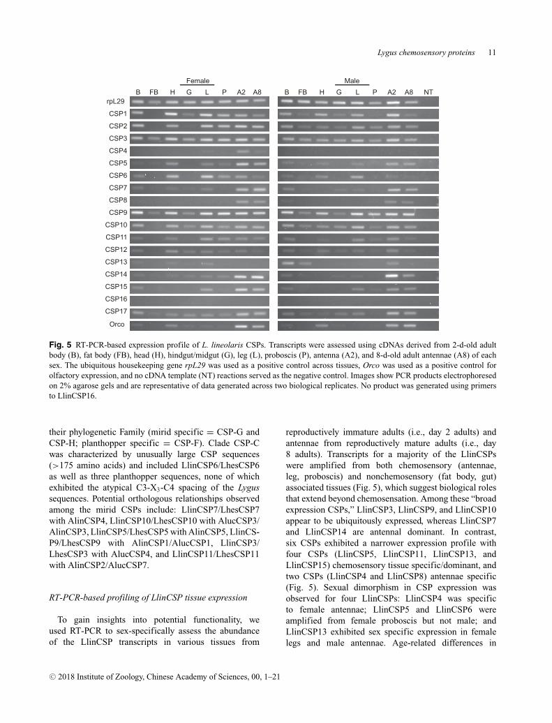
Fig. 5 RT-PCR-based expression profile of L. lineolaris CSPs. Transcripts were assessed using cDNAs derived from 2-d-old adultbody (B), fat body (FB), head (H), hindgut/midgut (G), leg (L), proboscis (P), antenna (A2), and 8-d-old adult antennae (A8) of eachsex. The ubiquitous housekeeping gene rpL29 was used as a positive control across tissues, Orco was used as a positive control forolfactory expression, and no cDNA template (NT) reactions served as the negative control. Images show PCR products electrophoresedon 2% agarose gels and are representative of data generated across two biological replicates. No product was generated using primersto LlinCSP16.
their phylogenetic Family (mirid specific = CSP-G andCSP-H; planthopper specific = CSP-F). Clade CSP-Cwas characterized by unusually large CSP sequences(>175 amino acids) and included LlinCSP6/LhesCSP6as well as three planthopper sequences, none of whichexhibited the atypical C3-X3-C4 spacing of the Lygussequences. Potential orthologous relationships observedamong the mirid CSPs include: LlinCSP7/LhesCSP7with AlinCSP4, LlinCSP10/LhesCSP10 with AlucCSP3/AlinCSP3, LlinCSP5/LhesCSP5 with AlinCSP5, LlinCS-P9/LhesCSP9 with AlinCSP1/AlucCSP1, LlinCSP3/LhesCSP3 with AlucCSP4, and LlinCSP11/LhesCSP11with AlinCSP2/AlucCSP7.
RT-PCR-based profiling of LlinCSP tissue expression
To gain insights into potential functionality, weused RT-PCR to sex-specifically assess the abundanceof the LlinCSP transcripts in various tissues from
reproductively immature adults (i.e., day 2 adults) andantennae from reproductively mature adults (i.e., day8 adults). Transcripts for a majority of the LlinCSPswere amplified from both chemosensory (antennae,leg, proboscis) and nonchemosensory (fat body, gut)associated tissues (Fig. 5), which suggest biological rolesthat extend beyond chemosensation. Among these “broadexpression CSPs,” LlinCSP3, LlinCSP9, and LlinCSP10appear to be ubiquitously expressed, whereas LlinCSP7and LlinCSP14 are antennal dominant. In contrast,six CSPs exhibited a narrower expression profile withfour CSPs (LlinCSP5, LlinCSP11, LlinCSP13, andLlinCSP15) chemosensory tissue specific/dominant, andtwo CSPs (LlinCSP4 and LlinCSP8) antennae specific(Fig. 5). Sexual dimorphism in CSP expression wasobserved for four LlinCSPs: LlinCSP4 was specificto female antennae; LlinCSP5 and LlinCSP6 wereamplified from female proboscis but not male; andLlinCSP13 exhibited sex specific expression in femalelegs and male antennae. Age-related differences in
antennal expression were largely limited to LlinCSP4in female antennae and LlinCSP14 in male antennaewith each transcript more abundant in immature adults(Fig. 5). No LlinCSP16 amplimers were generated fromany of the tissues assayed (Fig. 5), and primers designedto amplify LlinCSP17 yielded LlinCSP7 sequences (datanot shown).
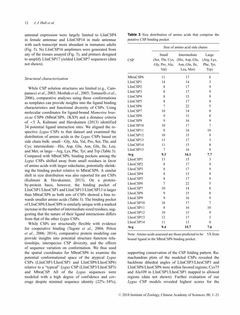
Structural characterization
While CSP solution structures are limited (e.g., Cam-panacci et al., 2003; Mosbah et al., 2003; Tomaselli et al.,2006), comparative analyses using those conformationsas templates can provide insights into the ligand bindingcharacteristics and functional diversity of CSPs. Usingmolecular coordinates for ligand-bound Mamestra bras-sicae CSP6 (MbraCSP6, 1KX9) and a distance criteriaof <5 A, Kulmuni and Havukainen (2013) identified34 potential ligand interaction sites. We aligned the re-spective Lygus CSPs to that dataset and examined thedistribution of amino acids in the Lygus CSPs based onside chain bulk: small—Gly, Ala, Val, Pro, Ser, Thr, andCys; intermediate—His, Asp, Glu, Asn, Gln, Ile, Leu,and Met; or large—Arg, Lys, Phe, Tyr, and Trp (Table 3).Compared with MbraCSP6, binding pockets among theLygus CSPs shifted away from small residues in favorof amino acids with larger sidechains, potentially shrink-ing the binding pocket relative to MbraCSP6. A similarshift in size distribution was also reported for ant CSPs(Kulmuni & Havukainen, 2013). On a protein-by-protein basis, however, the binding pocket ofLlinCSP1/LhesCSP1 and LlinCSP13/LlinCSP13 is largerthan MbraCSP6 as both sets of CSPs showed a bias to-wards smaller amino acids (Table 3). The binding pocketof LlinCSP6/LhesCSP6 is similarly unique with a markedincrease in the number of intermediate sized residues, sug-gesting that the nature of their ligand interactions differsfrom that of the other Lygus CSPs.
While CSPs are structurally flexible with evidencefor cooperative binding (Tegoni et al., 2004; Pelosiet al., 2006; 2014), comparative protein modeling canprovide insights into potential structure–function rela-tionships, interspecies CSP diversity, and the effectsof sequence variation on conformation. We thus usedthe spatial coordinates for MbraCSP6 to examine thepotential conformational space of the atypical LygusCSPs (LlinCSP1/LhesCSP1 and LlinCSP6/LhesCSP6)relative to a “typical” Lygus CSP (LlinCSP3/LhesCSP3)and MbraCSP. All of the Lygus sequences weremodeled with a high degree of confidence and cov-erage despite minimal sequence identity (22%–54%),
Table 3 Size distribution of amino acids that comprise theputative CSP binding pocket.
Note: Amino acids assessed are those predicted to be �5A frombound ligand in the MbraCSP6 binding pocket.
supporting conservation of the CSP folding pattern. Ra-machandran plots of the modeled CSPs revealed thebackbone dihedral angles of LlinCSP3/LhesCSP3 andLlinCSP6/LhesCSP6 were within favored regions; Cys75and Ala109 in LlinCSP1/LhesCSP1 mapped to allowedregions (data not shown). Further evaluation of ourLygus CSP models revealed highest scores for the
Fig. 6 Ribbon model representation of Lygus CSP structures. Homology-based conformations for a typical (LlinCSP3/LhesCSP3)and two atypical (LlinCSP1/LhesCSP1 and LlinCSP6/LhesCSP6) CSPs are shown along with the MbraCSP6 solution structure usedto generate the respective models. Helical segments are color-coded: helix 1, light blue; helix 2, light purple; helix 3, turquoise; helix4, orange; helix 5, light green, and helix 6, light pink. The upper panel shows different perspectives of the respective models with thefront and bottom views indicated. The Tyr residue in helix 2 that is predicted to form the bottom of the channel is colored bright pinkand depicted in stick mode. The Cys residues and accompanying disulfide bonds are colored yellow and are likewise depicted in stickmode. The lower panel shows differences in size of the putative binding pocket. The residues predicted to comprise the binding pocket,which is based on distance criteria (<5A) generated from a ligand bound MbraCSP6 structure, are shown in purple and depicted instick mode. The lack of a helix 6 and substitution of amino acids with smaller sidechains likely contributes to a larger binding pocketin LlinCSP1/LhesCSP1. Based on the spatial coordinates used to generate the respective models, the amino acid insertion between C3and C4 in LlinCSP6/LhesCSP6 prevented formation of the characteristic disulfide bridge. LlinCSP1 and LhesCSP1 are 100% identicalas is LlinCSP3 and LhesCSP3, thus only one model is shown for each set. Although LlinCSP6 and LhesCSP6 are 98% identical (fouramino acid differences), the residues affected reside in portions of the sequence that were not modeled. The figure was created usingSwiss-pbd viewer (Guex & Peitsch, 1997) and pov-ray (http://www.povray.org/).
“typical” LlinCSP3/LhesCSP3, which is consistent withthe higher predicted structural similarity. Coverage forthe atypical LlinCSP6/LhesCSP6 was lowest of the threeCSPs at � 50%. Although the resulting model lackedthe seventh predicted helix, it did encompass the pre-dicted binding pocket, consequently we concluded thatthe models, as well as those for LlinCSP1/LhesCSP1 andLlinCSP3/LhesCSP3, were of sufficient quality for thepurposes of our study.
Comparison of the three structures revealedLlinCSP1/LhesCSP1 had the smallest solvent accessiblesurface area (5821 A2), followed by LlinCSP6/LhesCSP6
(�6208 A2), and then LlinCSP3/LhesCSP3 (6511 A2),which is largely consistent with the predicted sizesof the proteins. The smaller surface area for the largeLlinCSP6/LhesCSP6 is likely due to incomplete modelingas the structural template lacked the seventh helix. Indeed,no template with the necessary sequence homology wasavailable in the databases to completely accommodatethe extended LlinCSP6/LhesCSP6 conformation. All ofthe Lygus CSP structures exhibited the typical CSP foldwith helices 1–2 and 4–5 forming a V-shaped structurecapped at one end by a perpendicular helix 3 (Fig. 6). InLlinCSP3/LhesCSP3 and LlinCSP6/LhesCSP6, helix 6
is parallel to helix 3 and partially occludes the carboxylterminal opening between helices 4–5. Consequently, theabsence of the sixth helix in LinCSP1/LhesCSP1 createsa larger opening, which may allow accommodation ofbulkier substrates. The Tyr in helix 2 (defined as Tyr26 inthe MbraCSP6 model) that caps one the end of the ligandpocket (Campanacci et al., 2003) is rotated away from thepocket in LinCSP1/LhesCSP1 and LinCSP6/LhesCSP6,but is facing the pocket in LinCSP3/LhesCSP3 (Fig. 6).This difference could indicate that LinCSP1/LhesCSP1and LinCSP6/LhesCSP6 accommodate longer/largerligands. Unexpectedly, the second disulfide bridgelinking C3 and C4 in LinCSP6/LhesCSP6 is not presentin our model (Fig. 6). We speculate that the structuralconstraints used to generate the models limited theconformational space available to the respective Cysresidues, which, because of the unique third amino acidinsertion, oriented the sidechains > 5A apart and thusprevented the linkage. While this almost certainly isnot the case with the actual solution structure, it furtherhighlights the divergence of the LinCSP6/LhesCSP6sequences.
To further investigate potential differences in ligandbinding among the three Lygus CSPs, we used the re-spective models to assess the effects that varied sidechain sizes have on the putative binding pockets. Asdiscussed above, the expansion of small residues in theLlinCSP1/LhesCSP1 pocket (Table 3) resulted in a morecompact structure with potentially greater ligand accessthan MbraCSP6 or LlinCSP3/LhesCSP3 (Fig. 6). In con-trast, the increased number of intermediate sized aminoacids lining the LlinCSP6/LhesCSP6 pocket yielded amore occluded pocket (Fig. 6). Consequently, it appearsthe variation in the ligand binding pocket residues affectsnot only the physicochemical properties of the respectiveCSPs, but also the size of potential ligands.
Discussion
To expand our knowledge of the Lygus chemosensory sys-tem, we used available transcriptome resources to identify17 CSP-like sequences in L. lineolaris and 14 sequencesin L. hesperus. Although the extent of the CSP repertoirevaries depending on species, the number identified in thetwo Lygus species is comparable to that reported for otherhemipterans: Acyrthosiphon pisum 12; Aphis gossypi 9; A.suturalis 8; A. lineolatus 8; N. lugens 11; S. furcifera 9; L.striatella 12; and A. lucorum 8. The increased number ofCSP in the Lygus species compared to other mirids likelyreflects methodological differences rather than geneexpansion. The L. lineolaris dataset was generated using
whole bodies across a range of developmental stages,and the L. hesperus assemblies were generated fromintact adults. In contrast, studies in other mirids focusedon either antenna (A. lineolatus and A. suturalis) or pro-boscis/leg (A. lucorum) specific transcriptomes/cDNAlibraries. Consequently, expanding those datasets toinclude other tissues and/or developmental stages wouldlikely reveal additional CSPs. Indeed, the absence ofthe evolutionarily conserved pentahelical CSPs (Fig. 4)suggests that those mirid datasets are incomplete.
Based on comparative analyses, the 14 L. hespe-rus sequences are likely orthologs of the L. lineolarisCSPs. Although LlinCSP15 was amplified from L. li-neolaris cDNAs using primers for LlinCSP4/LhesCSP4,we were unable to amplify the transcript from L. hespe-rus. This could indicate that LlinCSP15 transcription isspatially or temporally regulated. Alternatively, the ab-sence of the transcript could reflect physiological adap-tation and/or genetic differentiation of L. lineolaris. TheLlinCSP16 and LlinCSP17 sequence fragments were like-wise specific to the L. lineolaris datasets. However, thesequence similarity between LlinCSP16/LlinCSP2 andLlinCSP17/LlinCSP7, 8, and 14 could be indicative ofalternative splicing, which has been reported for L. line-olaris OBPs (Hull et al., 2014b) and CSPs in Holotrichiaparallela (Ju et al., 2014), or alternatively an example oftissue specific CSP RNA editing as reported in B. mori(Xuan et al., 2014).
LlinCSP1/LhesCSP1 and LlinCSP13/LhesCSP13 arestructurally and phylogenetically differentiated from theother Lygus CSPs. Unlike most CSPs, which are typically100–135 amino acid polypeptides characterized by six he-lices, LlinCSP1/LhesCSP1 and LlinCSP13/LhesCSP13are smaller pentahelical proteins. Furthermore, the con-served Tyr identified in MbraCSP6 (Tyr26 in the model,Tyr42 in the full-length sequence) that is critical forligand binding (Campanacci et al., 2003) has beenreplaced with Gln. The disparate properties of thesetwo residues could indicate different ligand interac-tion kinetics. Additionally, motif A in the pentaheli-cal CSPs, which is predicted to contribute to potentialprotein-protein interactions, is poorly conserved (14%sequence identity) relative to the other Lygus CSPs(43%–100% identity). As a result, the residues pre-dicted to comprise and/or surround the ligand-bindingpocket are smaller (Table 3) and may thus facilitate bind-ing of larger ligands (Fig. 6; Kulmuni & Havukainen,2013). Indeed, A. mellifera CSP2, which sorts to thesame monophyletic clade as LlinCSP1/LhesCSP1 andLlinCSP13/LhesCSP13, showed a preference for largeraromatic compounds (Dani et al., 2010). The presenceof this clade in virtually all taxa in which CSPs have
been annotated (Foret et al., 2007; Vieira & Rozas, 2011;Kulmuni & Havukainen, 2013; Pelosi et al., 2014) isconsistent with the nonchemosensory role proposed byKulmuni et al. (2013). In support of this, AmelCSP5has been shown to function in embryonic development(Maleszka et al., 2007) and AmelCSP2 and BmorCSP16(two other members of the pentahelical clade) are, likeLlinCSP1, broadly expressed (Foret et al., 2007; Qiaoet al., 2013). Similarly, FlyAtlas data indicate broadexpression for the pentahelical DmelCSP1 (Chintapalliet al., 2007). The discovery that all of the annotated CSPsin Daphnia pulex, an aquatic crustacean that is evolution-arily distant to insects, are pentahelical suggests that thepentahelical CSP clade pre-dates terrestrial colonizationand has thus likely gained new functionalities in devel-opment and cellular homeostasis. The presence of twodifferent genes encoding these CSPs in L. lineolaris andL. hesperus along with T. castaneum (Vieira & Rozas,2011), A. mellifera (Wanner et al., 2004; Foret et al.,2007; Vieira & Rozas, 2011), A. pisum (Vieira & Rozas,2011), Anopheles gambiae (Vieira & Rozas, 2011), andDiaphorina citri (Wu et al., 2016) suggests that the lonegenes reported in D. melanogaster (Vieira & Rozas, 2011)and Pediculus humanus (Vieira & Rozas, 2011) may bethe result of gene loss.
In addition to the pentahelical CSPs, we identified athird atypical CSP (LlinCSP6/LhesCSP6) in both Lygusspecies that is larger than normal (22.5 kDa, 196 aa),has seven helices (Fig. S1), and deviates from the highlyconserved C3-X2-C4 spacing motif. Although large CSPs(170–250 aa) have been identified in other species, theycomprise only a fraction of the annotated sequences inthe NCBI database. Curiously, the atypical CSPs in thedatabase largely consist of lepidopterans (four species)and hemipterans (five species). Whether or not this distri-bution is indeed order specific or is an artifact arising fromlimited datasets remains to be fully explored. However,it is interesting that among the hemipteran species thatcomprised our phylogenetic dataset we found six CSPs of170 aa or greater in length with three (NlugCSP9 189 aa,LstrCSP8 178 aa, and SfurCSP4 177 aa) forming a moder-ately supported clade (CSP-C) with LlinCSP6/LhesCSP6(Fig. 4). While all three are predicted to be heptahe-lical, none exhibit the C3-X3-C4 deviation present inLlinCSP6/LhesCSP6. These CSPs (i.e., LlinCSP6, S. fur-cifera CSP4 and CSP7; L. striatella CSP6 and CSP8) tendto be leg dominant (Fig. 6 and Zhou et al., 2015), suggest-ing a potential role in contact chemoreception. This tissuedistribution, however, is not shared by all large CSPs asthe 21.6 kDa BmorCSP9 (ABH88202, also referred toas BmCSP10) is broadly expressed (Qiao et al., 2013)and undergoes insecticide-dependent upregulation in
antennae and leg (Xuan et al., 2015). Similarly, the largeCSPs in Aphis gossypi (CSP1 and CSP9) are expressedin multiple tissues and developmental stages (Gu et al.,2013).
Deviations from the CSP Cys spacing (C1-X6-C2-X18-C3-X2-C4) proposed by Wanner et al. (2004) were ini-tially thought to be uncommon, but class/order-specificvariations have since been reported. Orthopteran CSPsfrequently have an insertion between C1 and C2 (C1-X8-C2-X18-C3-X2-C4), hymenopterans have an insertionbetween C2 and C3 (C1-X8-C2-X19-C3-X2-C4), and aCSP from Manduca sexta has a deletion between C2and C3 (C1-X8-C2-X17-C3-X2-C4). Consequently, Xuet al. (2009) proposed order-specific motifs that followthe general C1-X5-8-C2-X18-19-C3-X2-C4 pattern with theC3–C4 spacing the most highly conserved. Surprisingly,LlinCSP6/LhesCSP6 have an extra amino acid betweenthese two Cys, which yields a C3-X3-C4 motif (alsosee Figs. 1 and 2). Multiple independent clones am-plified from each species confirmed that the insertionwas not an artifact of transcriptome assembly. A searchof available databases identified only two additional se-quences, both derived from mirid species, with the C3-X3-C4 motif—a partial sequence from A. lineolatus (Al-inCSP9, accession no. AMD02858) and an unannotatedtranscript shotgun assembly sequence (GASV02024394)from Notostira elongate. This deviation was not reportedfor CSPs from the mirids A. suturalis (Cui et al., 2017)and A. lucorum (Hua et al., 2012, 2013). The absenceof LlinCSP6/LhesCSP6 orthologs in those datasets mayreflect spatial and/or temporal specific expression. Whilethe function of LlinCSP6/LhesCSP6 remains to be ad-dressed, the predicted binding pocket is likely larger thantypical CSPs (e.g., LlinCSP3/LhesCSP3), but smallerthan the pentahelical CSPs, and is thus expected to ac-commodate a different range of substrate sizes (Table 3).
The phylogenetic positioning of Lygus CSPs with othermirid CSPs (Fig. 4) may provide insights into the natureof possible ligands. LlinCSP3/LhesCSP3, LlinCSP5/LhesCSP5, LlinCSP9/LhesCSP9, LlinCSP10/LhesCSP10, and LlinCSP11/LhesCSP11 aligned with A.lineolatus and A. lucorum CSPs. LlinCSP9/LhesCSP9clustered with AlinCSP1 and AlucCSP1 (Fig. 4), whichare predominantly expressed in the antenna (Gu et al.,2012; Hua et al., 2012) but exhibit different ligandbinding profiles. AlinCSP1 binds a range of host plantvolatiles released in response to herbivore damage (Guet al., 2012), some of which have been shown to be attrac-tive to L. hesperus females (Blackmer et al., 2004) and/ortrigger positive electroantennograph responses (Williamset al., 2010). In contrast, AlucCSP1, which was clonedfrom a proboscis cDNA library, binds secondary cotton
metabolites (Hua et al., 2012), suggesting a gustatoryrole. LlinCSP10/LhesCSP10 aligned with AlinCSP3 andAlucCSP3. AlinCSP3 binds alfalfa and cotton volatiles(Gu et al., 2012), whereas AlucCSP3 binds secondarycotton metabolites (Hua et al., 2013). The two transcriptsare also differentially expressed with AlinCSP3 mostabundant in antennae (Gu et al., 2012) and AlucCSP3 infemale wings (Hua et al., 2012). LlinCSP11/LhesCSP11sorted with AlinCSP2 and AlucCSP7. AlinCSP2 is pre-dominantly expressed in the antennae and preferentiallybinds multiple green plant volatiles including linalool(Gu et al., 2012), an aliphatic terpenoid reported tohave repellency effects on Lygus males (Chinta et al.,1994; Williams et al., 2010). Tissue expression andfunctional characterization of the AlucCSP7 transcripthave yet to be determined. LlinCSP5/LhesCSP5 andLlinCSP3/LhesCSP3 are potentially orthologs of Al-inCSP5 and AlucCSP4, respectively. AlinCSP5 is anantenna dominant CSP with a relatively narrow ligandspectrum of green plant and cotton volatiles (Sun et al.,2015) that trigger L. hesperus female antennal responses(Williams et al., 2010). However, localization of Al-inCSP5 to the outer sensillum lymph of short sensillabasiconica, which lack neuron dendrites, suggests thatit may function as an odorant sink (Sun et al., 2015).AlucCSP4 is highly expressed in female wings andantennae, and, like the other characterized AlucCSPs,preferentially binds secondary cotton metabolites (Huaet al., 2013). LlinCSP7/LhesCSP7 sorted to clade CSP-Bwith AlinCSP4, an antenna dominant CSP that bindsmultiple compounds including various cotton and greenplant volatiles as well as components of the A. lineolatussex pheromone blend (Sun et al., 2015), some of which(i.e., trans-2-hexenyl-butyrate and hexyl butyrate) arealso active in L. lineolaris and L. hesperus (Byerset al., 2013). While the potential ligand specificities ofthe orthologous sequences are intriguing, conclusionswill require functional analyses of the Lygus CSPs.Furthermore, the variations in ligand binding reportedin the A. lineolatus and A. lucorum studies (Gu et al.,2012; Hua et al., 2012, 2013; Sun et al., 2015) maybe a result of methodological differences (the use ofdifferent fluorescent binding pocket probes) as opposedto actual biological/physiological differentiation of thephylogenetically related CSPs.
Acknowledgments
We thank Calvin A. Pierce III (USDA-ARS SIMRU)for assistance with insect rearing, RNA extractions, andcDNA synthesis. We also thank Lynn Jech (USDA-ARS
ALARC) for assistance with plasmid isolation and se-quencing. Mention of trade names or commercial prod-ucts in this article is solely for the purpose of providingspecific information and does not imply recommendationor endorsement by the U. S. Department of Agriculture.USDA is an equal opportunity provider and employer.
Disclosures
The authors declare no conflicts of interest.
References
Andersson, M.N. and Newcomb, R.D. (2017) Pest controlcompounds targeting insect chemoreceptors: Another silentspring? Frontiers in Ecology and Evolution, 5, 5.
Ban, L., Scaloni, A., Brandazza, A., Angeli, S., Zhang, L., Yan,Y. et al. (2003) Chemosensory proteins of Locusta migratoria.Insect Molecular Biology, 12, 125–134.
Berjanskii, M., Liang, Y., Zhou, J., Tang, P., Stothard, P., Zhou,Y. et al. (2010) PROSESS: a protein structure evaluation suiteand server. Nucleic Acids Research, 38, W633–640.
Bernado, P., Blackledge, M. and Sancho, J. (2006) Sequence-specific solvent accessibilities of protein residues in unfoldedprotein ensembles. Biophysical Journal, 91, 4536–4543.
Blackmer, J., Rodriguez-Saona, C., Byers, J., Shope, K. andSmith, J. (2004) Behavioral response of Lygus hesperus toconspecifics and headspace volatiles of alfalfa in a Y-tubeolfactometer. Journal of Chemical Ecology, 30, 1547–1564.
Brent, C.S. (2010) Reproduction of the western tarnished plantbug, Lygus hesperus, in relation to age, gonadal activity andmating status. Journal of Insect Physiology, 56, 28–34.
Briand, L., Eloit, C., Nespoulous, C., Bezirard, V., Huet, J.C.,Henry, C. et al. (2002) Evidence of an odorant-binding pro-tein in the human olfactory mucus: location, structural charac-terization, and odorant-binding properties. Biochemistry, 41,7241–7252.
Byers, J.A., Fefer, D. and Levi-Zada, A. (2013) Sex pheromonecomponent ratios and mating isolation among three Lygusplant bug species of North America. Naturwissenschaften,100, 1115–1123.
Campanacci, V., Lartigue, A., Hallberg, B.M., Jones, T.A.,Giudici-Orticoni, M.T., Tegoni, M. et al. (2003) Mothchemosensory protein exhibits drastic conformationalchanges and cooperativity on ligand binding. Proceedingsof the National Academy of Sciences USA, 100, 5069–5074.
Chinta, S., Dickens, J. and Aldrich, J. (1994) Olfactory recep-tion of potential pheromones and plant odors by tarnishedplant bug, Lygus lineolaris (Hemiptera: Miridae). Journal ofChemical Ecology, 20, 3251–3267.
Chintapalli, V.R., Wang, J. and Dow, J.A.T. (2007) Using Fly-Atlas to identify better Drosophila melanogaster models ofhuman disease. Nature Genetics, 39, 715–720.
Conesa, A., Gotz, S., Garcıa-Gomez, J.M., Terol, J., Talon, M.and Robles, M. (2005) Blast2GO: a universal tool for an-notation, visualization and analysis in functional genomicsresearch. Bioinformatics, 21, 3674–3676.
Crooks, G.E., Hon, G., Chandonia, J.M. and Brenner, S.E. (2004)WebLogo: a sequence logo generator. Genome Research, 14,1188–1190.
Cui, H.H., Gu, S.H., Zhu, X.Q., Wei, Y., Liu, H.W., Khalid,H.D., et al. (2017) Odorant-binding and chemosensory pro-teins identified in the antennal transcriptome of Adelphocorissuturalis Jakovlev. Comparative Biochemistry and PhysiologyPart D, Genomics and Proteomics, 24,139–145.
Dani, F.R., Iovinella, I., Felicioli, A., Niccolini, A., Calvello,M.A., Carucci, M.G. et al. (2010) Mapping the expression ofsoluble olfactory proteins in the honeybee. Journal of Pro-teome Research, 9, 1822–1833.
Dani, F.R., Michelucci, E., Francese, S., Mastrobuoni, G., Cap-pellozza, S., La Marca, G. et al. (2011) Odorant-binding pro-teins and chemosensory proteins in pheromone detection andrelease in the silkmoth Bombyx mori. Chemical Senses, 36,335–344.
Debolt, J.W. (1982) Meridic diet for rearing successive genera-tions of Lygus hesperus. Annals of the Entomological Societyof America, 75, 119–122.
Derks, M.F.L., Smit, S., Salis, L., Schijlen, E., Bossers, A.,Mateman, C. et al. (2015) The genome of winter moth (Oper-ophtera brumata) provides a genomic perspective on sexualdimorphism and phenology. Genome Biology and Evolution,7, 2321–2332.
Dickens, J., Callahan, F., Wergin, W. and Erbe, E. (1995) Ol-faction in a hemimetabolous insect: antennal-specific proteinin adult Lygus lineolaris (Heteroptera: Miridae). Journal ofInsect Physiology, 41, 857–867.
Drozdetskiy, A., Cole, C., Procter, J. and Barton, G.J. (2015)JPred4: a protein secondary structure prediction server. Nu-cleic Acids Research, 43, W389–W394.
Edgar, R.C. (2004) MUSCLE: multiple sequence alignmentwith high accuracy and high throughput. Nucleic Acids Re-search, 32, 1792–1797.
Ellsworth, P.C. and Barkley, V. (2001) Cost-effective Lygus man-agement in Arizona cotton. Cotton, A College of Agricultureand Life Sciences Report (ed. J.C. Silvertooth), pp. 299–307.University of Arizona, College of Agriculture and Life Sci-ences, Tucson, AZ.
Estrada, J., Bernado, P., Blackledge, M. and Sancho, J. (2009)ProtSA: a web application for calculating sequence specificprotein solvent accessibilities in the unfolded ensemble. BMCBioinformatics, 10, 1.
Finn, R.D., Clements, J. and Eddy, S.R. (2011) HMMER webserver: interactive sequence similarity searching. NucleicAcids Research, 39, W29–W37.
Fleming, D.E., Krishnan, N., Catchot, A.L., and Musser, F.R.(2016) Susceptibility to insecticides and activities of glu-tathione S-transferase and esterase in populations of Lyguslineolaris (Hemiptera: Miridae) in Mississippi. Pest Manage-ment Science, 72, 1595–1603.
Foret, S., Wanner, K.W. and Maleszka, R. (2007) Chemosen-sory proteins in the honey bee: insights from the annotatedgenome, comparative analyses and expressional profiling. In-sect Biochemistry and Molecular Biology, 37, 19–28.
Fountain, M., Jastad, G., Hall, D., Douglas, P., Farman, D. andCross, J. (2014) Further studies on sex pheromones of fe-male Lygus and related bugs: development of effective luresand investigation of species-specificity. Journal of ChemicalEcology, 40, 71–83.
Futahashi, R., Tanaka, K., Tanahashi, M., Nikoh, N., Kikuchi,Y., Lee, B.L. et al. (2013) Gene expression in gut symbioticorgan of stinkbug affected by extracellular bacterial symbiont.PLoS ONE, 8, e64557.
Gong, D.P., Zhang, H.J., Zhao, P., Lin, Y., Xia, Q.Y. and Xi-ang, Z.H. (2007) Identification and expression pattern of thechemosensory protein gene family in the silkworm, Bombyxmori. Insect Biochemistry and Molecular Biology, 37, 266–277.
Gonzalez, D., Zhao, Q., McMahan, C., Velasquez, D., Haskins,W.E., Sponsel, V. et al. (2009) The major antennal chemosen-sory protein of red imported fire ant workers. Insect Molecu-lar Biology, 18, 395–404.
Gotz, S., Garcıa-Gomez, J.M., Terol, J., Williams, T.D., Nagaraj,S.H., Nueda, M.J. et al. (2008) High-throughput functionalannotation and data mining with the Blast2GO suite. NucleicAcids Research, 36, 3420–3435.
Gu, S.H., Wang, S.Y., Zhang, X.Y., Ji, P., Liu, J.T. and Wang,G.R. (2012) Functional characterizations of chemosensoryproteins of the alfalfa plant bug Adelphocoris lineolatus in-dicate their involvement in host recognition. PLoS ONE, 7,e42871.
Gu, S.H., Wu, K.M., Guo, Y.Y., Field, L.M., Pickett, J.A., Zhang,Y.J. et al. (2013) Identification and expression profiling ofodorant binding proteins and chemosensory proteins betweentwo wingless morphs and a winged morph of the cotton aphidAphis gossypii Glover. PLoS ONE, 8, e73524.
Gu, X.C., Zhang, Y.N., Kang, K., Dong, S.L. and Zhang, L.W.(2015) Antennal transcriptome analysis of odorant recep-tion genes in the red turpentine beetle (RTB), Dendroctonusvalens. PLoS ONE, 10, e0125159.
Guex, N. and Peitsch, M.C. (1997) SWISS-MODEL and theSwiss-Pdb Viewer: an environment for comparative proteinmodeling. Electrophoresis, 18, 2714–2723.
Guo, W., Wang, X.H., Ma, Z.Y., Xue, L., Han, J.Y., Yu, D., et al.(2011) CSP and takeout genes modulate the switch betweenattraction and repulsion during behavioral phase change inthe migratory locust. PLoS Genetics, 7, e1001291.
Ho, H. and Millar, J. (2002) Identification, electroantennogramscreening, and field bioassays of volatile chemicals from Ly-gus hesperus Knight (Heteroptera: Miridae). Zoological Stud-ies, 41, 311–320.
Hou, C.X., Qin, G.X., Liu, T., Mei, X.L., Li, B., Shen, Z.Y.et al. (2013) Differentially expressed genes in the cuticle andhemolymph of the silkworm, Bombyx mori, injected with thefungus Beauveria bassiana. Journal of Insect Science, 13, 13.
Hua, J.F., Zhang, S., Cui, J.J., Wang, D.J., Wang, C.Y., Luo, J.Y.et al. (2012) Identification and binding characterization ofthree odorant binding proteins and one chemosensory proteinfrom Apolygus lucorum (Meyer-Dur). Journal of ChemicalEcology, 38, 1163–1170.
Hua, J.F., Zhang, S., Cui, J.J., Wang, D.J., Wang, C.Y., Luo, J.Y.et al. (2013) Functional characterizations of one odorant bind-ing protein and three chemosensory proteins from Apolyguslucorum (Meyer-Dur) (Hemiptera: Miridae) legs. Journal ofInsect Physiology, 59, 690–696.
Hull, J.J., Chaney, K., Geib, S.M., Fabrick, J.A., Brent, C.S.,Walsh, D. et al. (2014a) Transcriptome-based identificationof ABC transporters in the western tarnished plant bug Lygushesperus. PLoS ONE, 9, e113046.
Hull, J.J., Geib, S.M., Fabrick, J.A. and Brent, C.S. (2013) Se-quencing and de novo assembly of the western tarnished plantbug (Lygus hesperus) transcriptome. PLoS ONE, 8, e55105.
Hull, J.J., Hoffmann, E.J., Perera, O.P. and Snodgrass, G.L.(2012) Identification of the western tarnished plant bug (Ly-gus hesperus) olfactory co-receptor Orco: expression profileand confirmation of atypical membrane topology. Archives ofInsect Biochemistry and Physiology, 81, 179–198.
Hull, J.J., Perera, O.P. and Snodgrass, G.L. (2014b) Cloning andexpression profiling of odorant-binding proteins in the tar-nished plant bug, Lygus lineolaris. Insect Molecular Biology,23, 78–97.
Innocenzi, P.J., Hall, D., Cross, J.V. and Hesketh, H. (2005)Attraction of male European tarnished plant bug, Lygusrugulipennis to components of the female sex pheromonein the field. Journal of Chemical Ecology, 31, 1401–1413.
Iovinella, I., Dani, F.R., Niccolini, A., Sagona, S., Michelucci, E.,Gazzano, A. et al. (2011) Differential expression of odorant-binding proteins in the mandibular glands of the honey beeaccording to caste and age. Journal of Proteome Research,10, 3439–3449.
Jacquin-Joly, E., Vogt, R.G., Francois, M.C. and Nagnan-LeMeillour, P. (2001) Functional and expression pattern anal-ysis of chemosensory proteins expressed in antennae andpheromonal gland of Mamestra brassicae. Chemical Senses,26, 833–844.
Ju, Q., Li, X., Jiang, X.J., Qu, M.J., Guo, X.Q., Han, Z.J. et al.(2014) Transcriptome and tissue-specific expression analysisof OBP and CSP genes in the dark black chafer. Archives ofInsect Biochemistry and Physiology, 87, 177–200.
Kearse, M., Moir, R., Wilson, A., Stones-Havas, S., Cheung, M.,Sturrock, S. et al. (2012) Geneious Basic: an integrated andextendable desktop software platform for the organization andanalysis of sequence data. Bioinformatics, 28, 1647–1649.
Kitabayashi, A.N., Arai, T., Kubo, T. and Natori, S. (1998)Molecular cloning of cDNA for p10, a novel protein thatincreases in the regenerating legs of Periplaneta americana(American cockroach). Insect Biochemistry and MolecularBiology, 28, 785–790.
Kulmuni, J. and Havukainen, H. (2013) Insights into the evolu-tion of the CSP gene family through the integration of evo-lutionary analysis and comparative protein modeling. PLoSONE, 8, e63688.
Kulmuni, J., Wurm, Y. and Pamilo, P. (2013) Comparative ge-nomics of chemosensory protein genes reveals rapid evolu-tion and positive selection in ant-specific duplicates. Heredity,110, 538–547.
Lartigue, A., Campanacci, V., Roussel, A., Larsson, A.M., Jones,T.A., Tegoni, M. et al. (2002) X-ray structure and ligandbinding study of a moth chemosensory protein. Journal ofBiological Chemistry, 277, 32094–32098.
Li, H.L., Ni, C.X., Tan, J., Zhang, L.Y. and Hu, F.L. (2016)Chemosensory proteins of the eastern honeybee, Apis cer-ana: identification, tissue distribution and olfactory re-lated functional characterization. Comparative Biochemistryand Physiology B, Biochemistry and Molecular Biology,194, 11–19.
Lin, K., Simossis, V.A., Taylor, W.R. and Heringa, J. (2005)A simple and fast secondary structure prediction methodusing hidden neural networks. Bioinformatics, 21, 152–159.
Liu, G.X., Ma, H.M., Xie, H.Y., Xuan, N. and Picimbon, J.F.(2016a) Sequence variation of Bemisia tabaci chemosensoryprotein 2 in cryptic species B and Q: new DNA markers forwhitefly recognition. Gene, 576, 284–291.
tabaci chemosensory proteins: Role of CSP in insect defense.PLoS ONE, 11, e0154706.
Liu, Y.L., Guo, H., Huang, L.Q., Pelosi, P. and Wang, C.Z. (2014)Unique function of a chemosensory protein in the proboscisof two Helicoverpa species. Journal of Experimental Biology,217, 1821–1826.
Lovell, S.C., Davis, I.W., Arendall, W.B., de Bakker, P.I., Word,J.M., Prisant, M.G. et al. (2003) Structure validation by Cαgeometry: φ, ψ and Cβ deviation. Proteins, 50, 437–450.
Maleszka, J., Foret, S., Saint, R. and Maleszka, R. (2007) RNAi-induced phenotypes suggest a novel role for a chemosensoryprotein CSP5 in the development of embryonic integumentin the honeybee (Apis mellifera). Development Genes andEvolution, 217, 189–196.
Mezulis, S., Yates, C.M., Wass, M.N., Sternberg, M.J.E. and Kel-ley, L.A. (2015) The Phyre2 web portal for protein modeling,prediction and analysis. Nature Protocols, 10, 845–858.
Mosbah, A., Campanacci, V., Lartigue, A., Tegoni, M., Cam-billau, C. and Darbon, H. (2003) Solution structure of achemosensory protein from the moth Mamestra brassicae.Biochemical Journal, 369, 39–44.
Musser, F., Stewart, S., Bagwell, R., Lorenz, G., Catchot, A.,Burris, E. et al. (2007) Comparison of direct and indirectsampling methods for tarnished plant bug (Hemiptera: Miri-dae) in flowering cotton. Journal of Economic Entomology,100, 1916–1923.
Naranjo, S., Ellsworth, P., and Dierig, D. (2011) Impact of Lygusspp. (Hemiptera: Miridae) on damage, yield and quality oflesquerella (Physaria fendleri), a potential new oil-seed crop.Journal of Economic Entomology, 104, 1575–1583.
Oduol, F., Xu, J., Niare, O., Natarajan, R. and Vernick, K.D.(2000) Genes identified by an expression screen of the vectormosquito Anopheles gambiae display differential molecularimmune response to malaria parasites and bacteria. Proceed-ings of the National Academy of Sciences USA, 97, 11397–11402.
Ozaki, M., Wada-Katsumata, A., Fujikawa, K., Iwasaki, M.,Yokohari, F., Satoji, Y. et al. (2005) Ant nestmate and non-nestmate discrimination by a chemosensory sensillum. Sci-ence, 309, 311–314.
Patana, R. (1982) Disposable diet packet for feeding and ovipo-sition of Lygus hesperus (Hemiptera: Miridae). Journal ofEconomic Entomology, 75, 668–669.
Pelosi, P., Iovinella, I. and Felicioli, A. (2014) Soluble proteinsof chemical communication: an overview across arthropods.Frontiers in Physiology, 5, 320.
Pelosi, P., Zhou, J.J., Ban, L.P. and Calvello, M. (2006) Solu-ble proteins in insect chemical communication. Cellular andMolecular Life Sciences, 63, 1658–1676.
Petersen, T.N., Brunak, S., Heijne, G.V. and Nielsen, H. (2011)SignalP 4.0: discriminating signal peptides from transmem-brane regions. Nature Methods, 8, 785–786.
Picimbon, J.F., Dietrich, K., Krieger, J. and Breer, H. (2001)Identity and expression pattern of chemosensory proteinsin Heliothis virescens (Lepidoptera, Noctuidae). Insect Bio-chemistry and Molecular Biology, 31, 1173–1181.
Qiao, H.L., Deng, P.Y., Li, D.D., Chen, M., Jiao, Z.J., Liu,Z.C. et al. (2013) Expression analysis and binding exper-iments of chemosensory proteins indicate multiple rolesin Bombyx mori. Journal of Insect Physiology, 59, 667–675.
Ribeiro, J.M.C., Genta, F.A., Sorgine, M.H.F., Logullo, R.,Mesquita, R.D., Paiva-Silva, G.O. et al. (2014) An insight intothe transcriptome of the digestive tract of the bloodsuckingbug, Rhodnius prolixus. PLoS Neglected Tropical Diseases,8, e2594.
Ritter, R.A., Lenssen, A., Blodgett, S. and Taper, M.A. (2010)Regional assemblages of Lygus (Heteroptera: Miridae) inMontana canola fields. Journal of the Kansas Entomologi-cal Society, 83, 297–305.
Sabatier, L., Jouanguy, E., Dostert, C., Zachary, D., Dimarcq,J.L., Bulet, P. et al. (2003) Pherokine-2 and -3. Two Drosophilamolecules related to pheromone/odor-binding proteins in-duced by viral and bacterial infections. FEBS Journal, 270,3398–3407.
Schwartz, M.D. and Foottit, R.G. (1998) Revision of the Nearc-tic species of the genus Lygus Hahn, with a review of thePalaearctic species (Heteroptera: Miridae). Annals of the En-tomological Society of America, 91, 895–896.
Scott, D.R. (1977) An annotated listing of host plants of Ly-gus hesperus Knight. Bulletin of the Entomogical Society ofAmerica, 23, 19–22.
Sippl, M.J. (1993) Recognition of errors in three-dimensionalstructures of proteins. Proteins, 17, 355–362.
Snodgrass, G.L. (1996) Glass-vial bioassay to estimate insec-ticide resistance in adult tarnished plant bugs (Heteroptera:Miridae). Journal of Economic Entomoogy, 89, 1053–1059.
Snodgrass, G. and Scott, W. (2002) Tolerance to acephate intarnished plant bug (Heteroptera: Miridae) populations in theMississippi river delta. Southwestern Entomologist, 27, 191–199.
Snodgrass, G., Gore, J., Abel, C. and Jackson, R. (2009)Acephate resistance in populations of the tarnished plantbug (Heteroptera: Miridae) from the Mississippi River Delta.Journal of Economic Entomology, 102, 699–707.
Soffan, A., Antony, B., Abdelazim, M., Shukla, P., Witjaksono,W., Aldosari, S.A. et al. (2016) Silencing the olfactory co-receptor RferOrco reduces the response to pheromones in thered palm weevil, Rhynchophorus ferrugineus. PLoS ONE, 11,e0162203.
Song, Y.Q., Sun, H.Z. and Du, J. (2018) Identification and tis-sue distribution of chemosensory protein and odorant bindingprotein genes in Tropidothorax elegans Distant (Hemiptera:Lygaeidae). Scientific Reports, 8, 7803.
Strand, L. (2008) Integrated Pest Management for Strawberries,2nd edn. Agriculture and Natural Resources, University ofCalifornia, Oakland, CA.
Strong, F.E. (1970) Physiology of injury caused by Lygus hes-perus. Journal of Economic Entomology, 63, 808–814.
Sun, H.Y., Guan, L., Feng, H.L., Yin, J., Cao, Y.Z., Xi, J.H.et al. (2014) Functional characterization of chemosensoryproteins in the scarab beetle, Holotrichia oblita Faldermann(Coleoptera: Scarabaeida). PLoS ONE, 9, e107059.
Sun, L., Zhou, J.J., Gu, S.H., Xiao, H.J., Guo, Y.Y., Liu, Z.W.et al. (2015) Chemosensillum immunolocalization and ligandspecificity of chemosensory proteins in the alfalfa plant bugAdelphocoris lineolatus (Goeze). Scientific Reports, 5, 8073.
Tamura, K., Stecher, G., Peterson, D., Filipski, A. and Kumar,S. (2013) MEGA6: molecular evolutionary genetics analy-sis version 6.0. Molecular Biology and Evolution, 30, 2725–2729.
Tassone, E.E., Geib, S.M., Hall, B., Fabrick, J.A., Brent, C.S.and Hull, J.J. (2016) De novo construction of an expandedtranscriptome assembly for the western tarnished plant bug,Lygus hesperus. GigaScience, 5, 6.
Tegoni, M., Campanacci, V. and Cambillau, C. (2004) Structuralaspects of sexual attraction and chemical communication ininsects. Trends in Biochemical Sciences, 29, 257–264.
Tomaselli, S., Crescenzi, O., Sanfelice, D., Ab, E., Wechsel-berger, R., Angeli, S. et al. (2006) Solution structure of achemosensory protein from the desert locust Schistocercagregaria. Biochemistry, 45, 10606–10613.
Untergasser, A., Cutcutache, I., Koressaar, T., Ye, J., Faircloth,B.C., Remm, M. et al. (2012) Primer3-new capabilities andinterfaces. Nucleic Acids Research, 40, e115.
Vieira, F.G. and Rozas, J. (2011) Comparative genomics ofthe odorant-binding and chemosensory protein gene fami-lies across the Arthropoda: origin and evolutionary history ofthe chemosensory system. Genome Biology and Evolution, 3,476–490.
Vogel, H., Heidel, A.J., Heckel, D.G. and Groot, A.T. (2010)Transcriptome analysis of the sex pheromone gland of thenoctuid moth Heliothis virescens. BMC Genomics, 11, 29.
Vogt, R.G., Callahan, F.E., Rogers, M.E. and Dickens, J.C.(1999) Odorant binding protein diversity and distributionamong the insect orders, as indicated by LAP, an OBP-relatedprotein of the true bug Lygus lineolaris (Hemiptera, Het-eroptera). Chemical Senses, 24, 481–495.
Wang, R., Zhang, X.M., Li, H.L., Guo, X.J. and Luo, C. (2016)Identification and expression profiling of five chemosensoryprotein genes in the whitefly MED, Bemisia tabaci. Journalof Asia-Pacific Entomology, 19, 195–201.
Wanner, K.W., Isman, M.B., Feng, Q., Plettner, E. and Theil-mann, D.A. (2005) Developmental expression patterns offour chemosensory protein genes from the Eastern spruce
Wanner, K.W., Willis, L.G., Theilmann, D.A., Isman, M.B.,Feng, Q. and Plettner, E. (2004) Analysis of the insect os-d-like gene family. Journal of Chemical Ecology, 30, 889–911.
Wheeler, A.G. (2001) Biology of the Plant Bugs (Hemiptera:Miridae): Pests, Predators, Opportunists. Comstock Publish-ing Associates, Ithaca, NY, USA.
Wiederstein, M. and Sippl, M.J. (2007) ProSA-web: interactiveweb service for the recognition of errors in three-dimensionalstructures of proteins. Nucleic Acids Research, 35, W407–W410.
Williams, L., Blackmer, J.L., Rodriguez-Saona, C. and Zhu,S. (2010) Plant volatiles influence electrophysiological andbehavioral responses of Lygus hesperus. Journal of ChemicalEcology, 36, 467–478.
Wu, Z.Z., Zhang, H., Bin, S.Y., Chen, L., Han, Q.X. and Lin,J.T. (2016) Antennal and abdominal transcriptomes revealchemosensory genes in the Asian citrus psyllid, Diaphorinacitri. PLoS ONE, 11, e0159372–23.
Xu, Y.L., He, P., Zhang, L., Fang, S.Q., Dong, S.L., Zhang, Y.J.et al. (2009) Large-scale identification of odorant-bindingproteins and chemosensory proteins from expressed sequencetags in insects. BMC Genomics, 10, 632.
Xuan, N., Bu, X., Liu, Y.Y., Yang, X., Liu, G.X., Fan, Z.X.et al. (2014) Molecular evidence of RNA editing in Bombyxchemosensory protein family. PLoS ONE, 9, e86932.
Xue, W.X., Fan, J., Zhang, Y., Xu, Q.X., Han, Z.L., Sun, J.R. et al.(2016) Identification and expression analysis of candidateodorant-binding protein and chemosensory protein genes byantennal transcriptome of Sitobion avenae. PLoS ONE, 11,e0161839.
Yi, X., Wang, P.D., Wang, Z., Cai, J., Hu, M.Y. andZhong, G.H. (2014) Involvement of a specific chemosen-sory protein from Bactrocera dorsalis in perceivinghost plant volatiles. Journal of Chemical Ecology, 40,267–275.
Zhan, S., Merlin, C., Boore, J.L. and Reppert, S.M. (2011) Themonarch butterfly genome yields insights into long-distancemigration. Cell, 147, 1171–1185.
Zhang, S.F., Zhang, Z., Wang, H.B. and Kong, X.B. (2014)Antennal transcriptome analysis and comparison of olfactorygenes in two sympatric defoliators, Dendrolimus houi andDendrolimus kikuchii (Lepidoptera: Lasiocampidae). InsectBiochemistry and Molecular Biology, 52, 69–81.
Zhang, Y.N., Zhu, X.Y., Fang, L.P., He, P., Wang, Z.Q., Chen,G. et al. (2015) Identification and expression profiles of
sex pheromone biosynthesis and transport related genes inSpodoptera litura. PLoS ONE, 10, e0140019.
Zhou, J.J., Kan, Y., Antoniw, J., Pickett, J.A. and Field, L.M.(2006) Genome and EST analyses and expression of a genefamily with putative functions in insect chemoreception.Chemical Senses, 31, 453–465.
Zhou, J.J., Vieira, F.G., He, X.L., Smadja, C., Liu, R., Rozas,J. et al. (2010) Genome annotation and comparative analysesof the odorant-binding proteins and chemosensory proteins inthe pea aphid Acyrthosiphon pisum. Insect Molecular Biology,19(Suppl 2), 113–122.
Zhou, S.S., Sun, Z., Ma, W., Chen, W. and Wang, M.Q. (2014) Denovo analysis of the Nilaparvata lugens (Stal) antenna tran-scriptome and expression patterns of olfactory genes. Com-parative Biochemistry and Physiology Part D, Genomics andProteomics, 9, 31–39.
Zhou, W., Yuan, X., Qian, P., Cheng, J., Zhang, C., Gurr, G. et al.(2015) Identification and expression profiling of putativechemosensory protein genes in two rice planthoppers, Laodel-phax striatellus (Fallen) and Sogatella furcifera (Horvath).Journal of Asia-Pacific Entomology, 18, 771–778.
Zhou, X.H., Ban, L.P., Iovinella, I., Zhao, L.J., Gao, Q., Feli-cioli, A. et al. (2013) Diversity, abundance, and sex-specificexpression of chemosensory proteins in the reproductive or-gans of the locust Locusta migratoria manilensis. BiologicalChemistry, 394, 43–54.
Manuscript received August 13, 2018Final version received November 21, 2018Accepted November 26, 2018
Supporting Information
Additional supporting information may be found onlinein the Supporting Information section at the end of thearticle.
Fig. S1. Secondary structure prediction of L. lineo-laris and L. hesperus CSPs. Predictions performed us-ing JPRED4 (Drozdetskiy et al., 2015) (upper blue) andYASPIN (Lin et al., 2005) (lower red and orange). Puta-tive signal peptides are shown boxed, H indicates predictedhelical regions, E indicates predicted strand regions.
Table S1. Accession numbers of sequences used in phy-logenetic analyses.
Table S2. Oligonucleotide primers used in this study.Table S3. Heat map of L. lineolaris and L. hesperus
CSP percent amino identities.Table S4. TOP BLASTx hits for putative L. lineolaris
CSPs.Table S5. TOP BLASTx hits for putative L. hesperus