Received October 27, 2020, accepted November 3, 2020, date of publication November 5, 2020, date of current version November 17, 2020. Digital Object Identifier 10.1109/ACCESS.2020.3036219 Molecular Communication Aspects of Potassium Intracellular Signaling in Cardiomyocytes PENGFEI LU 1,2,3 , (Member, IEEE), MLADEN VELETIĆ 1,4 , JACOB BERGSLAND 1 , AND ILANGKO BALASINGHAM 1,5 , (Senior Member, IEEE) 1 The Intervention Centre, Oslo University Hospital, 0027 Oslo, Norway 2 School of Computer Science and Technology, Weinan Normal University, Weinan 714099, China 3 Faculty of Medicine, University of Oslo, 0315 Oslo, Norway 4 Faculty of Electrical Engineering, University of Banja Luka, 78000 Banja Luka, Bosnia and Herzegovina 5 Department of Electronic Systems, Norwegian University of Science and Technology, 7491 Trondheim, Norway Corresponding author: Pengfei Lu ([email protected]) This work was supported in part by the European Commission (EU), Norway, (EU-H2020:MSCA:ITN WiBEC–Wireless In-body Environment Communications) under Grant #675353, in part by the Research Council of Norway (RCN):WINNOW–Wireless In-body Sensor and Actuator Networks under Grant #270957, and in part by the RCN:CIRCLE–Communication Theoretical Foundation of Wireless Nanonetworks under Grant #287112. ABSTRACT Cardiovascular diseases continue to be a leading cause of morbidity and mortality worldwide. Cardiomyocytes, as the elementary heart components, play a crucial role in maintaining a healthy heart by coordinating contractions throughout the heart muscle that lead to a heartbeat. This study aims to characterize fine-grained ionic-level manipulation of cardiomyocytes for the controlled electrical activity that will offer new insights within the medical field. We explore the concept of Molecular Communications (MC) to analyze the propagation of potassium ions in the cardiomyocyte cytosol. By associating the number of the potassium ions in the cytosol with the membrane- and action potentials, we use metrics from the well-known Shannon’s information theory to optimize the ionic injection process and manipulate cardiomyocytes electrical activity. In case ON/OFF keying modulation is adopted as the potassium ion injection method, the optimal input distribution in terms of information capacity follows the derived Bernoulli distribution. This study offers underlying concepts that can be exploited in the creation of cardiomyocyte signals either for data communication via cellular infrastructure or heart pacing. The framework presented here needs to be upgraded in the following phases and made more physiologically plausible. INDEX TERMS Cardiomyocyte, channel capacity, intracellular communication, molecular communication, subthreshold communication. I. INTRODUCTION Pacemakers are permanent implants to treat patients with irregular heartbeats by injecting current to stimulate the heart in atrium and ventricle using electrodes (leads) [1]. Leads can cause infections and have led to the development of leadless pacemakers. These are small capsules-like devices containing sensors, current injectors, microcontrollers, wire- less transceivers, and batteries. Due to the requirement of small physical size and lifelong operation, the researchers are now looking for solutions beyond micro- and nanotechnology fields. Interestingly, biologists, inspired by the electronic industry and device development, are designing synthetic cells, inherently biocompatible and able to function The associate editor coordinating the review of this manuscript and approving it for publication was Wei-Wen Hu . like electronic devices or chords to perform key functions like sensing, computing, actuation, and signaling [2]. The advent of synthetic biology, in turn, has inspired communications engineers to develop new models and methods for intracel- lular and cell-to-cell communication using information and communication theoretical approaches. In a concept of the multi-nodal leadless pacemaker which we have recently proposed [3], communication of sensed data and commands for current injections between synthetic cells or capsules placed in atrial or ventricle can be realized utiliz- ing cardiomyocytes, thus enabling an alternative transmission pathway and connectivity which bypass the damaged natural conduction system. Intercellular cardiomyocyte signal trans- mission provides interesting insights into data transmission and scheduling using the cardiomyocyte system as a trans- mission channel without interrupting the natural, ongoing 201770 This work is licensed under a Creative Commons Attribution 4.0 License. For more information, see https://creativecommons.org/licenses/by/4.0/ VOLUME 8, 2020
Transcript
Received October 27, 2020, accepted November 3, 2020, date of publication November 5, 2020, date of current version November 17, 2020.
Digital Object Identifier 10.1109/ACCESS.2020.3036219
Molecular Communication Aspects of PotassiumIntracellular Signaling in CardiomyocytesPENGFEI LU 1,2,3, (Member, IEEE), MLADEN VELETIĆ 1,4, JACOB BERGSLAND 1,AND ILANGKO BALASINGHAM 1,5, (Senior Member, IEEE)1The Intervention Centre, Oslo University Hospital, 0027 Oslo, Norway2School of Computer Science and Technology, Weinan Normal University, Weinan 714099, China3Faculty of Medicine, University of Oslo, 0315 Oslo, Norway4Faculty of Electrical Engineering, University of Banja Luka, 78000 Banja Luka, Bosnia and Herzegovina5Department of Electronic Systems, Norwegian University of Science and Technology, 7491 Trondheim, Norway
This work was supported in part by the European Commission (EU), Norway, (EU-H2020:MSCA:ITN WiBEC–Wireless In-bodyEnvironment Communications) under Grant #675353, in part by the Research Council of Norway (RCN):WINNOW–Wireless In-bodySensor and Actuator Networks under Grant #270957, and in part by the RCN:CIRCLE–Communication Theoretical Foundation ofWireless Nanonetworks under Grant #287112.
ABSTRACT Cardiovascular diseases continue to be a leading cause of morbidity and mortality worldwide.Cardiomyocytes, as the elementary heart components, play a crucial role in maintaining a healthy heart bycoordinating contractions throughout the heart muscle that lead to a heartbeat. This study aims to characterizefine-grained ionic-level manipulation of cardiomyocytes for the controlled electrical activity that will offernew insights within the medical field. We explore the concept of Molecular Communications (MC) toanalyze the propagation of potassium ions in the cardiomyocyte cytosol. By associating the number of thepotassium ions in the cytosol with the membrane- and action potentials, we use metrics from the well-knownShannon’s information theory to optimize the ionic injection process and manipulate cardiomyocyteselectrical activity. In case ON/OFF keying modulation is adopted as the potassium ion injection method,the optimal input distribution in terms of information capacity follows the derived Bernoulli distribution.This study offers underlying concepts that can be exploited in the creation of cardiomyocyte signals eitherfor data communication via cellular infrastructure or heart pacing. The framework presented here needs tobe upgraded in the following phases and made more physiologically plausible.
INDEX TERMS Cardiomyocyte, channel capacity, intracellular communication, molecular communication,subthreshold communication.
I. INTRODUCTIONPacemakers are permanent implants to treat patients withirregular heartbeats by injecting current to stimulate the heartin atrium and ventricle using electrodes (leads) [1]. Leadscan cause infections and have led to the development ofleadless pacemakers. These are small capsules-like devicescontaining sensors, current injectors, microcontrollers, wire-less transceivers, and batteries. Due to the requirement ofsmall physical size and lifelong operation, the researchersare now looking for solutions beyond micro- andnanotechnology fields. Interestingly, biologists, inspired bythe electronic industry and device development, are designingsynthetic cells, inherently biocompatible and able to function
The associate editor coordinating the review of this manuscript and
approving it for publication was Wei-Wen Hu .
like electronic devices or chords to perform key functions likesensing, computing, actuation, and signaling [2]. The adventof synthetic biology, in turn, has inspired communicationsengineers to develop new models and methods for intracel-lular and cell-to-cell communication using information andcommunication theoretical approaches.
In a concept of the multi-nodal leadless pacemaker whichwe have recently proposed [3], communication of sensed dataand commands for current injections between synthetic cellsor capsules placed in atrial or ventricle can be realized utiliz-ing cardiomyocytes, thus enabling an alternative transmissionpathway and connectivity which bypass the damaged naturalconduction system. Intercellular cardiomyocyte signal trans-mission provides interesting insights into data transmissionand scheduling using the cardiomyocyte system as a trans-mission channel without interrupting the natural, ongoing
201770 This work is licensed under a Creative Commons Attribution 4.0 License. For more information, see https://creativecommons.org/licenses/by/4.0/ VOLUME 8, 2020
P. Lu et al.: Molecular Communication Aspects of Potassium Intracellular Signaling in Cardiomyocytes
FIGURE 1. (a) Cardiac channelopathies and (b) the normalized cardiomyocyte membrane potential, sodium current, slowinward calcium current, and potassium current change with time. The ionic currents synchronously depolarize the membraneand evoke membrane/action potentials.
cell-to-cell communication needed for heart function [4]–[7].In this regard, we have proposed and analyzed theresting-state (subthreshold) cardiomyocyte communicationmethod within an intracardiac communication system [7].
In the intracardiac communication system, a cardiomy-ocyte is an elementary building block, where ions such assodium (Na+), calcium (Ca2+) and potassium (K+) ionsfurther play a crucial role in defining electrophysiologicalactivity. This activity is, in turn, essential for encoding datavia the subthresholdmembrane potential fluctuations [7]. Theions are dynamically exchanged between the intracellular andextracellular space (Figure 1a), which leads to the creationof the ionic currents (Figure 1b): sodium current (INa), slowinward calcium current (ICa), and potassium current (IK),among others. The latter integrates the transient outwardpotassium current (Ito), the outward ultrarapid rectifier cur-rent (IKur), the outward rapid rectifier current (IKr) and theoutward slow rectifier current (IKs), and the inward recti-fying current (IK1) [8]. Although the ionic currents coordi-nately contribute the cardiomyocyte to generate membraneand action potentials, as shown in Figure 1b, their effects canbe studied independently. Thus, it is required to separatelyinvestigate the effects of ionic movements/currents and theirassociation with cellular electrophysiological activity beforeconducting further relevant analysis and experimental trialsin association with the proposed communication method.
Potassium ions are the first candidate whose dynamicscan be analyzed in a straightforward manner. Compared tosodium and/or calcium dynamics, potassium dynamicswithincardiomyocytes can be easily described. Although present inthe intracellular space where they hardly propagate/diffuselongitudinally, sodium ions are predominately concentratedin the extracellular space [9]. Although exist in the cytosolwhere they play crucial roles, calcium ions dynamics is morecomplex. This is particularly valid for membrane poten-tials when the calcium-induced-calcium-release (CICR)
mechanism in the cytosol is activated and calcium ionsare released from internal stores, e.g., endoplasmic retic-ulum, in addition to calcium influx from the extracellularspace [10].
Potassium ions in the cytosol are abundant compared totheir concentration in the extracellular space and intracel-lular concentrations of other ions. Besides, potassium ions1) have the potential to propagate/diffuse intracellularly in thelongitudinal direction1 either in the resting-, depolarization-,plateau-, and repolarization periods [12], and 2) are notbuffered intracellularly (like calcium ions), whereas onlyphysical barriers and local charges or components like mem-branes could restrict their propagation/diffusion [13], [14].Ultimately, adequate injection of potassium ions into theintracellular space depolarizes the cardiomyocyte’s mem-brane, which can be utilized for creation of signals forcommunication of sensed data and/or commands betweensynthetic cells or capsules.
The listed properties prompt us to deploy Molecular Com-munication (MC) paradigm and the Shannon’s informationtheory to
• analyze the potassium-based signaling (sub)-system,and
• propose a novel way of associating the intracellularlytransmitted ions with the membrane potential fluctu-ations relevant for encoding data via the resting-statecardiomyocyte communication method [7], [15]–[19].
The diffusion-based MC framework has been previouslyused to study the leadless pacemaker communications inthe heart chambers [20]. In that scenario, the communica-tion is based on pheromone transmission using unspecifiedmolecules which diffuse through the blood medium, where
1Potassium ion diffusion in the longitudinal direction is about 5000 timesgreater than the permeability of the surface membrane to outwardmovement [11].
VOLUME 8, 2020 201771
P. Lu et al.: Molecular Communication Aspects of Potassium Intracellular Signaling in Cardiomyocytes
the propagation distance is larger than the length of a singlecell [20], [21]. We describe the potassium ion propagationwithin the cardiomyocyte cytosol with the diffusion-basedMC models [22]–[25] and ground this study on the systemmodel presented in [23]. We assume that 1) the potassiumion transmitter is a point source which integrates the ionstransmitted via gap junctions from the neighboring cellsand/or externally injected ions (e.g., via electrophoresis),2) the potassium ions movement in the intracellular space canbe characterized by the diffusion law, and 3) the potassiumion receiver absorbs or accumulates the ions. Finally, we usethe information theory metrics such as the channel capacityto characterize the performance of the potassium-based intra-cellular signaling (sub)-system. Unlike in the existing works,e.g., [26]–[28], here we associate the concept of Shannon’sinformation capacity with the cardiomyocyte intracellularpotassium, with the objective to optimize the ionic injectionprocess and manipulate cardiomyocytes electrical activity.The concept of information theory can further be used toderive measures to investigate, diagnose, or treat cardiacdiseases in nanomedicine [5], [6].
The rest of the paper is organized as follows. Section IIintroduces the potassium-based intracellular signalingmodel.Section III characterizes the channel capacity of the proposedsystem. Section IV presents the numerical simulations andresults. Finally, Section V discusses and concludes the study.
II. POTASSIUM-BASED INTRACELLULAR SIGNALING(SUB)-SYSTEMWeidmann’s use of multiple compartment methods showedthat potassium ions could diffuse through multiple cardiaccells in the longitudinal direction [11], [29]. Besides, the dif-fusion process is divided into two steps: 1) diffusion throughthe intracellular space, and 2) diffusion across the gap junc-tions between two cells. Potassium ions diffusion in theintracellular space could be considered as a source-sinkcommunication [30] where the ions move from one selectedcompartment to another. Due to the similarities of the ionicmovement and molecular diffusion, we adapt the existingbasic MC concepts developed by the communications engi-neering community to model the potassium-based intracellu-lar signaling in cardiomyocytes.
A. BASIC MC MODELThe conventional MC system uses molecules/ions to transmitinformation between its peers. Figure 2 shows a generaldiffusion-based MC model which consists of source encod-ing, sending (emission), propagation (diffusion), reception(absorption), and source decoding [31], [32]:• Encoding: the transmitter encodes the signal related datainto the specific number of molecules/ions,
• Sending: the transmitter emits informationmolecules/ionsinto the channel,
• Propagation: the emitted molecules/ions roam in thecommunication channel between the transmitter andreceiver,
FIGURE 2. Basic diffusion-based MC system.
• Reception: the receiver absorbs the informationmolecules/ions from the communication channel,
• Decoding: the receiver reacts to the molecules/ions.
B. POTASSIUM-BASED INTRACELLULAR SIGNALING(SUB)-SYSTEM MODELSince the flow of potassium ions in cardiomyocytes can beconsidered as propagation from the source/emission point tothe sink/receiver point, we conceptualize the potassium-basedintracellular signaling (sub)-system model as shownin Figure 3. While establishing a potassium-based intracel-lular signaling system, we:
• consider the potassium ions diffusion in a three-dimensional space with a point source and athree-dimensional receiving sphere with the radius r thatequals the cardiomyocyte’s radius; it is reasonable tocount the received ions in a sphere as adult cardiomy-ocytes exhibit a rod shape which could be taken as acurve surface in a three-dimensional space [33],
• assume the homogeneous cytosolic milieu whereorganelles do not interrupt the propagation of ions, and
• neglect the impact of other ions.
The corresponding system thus consists of threemain com-partments: the transmitter, the channel, and the receiver.• The transmitter emits potassium ions. The ions sourcepresumably comes either from 1) neighboring cells orionic exchange between the intracellular and extracellu-lar space, or 2) the external (coordinated) electrophoreticinjection [34]. The transmitter ‘‘occupies’’ the area closeto the cell membrane, as shown in Figure 3. In this study,the transmitter is abstracted as a point source to simplifythe analysis.
• The channel allows for the emitted ions to propagate inthe intracellular space following the diffusion law. Thechannel ‘‘occupies’’ the cytoplasm of the communicat-ing cell.
• The receiver abstracts as a sphere receptor/nanosensorwhich detects the ions. According to the received ions,we quantify the encoding membrane potential whichhelps us decide whether we should stimulate the cellwith potassium injection or electrical stimulation andhow strong the stimulus should be to successfully prop-agate information signals to other cells/nodes via gapjunctions. The receiver ‘‘occupies’’ the distal segment ofthe cell in the longitudinal direction, as shown Figure 3.
The conceptual division in compartments helps us abstractand understand the intracellular communication system.
201772 VOLUME 8, 2020
P. Lu et al.: Molecular Communication Aspects of Potassium Intracellular Signaling in Cardiomyocytes
C. DIFFUSION EQUATIONThe diffusion equation is applied to characterize any sub-stance diffusing in intracellular space (e.g., ions or smallmolecules [35]). In general, the diffusion could be com-plex and anisotropic, and is affected by the cytosolic milieu.We model the potassium ions diffusion in cardiomyocyteswith a point emitter and a sphere receiver [23], as shownin Figure 3, and assume that 1) the cardiomyocyte is cylin-drically rod shaped, 2) the potassium ions propagate in thelongitudinal direction since the length of a cardiomyocyte isusually about ten times bigger than the radius [36], and 3) thepotassium diffusion coefficient in the longitudinal- is higherthan in the radial-direction [12].
The transmembrane efflux of the potassium ions affectsthe concentration of the potassium ions movement in thelongitudinal direction. Thereby, by taking into account thepotassium efflux, the potassium ions concentration variation(C(x, t)) is described as [37]
∂C(x, t)∂t
= D∂2C(x, t)∂x2
− kC(x, t), (1)
where x is the propagation distance, t is the propagation time,D is the diffusion coefficient of the potassium ions, and k isthe rate constant of transmembrane efflux in ms−1 (k = 0denotes that none of the ions move out of the intracellularspace, and the permeability of the cell membrane is very low,whereas k>0 denotes that some of the ions move from theintracellular- to the extracellular space). The rate constant kis described as [37]
k = −Mout
C(x, t)AcellVcell
, (2)
whereMout is the efflux in mmol/(µm2·ms), and Acell/Vcell is
the surface-to-volume ratio of the considered cardiomyocytesin cm−1. As the efflux is hardlymeasured, we use the half-lifecycle (t1/2) of the potassium ions to calculate the effluxrate [38], [39], which is written as
k =ln(2)t1/2
. (3)
To solve (1), we need to set the initial- and the boundarycondition. When considering that the propagation channel isinfinite, and the potassium ions are emitted at x = 0 with aninitial number Q0, we yield
C(x, t) = Q0(4πDt)−3/2 exp[−x2
4Dt− kt
]. (4)
Of note, Q0 is the initial value of the potassium ions in thesystem and is changed depending on the setup.
D. RELATION BETWEEN THE IONIC INJECTION AND THEMEMBRANE POTENTIAL CHANGEThe lipid bilayer of the cardiomyocyte membrane formsa capacitor that isolates the intracellular- and extracellularspace. In the resting state, ions accumulate on both sidesof the layer and keep the balance. The balance is disruptedwhen an external stimulation or physiological environmentchanges. Injecting cations into the intracellular space depo-larizes the membrane and creates a potential difference. Themembrane potential difference caused by the number ofinjected ions is described as [40]
Vd =eQ0
CmAcap, (5)
where e denotes the elementary charge, Cm denotes the spe-cific capacitance of the cardiomyocyte membrane in the unitarea, and Acap denotes the capacitive membrane area.With the membrane potential difference, the actual mem-
brane potential Vm is then calculated as
Vm = Vd + Vrest, (6)
where Vrest is the membrane potential in the resting state.When cations are continuously injected into the intracel-
lular space, the membrane continuously depolarizes whilethe membrane potential increases reaching the membranepotential threshold value (Vth) and, ultimately, the maximummembrane potential value (Vmax). Therefore, we derive thethreshold (Qth) and the maximum (Qmax) as of the number of
VOLUME 8, 2020 201773
P. Lu et al.: Molecular Communication Aspects of Potassium Intracellular Signaling in Cardiomyocytes
injected cations according to (5) and (6), respectively, as
Qth =CmACap
e(Vth − Vrest)
Qmax =CmACap
e(Vmax − Vrest). (7)
III. CHANNEL CAPACITYThe channel capacity is one of the most-frequently-usedmetrics to characterize the communication channel’s datatransmission. We use the channel capacity in this study toevaluate the potassium-based intracellular signaling [41].
We analyze the ionic transmission within time slots. Thetransmitter emits a certain number of potassium ions ineach time slot. However, in the diffusion-based MC system,the inter-symbol interference (ISI) is generated at the receiverpoint due to residual molecules/ions originating from theprevious time slots. The ISI can be eliminated unless thesignal propagation duration is infinite. One approach is to usea dynamic threshold detection technique [42]. We considerthe ISI by taking into account the impact of the previouslyemitted ions, but simplify the detection procedure with apredefined threshold detection to avoid the computationalburden.
A. CHANNEL MODELIn a time-slotted system, the ionic diffusion happens withintime T = nTd , where n denotes the number of time slots andTd the duration of each time slot. We consider the ON/OFFkeying modulation method. The transmitter emits M potas-sium ions when sending bit 1, and none when sending bit 0.
The probability P(x, t) of the ion at distance x and time tis given as [43]
P(x, t) =∫ t
0f (x, t ′)
∫∞
t ′g(u)dudt ′, (8)
where f (x, t ′) denotes the probability density function (PDF)of one ion arriving at the receiver at distance x and time t ′,g(u) denotes the PDF that characterizes the transmembraneefflux of ions, and is an exponential distribution function(g(u) = k exp [−ku]). In our scenario, we define f (x, t ′)as [19], [43]
f (x, t ′) =
{0 t ′ = 0rr0
x√
4πDt ′3exp
[−
x24Dt ′
]t ′ > 0,
(9)
where x is the distance from the transmitter to the surfaceof the receiver, r is the radius of the receiver sphere, andr0 = x + r is the distance from the transmitter to the centerof the receiver (Figure 3).
B. ISI ANALYSISAt the start of each time slot i ∈ [1, n], the transmitter sendsbit 1 by emittingM ions with the transmission probability pi.The transmitter thus sends bit 0 by emitting no ions withprobability (1-pi). All the ions diffuse independently, with thebinary state when reaching the receiver. Therefore, to decode
bit 1, the receiver successfully receives the ions withthe probability piP(x,Td ), where P(x,Td ) stems from (8).The receiver fails to receive the ions with the probabilitypi(1− P(x,Td )).
The number (Nc) of the received ions emitted by the trans-mitter within time slot n follows the Binomial distribution
Nc ∼ B(M ,P(x,Td )). (10)
A binomial distribution B(n, p) can be approximated witha normal distribution N (np, np(1 − p)), when n is greaterthan 50 [25], [44]. Since in the considered scenario n issignificantly higher than 50, as shown in Figure 8a, eq. (10)is approximated as
Nc ∼ N (µ, δ2), (11)
where
µ = MP(x,Td ),
δ2 = MP(x,Td )(1− P(x,Td )).
Further, we denote with Pi,n(1 ≤ i ≤ n) the probabilityof a single ion to be received in time slot n when emitting Mions in time slot i, and define as
From (10) to (12), we denote the residual ions from theprevious (n − 1) time slots in the current time slot with thefollowing distribution
NISI ∼
n−1∑i=1
pi(B(M ,P(x, (n− i+ 1)Td ))
−B(M ,P(x, (n− i)Td ))). (13)
Since all the ions independently propagate in the channel,eq. (13) is approximated from (10) and (11) as a normaldistribution
NISI ∼
n−1∑i=1
pi(N (µa, δa2)−N (µb, δb2)
)=
n−1∑i=1
pi(N (µa − µb, δa2 + δb2)
)= N
(n−1∑i=1
pi(µa − µb),n−1∑i=1
pi2(δa2 + δb2)
), (14)
where
µa = MP(x, (n− i+ 1)Td ),
δa2= MP(x, (n− i+ 1)Td )(1− P(x, (n− i+ 1)Td )),
µb = MP(x, (n− i)Td ),
δb2= MP(x, (n− i)Td )(1− P(x, (n− i)Td )).
201774 VOLUME 8, 2020
P. Lu et al.: Molecular Communication Aspects of Potassium Intracellular Signaling in Cardiomyocytes
C. DETECTIONWith the hypotheses H0 and H1 (Figure 4), we denotethe numbers of the received ions N0 when the transmittersends 0 and N1 when the transmitter sends 1 in the timeslot n, respectively. N0 and N1 follow the normal distribution,respectively,
N0 = NISI ∼
n−1∑i=1
pi(N (µa − µb, δ2a + δ2b))
∼ N (µ0, δ20), (15)
FIGURE 4. Binary test channel of the potassium-based intracellularsignaling model.
where
µ0 =
n−1∑i=1
pi(µa − µb),
δ20 =
n−1∑i=1
p2i (δ2a + δ
2b),
and
N1 ∼ N (µ, δ2)+n−1∑i=1
pi(N (µa, δ2a)−N (µb, δ2b))
∼ N (µ1, δ21), (16)
where
µ1 = µ+
n−1∑i=1
pi(µa − µb),
δ1 = δ2+
n−1∑i=1
p2i (δ2a + δ
2b).
To reduce the ISI, we set the threshold θ to a predefinedvalue. The probability (P(θ |H1)) that the hypothesis H1 hap-pens and the probability (P(θ |H0)) that the hypothesis H0happens can then be calculated from the cumulative distri-bution function of the normal distribution, F(θ, µ1, δ
21) and
F(θ, µ0, δ20), respectively. Therefore, the false alarm proba-
bility PF and the detection probability PD are given as
PF = Pr(N ≥ θ |X = 0) = 1− F(θ;µ0, δ20),
PD = Pr(N ≥ θ |X = 1) = 1− F(θ;µ1, δ21),
Pr(Y = 1|X = 0) = PF ,
Pr(Y = 1|X = 1) = PD,
Pr(Y = 0|X = 0) = 1− PF ,
Pr(Y = 0|X = 1) = 1− PD. (17)
Ultimately, we resort to the error probability to findthe proper detecting threshold θ using numerical methods(Section IV). The error probability of transmitting the randombit 0/1 in the current time slot n is written as
Pe = pc(1− PD)+ (1− pc)PF , (18)
where pc is the probability of transmitting bit 1. As shownin Figure 5, the error probability highly depends on the detect-ing threshold θ .
FIGURE 5. The error probability Pe versus threshold values withTd = 0.8 s, k = 0.005ms−1 and Q0 = 3× 10⁵.
D. CHANNEL CAPACITYFrom the binary communication channel (Figure 4), themutual information can be expressed as [45]
I (X;Y ) = H (Y )− H (Y |X )
=
1∑X=0
1∑Y=0
P(Y |X )P(X ) log2P(Y |X )P(Y )
= H (pc(1− PD)+ (1− pc)(1− PF ))
−(1− pc)H (PF )− pcH (1− PD), (19)
where H (x) is the entropy of x, and it is given as H (x) =−x log2(x)− (1− x) log2(1− x).Subsequently, we define the information capacity CK
as [45]
CK = maxpc
(I (X;Y ))
= log2(1+ z)−PF
PF − PDH (1− PD)
+PD
PF − PDH (PF ), (20)
where z = 2H (1−PD)−H (PF )
PF−PD . Note that the input distribution atthe transmitter follows the Bernoulli distribution, owing to thepre-selected ON/OFF keying modulation method.
VOLUME 8, 2020 201775
P. Lu et al.: Molecular Communication Aspects of Potassium Intracellular Signaling in Cardiomyocytes
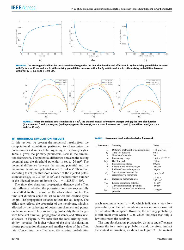
FIGURE 6. The arriving probabilities for potassium ions change with the time slot duration and efflux rate k : a) the arriving probabilities increasewith Td for x = 80 µm and k = 0; b) the arriving probabilities decrease with x for Td = 0.8 s and k = 0; c) the arriving probabilities decreasewith k for Td = 0.8 s and x = 80 µm.
FIGURE 7. When the emitted potassium ions is 2× 10⁷, the channel mutual information changes with (a) the time slot duration(k = 0.005 ms−1 and x = 80 µm), (b) the propagation distance (Td = 0.8 s and k = 0.005 ms−1) and (c) the efflux rate (Td = 0.8 sand x = 80 µm).
IV. NUMERICAL SIMULATION RESULTSIn this section, we present the numerical results from thecomputational simulations performed to characterize thepotassium-based intracellular signaling in cardiomyocytes.Table 1 gives the primary parameters used in the simula-tion framework. The potential difference between the restingpotential and the threshold potential is set to 24 mV. Thepotential difference between the resting potential and themaximum membrane potential is set to 124 mV. Therefore,according to (7), the threshold number of the injected potas-sium ions isQth = 2.30100×10⁷, and the maximum numberof the injected potassium ions is Qmax = 1.18885× 10⁸.The time slot duration, propagation distance and efflux
rate influence whether the potassium ions are successfullytransmitted to the receiver at the observation points. Thetime slot duration could be set to reflect the cardiac cyclelength. The propagation distance reflects the cell length. Theefflux rate reflects the properties of the membrane, which isaffected by the pathology of potassium channels and pumpson the membrane. The ions arriving probability thus changeswith time slot duration, propagation distance and efflux rate,as shown in Figure 6. We infer that the ions arriving prob-ability increases for higher values of the time slot duration,shorter propagation distance and smaller values of the effluxrate. Concerning the efflux rate, the arriving probabilities
TABLE 1. Parameters used in the simulation framework.
reach maximum when k = 0, which indicates a very lowpermeability of the cell membrane when no ions move outof the intracellular space. However, the arriving probabilityis still small even when k = 0, which indicates that only afew ions reach the receiver.
The time slot duration, propagation distance and efflux ratechange the ions arriving probability and, therefore, impactthe mutual information, as shown in Figure 7. The mutual
201776 VOLUME 8, 2020
P. Lu et al.: Molecular Communication Aspects of Potassium Intracellular Signaling in Cardiomyocytes
FIGURE 8. a) The reliability of at least one potassium ion to reach the receiver. b) The relation between the number ofemitted ions and the detection threshold.
information, in turn, reflects how much information in trans-mitted, on average, through the potassium-based signaling(sub)-system. Although the arriving probabilities increase,the mutual information decreases when the time slot durationincreases (Figure 7a) and the propagation distance decreases(Figure 7b). One explanation is that more error bits arereceivedwhen the time slot is longer and propagation distanceis shorter because of the ISI. Intuitively, the mutual infor-mation decreases when the efflux rate increases (Figure 7c)because less ions are received.
Further, we show the reliability of at least one of the emit-tedM ions to reach the receiver in Figure 8a. We observe thatthe reliability increases with the number of emitted ions with-out considering the efflux. The reliability almost reaches 1when the transmitter emits more than 104 potassium ions.However, the error probability could be very high
when the transmitter emits 104 ions. In such scenarios,a dynamic detecting threshold should be deployed at thereceiver. An inappropriate detecting threshold causes erro-neous detections. For example, if the threshold is too high,the receiver may decode bit 0 when the transmitter sendsbit 1 because the accumulated ions in the current time slotdo not reach the threshold; conversely, if the threshold is toolow, the receiver may decode bit 1 when the transmitter sendsbit 0 because the accumulated ions from the previous timeslots reach the threshold. We restrict the time slot durationTd = 0.8 s, propagation distance x = 80 µm and effluxrate k = 0.005 ms−1 to find the relationship between thenumber of emitted ions and the optimal detecting threshold.This relation is shown in Figure 8b. We then vary the numberof emitted potassium ions to test the error probabilities ofthe system, and select the detecting thresholds when the errorprobability has the lowest value. By using the fitting method,we yield the following expression
θ = 0.2223 exp[1.406 log10 Q0
], (21)
where Q0 denotes the number of the emitted potassium ionsfrom the transmitter, and θ denotes the corresponding optimaldetecting threshold.
Regarding (21), we experimented with different curves tofit the simulated data. Only the exponential curve and thepower curve have a reasonably good fit. We have, however,selected the exponential curve due to the following two rea-sons: 1) The exponential curve is commonly used in theliterature. With the exponential curve fitting, the confidencebound was 95%, R-square (coefficient of determination) was0.9974, and adjusted R-square was 0.9971. Both R-squareand adjusted R-square normally take values less than orequal to 1, with a value closer to 1 indicating a better fit.2) The exponential curve is a natural fit for the consideredphenomenon. When there is a large number of transmittedpotassium ions, the distribution of the received ions at thereceiver can be approximated as a Poisson distribution [42].This distribution belongs to the class of exponential familiesof distributions.
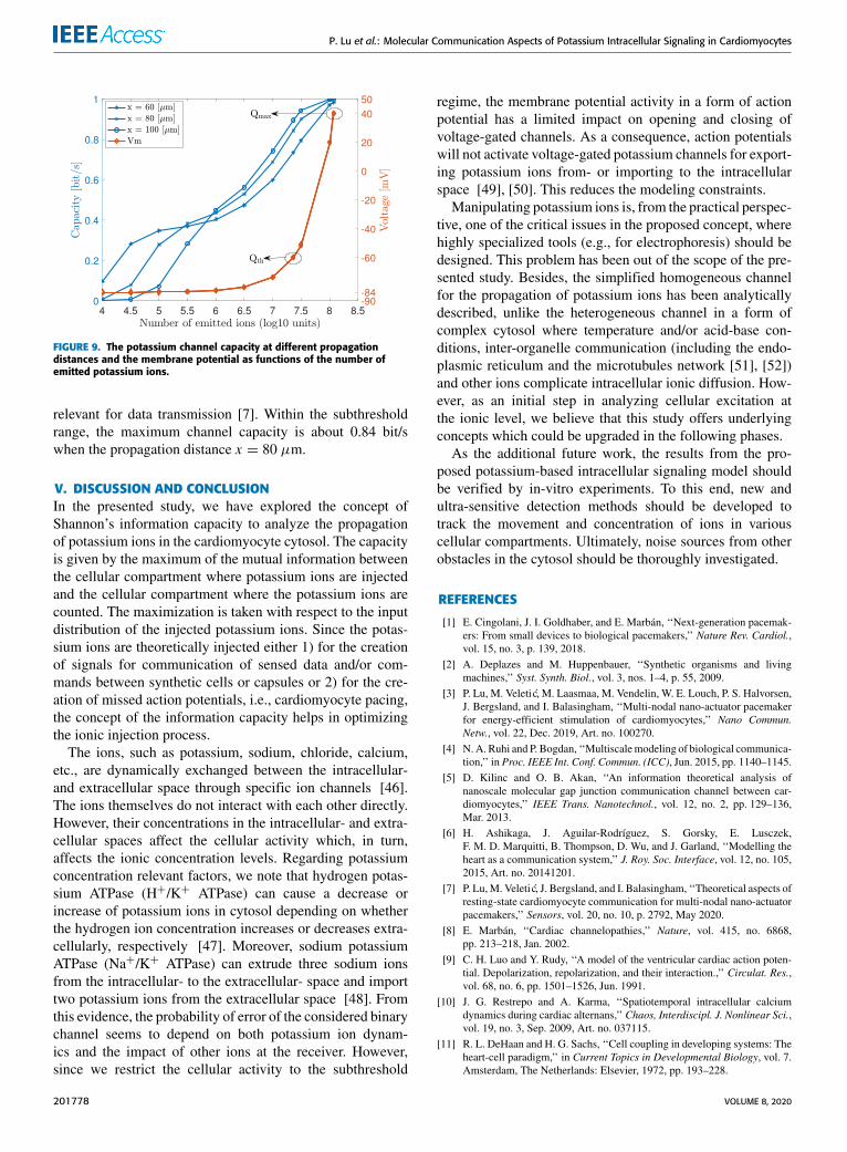
Finally, the number of emitted potassium ions affectsthe detecting threshold θ , which then impacts the mutualinformation and channel capacity. We infer how the channelcapacity changes with the number of injected potassium ionsaccording to (20). As shown in Figure 9, we observe that boththe channel capacity at different propagation distances andthe membrane potential increase when the number of emittedpotassium ions increases.2 The capacity reaches nearly 1 bit/swhen the number of emitted ions is Qmax = 1.18885 × 10⁸.The membrane potential then reaches nearly 40 mV. Practi-cally though, the cell membrane reaches 40 mV with signifi-cantly less number of the emitted ions (i.e., Qth) sufficient tobring the cell membrane to the threshold potential. When thenumber of injected ions is lower than Qth, the cell membranegenerates membrane potentials in the subthreshold range
2Here we assume that the capacitive membrane area Acap in (5) does notchange when the propagation distance changes.
VOLUME 8, 2020 201777
P. Lu et al.: Molecular Communication Aspects of Potassium Intracellular Signaling in Cardiomyocytes
FIGURE 9. The potassium channel capacity at different propagationdistances and the membrane potential as functions of the number ofemitted potassium ions.
relevant for data transmission [7]. Within the subthresholdrange, the maximum channel capacity is about 0.84 bit/swhen the propagation distance x = 80 µm.
V. DISCUSSION AND CONCLUSIONIn the presented study, we have explored the concept ofShannon’s information capacity to analyze the propagationof potassium ions in the cardiomyocyte cytosol. The capacityis given by the maximum of the mutual information betweenthe cellular compartment where potassium ions are injectedand the cellular compartment where the potassium ions arecounted. The maximization is taken with respect to the inputdistribution of the injected potassium ions. Since the potas-sium ions are theoretically injected either 1) for the creationof signals for communication of sensed data and/or com-mands between synthetic cells or capsules or 2) for the cre-ation of missed action potentials, i.e., cardiomyocyte pacing,the concept of the information capacity helps in optimizingthe ionic injection process.
The ions, such as potassium, sodium, chloride, calcium,etc., are dynamically exchanged between the intracellular-and extracellular space through specific ion channels [46].The ions themselves do not interact with each other directly.However, their concentrations in the intracellular- and extra-cellular spaces affect the cellular activity which, in turn,affects the ionic concentration levels. Regarding potassiumconcentration relevant factors, we note that hydrogen potas-sium ATPase (H+/K+ ATPase) can cause a decrease orincrease of potassium ions in cytosol depending on whetherthe hydrogen ion concentration increases or decreases extra-cellularly, respectively [47]. Moreover, sodium potassiumATPase (Na+/K+ ATPase) can extrude three sodium ionsfrom the intracellular- to the extracellular- space and importtwo potassium ions from the extracellular space [48]. Fromthis evidence, the probability of error of the considered binarychannel seems to depend on both potassium ion dynam-ics and the impact of other ions at the receiver. However,since we restrict the cellular activity to the subthreshold
regime, the membrane potential activity in a form of actionpotential has a limited impact on opening and closing ofvoltage-gated channels. As a consequence, action potentialswill not activate voltage-gated potassium channels for export-ing potassium ions from- or importing to the intracellularspace [49], [50]. This reduces the modeling constraints.
Manipulating potassium ions is, from the practical perspec-tive, one of the critical issues in the proposed concept, wherehighly specialized tools (e.g., for electrophoresis) should bedesigned. This problem has been out of the scope of the pre-sented study. Besides, the simplified homogeneous channelfor the propagation of potassium ions has been analyticallydescribed, unlike the heterogeneous channel in a form ofcomplex cytosol where temperature and/or acid-base con-ditions, inter-organelle communication (including the endo-plasmic reticulum and the microtubules network [51], [52])and other ions complicate intracellular ionic diffusion. How-ever, as an initial step in analyzing cellular excitation atthe ionic level, we believe that this study offers underlyingconcepts which could be upgraded in the following phases.
As the additional future work, the results from the pro-posed potassium-based intracellular signaling model shouldbe verified by in-vitro experiments. To this end, new andultra-sensitive detection methods should be developed totrack the movement and concentration of ions in variouscellular compartments. Ultimately, noise sources from otherobstacles in the cytosol should be thoroughly investigated.
REFERENCES
[1] E. Cingolani, J. I. Goldhaber, and E. Marbán, ‘‘Next-generation pacemak-ers: From small devices to biological pacemakers,’’ Nature Rev. Cardiol.,vol. 15, no. 3, p. 139, 2018.
[2] A. Deplazes and M. Huppenbauer, ‘‘Synthetic organisms and livingmachines,’’ Syst. Synth. Biol., vol. 3, nos. 1–4, p. 55, 2009.
[3] P. Lu, M. Veletić, M. Laasmaa, M. Vendelin, W. E. Louch, P. S. Halvorsen,J. Bergsland, and I. Balasingham, ‘‘Multi-nodal nano-actuator pacemakerfor energy-efficient stimulation of cardiomyocytes,’’ Nano Commun.Netw., vol. 22, Dec. 2019, Art. no. 100270.
[4] N.A. Ruhi and P. Bogdan, ‘‘Multiscalemodeling of biological communica-tion,’’ in Proc. IEEE Int. Conf. Commun. (ICC), Jun. 2015, pp. 1140–1145.
[5] D. Kilinc and O. B. Akan, ‘‘An information theoretical analysis ofnanoscale molecular gap junction communication channel between car-diomyocytes,’’ IEEE Trans. Nanotechnol., vol. 12, no. 2, pp. 129–136,Mar. 2013.
[6] H. Ashikaga, J. Aguilar-Rodríguez, S. Gorsky, E. Lusczek,F. M. D. Marquitti, B. Thompson, D. Wu, and J. Garland, ‘‘Modelling theheart as a communication system,’’ J. Roy. Soc. Interface, vol. 12, no. 105,2015, Art. no. 20141201.
[7] P. Lu,M. Veletić, J. Bergsland, and I. Balasingham, ‘‘Theoretical aspects ofresting-state cardiomyocyte communication for multi-nodal nano-actuatorpacemakers,’’ Sensors, vol. 20, no. 10, p. 2792, May 2020.
[9] C. H. Luo and Y. Rudy, ‘‘A model of the ventricular cardiac action poten-tial. Depolarization, repolarization, and their interaction.,’’ Circulat. Res.,vol. 68, no. 6, pp. 1501–1526, Jun. 1991.
[10] J. G. Restrepo and A. Karma, ‘‘Spatiotemporal intracellular calciumdynamics during cardiac alternans,’’ Chaos, Interdiscipl. J. Nonlinear Sci.,vol. 19, no. 3, Sep. 2009, Art. no. 037115.
[11] R. L. DeHaan and H. G. Sachs, ‘‘Cell coupling in developing systems: Theheart-cell paradigm,’’ in Current Topics in Developmental Biology, vol. 7.Amsterdam, The Netherlands: Elsevier, 1972, pp. 193–228.
201778 VOLUME 8, 2020
P. Lu et al.: Molecular Communication Aspects of Potassium Intracellular Signaling in Cardiomyocytes
[12] S. Weidmann and A. L. Hodgkin, ‘‘The diffusion of radiopotassium acrossintercalated disks of mammalian cardiac muscle,’’ J. Physiol., vol. 187,no. 2, pp. 323–342, Nov. 1966.
[13] H. Haljamäe, B. Johansson, O. Jonsson, and H. Röcker, ‘‘The distributionof sodium, potassium and chloride in the smooth muscle of the rat por-tal vein,’’ Acta Physiologica Scandinavica, vol. 78, no. 2, pp. 255–268,Feb. 1970.
[14] M. M. Civan, ‘‘Intracellular activities of sodium and potassium,’’ Amer. J.Physiol.-Renal Physiol., vol. 234, no. 4, pp. F261–F269, Apr. 1978.
[15] T. Nakano, T. Suda, M. Moore, R. Egashira, A. Enomoto, and K. Arima,‘‘Molecular communication for nanomachines using intercellular calciumsignaling,’’ in Proc. 5th IEEE Conf. Nanotechnol., 2005, pp. 478–481.
[16] I. Llatser, A. Cabellos-Aparicio, and E. Alarcon, ‘‘Networking challengesand principles in diffusion-based molecular communication,’’ IEEE Wire-less Commun., vol. 19, no. 5, pp. 36–41, Oct. 2012.
[17] M. Kuran, T. Tugcu, and B. Edis, ‘‘Calcium signaling: Overview andresearch directions of a molecular communication paradigm,’’ IEEE Wire-less Commun., vol. 19, no. 5, pp. 20–27, Oct. 2012.
[18] N. Farsad and A. Goldsmith, ‘‘A molecular communication system usingacids, bases and hydrogen ions,’’ in Proc. IEEE 17th Int. Workshop SignalProcess. Adv. Wireless Commun. (SPAWC), Jul. 2016, pp. 1–6.
[19] N. Farsad, H. B. Yilmaz, A. Eckford, C.-B. Chae, and W. Guo,‘‘A comprehensive survey of recent advancements in molecular commu-nication,’’ IEEE Commun. Surveys Tuts., vol. 18, no. 3, pp. 1887–1919,3rd Quart., 2016.
[20] E. U. Thodesen, ‘‘Diffusion-based molecular communications in wirelesspacemakers,’’ M.S. thesis, NTNU, Trondheim, Norway, 2016.
[21] B. D. Unluturk and I. F. Akyildiz, ‘‘An End-to-End model of plantpheromone channel for long range molecular communication,’’ IEEETrans. Nanobiosci., vol. 16, no. 1, pp. 11–20, Jan. 2017.
[22] I. Llatser, A. Cabellos-Aparicio, M. Pierobon, and E. Alarcon, ‘‘Detectiontechniques for diffusion-based molecular communication,’’ IEEE J. Sel.Areas Commun., vol. 31, no. 12, pp. 726–734, Dec. 2013.
[23] H. B. Yilmaz, A. C. Heren, T. Tugcu, and C.-B. Chae, ‘‘Three-dimensionalchannel characteristics for molecular communications with an absorbingreceiver,’’ IEEE Commun. Lett., vol. 18, no. 6, pp. 929–932, Jun. 2014.
[24] A. Zare and A. Jamshidi, ‘‘Receiver design and performance analysis forpulse position modulation technique in diffusion-based molecular commu-nication,’’ Nano Commun. Netw., vol. 21, Sep. 2019, Art. no. 100256.
[25] A. O. Kislal, H. B. Yilmaz, A. E. Pusane, and T. Tugcu, ‘‘ISI-aware channelcode design for molecular communication via diffusion,’’ IEEE Trans.Nanobiosci., vol. 18, no. 2, pp. 205–213, Apr. 2019.
[26] A. Gohari, M. Mirmohseni, and M. Nasiri-Kenari, ‘‘Information theory ofmolecular communication: Directions and challenges,’’ IEEE Trans. Mol.,Biol. Multi-Scale Commun., vol. 2, no. 2, pp. 120–142, Dec. 2016.
[27] H. Awan and C. T. Chou, ‘‘Molecular communications with molecu-lar circuit-based transmitters and receivers,’’ IEEE Trans. Nanobiosci.,vol. 18, no. 2, pp. 146–155, Apr. 2019.
[28] N. Abadi, A. A. Gohari, M. Mirmohseni, and M. Nasiri-Kenari, ‘‘Zero-error codes for multi-type molecular communication in random delaychannel,’’ in Proc. Iran Workshop Commun. Inf. Theory (IWCIT),Apr. 2018, pp. 1–6.
[29] L. Cleemann and M. J. Gaughan, ‘‘Measurement of intracellular 42K dif-fusion in frog ventricular strips,’’ Pflügers Archiv Eur. J. Physiol., vol. 401,no. 1, pp. 101–103, May 1984.
[30] S.-M. Yu, S.-F. Lo, and T.-H.-D. Ho, ‘‘Source–sink communication: Regu-lated by hormone, nutrient, and stress cross-signaling,’’ Trends Plant Sci.,vol. 20, no. 12, pp. 844–857, Dec. 2015.
[31] T. Nakano, A. W. Eckford, and T. Haraguchi, Molecular Communication.Cambridge, U.K.: Cambridge Univ. Press, 2013.
[32] P. Lu, Z. Wu, and B. Liu, ‘‘A vertical channel model of molecular commu-nication and its test-bed,’’ EAI Endorsed Trans. Pervas. Health Technol.,vol. 3, no. 9, Mar. 2017, Art. no. 152390.
[33] Y. Guo and W. T. Pu, ‘‘Cardiomyocyte maturation,’’ Circulat. Res.,vol. 126, no. 8, pp. 1086–1106, Apr. 2020.
[34] W. DeMello, Intercellular Communication. Cham, Switzerland: Springer,2013.
[35] C. Koch, Biophysics of Computation: Information Processing in SingleNeurons. Oxford, U.K.: Oxford Univ. Press, 2004.
[36] C. H. Luo and Y. Rudy, ‘‘A dynamic model of the cardiac ventricular actionpotential. I. Simulations of ionic currents and concentration changes.,’’Circulat. Res., vol. 74, no. 6, pp. 1071–1096, Jun. 1994.
[37] R.Weingart, ‘‘The permeability to tetraethylammonium ions of the surfacemembrane and the intercalated disks of sheep and calf myocardium,’’J. Physiol., vol. 240, no. 3, pp. 741–762, Aug. 1974.
[38] J. F. Lamb and J. A. S. McGuigan, ‘‘The efflux of potassium, sodium,chloride, calcium and sulphate ions and of sorbitol and glycerol during thecardiac cycle in frog’s ventricle,’’ J. Physiol., vol. 195, no. 2, pp. 283–315,Mar. 1968.
[39] A. C. Heren, H. B. Yilmaz, C.-B. Chae, and T. Tugcu, ‘‘Effect of degra-dation in molecular communication: Impairment or enhancement?’’ IEEETrans. Mol., Biol. Multi-Scale Commun., vol. 1, no. 2, pp. 217–229,Jun. 2015.
[40] A. Moorhouse, ‘‘Membrane potential: Concepts,’’ in Encyclopedia of CellBiology, R. A. Bradshaw and P. D. Stahl, Eds. Waltham, MA, USA:Academic, 2016, pp. 218–236.
[41] T. Nakano, M. J. Moore, F. Wei, A. V. Vasilakos, and J. Shuai, ‘‘Molecularcommunication and networking: Opportunities and challenges,’’ IEEETrans. Nanobiosci., vol. 11, no. 2, pp. 135–148, Jun. 2012.
[42] Z. Cheng, Y. Zhu, K. Chi, Y. Li, and M. Xia, ‘‘Capacity analysis fordiffusive molecular communication with ISI channel,’’ Nano Commun.Netw., vol. 13, pp. 43–50, Sep. 2017.
[43] T. Nakano, Y. Okaie, and J.-Q. Liu, ‘‘Channel model and capacity analysisof molecular communication with brownian motion,’’ IEEE Commun.Lett., vol. 16, no. 6, pp. 797–800, Jun. 2012.
[44] H. B. Yilmaz, C.-B. Chae, B. Tepekule, and A. E. Pusane, ‘‘Arrivalmodeling and error analysis for molecular communication via diffusionwith drift,’’ in Proc. 2nd Annu. Int. Conf. Nanosc. Comput. Commun.(NANOCOM), 2015, pp. 1–6.
[45] R. W. Yeung, A First Course in Information Theory. Cham, Switzerland:Springer, 2012.
[46] B. Arnold, C. A. Kaiser, H. Lodish, A. Amon, H. Ploegh, A. Bretscher,M. Krieger, and K. C. Martin,Molecular Cell Biology. San Francisco, CA,USA: Freeman, 2008.
[47] I. R. Driel and J. M. Callaghan, ‘‘Proton and potassium transport byH+/K+-ATPases,’’ Clin. Experim. Pharmacol. Physiol., vol. 22, no. 12,pp. 952–960, Dec. 1995.
[48] Z. Xie and A. Askari, ‘‘Na+ /K+ -ATPase as a signal transducer,’’ Eur. J.Biochem., vol. 269, no. 10, pp. 2434–2439, May 2002.
[49] F. Bezanilla, ‘‘Voltage-gated ion channels,’’ in Biological Membrane IonChannels. Cham, Switzerland: Springer, 2007, pp. 81–118.
[50] M. P. Mahaut-Smith, T. J. Rink, S. C. Collins, and S. O. Sage, ‘‘Voltage-gated potassium channels and the control of membrane potential in humanplatelets.,’’ J. Physiol., vol. 428, no. 1, pp. 723–735, Sep. 1990.
[51] N. Panayotis, A. Karpova, M. R. Kreutz, and M. Fainzilber, ‘‘Macro-molecular transport in synapse to nucleus communication,’’ Trends Neu-rosciences, vol. 38, no. 2, pp. 108–116, Feb. 2015.
[52] A. Enomoto, M. J. Moore, T. Suda, and K. Oiwa, ‘‘Design of self-organizing microtubule networks for molecular communication,’’ NanoCommun. Netw., vol. 2, no. 1, pp. 16–24, Mar. 2011.
PENGFEI LU (Member, IEEE) received the bach-elor’s degree in computer science and technol-ogy from the College of Information Science andTechnology, Beijing University of Chemical Tech-nology, China, in 2013, and the master’s degreein computer system structure from the School ofComputer Science, Shaanxi Normal University,China. He is currently pursuing the Ph.D. degreewith the Department of Clinical Medicine, Facultyof Medicine, University of Oslo. He is also doing
his Ph.D. degree research work with the Intervention Center, Oslo UniversityHospital, Norway. His research interests include channel model, molecu-lar communication, nanonetworks, leadless pacemaker communication, andheart synchronization.
VOLUME 8, 2020 201779
P. Lu et al.: Molecular Communication Aspects of Potassium Intracellular Signaling in Cardiomyocytes
MLADEN VELETIĆ received the B.Sc. and M.Sc.degrees in electronics and telecommunicationsfrom the Faculty of Electrical Engineering, Uni-versity of Banja Luka (UNIBL), Bosnia andHerzegovina, in 2010 and 2012, respectively, andthe Ph.D. degree in telecommunications from theDepartment of Electronic Systems, NorwegianUniversity of Science and Technology (NTNU),Norway, and the Faculty of Electrical Engineering,UNIBL. From 2011 to 2017, he worked as a Senior
Teaching and a Research Assistant with the University of Banja Luka. He iscurrently a Postdoctoral Research Scientist with the Intervention Center,Oslo University Hospital. He was awarded a Gold Plaque from the UNIBLfor his achievements throughout the undergraduate education. His researchinterests include molecular and nano-neural communications, wireless com-munications, and positioning in cellular networks.
JACOB BERGSLAND received the medical andPh.D. degrees from Oslo University, in 1973 and2011, respectively. After internship in Norway,he moved to USA and became a Specialistin General Surgery and Cardiothoracic Surgery,in 1981 and 1983. He was the Director of cardiacsurgery with Buffalo Veterans Hospital and theDirector of the Cardiac Transplantation and Min-imally Invasive Cardiac Surgery, Buffalo GeneralHospital. He was a Clinical Associate Professor of
surgery with SUNY at Buffalo. He initiated the Partnership between BuffaloGeneral Hospital and the Tuzla University Medical Center, Bosnia, wherehe started the country’s first cardiac program. He co-initiated the BH HeartCenter, Tuzla. He has served as a Medical Director until 2019. In addition,he has been working as a Researcher with the Intervention Center, OsloUniversity Hospital, since 2001. He is currently the Founder and a MedicalDirector of a Medical Device start-up, Cardiomech AS.
ILANGKO BALASINGHAM (Senior Member,IEEE) received theM.Sc. and Ph.D. degrees in sig-nal processing from the Department of Electronicsand Telecommunications, Norwegian Universityof Science and Technology (NTNU), Trondheim,Norway, in 1993 and 1998, respectively. He per-formed the master’s degree thesis with the Depart-ment of Electrical and Computer Engineering,University of California at Santa Barbara, SantaBarbara, CA, USA. From 1998 to 2002, he was
a Research Engineer developing image and video streaming solutions formobile handheld devices with the Fast Search and Transfer ASA, Oslo,Norway, which is currently a part of Microsoft Inc. Since 2002, he has beena Senior Research Scientist with the Intervention Center, Oslo UniversityHospital, Oslo, where he heads the Wireless Sensor Network ResearchGroup. He was appointed as a Professor in signal processing in medicalapplications with NTNU, in 2006. From 2016 to 2017, he was a Professorby courtesy with the Frontier Institute, Nagoya Institute of Technology,Japan. He has authored or coauthored more than 200 journals and conferencepapers, seven book chapters, 42 abstracts, five patents, and 16 articles inpopular press. His research interests include super robust short range com-munications for both inbody and onbody sensors, body area sensor networks,microwave short range sensing of vital signs, short range localization andtracking mobile sensors, and nanoscale communication networks. He hasgiven 16 invited/keynotes at the international conferences. In addition, he isactive in organizing conferences (has also been a Steering Committee mem-ber of ACM NANOCOM, since 2018, the General Chair: the 2019 IEEEInternational Symposium of Medical ICT and the 2012 Body Area NetworksConference, and the TPC Chair of the 2015 ACM NANOCOM), and anEditorial Board (has been an Area Editor of Nano Communication Networks(Elsevier), since 2013).


![Regulation of the intracellular Ca2+. Regulation of intracellular [H]:](https://static.documents.pub/doc/80x56/5a4d1b717f8b9ab0599b56a5/regulation-of-the-intracellular-ca2-regulation-of-intracellular-h.jpg)


