Revista de Biología Tropical ISSN: 0034-7744 [email protected]Universidad de Costa Rica Costa Rica Gautam, Ragini; Singh, S. K.; Sharma, Vinay Molecular diagnosis and intraspecific genetic variability of root pathogens of arid legumes in Western Rajasthan, India Revista de Biología Tropical, vol. 64, núm. 4, 2016, pp. 1505-1518 Universidad de Costa Rica San Pedro de Montes de Oca, Costa Rica Available in: http://www.redalyc.org/articulo.oa?id=44947539012 How to cite Complete issue More information about this article Journal's homepage in redalyc.org Scientific Information System Network of Scientific Journals from Latin America, the Caribbean, Spain and Portugal Non-profit academic project, developed under the open access initiative
1505Rev. Biol. Trop. (Int. J. Trop. Biol. ISSN-0034-7744) Vol. 64 (4): 1505-1518, December 2016
Molecular diagnosis and intraspecific genetic variability of root pathogens of arid legumes in Western Rajasthan, India
Ragini Gautam1, S. K. Singh1 & Vinay Sharma2
1. Central Arid Zone Research Institute, Jodhpur- 342003, Rajasthan, India; [email protected], [email protected]. Department of Biosciences and Biotechnology, Banasthali Vidhyapeeth, Banasthali-304022, Rajasthan, India; [email protected]
Received 26-II-2016. Corrected 03-VII-2016. Accepted 04-VIII-2016.
Abstract: The productivity of arid legumes, such as Clusterbean (Cyamopsis tetragonoloba), Cowpea (Vigna unguiculata), Moth bean (Vigna aconitifolia) and Horse gram (Macrotyloma uniflorum), may remain stagnant over decades because of their high susceptibility to root diseases. Besides, there is a limitation on the infor-mation about molecular diagnosis and intraspecific genetic variability of root pathogens in arid legumes. To contribute in this field, we assessed a total of 52 isolates from 88 root samples that were found infected with fungal pathogens in Jodhpur, Jaipur and Bikaner Districts of Rajasthan. Diseased roots samples were analyzed following standard microbiological methods for fungus extraction and purification, and for genetic studies. Irrespective of the geographical location from where the diseased samples were collected, all pathogen isolates were clustered in RAPD dendrograms as per their respective genera. Phylogram, based on multiple sequence alignment, revealed that different genera (i.e. Fusarium, Neocosmospora and Syncephalastrum), separated from each other, and species within the same genera, clustered together with their reference sequences with apreciable bootstrap values. Out of 20 representative isolates representing each cluster and all outgroups sequenced, eight were molecularly identified as Neocosmospora vasinfecta, five as Fusarium solani, two as Neocosmospora striata, two as Fusarium acutatum, one as Syncephalastrum monosporum, one as Fusarium oxysporum and one as Fusarium species. The root pathogens of the arid legumes were found neither restricted to a geographi-cal location nor were host specific in nature. Fusarium solani wilt in cowpea and seedling rot in moth bean, F. oxysporum wilt in moth bean, F. acutatum damping off in cowpea and Clusterbean, Fusarium sp. seedling rot in Clusterbean, Neocosmospora striata root rot in cowpea and wilt in Clusterbean and Syncephalastrum monospo-rum root rot in Clusterbean were molecularly identified as new fungal records as pathogens causing root diseases in arid legumes. Rev. Biol. Trop. 64 (4): 1505-1518. Epub 2016 December 01.
In India, arid legumes are grown in about 5 million hectares area with approximately 0.2 million tons production of grains annually. The major arid legumes are Clusterbean (Cya-mopsis tetragonoloba), Cowpea (Vigna ungui-culata), Moth bean (Vigna aconitifolia) and Horse gram (Macrotyloma uniflorum) which are predominantly grown in arid and semiarid tracks of Indian subcontinents. Due to inherent drought hardy characteristics arid legumes are mostly grown where land conditions are not
suitable for cultivation of cereals mostly under rain fed conditions. During rainy season, high temperature coupled with moisture stress put them into abiotic and biotic stresses. Besides, blights, leaf spots, powdery mildews and viral diseases, these arid legumes are attacked by serious soil-borne pathogens with a very wide host range causing substantial damages and yield reduction in arid legumes. The infor-mation on molecular diagnosis and intras-pecific genetic variability of root pathogens
1506 Rev. Biol. Trop. (Int. J. Trop. Biol. ISSN-0034-7744) Vol. 64 (4): 1505-1518, December 2016
of arid legumes of the region is limited (Gau-tam, Singh, & Sharma, 2013).
The most common diseases caused by these pathogens are charcoal rot (damping off), dry root rot, wilt, leaf blight and ashy stem blight. Efforts have been made to characte-rize the fungus populations in different parts of the world. This is based on its pathogenic variability, the morphological characteristics, as well as the molecular characteristics (Jana, Sharma, Prasad, & Arora, 2003). Recently Ran-dom Amplified Polymorphic DNA (RAPD) and nuclear rDNA Internal Transcribed Spacer (ITS) polymorphism within Macrophomina phaseolina isolated from arid legumes of Wes-tern Rajasthan revealed genetic diversity among 33 isolates (Gautam et al., 2013). Species of Fusarium causes both major and minor disea-ses in these legumes (Aigbe & Fawole, 2010).
The productivity of these arid legumes remains virtually stagnant over decades becau-se of their susceptibility to root diseases. Most of the root pathogens are soil or seed borne, and colonize the xylem vessels by clogging and blocking completely to effect wilting. Accu-rate and rapid identification of pathogens is necessary for appropriate management of plant diseases (Narayanasamy, 2001).
The classical biological pathotyping tech-niques alone are not enough for a reliable identification and characterization of fungal pathotypes and population (Jamil et al., 2000). Therefore, present study was carried out to identify the cause of the disease (Koch’s pos-tulates), molecularly identify and characterize root pathogens of arid legumes using RAPD and ITS rDNA polymorphism to reveal inter and intra specific genetic relationships among and within root pathogens, to facilitate effecti-ve management of major diseases.
MATERIALS AND METHODS
Isolation of pathogens: Surveys were con-ducted to collect diseased plants of Clusterbean (Cyamopsis tetragonoloba), Cowpea (Vigna unguiculata), Moth bean (Vigna aconitifolia) and Horse gram (Macrotyloma uniflorum)
from Jodhpur, Jaipur and Bikaner Districts of Rajasthan during the months of August and September, 2011. A total of 88 infected plants i.e., Clusterbean (38), Cowpea (16), Moth bean (24) and Horse gram (10) were collected, but this report only assessed those infections diffe-rent from Macropomina phaseolina. To obtain pure cultures of root pathogens, these infected roots were cut into small pieces and surface sterilized using 5 % sodium hypochlorite for three minutes, washed thrice in sterilized disti-lled water and air dried. Two or three infected root bits from each infected root sample were then aseptically transferred onto Petri plates containing PDA (39 g PDA, HiMedia Com-pany) culture medium, containing 150 mg/litre of streptomycin sulphate (to avoid bacterial contamination) and incubated in an incubator (Jindal, S.M. Scientific Instruments (P) Ltd, New Delhi, India) at 25 ± 2°C for five days. To raise pure cultures, the mycelia from the growing margins of each culture along with a piece of media from Petri plates were aseptica-lly transferred into test tubes containing PDA, and incubated at 25 ± 2 °C for a week. The 52 pure cultures obtained were stored at 4°C in a refrigerator until use.
Pathogenicity tests: Sandy loam soil was sterilized by autoclaving it (Apex, Conica Enterprises, New Delhi, India), at 121 °C for one hour. PVC plastic pots were filled with two kg of sterilized soil, and a pathogenecity test was set in a complete randomized block design, considering legumes origin and all 20 isola-tes: JD-HG10, JAI-CP11, JAI-CP16, JD-CB7, JD-CB9, BK-CB15, BK-CB17, BK-CB20, BK-CB26, JAI-CB37, JAI-MB22, JAI-MB24, BK-CB18, JD-CB10, BK-CB30, BK-CB22, JAI-CB34, JAI-CP13, JAI-CB36 and JAI-MB21. Each isolated pathogen was raised in conical flasks on liquid Malt Extract Dextrose Broth culture medium (Malt Extract-10 g; Dex-trose-5 g with antibacterial agent streptomycin 150 mg; 1 litre of sterilized distilled water) for 10 days. Conidial suspension was obtained by filtering mycelial mats and blending it with sterilized distilled water and again filtering it
1507Rev. Biol. Trop. (Int. J. Trop. Biol. ISSN-0034-7744) Vol. 64 (4): 1505-1518, December 2016
through two layers of sterile cheesecloth. Two week old plants of arid legumes i.e., cluster-bean, cowpea, moth bean and horse gram, were then inoculated with conidial suspension of specified isolate (from which it was originally isolated) at the base by disturbing the soil. The inoculated plants were incubated in green house under high temperature (28-300C) and moisture stress conditions (100 mL water every 4th day and humidity 60-70 %) to facilitate disease development. A total of five pots were inoculated with each isolate. Plant symptoms were recorded and the pathogen reisolated. Besides, upon microscopic examination, the pathogenicity of each isolated pathogen was confirmed. Identifications to genus level were carried following Barnett and Hunter (1990).
DNA isolation: For DNA isolation, a small piece of growing mycelia from each of the 52 isolated cultures was aseptically trans-ferred to separate Malt Extract Dextrose Broth culture medium and incubated in a BOD incu-bator at 25 ± 2 °C for seven days. The fungal mycelial net from each raised pure culture was filtered using filter paper through funnel. The genomic DNA was extracted from approxima-tely 100 mg of fresh mycelium by crushing it in conical micro centrifuge tubes using micro-pestles in liquid nitrogen. The HiPura kit of HiMedia Company and protocols suggested by Birren and Lai (1993), Sambrook, Fritsch and Maniatis (1989) were followed for genomic DNA isolation. Finally, the DNA was eluted in 200µL of Tris EDTA buffer (TE buffer).
RAPD analysis: Eighteen decamer ran-dom primers of OPA, OPB and OPP series (Operon Technologies, USA: http://www.ope-ron.com) were used for initial screening of all the isolates. Finally, the data of ten RAPD primers exhibiting consistent reproducible amplified products on gel electrophoresis were used for DNA fingerprinting. Each PCR was performed in a total reaction mixture of 25 µL following Gautam et al. (2013). Amplicons were separated on 1.5 % agarose gel (Sigma) pre-stained with ethidium bromide solution
using 1 × TAE (Tris Acetic acid EDTA, Hime-dia) buffer. The gel was run for 3 h at 50V and the RAPD amplicons profiles were recorded using Syngene Gel Documentation System with Genesnap software. The size of amplified fragments was determined using 100 bp plus ladder (MBI Fermentas). All RAPD reactions were performed twice to test the reproducibility of the amplicons profile.
Internal transcribed spacer (ITS) ampli-fication: The universal primers ITS-1 (5’ TCC GTA GGT GAA CCT GCG G 3’) and ITS-4 (5’ TCC TCC GCT TAT TGA TAT GC 3’) were used for the amplification of ITS-1-5.8S-ITS-4 region. Each PCR amplification was performed in a total volume of 50 µL following Gautam et al. (2013). Amplified ITS regions were sequenced employing ABI Prism DNA sequen-cer (Applied Biosystems, Carlsbad, CA, USA) using ITS-1 and ITS-4 primers separately for DNA labeling by the BigDye terminator method (Applied Biosystems, Foster City, CA, USA). Molecular identification of each fungal isolate was done on the basis of similarity with the best aligned sequence of BLAST search and accordingly species designated.
Molecular data analysis: The RAPD amplification products were scored as present (1) and absent (0) of scorable loci for each pri-mer isolate combinations. Molecular data were entered into a binomial matrix and were used to determine Jaccard’s similarity coefficient with NTSYS-pc software (Rohlf, 1997).
To perform analysis of molecular variance (AMOVA), the 52 isolates of root pathogens from arid legumes were divided into four populations based on the arid legume crop from which they were isolated i.e., population one (clusterbean), population two (horse gram), population three (cowpea) and population four (moth bean). Principal Component Analysis (PCA) via covariance matrix was calculated using GenALEx 6 software. Whereas, diversity in the frequency of fragment size of RAPD pat-terns was apportioned within and among root pathogen isolates using Shannon’s information
1508 Rev. Biol. Trop. (Int. J. Trop. Biol. ISSN-0034-7744) Vol. 64 (4): 1505-1518, December 2016
index (i) and gene diversity index (h) following using PopGen 32 programme.
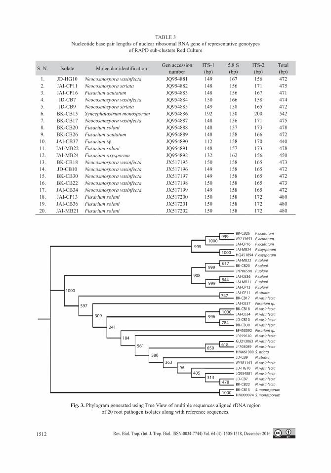
The multiple sequence alignment of ITS region (ITS-1, 5.8S r-RNA gene and ITS-2) of all the 20 representative root pathogen isolates was performed using ClustalX 2.0.11 software. The phylogenetic relationships of root rot pathogens were established by mul-tiple alignment of sequences and generating phylogram depicting bootstrap values using NJ plot software based on Single Nucleotide Polymorphisms (SNPs), insertions/deletions (INDELS), and or length diversity in the ITS and 5.8S nuclear rDNA regions. To assess the robustness of phylogenetic relationships, the best aligned reference sequence represen-ting of each species from GenBank database was downloaded in fasta format. A compo-site phylogenetic tree with bootstrap values showing grouping of 20 isolates sequenced with 11 reference sequences was generated to measure phylogenetic accuracy.
RESULTS
Isolation tests results: All the four steps of Koch’s postulates were successfully com-pleted for each test pathogen in a replicated trial. The details of fungal isolates, host, dis-trict and colony characteristics are shown at table 1. A total of 52 root samples were found infected with fungal pathogens belonging to genus Fusarium, Neocosmospora and Syn-cephalastrum. In all, four types of colony and growth patterns i.e. feathery, dense, fluffy and restricted, were recorded from different isola-tes. Similarly disease symptoms from seedling wilt (damping off) to seedling rot, wilt and root rot were recorded. Light pink to dark red pigmentations were also recorded in different cultures. Based on microscopic examinations, genus level identifications revealed that from the total of 52 diseased root samples consi-dered in this study, 26 samples were identi-fied as Neocosmospora, 25 as Fusarium and 1 as Syncephalastrum.
RAPD analysis: Out of 18 decamer ran-dom primers, 10 primers detected intraspecific variations generating scorable amplicons and reproducible patterns that have generated 147 marker bands in the range of 250 bp to 5 000 bp. Of which, 146 marker bands were poly-morphic amounting to 99.41 % polymorphism ranging from 94.11 % to 100 %. The number of PCR amplified products ranged from 11 (OPP-16) to 18 (OPB-04) with an average of about 14.7 bands/primer (Table 2). The PIC values varied from 84 % to 92 % with an average of 90 %. The primer OPB-06 and OPB-14 exhi-bited the maximum PIC value of 92 % which was closely followed by OPA-09, OPA-13 and OPB-05 with 91 %. A representative RAPD profile generated using OPB-06 exhibiting the maximum PIC value is shown vide figure. 1A, figure 1B and figure 1C.
A cumulative RAPD dendrogram deli-neated all the 52 isolates into sub-clusters ranging from 32 to 95 % similarity. In all, the isolates can be seen in eight distinct clusters and seven isolates as out groups to these clus-ters (Fig. 2). Cluster one included two, cluster two contained nine, cluster three compiled 10, cluster four comprised seven, cluster five included six, cluster six and seven comprised four isolates each, and cluster eight compiled of three isolates.
Irrespective of the geographical loca-tion from where the diseased samples were collected, all isolates belonging to the genus Neocosmospora, aligned from cluster one to cluster five including all outgroups between these clusters. Whereas, all isolates of genus Fusarium converged into cluster six to eight and flanking outgroups including one iso-late of Syncephalastrum.
ITS amplification: Based on the clus-tering patterns of different isolates of root pathogens of arid legumes, 20 representative isolates from all RAPD clusters and distinct isolates were selected for nuclear rDNA ITS region sequencing. All the isolates generated
1509Rev. Biol. Trop. (Int. J. Trop. Biol. ISSN-0034-7744) Vol. 64 (4): 1505-1518, December 2016
TABLE 1Morphological characterization of fungal pathogens causing root diseases in arid legumes
S. No. Isolate Host District Colony Growth Pattern Colony Colour Pigmentation
in medium Genus Disease Symptom
1. JD-CB7 Clusterbean Jodhpur Feathery Off White Light Pink Neocosmospora Wilt NV2. JD-CB8 Clusterbean Jodhpur Feathery Off White Light Pink Neocosmospora Wilt3. JD-CB9 Clusterbean Jodhpur Restricted Light Purple Dark Pink Neocosmospora Wilt NS4. JD-CB10 Clusterbean Jodhpur Restricted Off White Light Pink Neocosmospora Wilt NV5. JD-CB11 Clusterbean Jodhpur Restricted Off White Light Pink Neocosmospora Damping off6. JD-CB12 Clusterbean Jodhpur Dense Off White Light Pink Neocosmospora Wilt7. JD-CB13 Clusterbean Jodhpur Fluffy Off White Light Pink Neocosmospora Wilt8. JD-CB14 Clusterbean Jodhpur Dense Off White Light Pink Neocosmospora Wilt9. JD-HG9 Horse gram Jodhpur Dense Off White Light Pink Neocosmospora Wilt10. JD-HG10 Horse gram Jodhpur Restricted Off White Light Pink Neocosmospora Wilt NV11. JD-CP9 Cowpea Jodhpur Dense Light Pink Dark Pink Neocosmospora Wilt12. JD-MB14 Moth bean Jodhpur Feathery Dark Pink Dark Pink Neocosmospora Wilt13. JD-MB15 Moth bean Jodhpur Feathery Off White Light Pink Neocosmospora Wilt14. BK-CP10 Cowpea Bikaner Feathery Off White Dark Pink Neocosmospora Seedling rot15. BK-MB16 Moth bean Bikaner Restricted Dark Pink Dark Pink Neocosmospora Wilt16. BK-MB17 Moth bean Bikaner Fluffy Dark Pink Light Pink Neocosmospora Wilt17. BK-MB18 Moth bean Bikaner Fluffy Off White Light Pink Neocosmospora Seedling rot18. BK-CB15 Clusterbean Bikaner Dense Off White Dark Pink Syncephalastrum Root rot SM19. BK-CB16 Clusterbean Bikaner Restricted Off White Light Pink Neocosmospora Wilt20. BK-CB17 Clusterbean Bikaner Restricted Light Pink Light Pink Neocosmospora Wilt NV21. BK-CB18 Clusterbean Bikaner Feathery Off White Dark Red Neocosmospora Wilt NV22. BK-CB19 Clusterbean Bikaner Fluffy Off White Light Red Neocosmospora Wilt23. BK-CB20 Clusterbean Bikaner Restricted Off White Light Pink Fusarium Wilt FS24. BK-CB21 Clusterbean Bikaner Fluffy Pink Light Red Neocosmospora Root rot25. BK-CB22 Clusterbean Bikaner Restricted Off White Light Red Neocosmospora Root rot26. BK-CB23 Clusterbean Bikaner Restricted Pink Light Red Fusarium Wilt NV27. BK-CB24 Clusterbean Bikaner Dense Off White Light Red Fusarium Wilt28. BK-CB25 Clusterbean Bikaner Dense Off White Light Red Fusarium Wilt29. BK-CB26 Clusterbean Bikaner Restricted Light Pink Light Red Fusarium Damping off FA30. BK-CB27 Clusterbean Bikaner Restricted Off White Dark Pink Fusarium Damping off31. BK-CB28 Clusterbean Bikaner Restricted Off White Dark Pink Fusarium Damping off32. BK-CB29 Clusterbean Bikaner Restricted Off White Dark Pink Fusarium Root rot33. BK-CB30 Clusterbean Bikaner Dense Off White Dark Pink Neocosmospora Root rot NV34. JAI-CP11 Cowpea Jaipur Restricted Light Pink Dark Pink Neocosmospora Root rot35. JAI-CP12 Cowpea Jaipur Feathery Off White Light Pink Fusarium Wilt36. JAI-CP13 Cowpea Jaipur Feathery Off White Light Pink Fusarium Wilt FS37. JAI-CP14 Cowpea Jaipur Feathery Off White Light Pink Fusarium Wilt38. JAI-CP15 Cowpea Jaipur Feathery Off White Light Pink Fusarium Wilt39. JAI-CP16 Cowpea Jaipur Dense Off White Light Red Fusarium Damping off FA40. JAI-MB19 Moth bean Jaipur Feathery Pink Dark Red Fusarium Wilt41. JAI-MB20 Moth bean Jaipur Fluffy Off White Dark Red Fusarium Wilt42. JAI-MB21 Moth bean Jaipur Fluffy Dark pink Light Pink Fusarium Seedling rot FS43. JAI-MB22 Moth bean Jaipur Dense Purple Dark Red Fusarium Wilt F OX44. JAI-MB24 Moth bean Jaipur Dense Pink Light Red Fusarium Wilt45. JAI-CB31 Clusterbean Jaipur Dense Off White Light Red Fusarium Wilt46. JAI-CB32 Clusterbean Jaipur Dense Off White Light Red Fusarium Wilt47. JAI-CB33 Clusterbean Jaipur Restricted Pink Light Red Fusarium Wilt48. JAI-CB34 Clusterbean Jaipur Restricted Off White Light Pink Neocosmospora Seedling rot NV49. JAI-CB35 Clusterbean Jaipur Restricted Off White Dark Red Fusarium Wilt50. JAI-CB36 Clusterbean Jaipur Dense Off White Dark Red Fusarium Wilt FS51. JAI-CB37 Clusterbean Jaipur Dense Off White Light Pink Fusarium Seedling rot F Sp52. JAI-CB38 Clusterbean Jaipur Dense Off White Dark Red Fusarium Wilt
1510 Rev. Biol. Trop. (Int. J. Trop. Biol. ISSN-0034-7744) Vol. 64 (4): 1505-1518, December 2016
TABLE 2Details of primer code, GC content, per cent polymorphism and PIC values of RAPD primers
S.N. Primer Code Primer Sequence GC (%) No. of Bands
No. of Polymorphic bands
Polymorphism (%) PIC Values
1. OPA-02 TGC CGA GCT G 70 13 13 100 90 %2. OPA-09 GGG TAA CGC C 70 15 15 100 91 %3. OPA-13 CAG CAC CCA C 70 13 13 100 91 %4. OPB-04 GGA CTG GAG T 60 18 18 100 90 %5. OPB-05 TGC GCC CTT C 70 12 12 100 91 %6. OPB-06 TGC TCT GCC C 70 17 16 94.11 92 %7. OPB-10 CTG CTG GGA C 70 16 16 100 90 %8. OPB-13 TTC CCC CGC T 70 14 14 100 89 %9. OPB-14 TCC GCT CTG G 70 18 18 100 92 %10. OPP-16 CCA AGC TGC C 70 11 11 100 84 %
Total 147 146Average 99.41 90.00
Fig. 1(A,B,C). RAPD profiles of root pathogens amplified by OPB-06 primer (a) Jodhpur, (b) Bikaner and (c) Jaipur.
1511Rev. Biol. Trop. (Int. J. Trop. Biol. ISSN-0034-7744) Vol. 64 (4): 1505-1518, December 2016
a single prominent band on agarose gel which included partial sequence of 18S gene, com-plete sequence of ITS-1, 5.8S gene, ITS-2 and partial sequence of 28S gene. The nucleotide sequences were subjected to BLAST search using NCBI, USA databases. The species level designation were assigned to each isolate con-sidering the maximum identities with that of available reference sequences of NCBI databa-ses (Table 3).
Molecular data analysis: The novel gene sequences were submitted to NCBI and gene bank accession numbers obtained. In order to detect SNP’s, insertions and deletions
(INDELS) and total length polymorphism of rDNA region, all the isolates were subjected to multiple sequence alignment using clustal X software programme. A phylogram was gene-rated using tree view software and boot strap values determined using NJ plot programme (Fig. 3) to validate the species designation and to reveal the nucleotide polymorphism vis-à-vis reference sequence available in NCBI databa-ses. The best aligned reference sequences were also downloaded and subjected to multiple sequence alignment to establish lineages. It is clear from the phylogram that different genera (i.e. Fusarium, Neocosmospora and Syncepha-lastrum) separated from each other and species
Fig. 2. Dendrogram of 52 isolates of root pathogens based on 10 RAPD informative primers.
Fig. 3. Phylogram generated using Tree View of multiple sequences aligned rDNA region of 20 root pathogen isolates along with reference sequences.
1513Rev. Biol. Trop. (Int. J. Trop. Biol. ISSN-0034-7744) Vol. 64 (4): 1505-1518, December 2016
within the genera converged together and grou-ped with respective reference sequences with appreciable bootstrap values.
Out of 20 isolates, eight were molecularly identified as Neocosmospora vasinfecta, five as Fusarium solani, two as Neocosmospora striata, two as Fusarium acutatum, one as Syncephalastrum monosporum, one as Fusa-rium oxysporum and one as Fusarium species. AMOVA revealed 100 % variation within population of root rot pathogens isolated from arid legumes. All the four populations of arid legumes, root pathogens were subjected to PCA using GenaLX. The first three principle coordinates accounted for 44.74, 17.78 and 15.03 respective amounting to a total of 77.55 % of total variance. The eigen vector analysis indicated that the contribution of the first three factors were 38.2, 15.2 and 12.8 respectively explaining a total of 66.2 % of total variabi-lity. The analysis of RAPD binary data using Popgene revealed that the coefficient of gene differentiation between populations (Gst) was 2.0170 among all the populations. The genetic analysis of RAPD data of all the 52 isolates
of root pathogens isolated were performed by dividing the isolates on the basis of host from which they were initially isolated. For example, population one (32 isolates of Clusterbean), population two (two isolates of horse gram), population three (eight isolates of cow pea), and population four (10 isolates of moth bean).
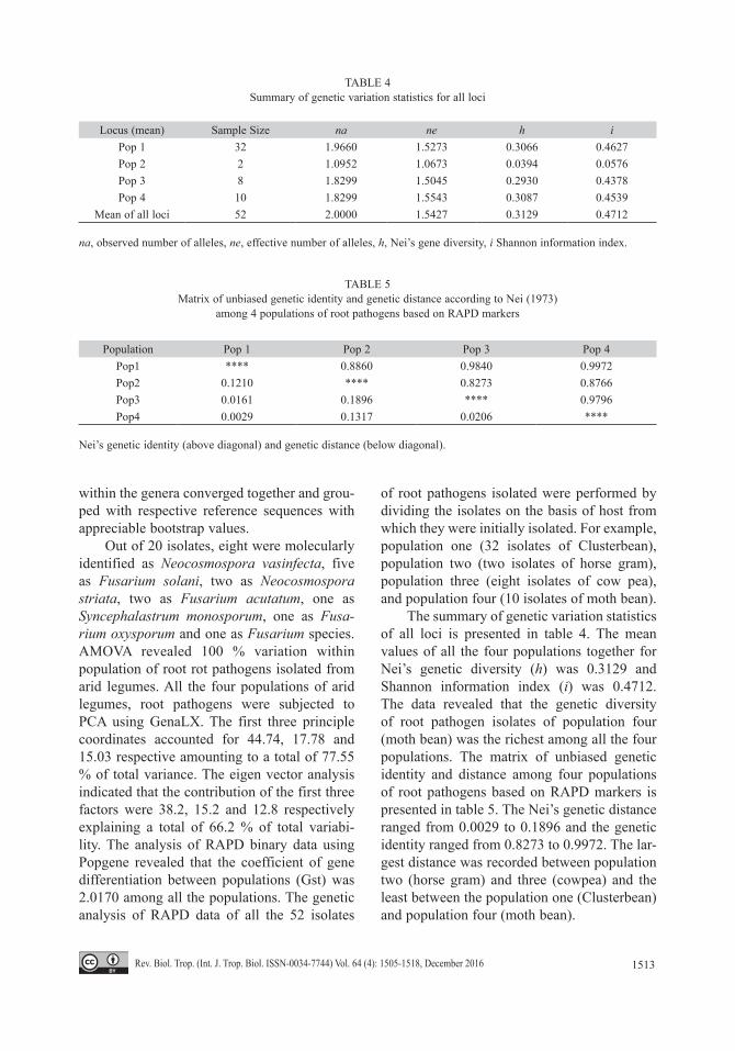
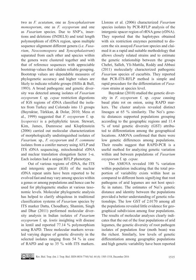
The summary of genetic variation statistics of all loci is presented in table 4. The mean values of all the four populations together for Nei’s genetic diversity (h) was 0.3129 and Shannon information index (i) was 0.4712. The data revealed that the genetic diversity of root pathogen isolates of population four (moth bean) was the richest among all the four populations. The matrix of unbiased genetic identity and distance among four populations of root pathogens based on RAPD markers is presented in table 5. The Nei’s genetic distance ranged from 0.0029 to 0.1896 and the genetic identity ranged from 0.8273 to 0.9972. The lar-gest distance was recorded between population two (horse gram) and three (cowpea) and the least between the population one (Clusterbean) and population four (moth bean).
TABLE 4Summary of genetic variation statistics for all loci
Locus (mean) Sample Size na ne h iPop 1 32 1.9660 1.5273 0.3066 0.4627Pop 2 2 1.0952 1.0673 0.0394 0.0576Pop 3 8 1.8299 1.5045 0.2930 0.4378 Pop 4 10 1.8299 1.5543 0.3087 0.4539
Mean of all loci 52 2.0000 1.5427 0.3129 0.4712
na, observed number of alleles, ne, effective number of alleles, h, Nei’s gene diversity, i Shannon information index.
TABLE 5Matrix of unbiased genetic identity and genetic distance according to Nei (1973)
among 4 populations of root pathogens based on RAPD markers
Population Pop 1 Pop 2 Pop 3 Pop 4Pop1 **** 0.8860 0.9840 0.9972 Pop2 0.1210 **** 0.8273 0.8766 Pop3 0.0161 0.1896 **** 0.9796 Pop4 0.0029 0.1317 0.0206 ****
Nei’s genetic identity (above diagonal) and genetic distance (below diagonal).
1514 Rev. Biol. Trop. (Int. J. Trop. Biol. ISSN-0034-7744) Vol. 64 (4): 1505-1518, December 2016
DISCUSSION
Besides dry root rot caused by Macropho-mina phaseolina (Gautam et al., 2013), 52 root samples were found infected with fungal patho-gens. Irrespective of the geographical location from where the diseased samples were collec-ted, all pathogen isolates clustered in RAPD dendrogram as per their respective genera. An insight of morphological characters and RAPD dendrogram revealed no consistency in grou-ping root pathogen isolates as morphologica-lly similar isolates were genetically cataloged into different RAPD clusters. For instance, N. vasinfecta isolates with identical morphologi-cal characters such as restricted colony growth pattern, off white colony color and light pink pigmentation in the culture medium were gene-tically cataloged into distinct RAPD clusters i.e. JD-HG10 (cluster two), JAI-CB34 (cluster five) and JD-CB10 as out group between clus-ters three and four. Similarly, the phylogenetic clustering of isolates was also not in accordan-ce with the host from which they were isolated. The isolates belonging to different hosts were well distributed among different RAPD phylo-genetic clusters. For example, isolates from Bikaner aligned in cluster three, four and bet-ween cluster four and five, six and seven and seven and eight clusters as out groups.
The results indicated that the measures of relative genetic distances among populations did not completely correlate the geographical distances of places of their isolations and or hosts suggesting that the root pathogens of the arid legumes are neither confined to a geographical location and or are host specific in nature. Present results are in agreement with earlier molecular studies suggesting that grouping of Macrophomina phaseolina isolates independent of host and geography (Gautam et al., 2013) and DNA polymorphism in Fusa-rium spp. failed to show a positive correlation with geographical origin (Gargouri, Bernier, Hajlaoui, & Marrakchi, 2003) performed geno-typing with RAPD markers resolves pathotype diversity in the ascochyta blight and Fusarium wilt pathogens of chickpea in Pakistan. By
contrast, they reported different levels of viru-lence among F. oxysporum f. sp. ciceri collec-ted from different geographical locations.
RAPD technique has been used for the characterization of the microbes and detection of microbial diversity (Gautam et al., 2013; Sharma, Verma, & Sharma, 2013). Under pre-sent study, RAPD revealed high levels of polymorphism among root pathogens coupled with higher PIC percentage due to discretio-nary ability of selective primers in evaluating genetic diversity of root pathogens in arid legumes. Further genus wise RAPD cluste-ring and alignment of isolates belonging to the genus Neocosmospora from cluster 1 to 5 including all outgroups between these clusters and Fusarium isolates aggregating from cluster 6 to 8 and flanking outgroups including one isolate of Syncephalastrum revealed both inter and intraspecific genetic diversity among root pathogen isolates.
Fusarium taxonomy and species designa-tion based on morphological characterization is most frustrating (Brayford, 1989). More than 120 different formae speciales of F. oxysporum have been identified based on specificity to host species belonging to a wide range of plant families some formae speciales are not pri-marily vascular pathogens but cause foot and root rot or bulb rot (Pietro, Madrid, Caracuel, Jarana, & Roncero, 2003).
Riveros, Munoz, Gonzalez, Rojas and Hin-richsen (2001) compared RAPD-PCR with classical taxonomy, morphology, pathogenecity of Fusarium strains and found that the obtained results were inconsistent. Different workers (Jana et al., 2003; Ibrahim & Nirenberg, 2000) have grouped Fusarium spp. population from different plant host by using RAPD analysis and suggested that RAPD markers can be a quick and reliable alternative for differentiating isolates of Fusarium spp.
A high degree of nucleotide variations in the nuclear rDNA ITS region allowed separa-tion of 20 representative isolates of all RAPD clusters and distinct isolates and of which eight were molecularly identified as N. vasin-fecta, five as F. solani, two as N. striata,
1515Rev. Biol. Trop. (Int. J. Trop. Biol. ISSN-0034-7744) Vol. 64 (4): 1505-1518, December 2016
two as F. acutatum, one as Syncephalastrum monosporum, one as F. oxysporum and one as Fusarium species. Due to SNP’s, inser-tions and deletions (INDELS) and total length polymorphism of rDNA region, upon multiple sequence alignment different genera (i.e. Fusa-rium, Neocosmospora and Syncephalastrum) separated from each other and species within the genera were clustered together and with that of reference sequences with appreciable bootstrap values that validate reliable grouping. Bootstrap values are dependable measures of phylogenetic accuracy and higher values are likely to indicate reliable groups (Hillis & Bull, 1993). A broad pathogenic and genetic diver-sity was detected among isolates of Fusarium oxysporum f. sp. cepae restriction digestions of IGS regions of rDNA classified the isola-tes from Turkey and Colorado into 13 groups (Bayraktar, Türkkan, & Dolar, 2010; Mess et al., 1999) suggested that F. oxysporum f. sp. lycopersici is a polyphyletic taxon. Stewart, Kim, James, Dumroese and Klopfenstein (2006) carried out molecular characterization of morphologically undistinguished isolates of Fusarium. sp., F. oxysporum and F. commune isolates from a conifer nursery using AFLP and ITS rDNA sequencing, mitochondrial rDNA and nuclear translation elongation factor 1-α. Each isolates had a unique RFLP phenotype.
Out of various regions of rDNA, the ITS and intergenic spacer (IGS) of the nuclear rDNA repeat units have been reported to be evolved fast and may vary among species within a genus or among populations and hence can be used for phylogenetic studies at various taxo-nomic levels. Molecular phylogenetic analysis has helped to clarify ubiquitous in traditional classification systems of Fusarium species by ITS marker Datta, Choudhary, Shamim, Singh and Dhar (2011) performed molecular diver-sity analysis in Indian isolates of Fusarium oxysporum f. sp. lentis insighting wilt disease in lentil and reported 77.14 % polymorphism using RAPD. Three molecular markers revea-led varying degree of genetic diversity in the selected isolates ranging from 54 % in case of RAPD and up to 35 % with ITS markers.
Llorens et al. (2006) characterized Fusarium species isolates by PCR-RFLP analysis of the intergenic spacer region of rRNA gene (rDNA). They reported that the haplotypes obtained with six restriction enzymes permitted to dis-cern the six assayed Fusarium species and clai-med it as a rapid and suitable methodology that allows closely related strains and to estimate the genetic relationship between the groups Chehri, Salleh, Yli-Mattila, Reddy and Abbasi (2011) molecularly characterized pathogenic Fusarium species of cucurbits. They reported that PCR-ITS-RFLP method is simple and rapid procedure for the differentiation of Fusa-rium strains at species level.
Bayraktar (2010) studied the genetic diver-sity in F. oxysporum f. sp. cepae causing basal plate rot on onion, using RAPD mar-kers. The cluster analysis revealed distinct lineages at 65 % similarity. The Nei’s gene-tic distances supported populations grouping according to the geographic regions and 11.4 % of total genetic diversity (GST) attribu-ted to differentiation among the geographical locations. AMOVA confirmed that there were low genetic differences among populations. Their results suggest that RAPD-PCR is a useful method for analyzing genetic variation within and between populations of Fusarium oxysporum f. sp. cepae.
The AMOVA revealed 100 % variation within population indicating that the total pro-portion of variability exists within host as compared to different hosts signifying that root pathogens of arid legumes are not host speci-fic in nature. The estimates of Nei’s genetic distance and identity between the populations provided a better understanding of genetic rela-tionships. The low GST of 2.0170 among all the populations revealed little evidence for geo-graphical subdivision among four populations. The results of molecular analyses clearly indi-cates that the out of the four populations of arid legumes, the genetic diversity of root pathogen isolates of population four (moth bean) was the richest. Similarly, low levels of genetic differentiation among geographic populations and high genetic variability have been reported
1516 Rev. Biol. Trop. (Int. J. Trop. Biol. ISSN-0034-7744) Vol. 64 (4): 1505-1518, December 2016
on asexually reproducing fungi such as F. oxys-porum f. sp. vasinfectum (Wang et al., 2006).
The diversity analysis based on DNA sequence polymorphisms existing within highly conserved regions of the nuclear ribo-somal DNA, such as the internal transcribed spacer or the intergenic spacer region (Edel, Steinberg, Gautheron, Recorbet, & Alabouvet-te, 2000). Zaccardelli, Vitale, Luongo, Merighi, & Corazza (2008) performed molecular and morphological characterization of Fusarium solani isolates based on host range tests. F. solani were subdivided into different formae speciales and varieties while DNA sequence of 28S rDNA, ITS-rDNA and elongation factor (EF-1α) distinguished F. solani complex in 50 subspecific lineages.
The documented ‘Fungi of India’ records of fungi are mostly reported with identifi-cations based on microscopic characteristics following traditional and obsolete keys of classification. Fungi reported herein were iden-tified on the basis of both morphological and nuclear rDNA analysis. The available ‘Fungi of India’ records and internet searches revealed that Fusarium solani in India as pathogen on leguminous crop was first reported causing root rot of pea (Sen, Lal, & Majumdar, 1970) and on guar (Satyaprasad & Ramarao, 1981).
Neocosmospora vasinfecta, from rhizos-phere of peanut (Arachis hypogaea L.), as a pathogen of new wild disease of horse gram in India, was first reported by Mishra (1988). N. vasinfecta has a wide distribution in tropical and temperate regions and is commonly asso-ciated with roots of leguminous crops and has been isolated from soil. N. vasinfecta an inci-dent of wilt of Clusterbean have been reported by Patel, Patel, Desai and Khandar (1998). N. vasinfecta associated with the root rot comple-xes of peanuts has been reported in Vietnam (Dau et al., 2010) and China (Huang, Chen, & Chung, 1992). Cornely et al. (2001) reported N. vasinfecta as human pathogenic fungi in a patient with acute non lymphocytic leukemia on the basis of 5.8S nuclear rDNA region. They reported SNP’s at two nucleotide positions and on insertion of a single base pair.
The root pathogens of the arid legumes were found neither restricted to a geographical location and nor were host specific in nature. A high degree of nucleotide variations in the nuclear rDNA ITS region allowed separation of different genera and species within the genera clustered together and with that of reference sequences validating reliable grouping. We report association of several fungi for the first time as pathogens causing root diseases in arid legumes. Precisely, F. solani wilt in cowpea and seedling rot in moth bean, F. oxysporum wilt in moth bean, F. acutatum damping off in cowpea and Clusterbean, Fusarium sp. seed-ling rot in clusterbean, Neocosmospora striata root rot in cowpea and wilt in Clusterbean and S. monosporum root rot in Clusterbean. The present findings reveal a complete spectrum of major root pathogens associated with the arid legume and warrant concerted research towards effective management of these pathogens.
ACKNOWLEDGMENTS
The authors are thankful to the Director, Central Arid Zone Research Institute, Jodhpur for providing necessary laboratory and field facilities. First author is grateful to the Univer-sity Grants Commission for providing financial assistance in the form of fellowship to carry out this study.
RESUMEN
Diagnóstico molecular y la variabilidad genética intraespecífica de patógenos de raíces en leguminosas resistentes a sequías del oeste de Rajasthan, India. La producción de leguminosas resistentes a sequías como Cyamopsis tetragonoloba, Vigna unguiculata, Vigna aco-nitifolia y Macrotyloma uniflorum, puede permanecer inactiva durante décadas debido a su alta susceptibilidad a enfermedades en las raíces. Además, hay información limitada relacionada con el diagnóstico molecular y la variabilidad genética intraespecífica de patógenos de raíces en estas leguminosas resistentes a sequías. Para contribuir en esta área, evaluamos un total de 52 extractos de 88 raíces infectadas con patógenos fúngicos en los distritos de Jodhpur, Jaipur y Bikaner de Rajastán. Las muestras de raíces infectadas se analizaron siguiendo los métodos estándar de microbiología para extracción y purificación
1517Rev. Biol. Trop. (Int. J. Trop. Biol. ISSN-0034-7744) Vol. 64 (4): 1505-1518, December 2016
de hongos y para estudios genéticos. Independientemente del sitio donde se recolectaron las muestras contaminadas, todos los extractos patógenicos se agruparon en dendro-gramas RAPD en cada uno de sus respectivos géneros. El filograma, basado en alineamiento de secuencias múltiples reveló que distintos géneros (Fusarium, Neocosmospora y Syncephalastrum) separados entre ellos y especies del mismo género se agrupan con sus secuencias de referen-cia con valores de bootstrap significativos. De cada 20 extractos representantes de cada agrupamiento y todos los grupos externos secuenciados, ocho fueron identificados molecularmente como Neocosmospora vasinfecta, dos como Fusarium acutatum, una como Syncephalastrum monosporum, una como Fusarium oxysporum y una como Fusarium. Los patógenos de estas leguminosas resistentes a sequías no están restringidos por la localidad ni por un hospedero específico. Fusarium solani que marchita el fri-jol de vaca y pudre la semilla de Vigna aconitifolia, F. oxys-porum que marchita a Vigna aconitifolia, F. acutatum que marchita a Vigna unguiculata y Cyamopsis tetragonoloba, Fusarium sp. que pudre la semilla de Cyamopsis tetrago-noloba, Neocosmospora striata que pudre la raíz de Vigna unguiculata y marchita a Cyamopsis tetragonoloba y, Syn-cephalastrum monosporum que pudre la raíz en Cyamopsis tetragonoloba, fueron identificados molecularmente como nuevos registros de patógenos fúngicos que causan daños en las raíces de leguminosas resistentes a sequías.
Aigbe, S. O., & Fawole, B. (2010). An efficient labo-ratory screening method for Fusarium oxysporum of cowpea. Nigerian Annals of Natural Sciences, 10(1), 53-59.
Barnett, H. L., & Hunter, B. B. (1990). Illustrated genera of imperfect fungi. Third edition. Minneapolis, Min-nesota: Burgess Publishing Company.
Bayraktar, H. (2010). Genetic diversity and population structure of Fusarium oxysporum f. sp. cepae, the causal agent of Fusarium basal plate rot on onion, using RAPD markers. Journal of Agricultural Scien-ces, 16, 139.
Bayraktar, H., Türkkan, M., & Dolar, F. S. (2010). Charac-terization of Fusarium oxysporum f. sp. cepae from onion in Turkey based on vegetative compatibility and rDNA RFLP analysis. Journal of Phytopatholo-gy, 158, 691-697.
Birren, B., & Lai, E. (1993). Pulsed field gel electropho-resis: a practical guide. San Diego: Academic Press.
Brayford, D. (1989). Progress in the study of Fusarium and some related genera. Journal of Applied Bacteriology, 67(1), 475-605.
Chehri, K., Salleh, B., Yli-Mattila, T., Reddy, K. R. N., & Abbasi, S. (2011). Molecular characterization of pathogenic Fusarium species in cucurbit plants from Kermanshah province. Iran Saudi Journal of Biologi-cal Sciences, 18(4), 341-351.
Cornely, O. A., Chemnitz, J., Brochhagen, H. G., Lemmer, K., Schütt, H., Söhngen, D., Staib, P., Wickenhauser, C., Diehl, V., & Tintelnot, K. (2001). Disseminated Neocosmospora vasinfecta infection in a patient with acute nonlymphocytic leukemia. Emerging Infectious Diseases, 7(1), 149-15.
Datta, S., Choudhary, R. G., Shamim, M. D., Singh, R. K., & Dhar, V. (2011). Molecular diversity in Indian isolates of Fusarium oxysporum f.sp. lentis inciting wilt disease in lentil (Lens culinaris Medik). African Journal of Biotechnology, 10(38), 7314-7323.
Dau, V. T., Pham, L. T., Luong, T. M., Huynh, L. M. T., Tran, N. T., Ho, T. D., Hoang, H. M. T., Phan, H. T. & Burgess, L. W. (2010). First report of Neocosmos-pora vasinfecta associated with the root rot complex of peanuts in Vietnam. Australasian Plant Disease Note, 5, 79-81.
Edel, V., Steinberg, C., Gautheron, N., Recorbet, G., & Ala-bouvette, C. (2000). Genetic diversity of Fusarium oxysporum populations isolated from different soils in France. FEMS Microbiology Ecology, 36, 61-71.
Gargouri, S., Bernier, L., Hajlaoui, M. R., & Marrakchi, M. (2003). Genetic variability and population structure of the wheat foot rot fungus, Fusarium culmorum, in Tunisia. European Journal of Plant Pathology, 109, 807-815.
Gautam, R., Singh, S. K., & Sharma, V. (2013). RAPD and nuclear rDNA ITS polymorphism within Macropho-mina phaseolina isolated from arid legumes of wes-tern Rajasthan. Proceedings of the National Academy of Sciences, India Section B: Biological Sciences, 84(1), 171-181.
Hillis, D. M., & Bull, J. J. (1993). An empirical test of bootstrapping as a method for assessing confiden-ce in phylogenetic analysis. Systematic Biology, 42(2), 182-192.
Huang, J. W., Chen, S. S., & Chung, W. C. (1992). Neocos-mospora foot rot of peanut in Taiwan. Plant Patholo-gy Bulletin, 1, 203-205.
Ibrahim, G., & Nirenberg, H. I. (2000). Recent studies on Fusarium vascular wilt of cotton at the Federal Bio-logical Center for Agricultural and Forstery (BBA), Berlin. Mitt Biol BundAnst Ld.- u. Forstwirtsch H, 377, 87-88.
1518 Rev. Biol. Trop. (Int. J. Trop. Biol. ISSN-0034-7744) Vol. 64 (4): 1505-1518, December 2016
Jamil, F. F., Sarwar, N., Sarwar, M., Khan, J. A., Geistlin-ger, J., & Kahl, G. (2000). Genetic and pathogenic diversity within Ascochyta rabiei (Pass.) Lab. popu-lations in Pakistan causing blight of chickpea (Cicer arietinum L.). Physiological and Molecular Plant Pathology, 57, 243-257.
Jana, T. K., Sharma, T. R., Prasad, R. D., & Arora, D. K. (2003). Molecular characterization of Macrophomina phaseolina and Fusarium species by using a single primer RAPD technique. Microbiological Research, 158, 249-57.
Llorens, A., Hinojo, M. J., Mateo, R., González-Jaén, M. T., Valle-Algarra, F. M., Logrieco, A., & Jiménez, M. (2006). Characterization of Fusarium spp. isolates by PCR-RFLP analysis of the intergenic spacer region of the rRNA gene (rDNA). International journal of Food Microbiology, 106(3), 297-306.
Mes, J. J., Weststeijn, E. A., Herlaar, F., Lambalk, J. J. M., Wijbrandi, J., Haring, M. A., & Cornelissen, B. J. C. (1999). Biological and molecular characterization of Fusarium oxysporum f. sp. lycopersici divides race 1 isolates into separate virulence groups. Phytopatho-logy, 89, 156-160.
Mishra, D. (1988). A new wild disease of horse gram in India. Current Science, 57, 898.
Narayanasamy, P. (2001). Plant Pathogen detection and disease diagnosis. New York, USA.: Marcel Dekker, Inc.
Nei, M. (1973). Analysis of gene diversity in subdivided populations. Proceedings of the National Academy of Sciences, USA, 70, 3321-3323.
Patel, D. S., Patel, S. I., Desai, B. G., & Khandar, R. R. (1998). Neocosmospora vasinfecta an incitant of wilt of clusterbean. Indian Phytopathology, 51(3), 305.
Pietro, A. D., Madrid, M. P., Caracuel, Z., Jarana, J. D., & Roncero, M. I. G. (2003). Fusarium oxysporum: exploring the molecular arsenal of a vascular wilt fungus. Molecular Plant Pathology, 4(5), 315-25.
Riveros, F., Munoz, G., Gonzalez, L., Rojas, A. M., & Hinrichsen, P. (2001). Comparison between DNA and morphological analysis for identification of Fusarium species isolated from muskmelon (Cucumis melo L.). Agriculture Tecnica, 61(3), 281-293.
Rohlf, F. J. (1997). NTSYS pc: Numerical taxonomy and multivariate analysis system version 2.02 h. Exeter Software, New York.
Sambrook, J., Fritsch, E. F., & Maniatis, T. (1989). Mole-cular cloning: a laboratory manual, 2nd edn. Plain-view: Cold Spring Harbor Laboratory Press.
Satyaprasad, K., & Ramarao, P. (1981). Root rot of guar caused by Fusarium solani. Indian Phytopathology, 34, 523-524.
Sen, B., Lal, S. P., & Majumdar, M. (1970). Fusarium root rot of pea. Indian Phytopathology, 23, 727-728.
Sharma, G., Verma, H. N., & Sharma, R. (2013). RAPD Analysis to Study Metagenome Diversity in Soil Microbial Community of Arid Zone Plants. Proce-edings of the National Academy of Sciences, India Section B: Biological Sciences, 83, 135-139.
Stewart, J. E., Kim, M. S., James, R. L., Dumroese, R. K., & Klopfenstein, N. B. (2006). Molecular characteri-zation of Fusarium oxysporum and Fusarium com-mune isolates from a conifer nursery. Phytopathology, 96, 1124-1133.
Wang, B., Brubaker, C. L., Tate, W., Woods, M. J., Mathe-son, B. A., & Burdon, J. J. (2006). Genetic variation and population structure of Fusarium oxysporum f. sp. vasinfectum in Australia. Plant Pathology, 55, 746-755.
Zaccardelli, M., Vitale, S., Luongo, L., Merighi, M., & Corazza, L. (2008). Morphological and molecular characterization of Fusarium solani isolates. Journal of Phytopathology, 156(9), 534-541.