Antonie van Leeuwenhoek 83: 293–303, 2003. 293 2003 Kluwer Academic Publishers. Printed in the Netherlands. Molecular diversity of oligotrophic and neurotropic members of the black yeast genus Exophiala, with accent on E. dermatitidis 1,3 2 1 1,4, * T. Matos , G. Haase , A.H.G. Gerrits van den Ende and G.S. de Hoog 1 2 Centraalbureau voor Schimmelcultures, P .O. Box 85167, NL-3508 AD Utrecht, The Netherlands; Institute of Medical Microbiology, University Hospital RWTH Aachen, Pauwelstr. 30, D-52057 Aachen, Germany; 3 Medical Faculty, Institute of Microbiology and Immunology, Zaloska 4, SI-1000 Ljubljana, Slovenia; 4 Institute for Biodiversity and Ecosystem Dynamics, University of Amsterdam, Kruislaan 318, NL-1098 SM * Amsterdam, The Netherlands; Author for correspondence Received 28 January 2002; accepted in revised form 18 September 2002 Key words: Black yeasts, Exophiala, ITS rDNA sequencing, M-13 Fingerprint, Neurotropism, Oligotrophic fungi, Sarcinomyces, Taxonomy Abstract Analysis of ITS rDNA of the black yeast Exophiala dermatitidis revealed a close phylogenetic relationship to the meristematic fungus Sarcinomyces phaeomuriformis. As most strains of S. phaeomuriformis have a yeast-like phenotype corresponding to the anamorph genus Exophiala, a new combination in Exophiala is proposed. On the basis of ITS sequence, M-13 fingerprint and SSU intron data, two main entities could be distinguished within E. dermatitidis. One of these (B) contained prevalently strains from environmental sources, while the other (A) mainly comprised strains from clinical sources. This may be due to a difference in virulence. All strains from severe brain and disseminated infections in East Asia clustered in group A. However, strains of group A caused a relatively mild fungemia in patients outside East Asia. Introduction Asia, infections are mainly sequelae of trauma (Crosby et al. 1989), although subclinical pulmonary Neurotropism of the black yeast Exophiala der- colonizations of patients with cystic fibrosis are also matitidis (Kano) de Hoog has been proven by animal elucidated (Haase et al. 1991). Disseminated cases do experiments (Nishimura and Miyaji 1983; Dixon et occur in immunocompromised patients outside Asia al. 1992). The species is distributed world wide, but (Weissbrodt et al. 1994; Kabel et al. 1994; Blaschke- nevertheless cerebral cases are restricted to East Asia Hellmessen et al. 1994), but in none of these cases is ´ (Horre and De Hoog 1999). The rarity of the fungus any neurotropism seen. hampers statistical confirmation of this phenomenon: The apparent uneven geographical distribution of twelve neurological cases were listed by Hiruma et al. neurotropism in E. dermatitidis remains difficult to (1993), Matsumoto et al. (1993), while Chang et al. explain. Matos et al. (2002) investigated the possi- (2000) described a further case from Korea. Only two bility of unequal exposure to the fungus, due to cases have been reported from outside the Far East. differences in bathing culture between Asia and else- Ajanee et al. (1996) isolated the species from a fatal where. The fungus, however, was found to be abun- brain infection in an immunocompetent patient in dant in the commonly visited public steam baths in Pakistan, and Kenney et al. (1992) described an Northwest Europe as well as in Asia. Although no infection with neurological involvement from the quantitative data are available, differences in expo- U.S.A. Both patients were non-Asian. However, the sure are not a likely cause of the uneven distribution strains involved have not been preserved and hence of cerebral cases. verification of their identity is impossible. Outside Uijthof et al. (1994, 1998) investigated whether
Transcript
Antonie van Leeuwenhoek 83: 293–303, 2003. 293 2003 Kluwer Academic Publishers. Printed in the Netherlands.
Molecular diversity of oligotrophic and neurotropic members of theblack yeast genus Exophiala, with accent on E. dermatitidis
1,3 2 1 1,4,*T. Matos , G. Haase , A.H.G. Gerrits van den Ende and G.S. de Hoog1 2Centraalbureau voor Schimmelcultures, P.O. Box 85167, NL-3508 AD Utrecht, The Netherlands; Instituteof Medical Microbiology, University Hospital RWTH Aachen, Pauwelstr. 30, D-52057 Aachen, Germany;3Medical Faculty, Institute of Microbiology and Immunology, Zaloska 4, SI-1000 Ljubljana, Slovenia;4Institute for Biodiversity and Ecosystem Dynamics, University of Amsterdam, Kruislaan 318, NL-1098 SM
*Amsterdam, The Netherlands; Author for correspondence
Received 28 January 2002; accepted in revised form 18 September 2002
Key words: Black yeasts, Exophiala, ITS rDNA sequencing, M-13 Fingerprint, Neurotropism, Oligotrophic fungi,Sarcinomyces, Taxonomy
Abstract
Analysis of ITS rDNA of the black yeast Exophiala dermatitidis revealed a close phylogenetic relationship to themeristematic fungus Sarcinomyces phaeomuriformis. As most strains of S. phaeomuriformis have a yeast-likephenotype corresponding to the anamorph genus Exophiala, a new combination in Exophiala is proposed. On thebasis of ITS sequence, M-13 fingerprint and SSU intron data, two main entities could be distinguished within E.dermatitidis. One of these (B) contained prevalently strains from environmental sources, while the other (A)mainly comprised strains from clinical sources. This may be due to a difference in virulence. All strains fromsevere brain and disseminated infections in East Asia clustered in group A. However, strains of group A caused arelatively mild fungemia in patients outside East Asia.
Introduction Asia, infections are mainly sequelae of trauma(Crosby et al. 1989), although subclinical pulmonary
Neurotropism of the black yeast Exophiala der- colonizations of patients with cystic fibrosis are alsomatitidis (Kano) de Hoog has been proven by animal elucidated (Haase et al. 1991). Disseminated cases doexperiments (Nishimura and Miyaji 1983; Dixon et occur in immunocompromised patients outside Asiaal. 1992). The species is distributed world wide, but (Weissbrodt et al. 1994; Kabel et al. 1994; Blaschke-nevertheless cerebral cases are restricted to East Asia Hellmessen et al. 1994), but in none of these cases is
´(Horre and De Hoog 1999). The rarity of the fungus any neurotropism seen.hampers statistical confirmation of this phenomenon: The apparent uneven geographical distribution oftwelve neurological cases were listed by Hiruma et al. neurotropism in E. dermatitidis remains difficult to(1993), Matsumoto et al. (1993), while Chang et al. explain. Matos et al. (2002) investigated the possi-(2000) described a further case from Korea. Only two bility of unequal exposure to the fungus, due tocases have been reported from outside the Far East. differences in bathing culture between Asia and else-Ajanee et al. (1996) isolated the species from a fatal where. The fungus, however, was found to be abun-brain infection in an immunocompetent patient in dant in the commonly visited public steam baths inPakistan, and Kenney et al. (1992) described an Northwest Europe as well as in Asia. Although noinfection with neurological involvement from the quantitative data are available, differences in expo-U.S.A. Both patients were non-Asian. However, the sure are not a likely cause of the uneven distributionstrains involved have not been preserved and hence of cerebral cases.verification of their identity is impossible. Outside Uijthof et al. (1994, 1998) investigated whether
294
local Asian populations of E. dermatitidis with differ- 220 8C for at least 30 min. The pellet, obtained byent neurotropic pathology exist. One of their popula- centrifugation for 5 min at 14,000 r.p.m., was washedtions, defined by RAPD profiles, contained cerebral twice with 500 mL ethanol 70% at 220 8C. DNA wasAsian strains as well as subclinical European strains dried overnight at room temperature and suspended infrom cystic fibrosis (CF) patients, and thus the possi- 97.5 mL TE-buffer (10 mM Tris, 10 mM Na-EDTA,bility that the former strains were distinct was not pH 8.0) with 2.5 mL RNAse-solution (10 mg pan-supported. Statistical support for their conclusions creatic RNAse 20 U/mg in 1 mL 0.01 M Na-acetate;remained insufficient because the number of strains pH 7.4). Samples were incubated for 5–30 min at 37studied was low. 8C, cleaned over GFX columns (Amersham Bio-
In recent years a larger set of strains has become sciences) and stored at 4 8C.available. These have been isolated from diverse PCR fingerprinting. The core sequence of the
¨environments. The habitats involved are by no means phage M13 (59-gAg ggT ggC ggT TCT; Graser et al.randomly distributed. This suggests the presence of a (1999b)) was used as a single primer. Amplificationspecial, as yet undisclosed ecological niche of the was performed in 50 mL volumes containing 25 ng offungus. Matos et al. (2002) were able to partially template DNA, 5 mL reaction buffer (0.1 M Tris-HClcharacterize the preferred niches as low-nitrogen en- [pH 8.0], 0.5 M KCl, 15 mM MgCl , 0.1% gelatin and2
vironments. Growth in such habitats may be com- 1% Triton X-100), 200 mM of each dATP, dCTP,bined with occasional passage through the intestinal dGTP, dTTP (Pharmacia LKB Biotechnology Inc.,tract of animals. In the present paper we report on the NJ, USA) and 2.0 U Taq DNA polymerase (ITKoccurrence of genotypes within the species, as a Diagnostics, Leiden, The Netherlands). The primerfurther step towards understanding the behaviour of was added at a final concentration of 25 pmol. Sam-the latter in nature. We used ITS sequencing, finger- ples were amplified through 40 cycles in a Gene Ampprinting and SSU rDNA intron detection. A com- thermocycler, as follows: predwell 94 8C, 29, denatu-parison is made with Sarcinomyces phaeomuriformis, ration (94 8C, 200), annealing (50 8C, 19), elongationa closely related thermotolerant black yeast which is (72 8C, 209), postdwell (72 8C, 69). Amplicons wereknown from very similar environments. The relation- electrophoresed in 1.2% agarose gels (Gibco BRL,ship of the two species to the teleomorph genus Life Technologies) for 7 h at 60 V in TAE buffer (40Capronia (Ascomycota, Chaetothyriales, Herpotrich- mM Tris, 20 mM acetic acid, 1 mM EDTA, pH 8.3).iellaceae) is considered. Gels were stained with ethidium bromide and photo-
graphed for further analyses. All tests were done induplicate.
Material and Methods Comparison of fingerprinting data. Gels were im-ported in BioNumerics version 2.5 (Applied Maths,
Strains and culture conditions. Strains studied are Kortrijk, Belgium) and compared using the GelCom-listed in Table 1 and were grown on Potato Dextrose par option. Trees were made using the UPGMAAgar (PDA) slants for 2–3 wk at 24 8C. The presence algorithm with Jaccard’s correction and 5% bandor absence of extracellular polysaccharides (EPS) was position tolerance; uncertain bands were ignored.verified microscopically in Indian Ink. Sequencing. rDNA ITS domains were amplified
DNA extraction. About 1 g of mycelium was with 30 cycles in an Amplitron thermocycler usingtransferred to a 2:1 mixture of silicagel and Celite 545 primers V9G and LS266 (De Hoog and Gerrits vanwith 300 mL CTAB-buffer (Tris.HCl, 200 mM, pH den Ende 1998), as follows: predwell (94 8C, 59),7.5; Na-EDTA, 200 mM; NaCl 8.2%; CTAB 2%). denaturation (94 8C, 459), annealing (52 8C, 309),The material was ground with a micropestle (Eppen- elongation (72 8C, 29), postdwell (72 8C, 69). One unitdorf, Hamburg, Germany). After adding 200 mL Super-Taq polymerase (SphaeroQ, Leiden, TheCTAB-buffer and vigorous shaking, the sample was Netherlands) was used per reaction. Sequencing prim-incubated for 10 min in a 65 8C water bath. 500 mL ers were ITS1, ITS4 and ITS5 (White et al. 1990) andChloroform was added, followed by vortexing and amplification was done as follows: 96 8C, 100; 50 8C,centrifugation for 5 min at 14,000 r.p.m. The aqueous 50; 60 8C, 49 (25 cycles), carried out with 15–50 ng ofsupernatant was transferred to a new Eppendorf tube, DNA for a 10 mL reaction mixture including 4 pmol2 volumes (|800 mL) ethanol 96%, 220 8C were primer and 4 mL BigDye RR Mix (Applied Bio-added and mixed gently. The DNA was precipitated at systems, Nieuwerkerk a /d IJssel, The Netherlands).
295
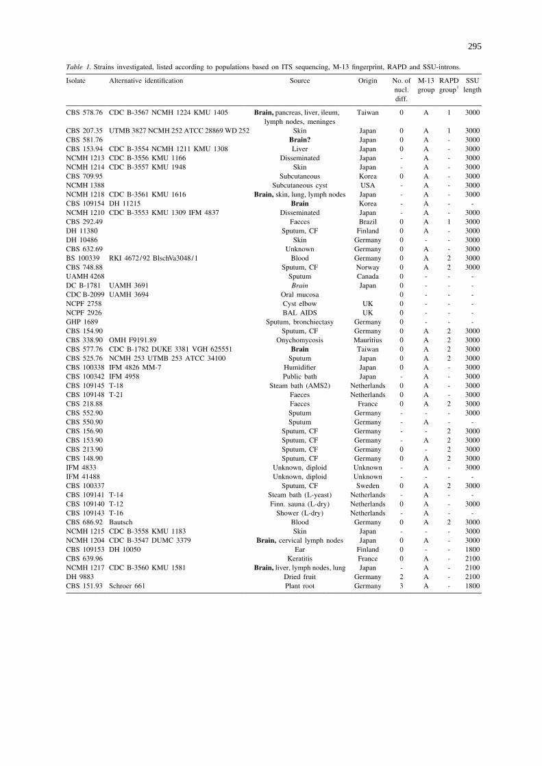
Table 1. Strains investigated, listed according to populations based on ITS sequencing, M-13 fingerprint, RAPD and SSU-introns.
Isolate Alternative identification Source Origin No. of M-13 RAPD SSU1nucl. group group length
diff.
CBS 578.76 CDC B-3567 NCMH 1224 KMU 1405 Brain, pancreas, liver, ileum, Taiwan 0 A 1 3000lymph nodes, meninges
CBS 207.35 UTMB 3827 NCMH 252 ATCC 28869 WD 252 Skin Japan 0 A 1 3000CBS 581.76 Brain? Japan 0 A - 3000CBS 153.94 CDC B-3554 NCMH 1211 KMU 1308 Liver Japan 0 A - 3000NCMH 1213 CDC B-3556 KMU 1166 Disseminated Japan - A - 3000NCMH 1214 CDC B-3557 KMU 1948 Skin Japan - A - 3000CBS 709.95 Subcutaneous Korea 0 A - 3000NCMH 1388 Subcutaneous cyst USA - A - 3000NCMH 1218 CDC B-3561 KMU 1616 Brain, skin, lung, lymph nodes Japan - A - 3000CBS 109154 DH 11215 Brain Korea - A - -NCMH 1210 CDC B-3553 KMU 1309 IFM 4837 Disseminated Japan - A - 3000CBS 292.49 Faeces Brazil 0 A 1 3000DH 11380 Sputum, CF Finland 0 A - 3000DH 10486 Skin Germany 0 - - 3000CBS 632.69 Unknown Germany 0 A - 3000BS 100339 RKI 4672/92 BlschVa3048/1 Blood Germany 0 A 2 3000CBS 748.88 Sputum, CF Norway 0 A 2 3000UAMH 4268 Sputum Canada 0 - - -DC B-1781 UAMH 3691 Brain Japan 0 - - -CDC B-2099 UAMH 3694 Oral mucosa 0 - - -NCPF 2758 Cyst elbow UK 0 - - -NCPF 2926 BAL AIDS UK 0 - - -GHP 1689 Sputum, bronchiectasy Germany 0 - - -CBS 154.90 Sputum, CF Germany 0 A 2 3000CBS 338.90 OMH F9191.89 Onychomycosis Mauritius 0 A 2 3000CBS 577.76 CDC B-1782 DUKE 3381 VGH 625551 Brain Taiwan 0 A 2 3000CBS 525.76 NCMH 253 UTMB 253 ATCC 34100 Sputum Japan 0 A 2 3000CBS 100338 IFM 4826 MM-7 Humidifier Japan 0 A - 3000CBS 100342 IFM 4958 Public bath Japan - A - 3000CBS 109145 T-18 Steam bath (AMS2) Netherlands 0 A - 3000CBS 109148 T-21 Faeces Netherlands 0 A - 3000CBS 218.88 Faeces France 0 A 2 3000CBS 552.90 Sputum Germany - - - 3000CBS 550.90 Sputum Germany - A - -CBS 156.90 Sputum, CF Germany - - 2 3000CBS 153.90 Sputum, CF Germany - A 2 3000CBS 213.90 Sputum, CF Germany 0 - 2 3000CBS 148.90 Sputum, CF Germany 0 A 2 3000IFM 4833 Unknown, diploid Unknown - A - 3000IFM 41488 Unknown, diploid Unknown - - - -CBS 100337 Sputum, CF Sweden 0 A 2 3000CBS 109141 T-14 Steam bath (L-yeast) Netherlands - A - -CBS 109140 T-12 Finn. sauna (L-dry) Netherlands 0 A - 3000CBS 109143 T-16 Shower (L-dry) Netherlands - A - -CBS 686.92 Bautsch Blood Germany 0 A 2 3000NCMH 1215 CDC B-3558 KMU 1183 Skin Japan - - - 3000NCMH 1204 CDC B-3547 DUMC 3379 Brain, cervical lymph nodes Japan 0 A - 3000CBS 109153 DH 10050 Ear Finland 0 - - 1800CBS 639.96 Keratitis France 0 A - 2100NCMH 1217 CDC B-3560 KMU 1581 Brain, liver, lymph nodes, lung Japan - A - 2100DH 9883 Dried fruit Germany 2 A - 2100CBS 151.93 Schroer 661 Plant root Germany 3 A - 1800
296
Table 1. (continued)
Isolate Alternative identification Source Origin No. of M-13 RAPD SSU1nucl. group group length
diff.
CBS 424.67 Skin Germany 0 A* - 3000CBS 736.87 Beer Ireland 21121 A* - 2100IFM 45986 Tap water Japan 21121 - - -CBS 109139 T-10 Steam bath (L) Netherlands 21121 - - 2100CBS 109136 T-7 Steam bath (H) Netherlands 3 B - 2100CBS 109149 T-22 Steam bath Slovenia 3 B - 2100CDC B-1778 UAMH 3700 Blood USA 3 - - -NCPF 2924 Sinus UK 321 - - -CBS 109142 T-15 Fruit Netherlands 3 B - -GHP 944 Ceiling Germany 3 - - -CBS 109144 T-17 Steam bath (AMS1) Netherlands 3 - - 1800CBS 109138 T-9 Hall sauna (H) Netherlands 3 B - -CBS 109134 T-1 Steam bath (H) Netherlands 3 B - -DH 11823 T-5 Steam bath (H) Netherlands - B - -CBS 549.90 Sputum Germany - - - -CBS 971.87 Unknown Iraq - B 5 1800CBS 100341 RKI 4669/92 BlschVa300/8 Blood Germany 3 B 6 1800CBS 100340 IFM 4848 INPA 109 Liver of bat Brazil 3 B - 1800CBS 106.92 DBVPG 4256 Grape Italy 3 B 7 1800CBS 149.90 Sputum, CF Germany 1 B 4 1800CDC B-4584 Pinus Unknown 1 - - -IFM 41823 CBS 109152 Cactus Venezuela 21 C - 1800CBS 150.90 Sputum Netherlands 30 D 3 1800
1- 5 not determined; Data taken from Uijthof et al. (1998). Numbers of ITS nucleotide differences are compared with CBS 207.35 (T) and arelimited to phylogenetically informative sites which are identical in each group. 1 and 2 are additional differences or deletions. Ams1 and 2 5
two sauna complexes in Amsterdam; H 5 Hilversum; L 5 Laren.
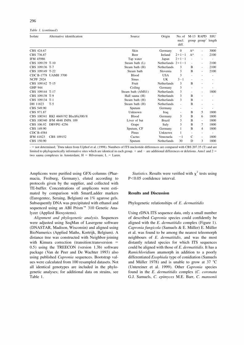
2Amplicons were purified using GFX-columns (Phar- Statistics. Results were verified with x tests usingmacia, Freiburg, Germany), eluted according to P,0.05 confidence interval.protocols given by the supplier, and collected withTE-buffer. Concentrations of amplicons were esti-mated by comparison with SmartLadder markers Results and Discussion(Eurogentec, Seraing, Belgium) on 1% agarose gels.Subsequently DNA was precipitated with ethanol and Phylogenetic relationships of E. dermatitidissequenced using an ABI Prism� 310 Genetic Ana-lyzer (Applied Biosystems). Using rDNA ITS sequence data, only a small number
Alignment and phylogenetic analysis. Sequences of described Capronia species could confidently bewere adjusted using SeqMan of Lasergene software aligned with the E. dermatitidis complex (Figure 1).
¨ ¨(DNASTAR, Madison, Wisconsin) and aligned using Capronia fungicola (Samuels & E. Muller) E. MullerBioNumerics (Applied Maths, Kortrijk, Belgium). A et al. was found to be among the nearest teleomorphdistance tree was constructed with Neighbor-joining neighbours of E. dermatitidis, and was the mostwith Kimura correction (transition:transversion 5 distantly related species for which ITS sequences0.5) using the TREECON (version 1.3b) software could be aligned with those of E. dermatitidis. It has apackage (Van de Peer and De Wachter 1993) also Ramichloridium anamorph in addition to a poorlyusing published Capronia sequences. Bootstrap val- differentiated Exophiala type of conidiation (Samuels
¨ues were calculated from 100 resampled datasets. Not and Muller 1978) and is unable to grow at 37 8Call identical genotypes are included in the phylo- (Untereiner et al. 1999). Other Capronia speciesgenetic analyses; for additional data on strains, see found in the E. dermatitidis complex (C. coronataTable 1. G.J. Samuels, C. epimyces M.E. Barr, C. mansonii
297
Figure 1. Distance tree of the Exophiala dermatitidis complex based on confidently aligned ITS rDNA sequences, constructed with Neighborjoining with Kimura correction, taking Capronia epimyces as outgroup. Bootstrap values .90 of 100 resampled datasets are shown.Summarized ecological source of each strain is listed in brackets. Populations with numbers of ITS changes are listed as A-D. Strains withconflicting fingerprint data are mentioned in italics. The right panel shows the M-13 fingerprint data of the E. dermatitidis strains, with listingof populations in italics. Strains with conflicting ITS sequencing data are mentioned left of the vertical bars.Abbreviations of names: C 5
Capronia, E 5 Exophiala, S 5 Sarcinomyces
¨(Schol-Schwarz) E. Muller et al. and C. munkii Un- phaeomuriformis was considerable (26 bases; Uijthoftereiner) are all relatively close to Sarcinomyces et al. (1998)), but nevertheless the two species couldphaeomuriformis Matsumoto et al. The anamorph of be confidently aligned over the entire spacer domain.C. epimyces is an Exophiala species (Untereiner In the two ex-type strains, the SSU rDNA genes,1995) and has a phialidic synanamorph (Untereiner including the introns, were identical (Haase et al.and Naveau 1999). Capronia coronata, C. mansonii 1999). Some physiological features considered to beand C. munkii have only Exophiala anamorphs. Cap- diagnostic for E. dermatitidis, such as growth withronia epimyces and C. munkii are the only Capronia nitrate, nitrite, creatine and creatinine, are also charac-species able to grow at 37 8C (Untereiner et al. 1999). teristic of S. phaeomuriformis (Uijthof et al. 1998).
The distance between E. dermatitidis and S. The latter species was described originally (Mat-
298
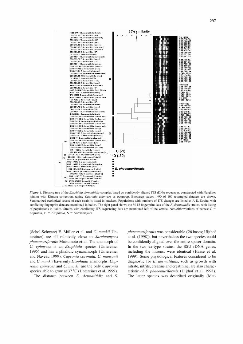
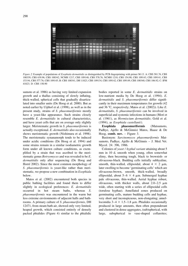
Figure 2. Example of populations of Exophiala dermatitidis as distinguished by PCR-fingerprinting with primer M-13. A: CBS 581.76, CBS100339, CBS 639.96, CBS 100342, NCMH 1217, CBS 109148, CBS 578.76, NCMH 1210, CBS 154.90, CBS 109143, CBS 109141, CBS153.94, CBS 577.76, CBS 109145; B: CBS 100341, DH 11823, CBS 109134, CBS 109142, CBS 109149, CBS 100340, CBS 106.92; C: IFM41823; D: CBS 150.90
sumoto et al. 1986) as having very limited expansion bodies reported in some E. dermatitidis strains ongrowth and a thallus consisting of slowly inflating, low-nutrient media by De Hoog et al. (1994). E.thick-walled, spherical cells that gradually disarticu- dermatitidis and S. phaeomuriformis differ signifi-lated into smaller units (De Hoog et al. 2000). But as cantly in their maximum temperatures for growth (42noted earlier by Uijthof et al. (1998), as well as in the and 36 8C, respectively; Matos et al. (2002)). Like E.present study, strains of S. phaeomuriformis mostly dermatitidis, S. phaeomuriformis can be involved inhave a yeast-like appearance. Such strains closely superficial and systemic infections in humans (Mori etresemble E. dermatitidis in cultural characteristics, al. (1961), as Hormiscium dermatitidis; Gold et al.and have yeast cells that are on average only slightly (1994), as Exophiala castellanii).larger. Meristematic growth in S. phaeomuriformis is Exophiala phaeomuriformis (Matsumoto,actually exceptional. E. dermatitidis also occasionally Padhye, Ajello & McGinnis) Matos, Haase & Deshows meristematic growth (Nishimura et al. 1998). Hoog, comb. nov. – Figure 3.The meristematic synanamorph tends to be induced Basionym: Sarcinomyces phaeomuriformis Mat-under acidic conditions (De Hoog et al. 1994) and sumoto, Padhye, Ajello & McGinnis – J. Med. Vet.some strains remain in a similar isodiametric growth Mycol. 24: 396, 1986.form under all known culture conditions, as exem- Colonies of yeast / hyphal variant attaining about 5plified by a strain that was ascribed to the meri- mm in 10 d, smooth when young, often somewhatstematic genus Botryomyces and was revealed to be E. slimy, then becoming tough, black to brownish- ordermatitidis only after sequencing (De Hoog and olivaceous-black. Budding cells initially subhyaline,
´Horre 2002). Since the most common morphology of smooth, thin-walled, ellipsoidal, about 4 3 2 mm,S. phaeomuriformis is yeast-like rather than meri- later swelling to become ‘germinating cells’ which arestematic, we propose a new combination in Exophiala olivaceous-brown, smooth, thick-walled, broadlybelow. ellipsoidal, about 5–6 3 4 mm. Submerged hyphae
Matos et al. (2002) encountered both species in pale olivaceous, thin-walled. Aerial hyphae robust,public bathing facilities and found them to differ olivaceous, with thicker walls, about 2.0–2.5 mmslightly in ecological preferences: E. dermatitidis wide, often starting with a series of ellipsoidal cellsoccurred in hot steam baths, whereas S. (torulose hyphae). Annellated zones produced onphaeomuriformis was encountered in the warm but germinating cells, mature budding cells and hyphae,less extreme environments of adjacent halls and bath- very short and inconspicuous, non-elongating; annel-rooms. A primary culture of S. phaeomuriformis, DH loconidia 3–4 3 1.5–1.8 mm. Phialides occasionally12471, from steam bath air, showed only very limited, produced in large amounts, then often preponderantheaped growth, which consisted entirely of densely and clustered in dense aggregates, (sub)spherical, withpacked phialides (Figure 4) similar to the phialidic large, subspherical to vase-shaped collarettes;
299
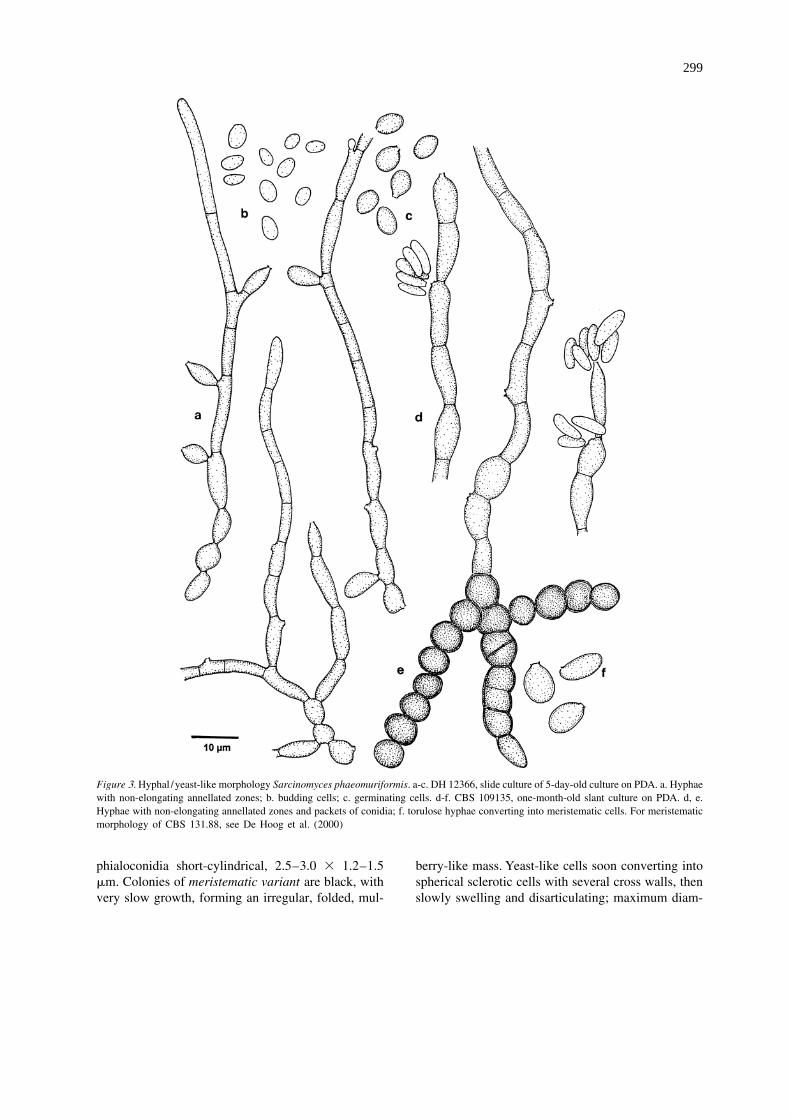
Figure 3. Hyphal /yeast-like morphology Sarcinomyces phaeomuriformis. a-c. DH 12366, slide culture of 5-day-old culture on PDA. a. Hyphaewith non-elongating annellated zones; b. budding cells; c. germinating cells. d-f. CBS 109135, one-month-old slant culture on PDA. d, e.Hyphae with non-elongating annellated zones and packets of conidia; f. torulose hyphae converting into meristematic cells. For meristematicmorphology of CBS 131.88, see De Hoog et al. (2000)
phialoconidia short-cylindrical, 2.5–3.0 3 1.2–1.5 berry-like mass. Yeast-like cells soon converting intomm. Colonies of meristematic variant are black, with spherical sclerotic cells with several cross walls, thenvery slow growth, forming an irregular, folded, mul- slowly swelling and disarticulating; maximum diam-
300
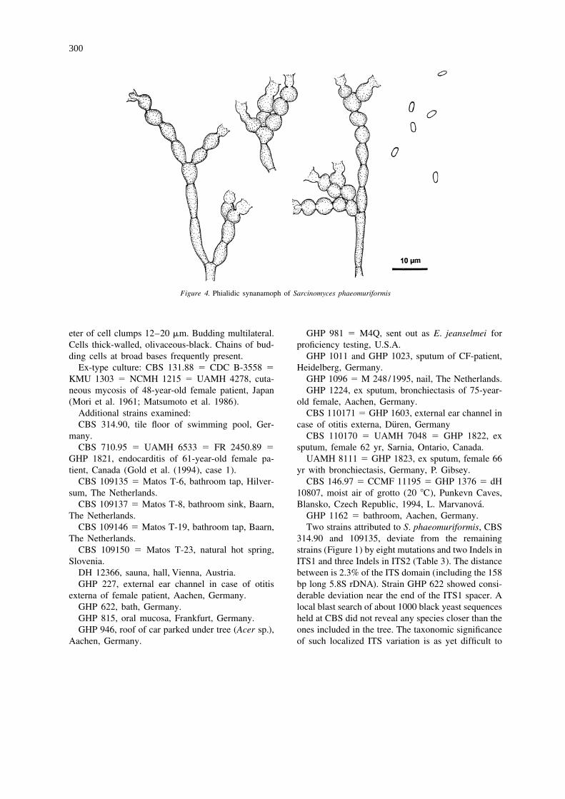
Figure 4. Phialidic synanamoph of Sarcinomyces phaeomuriformis
eter of cell clumps 12–20 mm. Budding multilateral. GHP 981 5 M4Q, sent out as E. jeanselmei forCells thick-walled, olivaceous-black. Chains of bud- proficiency testing, U.S.A.ding cells at broad bases frequently present. GHP 1011 and GHP 1023, sputum of CF-patient,
Ex-type culture: CBS 131.88 5 CDC B-3558 5 Heidelberg, Germany.KMU 1303 5 NCMH 1215 5 UAMH 4278, cuta- GHP 1096 5 M 248/1995, nail, The Netherlands.neous mycosis of 48-year-old female patient, Japan GHP 1224, ex sputum, bronchiectasis of 75-year-(Mori et al. 1961; Matsumoto et al. 1986). old female, Aachen, Germany.
Additional strains examined: CBS 110171 5 GHP 1603, external ear channel in¨CBS 314.90, tile floor of swimming pool, Ger- case of otitis externa, Duren, Germany
GHP 1821, endocarditis of 61-year-old female pa- UAMH 8111 5 GHP 1823, ex sputum, female 66tient, Canada (Gold et al. (1994), case 1). yr with bronchiectasis, Germany, P. Gibsey.
CBS 109135 5 Matos T-6, bathroom tap, Hilver- CBS 146.97 5 CCMF 11195 5 GHP 1376 5 dHsum, The Netherlands. 10807, moist air of grotto (20 8C), Punkevn Caves,
CBS 109146 5 Matos T-19, bathroom tap, Baarn, Two strains attributed to S. phaeomuriformis, CBSThe Netherlands. 314.90 and 109135, deviate from the remaining
CBS 109150 5 Matos T-23, natural hot spring, strains (Figure 1) by eight mutations and two Indels inSlovenia. ITS1 and three Indels in ITS2 (Table 3). The distance
DH 12366, sauna, hall, Vienna, Austria. between is 2.3% of the ITS domain (including the 158GHP 227, external ear channel in case of otitis bp long 5.8S rDNA). Strain GHP 622 showed consi-
externa of female patient, Aachen, Germany. derable deviation near the end of the ITS1 spacer. AGHP 622, bath, Germany. local blast search of about 1000 black yeast sequencesGHP 815, oral mucosa, Frankfurt, Germany. held at CBS did not reveal any species closer than theGHP 946, roof of car parked under tree (Acer sp.), ones included in the tree. The taxonomic significance
Aachen, Germany. of such localized ITS variation is as yet difficult to
301
evaluate. A similar phenomenon was noted in Au- to differences in M-13 banding profiles. Attribution ofreobasidium pullulans, where ITS2 was found to be strains to groups A or B is mostly possible by eitheralmost six times more variable than ITS1 (Yurlova et fingerprinting or by ITS sequencing. In this way weal. 1999). were able to categorize nearly all 75 strains listed in
Table 1, even though not all methods were applied toInfraspecific typing of E. dermatitidis strains all strains. The patterns seem to be stable, as can be
concluded from the fact that recent sauna strains fromThe present study included all E. dermatitidis strains The Netherlands, as well as a strain, CBS 100340,from cerebral cases that are known to be preserved. A isolated 25 years previously from bat liver in Brazil
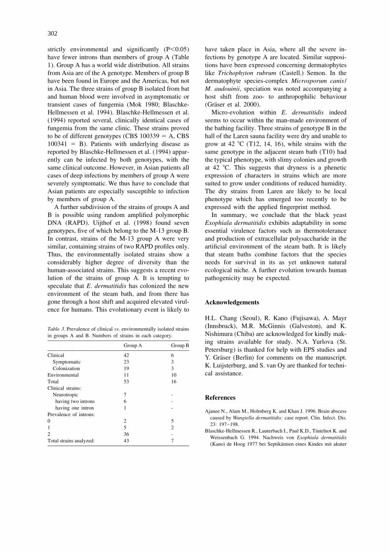
˜further strain published as Phialophora dermatitidis (Mok and Luizao 1981), were identical. Uijthof et al.by Jotisankasa et al. (1970) from brain in Japan, CBS (1994) found identical RAPD patterns in strains iso-579.76, was found to have an ITS sequence identical lated at time intervals of 15 to 50 yr (RAPD group 1).to that of Exophiala sp., CBS 725.88, reported by Nearly all members of group A had SSU ampliconTintelnot et al. (1991) from a fatal cerebral infection lengths of approximately 3000 bp, corresponding within a patient from Germany. This taxon will be de- the presence of two introns. Three strains, CBSscribed as a new species elsewhere (G.S. de Hoog, 639.96, NCMH 1217 and DH 9883, had only a singleunpublished data). Based on fingerprinting data, near- intron (2100 bp; Table 1). CBS 109153 was the onlyly all 75 strains definitely assigned to E. dermatitidis strain of group A without an intron. Most members ofcould be attributed to one of two main groups A and B group B lacked introns, some having a single intron(Table 1). The profiles closely resembled each other and none was observed to have two introns (Table 2).(Figure 2). Only three strains, CBS 151.93, IFM Thus it seems that groups A and B, though closely41823 and CBS 150.90, were clearly different, having interrelated, are well-supported.only few bands in common. These strains also de- Group A contained 43 strains (Table 3) originatingviated in their ITS sequences: CBS 150.90 had a from humans. Twenty-three of these strains weredeletion of 26 bp, IFM 41823 lacked a base on from symptomatic individuals and 19 were apparentlyposition 184 and CBS 151.93 had 3 substitutions. The harmless colonizers of lungs and intestinal tract.latter mutations were shared by all members of group Group A contained significantly more clinical strainsB (Figure 1). Remarkably, CBS 151.93 (ITS group B) than group B (P,0.05). All strains from cerebral,had an M-13 A-profile. Profile A was also found in liver and disseminated cases clustered in group A.CBS 109139 and DH 9883, which deviated by three Twenty-eight of the human-derived strains had twomutations. The incongruences show that members of introns (SSU approx. 3000 bp; Table 1), while threegroups A and B are very closely related. Some addi- had one intron or none. Conversely, of the 9 en-tional single nucleotide polymorphisms (SNPs) were vironmentally isolated strains of group A, four hadoccasionally noted in ITS. They were not connected one or no intron. In contrast, 10 strains of group B are
Table 2. Infraspecific variability in ITS1-2 domains of S. phaeomuriformis and E. dermatitidis.
Sarc* A T T T T A T C A - A - ASarc(T) C C C C G G C T T T - A -Exophiala dermatitidisITS1 (210) ITS2 (220)
62 123 162 184 196 16 198Ed(T) A T T - A - A1 A T T - A C A2 G C T - A - A21121 G C T - A C -3 A T C T C - A
ITS1 and 2 domains given with approximate total lengths in brackets. Sarc(T) 5 Sarcinomyces phaeomuriformis, group containing the ex-typestrain; Sarc* 5 Sarcinomyces cf. phaeomuriformis, deviating strains. Ed(T) 5 Exophiala dermatitidis, type strain
302
strictly environmental and significantly (P,0.05) have taken place in Asia, where all the severe in-have fewer introns than members of group A (Table fections by genotype A are located. Similar supposi-1). Group A has a world wide distribution. All strains tions have been expressed concerning dermatophytesfrom Asia are of the A genotype. Members of group B like Trichophyton rubrum (Castell.) Semon. In thehave been found in Europe and the Americas, but not dermatophyte species-complex Microsporum canis /in Asia. The three strains of group B isolated from bat M. audouinii, speciation was noted accompanying aand human blood were involved in asymptomatic or host shift from zoo- to anthropophilic behaviour
¨transient cases of fungemia (Mok 1980; Blaschke- (Graser et al. 2000).Hellmessen et al. 1994). Blaschke-Hellmessen et al. Micro-evolution within E. dermatitidis indeed(1994) reported several, clinically identical cases of seems to occur within the man-made environment offungemia from the same clinic. These strains proved the bathing facility. Three strains of genotype B in theto be of different genotypes (CBS 100339 5 A, CBS hall of the Laren sauna facility were dry and unable to100341 5 B). Patients with underlying disease as grow at 42 8C (T12, 14, 16), while strains with thereported by Blaschke-Hellmessen et al. (1994) appar- same genotype in the adjacent steam bath (T10) hadently can be infected by both genotypes, with the the typical phenotype, with slimy colonies and growthsame clinical outcome. However, in Asian patients all at 42 8C. This suggests that dryness is a pheneticcases of deep infections by members of group A were expression of characters in strains which are moreseverely symptomatic. We thus have to conclude that suited to grow under conditions of reduced humidity.Asian patients are especially susceptible to infection The dry strains from Laren are likely to be localby members of group A. phenotype which has emerged too recently to be
A further subdivision of the strains of groups A and expressed with the applied fingerprint method.B is possible using random amplified polymorphic In summary, we conclude that the black yeastDNA (RAPD). Uijthof et al. (1998) found seven Exophiala dermatitidis exhibits adaptability in somegenotypes, five of which belong to the M-13 group B. essential virulence factors such as thermotoleranceIn contrast, strains of the M-13 group A were very and production of extracellular polysaccharide in thesimilar, containing strains of two RAPD profiles only. artificial environment of the steam bath. It is likelyThus, the environmentally isolated strains show a that steam baths combine factors that the speciesconsiderably higher degree of diversity than the needs for survival in its as yet unknown naturalhuman-associated strains. This suggests a recent evo- ecological niche. A further evolution towards humanlution of the strains of group A. It is tempting to pathogenicity may be expected.speculate that E. dermatitidis has colonized the newenvironment of the steam bath, and from there hasgone through a host shift and acquired elevated virul- Acknowledgementsence for humans. This evolutionary event is likely to
H.L. Chang (Seoul), R. Kano (Fujisawa), A. Mayr(Innsbruck), M.R. McGinnis (Galveston), and K.Table 3. Prevalence of clinical vs. environmentally isolated strainsNishimura (Chiba) are acknowledged for kindly mak-in groups A and B. Numbers of strains in each category.ing strains available for study. N.A. Yurlova (St.
Group A Group BPetersburg) is thanked for help with EPS studies and
Clinical 42 6 ¨Y. Graser (Berlin) for comments on the manuscript.Symptomatic 23 3
K. Luijsterburg, and S. van Oy are thanked for techni-Colonization 19 3cal assistance.Environmental 11 10
Total 53 16Clinical strains:
Neurotropic 7 - Referenceshaving two introns 6 -having one intron 1 -
Ajanee N., Alam M., Holmberg K. and Khan J. 1996. Brain abscessPrevalence of introns:
caused by Wangiella dermatitidis: case report. Clin. Infect. Dis.0 2 5
23: 197–198.1 5 2
Blaschke-Hellmessen R., Lauterbach I., Paul K.D., Tintelnot K. and2 36 -
Weissenbach G. 1994. Nachweis von Exophiala dermatitidisTotal strains analyzed: 43 7
¨(Kano) de Hoog 1977 bei Septikamien eines Kindes mit akuter
303
¨lymphatischer Leukamie und bei Patienten mit Mukoviszidose. Matsumoto T., Matsuda T., McGinnis M.R. and Ajello L. 1993.Mycoses 37 (Suppl. 1): 89–96. Clinical and mycological spectra of Wangiella dermatitidis in-
Chang H.L., Kim D.S., Park D.J., Kim H.J., Lee C.H. and Shin H.J. fections. Mycoses 36: 145–155.2000. Acute cerebral phaeohyphomycosis due to Wangiella Matsumoto T., Padhye A.A., Ajello L. and McGinnis M.R. 1986.dermatitidis accompanied by cerebrospinal eosinophilia. J. Clin. Sarcinomyces phaeomuriformis: a new dematiaceous hy-Microbiol. 38: 1965–1966. phomycete. J. Med. Vet. Mycol. 24: 395–400.
Crosby J.H., O’Quinn M.H., Steele J.C.H. and Rao R.N. 1989. Mok W.Y. 1980. Ecology of Wangiella dermatitidis in the AmazonFine-needle aspiration of subcutaneous phaeohyphomycosis basin. Pan Am. Health Org. Sci. Publ. 396: 269–275.
˜caused by Wangiella dermatitidis. Diagn. Cytopath. 5: 293–297. Mok W.Y. and Luizao R.C.C. 1981. Serological analysis andDe Hoog G.S. and Gerrits van den Ende A.H.G. 1998. Molecular pathogenic potentials of Wangiella dermatitidis isolated from
diagnostics of clinical strains of filamentous basidiomycetes. bats. Mycopathologia 73: 93–99.Mycoses 41: 183–189. Mori A., Morikawa H. and Akagi M. 1961. A case of chromoblas-
´De Hoog G.S., Guarro J., Gene J. and Figueras M.J. 2000. Atlas of tomycosis due to Hormiscium dermatitidis. Skin Res., Osaka 3:Clinical Fungi. 2nd edn. Centraalbureau voor Schimmelcultures 158./ Universitat Rovira i Virgili, Utrecht /Reus, 1126 pp. Nishimura K. and Miyaji M. 1983. Defense mechanisms of mice
´De Hoog G.S. and Horre R. 2002. Molecular taxonomy of the against Exophiala dermatitidis infection. Mycopathologia 81:Alternaria and Ulocladium species from humans and their 9–21.identification in the routine laboratory. Mycoses 45: 259–276. Nishimura K., Miyaji M., Takeo K., Mikami Y., Kamei K.,
¨De Hoog G.S., Takeo K., Yoshida S., Gottlich E., Nishimura K. and Yokoyama K. et al. 1998. IFM List of Pathogenic Fungi andMiyaji M. 1994. Pleoanamorphic life cycle of Exophiala ( Wan- Actinomycetes with Photomicrographs. 2nd edn. Chiba Uni.,giella) dermatitidis. Antonie van Leeuwenhoek 65: 143–153. Chiba, 305 pp.
¨Dixon D.M., Migliozzi J., Cooper C.R., Solis O., Breslin B. and Samuels G.J. and Muller E. 1978. Life-histories of BrazilianSzaniszlo P.J. 1992. Melanized and non-melanized multicellular ascomycetes 3. Sydowia 31: 142–156.form mutants of Wangiella dermatitidis in mice: mortality and Tintelnot K., De Hoog G.S., Thomas E., Steudel W.-I.., Huebner K.histopathology studies. Mycoses 35: 17–21. and Seeliger H.P.R. 1991. Cerebral phaeohyphomycosis caused
Gold W.L., Vellend H., Salit I.E., Campbell I., Summerbell R., by an Exophiala species. Mycoses 34: 239–244.Rinaldi M. et al. 1994. Successful treatment of systemic and Uijthof J.M.J., De Hoog G.S., De Cock A.W.A.M., Takeo K. andlocal infections due to Exophiala species. Clin. Infect. Dis. 19: Nishimura K. 1994. PCR-based evaluation of pathology of339–341. strains of the black yeast Exophiala ( Wangiella) dermatitidis.¨Graser Y., Kuijpers A.F.A., Presber W. and De Hoog G.S. 1999b. Mycoses 37: 235–242.Molecular taxonomy of Trichophyton mentagrophytesand T. Uijthof J.M.J., Van Belkum A., De Hoog G.S. and Haase G. 1998.tonsurans. Med. Mycol. 37: 315–330. Exophiala dermatitidis and Sarcinomyces phaeomuriformis: IT-¨Graser Y., Kuijpers A.F.A., El Fari M., Presber W. and De Hoog S-sequencing and nutritional physiology. Med. Mycol. 36: 143–G.S. 2000. Molecular and conventional taxonomy of the Mi- 151.crosporum canis complex. Med. Mycol. 38: 143–153. Untereiner W.A. 1994. A simple method for the in vitro production
Haase G., Skopnik H., Groten T., Kusenbach G. and Posselt H.-G. of pseudothecia in species of Capronia. Mycologia 86: 290–295.1991. Long-term fungal cultures from patients with cystic fi- Untereiner W.A. 1995. Fruiting studies in species of Caproniabrosis. Mycoses 34: 373–376. (Herpotrichiellaceae). Antonie van Leeuwenhoek 68: 3–17.
Haase G., Sonntag L., Melzer-Krick B. and De Hoog G.S. 1999. Untereiner W.A., Gerrits van den Ende A.H.G. and De Hoog G.S.Phylogenetic inference by SSU-gene analysis of members of the 1999. Nutritional physiology of species of Capronia. Stud.Herpotrichiellaceae with special reference to human pathogenic Mycol. 43: 98–106.species. Stud. Mycol. 43: 80–97. Untereiner W.A. and Naveau F.A. 1999. Molecular systematics of
Hiruma M., Kawada A., Ohata H., Ohnishi Y., Takahashi H., the Herpotrichiellaceae with an assessment of the phylogeneticYamazaki M. et al. 1993. Systemic phaeohyphomycosis caused positions of Exophiala dermatitidis and Phialophora americana.by Exophiala dermatitidis. Mycoses 36: 1–7. Mycologia 91: 67–83.
´Horre R. and De Hoog G.S. 1999. Primary cerebral infections by Van de Peer Y. and De Wachter R. 1993. TREECON: a softwaremelanized fungi: a review. Stud. Mycol. 43: 176–193. package for the construction and drawing of evolutionary trees.
Jotisankasa V., Nielsen H.S. and Conant N.F. 1970. Phialophora Comput. Appl. Biosci. 9: 177–182.¨dermatitidis, its morphology and biology. Sabouraudia 8: 98– Weissbrodt H., Tummler B. and Bautsch W. 1994. Exophiala
107. dermatitidis in cystic fibrosis (CF-) and non-CF patients Proc.Kabel P.J., Illy K.E., Holl R.A., Buiting A.G.M. and Wintermans Deutsch. Ges. Hyg. Mikrobiol., Kiel P006., pp. 24–25.
R.G.F. 1994. Nosocomial intravascular infection with Exophiala White T.J., Bruns T., Lee S. and Taylor J.W. 1990. Amplificationdermatitidis. Lancet 344: 1167–1168. and direct sequencing of fungal ribosomal RNA genes for
Kenney R.T., Kwon-Chung K.J., Waytes A.T., Melnick D.A., Pass phylogenetics. In: Innis M.A. (ed.), PCR Protocols. Academic,H.I., Merino M.J. et al. 1992. Successful treatment of systemic San Diego, pp. 315–322.Exophiala dermatitidis infection in a patient with chronic Yurlova N.A., De Hoog G.S. and Gerrits van den Ende A.H.G.granulomatous disease. Clin. Infect. Dis. 14: 235–242. 1999. Taxonomy of Aureobasidium and allied genera. Stud.
Matos T., De Hoog G.S., De Boer A.G., De Crom I. and Haase G. Mycol. 43: 63–69.2002. High prevalence of the neurotropic black yeast Exophiala( Wangiella) dermatitidis in steam baths. Mycoses (in press).