of November 27, 2018. This information is current as Complement and Coagulation Systems Molecular Intercommunication between the Huber-Lang Nilsson, Florian Gebhard, John D. Lambris and Markus Klos, Hui Chen, Barbara Acker, Uwe B. Brückner, Bo Umme Amara, Michael A. Flierl, Daniel Rittirsch, Andreas ol.0903678 http://www.jimmunol.org/content/early/2010/09/24/jimmun published online 24 September 2010 J Immunol average * 4 weeks from acceptance to publication Fast Publication! • Every submission reviewed by practicing scientists No Triage! • from submission to initial decision Rapid Reviews! 30 days* • Submit online. ? The JI Why Subscription http://jimmunol.org/subscription is online at: The Journal of Immunology Information about subscribing to Permissions http://www.aai.org/About/Publications/JI/copyright.html Submit copyright permission requests at: Email Alerts http://jimmunol.org/alerts Receive free email-alerts when new articles cite this article. Sign up at: Print ISSN: 0022-1767 Online ISSN: 1550-6606. All rights reserved. 1451 Rockville Pike, Suite 650, Rockville, MD 20852 The American Association of Immunologists, Inc., is published twice each month by The Journal of Immunology by guest on November 27, 2018 http://www.jimmunol.org/ Downloaded from by guest on November 27, 2018 http://www.jimmunol.org/ Downloaded from
Transcript
of November 27, 2018.This information is current as
Complement and Coagulation SystemsMolecular Intercommunication between the
Huber-LangNilsson, Florian Gebhard, John D. Lambris and MarkusKlos, Hui Chen, Barbara Acker, Uwe B. Brückner, Bo Umme Amara, Michael A. Flierl, Daniel Rittirsch, Andreas
Email Alertshttp://jimmunol.org/alertsReceive free email-alerts when new articles cite this article. Sign up at:
Print ISSN: 0022-1767 Online ISSN: 1550-6606. All rights reserved.1451 Rockville Pike, Suite 650, Rockville, MD 20852The American Association of Immunologists, Inc.,
is published twice each month byThe Journal of Immunology
Molecular Intercommunication between the Complementand Coagulation Systems
Umme Amara,* Michael A. Flierl,† Daniel Rittirsch,* Andreas Klos,‡ Hui Chen,x
Barbara Acker,* Uwe B. Bruckner,* Bo Nilsson,{ Florian Gebhard,* John D. Lambris,x and
Markus Huber-Lang*
The complement system as well as the coagulation system has fundamental clinical implications in the context of life-threatening
tissue injury and inflammation. Associations between both cascades have been proposed, but the precise molecular mechanisms
remain unknown. The current study reports multiple links for various factors of the coagulation and fibrinolysis cascades with the
central complement components C3 and C5 in vitro and ex vivo. Thrombin, human coagulation factors (F) XIa, Xa, and IXa, and
plasmin were all found to effectively cleave C3 and C5. Mass spectrometric analyses identified the cleavage products as C3a and
C5a, displaying identical molecular weights as the native anaphylatoxins C3a and C5a. Cleavage products also exhibited robust
chemoattraction of human mast cells and neutrophils, respectively. Enzymatic activity for C3 cleavage by the investigated clotting
and fibrinolysis factors is defined in the following order: FXa > plasmin > thrombin > FIXa > FXIa > control. Furthermore, FXa-
induced cleavage of C3 was significantly suppressed in the presence of the selective FXa inhibitors fondaparinux and enoxaparin
in a concentration-dependent manner. Addition of FXa to human serum or plasma activated complement ex vivo, represented by
the generation of C3a, C5a, and the terminal complement complex, and decreased complement hemolytic serum activity that
defines exact serum concentration that results in complement-mediated lysis of 50% of sensitized sheep erythrocytes. Further-
more, in plasma from patients with multiple injuries (n = 12), a very early appearance and correlation of coagulation (thrombin–
antithrombin complexes) and the complement activation product C5a was found. The present data suggest that coagulation/
fibrinolysis proteases may act as natural C3 and C5 convertases, generating biologically active anaphylatoxins, linking both
cascades via multiple direct interactions in terms of a complex serine protease system. The Journal of Immunology, 2010,
185: 000–000.
Traditionally, the complement and coagulation systems aredescribed as separate cascades. As descendants of acommon ancestral pathway, both proteolytic cascades are
composed of serine proteases with common structural character-istics, such as highly conserved catalytic sites of serine, histidine,and aspartate (1, 2). Furthermore, both systems belong to a com-plex inflammatory network (3) and exhibit some similar charac-
teristics regarding the specialized functions of their activators andinhibitors. In particular, the clotting factor [human coagulationfactor (F)] XIIa can activate the complement factor C1r andthereby initiate the classical pathway of complement activation. Inturn, the C1 esterase inhibitor suppresses not only all threeestablished complement pathways (classical, lectin, and alterna-tive), but also the intrinsic coagulation cascade (kallikrein, FXIIa)(4, 5). Recently, it has been shown that thrombin is capable ofgenerating the complement activation product C5a in the absenceof C3 (6). In another study, Clark et al. (7) suggested that thrombinand plasmin may contribute to nontraditional complement acti-vation during liver regeneration even in the absence of C4 andduring inhibition of factor B. Thrombin may also act as a physi-ological agonist of the protein kinase C-dependent regulation ofthe complement decay-accelerating factor and thereby may pro-vide a negative-feedback loop helping to prevent thrombosisduring inflammation (8). In the setting of systemic inflammation,activation of the coagulation cascade is accompanied by a pro-found activation of the complement system, resulting in the gen-eration of the anaphylatoxins C3a and C5a (9, 10). According toa previous report, C5a induces tissue factor (TF) activity in humanendothelial cells (11) and may therefore be involved in the acti-vation of the extrinsic coagulation pathway. Furthermore, C5ahas been shown to stimulate the expression of TF on neutrophilsvia the C5aR, which was associated with a higher procoagulantactivity (12). Additional evidence of procoagulant effects bycomplement components has been provided by a recent reportdemonstrating in vitro that mannan-binding lectin-associated ser-ine protease 2 of the lectin pathway was capable of promotingfibrinogen turnover by cleaving prothrombin into thrombin (13).
*Department of Traumatology, Hand-, Plastic- and Reconstructive Surgery, Univer-sity Hospital of Ulm, Ulm; ‡Department of Medical Microbiology and HospitalEpidemiology, Hannover Medical School, Hannover, Germany; †Department of Or-thopaedic Surgery, Denver Health Medical Center, University of Colorado School ofMedicine, Denver, CO 80204 xDepartment of Pathology and Laboratory Medicine,University of Pennsylvania Medical School, Philadelphia, PA 19104; and {Divisionof Clinical Immunology, Rudbeck Laboratory, Uppsala University, Uppsala, Sweden
Received for publication November 16, 2009. Accepted for publication July 29,2010.
This work was supported by the I. European Shock Society Award Grant 2005, by theDeutsche Forschungsgemeinschaft HU 823/2, and partially by National Institutes ofHealth Grants GM062134, AI30040, and AI068730 as well as by Deutsche For-schungsgemeinschaft KFO 200 and HU 823/3-1.
Address correspondence and reprint requests to Dr. Markus Huber-Lang, Profes-sor of Clinical- and Experimental Trauma-Immunology, Head of Deutsche For-schungsgemeinschaft Emmy Noether Research Group, Department of Traumatology,Hand-, Plastic- and Reconstructive Surgery, University of Ulm Medical School,Steinhoevelstraße 9, 89075 Ulm, Germany. E-mail address: [email protected]
Abbreviations used in this paper: aPC, activated protein C; CH50, complementhemolytic serum activity that defines exact serum concentration that results incomplement-mediated lysis of 50% of sensitized sheep erythrocytes; DPBS, Dulbec-co’s PBS; F, human coagulation factor; MAC, membrane attack complex; MBL,mannan-binding lectin; MS, mass spectrometry; PK, prekallikrein; TAT, thrombin–antithrombin complex; TCC, terminal complement complex; TF, tissue factor.
Copyright� 2010 by The American Association of Immunologists, Inc. 0022-1767/10/$16.00
www.jimmunol.org/cgi/doi/10.4049/jimmunol.0903678
Published September 24, 2010, doi:10.4049/jimmunol.0903678 by guest on N
In 1986, Sims et al. (14) showed that the terminal complementcomplex (TCC; C5b-9) can catalyze prothrombin cleavage tothrombin even in the absence of FV and thereby specifically in-crease platelet prothrombinase activity. In contrast, C5a has beendescribed as having fibrinolytic effects by upregulating the plas-minogen activator inhibitor I expression in the human mast cellline HMC-1 (15).Thus, it is now becoming clear that both cascades may interact on
a much larger scale than previously anticipated (16, 17). However,many of the underlying molecular mechanisms remain poorly un-derstood. As indicated above, several factors of the coagulation/fibrinolysis cascade and components of the complement system dis-play similar serine protease properties. In the current study, we hy-pothesized that various serine protease components of the clotting/fibrinolytic cascade directly cleave complement proteins, chal-lenging the classic dogma that the two systems are separate cas-cades, and propose a model of a complex serine protease network.
Materials and MethodsReagents
Unless stated otherwise, reagents were purchased from Sigma-Aldrich(Taufkirchen, Germany). Purified human C3 and C5 were obtained fromQuidel (San Diego, CA). Human coagulation factors (F) VII, VIIa, IXa, Xa,Xia, and (activated) protein C were purchased from Calbiochem (Darm-stadt, Germany). rFX, rFXI, and rTF were acquired from R&D Systems(Wiesbaden, Germany). Sodium enoxaparin and sodium fondaparinuxwere obtained from Sanofi-Aventis (Frankfurt, Germany).
In vitro cleavage of C3 and C5
In vitro experiments were performed by incubating native C3 (100 mg/ml)or C5 (100 mg/ml) in Dulbecco’s PBS (DPBS) in the absence or presenceof various coagulation factors [FXa, rFX, FXIa, rFXI, FIXa, FVIIa, FVII,FVIII, TF, protein C, activated protein C, thrombin (FIIa), plasmin andplasminogen] at 37˚C in a dose- and time-dependent manner, followed byELISA and Western blot analyses. Experiments were repeated in the ab-sence or presence of selective FXa inhibitors (sodium enoxaparin andsodium fondaparinux). Human serum and plasma was incubated for 90 minat 37˚C with FXa (ranging from 0–100 mg/ml) and assessed for C3a andC5a production as well as the assembly of TCC using Western blot andELISA analysis.
Western blot analysis for C3a and C5a
Samples and controls were separated by SDS-PAGE under reducing con-ditions and transferred onto a polyvinylidene fluoride membrane (Schleicher& Schuell, Keene, NH). The blots were incubated overnight at 4˚C with1:1000 polyclonal rabbit anti-human C3a IgG (Calbiochem) or 1:1000 rab-bit anti-human C5a IgG (Calbiochem). After washing, membranes wereincubated for 1 h using alkaline phosphatase-conjugated goat anti-rabbitIgG (1:5000; Jackson ImmunoResearch Laboratories, West Grove, PA). Fordevelopment, alkaline phosphatase substrate color development buffer(Bio-Rad, Hercules, CA) was used.
ELISA analysis of C3a, C5a, and TCC (C5b-9)
For quantitative analysis of C3a, C5a, and TCC (C5b-9), commerciallyavailable ELISA kits (Quidel and DRG Diagnostics, Marburg, Germany)were used according to the manufacturers’ instructions.
Chemotaxis assay
HMC-1 (human mast cell line) and neutrophil chemotaxis assays wereperformed as previously described (18). Briefly, HMC-1 cells werefluorescein-labeled with 29, 79-bis (2-carboxyethyl)-5-(and 6)-carboxy-fluorescein acetoxymethyl ester (Molecular Probes, Eugene, OR) anda chemotaxis assay was performed using a 5-mm porosity filter (NeuroP-robe, Gaithersburg, MD). Labeled HMC-1 cells (5 3 106 cells/ml) wereloaded into the upper chamber of a 96-well device (NeuroProbe) with C3in the absence or presence of increasing concentrations of coagulation/fibrinolysis factors in the lower chamber. Glycosylated human C3a (100ng/ml) served as a positive control.
Human neutrophils were isolated from whole blood of healthy humanvolunteers (approved by the Independent Ethics Committee of the Uni-versity of Ulm, Ulm, Germany, No. 44/06, following written informed
consent of all individuals). Whole blood was drawn from the antecubitalvein into syringes containing the anticoagulant citrate dextrose (1:10;Baxter Health Care, Deerfield, IL). Neutrophils were isolated using Ficoll-Paque gradient centrifugation (Pharmacia Biotech, Stockholm, Sweden)followed by a dextran sedimentation step. After hypotonic lysis of residualRBCs, neutrophils were resuspended in HBSS and fluorescein labeled with29, 79-bis (2-carboxyethyl)-5-(and 6)-carboxy-fluorescein acetoxymethylester (Molecular Probes) for 30 min at 37˚C. Labeled neutrophils (5 3 106
cells/ml) were loaded into the upper chamber of a 96-well device (Neuro-Probe) and separated by a polycarbonate filter with a porosity of 3 mm(NeuroProbe). The lower chambers were loaded with recombinant hu-man C5a (100 ng/ml, positive control) or C5 in the absence or presenceof increasing concentrations of coagulation/fibrinolysis factors. Postincu-bation at 37˚C for 30 min, the number of cells that had migrated throughthe polycarbonate membrane was determined by cytofluorometry (Cyto-fluor II, Per Septive Biosystems, Framingham, MA).
Mass spectrometry analysis by MALDI-TOF
Deglycosylation by PNGase F. PNGase F was purchased fromNew EnglandBioLabs (Ipswich, MA). For glycan cleavage, the procedure followed theproduct’s guide. Protein samples were reduced with 5 mM DTT for 45 minat 56˚C. Afterwards, 1 ml enzyme PNGase F was added to the samples, andthe solutions were incubated at 37˚C overnight. After desalting and en-richment by C18 Ziptips (Millipore, Billerica, MA), the deglycosylatedproteins were ready for mass spectrometry (MS) analysis.
MALDI-TOFMS analysis. Protein samples were analyzed by MALDI-TOF.Spectra were acquired on a MALDI-Micro MX (Waters, Milford, MA) inlinear mode. The instrument is equipped with an N2 UV laser emitting at337 nm, a pulsed ion extraction source, an electrostatic reflectron of 2.3 meffective path length, a 2 GHz 8-bit transient analog to digital converterwith real time peak display and fast dual microchannel plate detectors.All sample solutions were mixed in a 1:2 volume ratio with a matrixsolution (10 mg/ml sinapinic acid in 4:6 acetonitrile/0.1% trifluoroaceticacid). Then, 0.8 ml each sample matrix mixture was spotted on the targetplate and allowed to dry under moderate vacuum for 1 min. Thirty single-shot mass spectra were summed to give a composite spectrum. All datawere reprocessed using Waters MassLynx software (Waters). The massscale was calibrated externally using a defined peptide mixture (insulin,cytochrome-c, hemoglobin, myoglobin, and trypsinogen).
Complement hemolytic serum activity
Hemolytic activity of human serum in the absence and presence of FXa(100 mg/ml) was assessed as previously described (19, 20). Briefly, sheeperythrocytes (Oxoid, Wesel, Germany) were sensitized with hemolysin(Colorado Serum Company, Denver, CO) and exposed to dilutions of se-rum samples in TBS (pH 7.35, 37˚C, 60 min). The complement reactionwas stopped by the addition of ice-cold TBS followed by a centrifugationstep (700 3 g, 5 min). Absorption values of the supernatant fluids weredetermined by spectrophotometry at 541 nm. The complement hemolyticserum activity (CH50) defines the exact serum concentration that results incomplement-mediated lysis of 50% of sensitized sheep erythrocytes.
Measurement of thrombin–antithrombin complexes and C5aearly after multiple injury in humans
Twelve patients after multiple injury (10 males, 2 females, median age: 38 y,range: 19–74 y) with a median injury severity score of 48 (range: 25–60) asdefined by the Consensus Criteria were enrolled in the study in accordanceto the Independent Local Ethics Committee of the University of Ulm(approval number 44/06). For all patients, informed consent was obtained.If the patient was incapable of making decisions because of sedation oraltered mental status, informed consent was obtained postrecovery. Bloodwas drawn within 1 h postinjury on admission to the emergency room atthe University Hospital Ulm, and the thrombin–antithrombin complex(TAT) as well as the anaphylatoxin C5a concentrations were determined(see above). Plasma levels of TAT were measured using a sandwichELISA. TAT was captured in wells coated with anti-human thrombin, andHRP-coupled anti-human antithrombin Ab was used for detection (bothAbs from Enzyme Research Laboratories, South Bend, IN). A standardprepared by diluting pooled human serum in normal citrate-phosphate-dextrose plasma was used. The standard was calibrated using TAT com-plexes produced from purified thrombin and antithrombin. Values wereexpressed as milligrams per liter.
Statistical analysis
All values are expressed as mean 6 SD. Data sets of ELISA and che-motaxis assays were analyzed by Kruskal-Wallis ANOVA on ranks;
differences in the mean values among the experimental groups were thencompared using the multiple comparison procedure (Dunn’s method). Forcorrelation analysis, the Pearson coefficient was determined. Results wereconsidered statistically significant when p , 0.05.
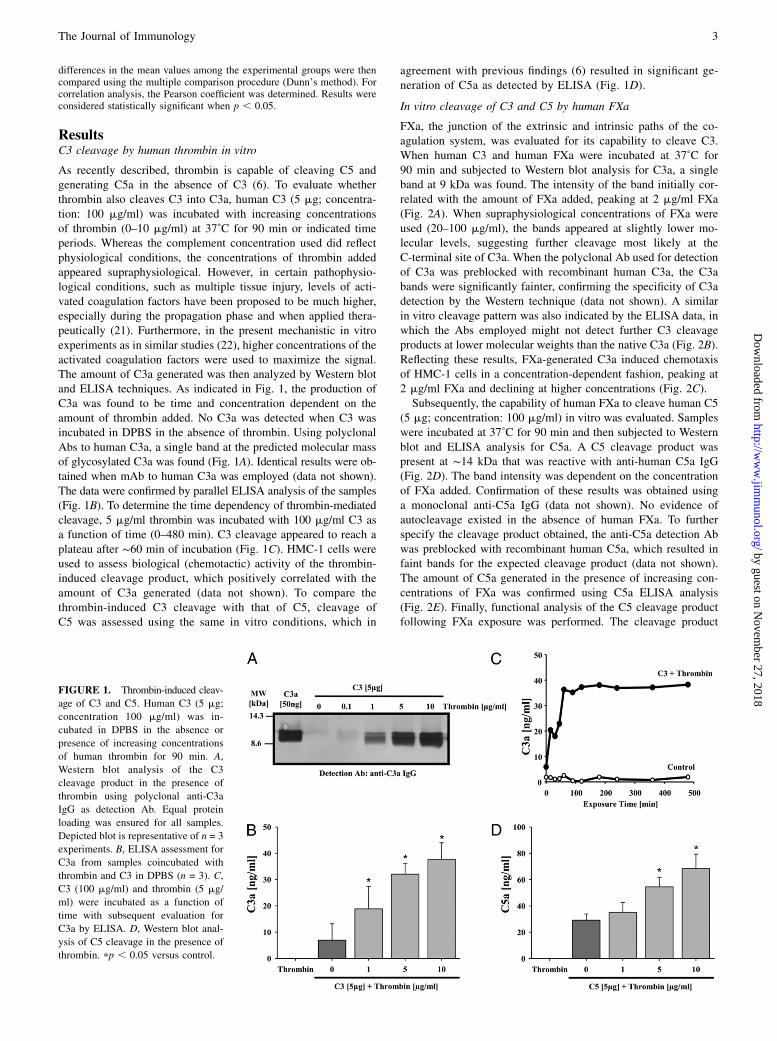
ResultsC3 cleavage by human thrombin in vitro
As recently described, thrombin is capable of cleaving C5 andgenerating C5a in the absence of C3 (6). To evaluate whetherthrombin also cleaves C3 into C3a, human C3 (5 mg; concentra-tion: 100 mg/ml) was incubated with increasing concentrationsof thrombin (0–10 mg/ml) at 37˚C for 90 min or indicated timeperiods. Whereas the complement concentration used did reflectphysiological conditions, the concentrations of thrombin addedappeared supraphysiological. However, in certain pathophysio-logical conditions, such as multiple tissue injury, levels of acti-vated coagulation factors have been proposed to be much higher,especially during the propagation phase and when applied thera-peutically (21). Furthermore, in the present mechanistic in vitroexperiments as in similar studies (22), higher concentrations of theactivated coagulation factors were used to maximize the signal.The amount of C3a generated was then analyzed by Western blotand ELISA techniques. As indicated in Fig. 1, the production ofC3a was found to be time and concentration dependent on theamount of thrombin added. No C3a was detected when C3 wasincubated in DPBS in the absence of thrombin. Using polyclonalAbs to human C3a, a single band at the predicted molecular massof glycosylated C3a was found (Fig. 1A). Identical results were ob-tained when mAb to human C3a was employed (data not shown).The data were confirmed by parallel ELISA analysis of the samples(Fig. 1B). To determine the time dependency of thrombin-mediatedcleavage, 5 mg/ml thrombin was incubated with 100 mg/ml C3 asa function of time (0–480 min). C3 cleavage appeared to reach aplateau after ∼60 min of incubation (Fig. 1C). HMC-1 cells wereused to assess biological (chemotactic) activity of the thrombin-induced cleavage product, which positively correlated with theamount of C3a generated (data not shown). To compare thethrombin-induced C3 cleavage with that of C5, cleavage ofC5 was assessed using the same in vitro conditions, which in
agreement with previous findings (6) resulted in significant ge-neration of C5a as detected by ELISA (Fig. 1D).
In vitro cleavage of C3 and C5 by human FXa
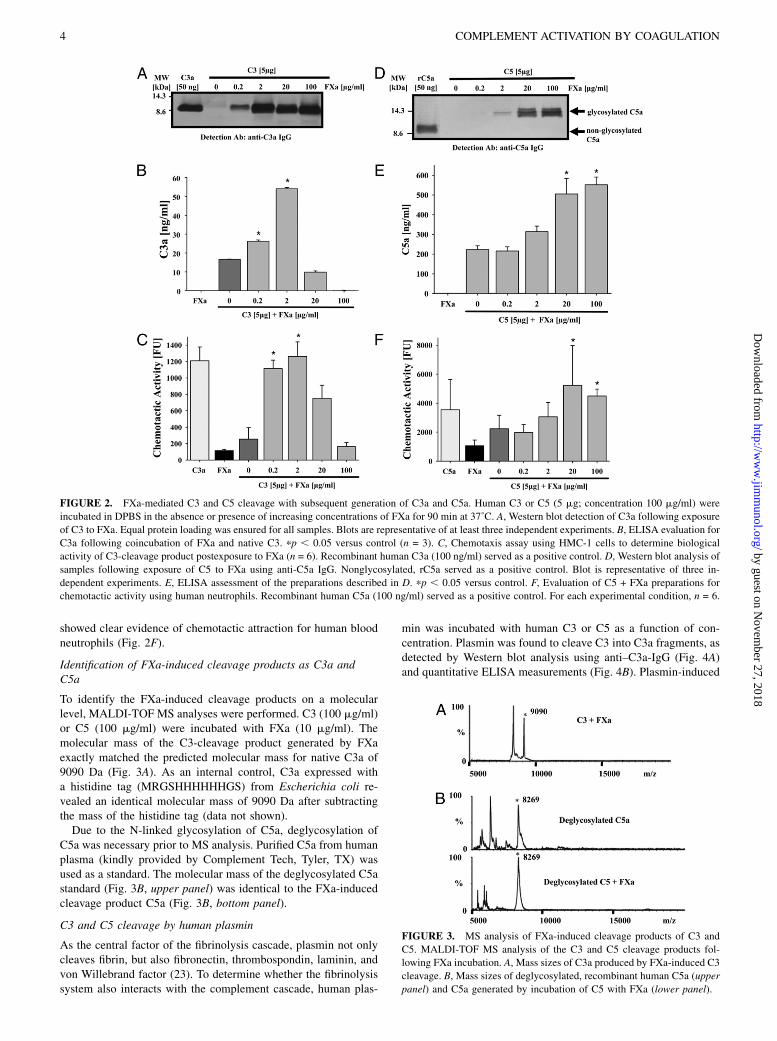
FXa, the junction of the extrinsic and intrinsic paths of the co-agulation system, was evaluated for its capability to cleave C3.When human C3 and human FXa were incubated at 37˚C for90 min and subjected to Western blot analysis for C3a, a singleband at 9 kDa was found. The intensity of the band initially cor-related with the amount of FXa added, peaking at 2 mg/ml FXa(Fig. 2A). When supraphysiological concentrations of FXa wereused (20–100 mg/ml), the bands appeared at slightly lower mo-lecular levels, suggesting further cleavage most likely at theC-terminal site of C3a. When the polyclonal Ab used for detectionof C3a was preblocked with recombinant human C3a, the C3abands were significantly fainter, confirming the specificity of C3adetection by the Western technique (data not shown). A similarin vitro cleavage pattern was also indicated by the ELISA data, inwhich the Abs employed might not detect further C3 cleavageproducts at lower molecular weights than the native C3a (Fig. 2B).Reflecting these results, FXa-generated C3a induced chemotaxisof HMC-1 cells in a concentration-dependent fashion, peaking at2 mg/ml FXa and declining at higher concentrations (Fig. 2C).Subsequently, the capability of human FXa to cleave human C5
(5 mg; concentration: 100 mg/ml) in vitro was evaluated. Sampleswere incubated at 37˚C for 90 min and then subjected to Westernblot and ELISA analysis for C5a. A C5 cleavage product waspresent at ∼14 kDa that was reactive with anti-human C5a IgG(Fig. 2D). The band intensity was dependent on the concentrationof FXa added. Confirmation of these results was obtained usinga monoclonal anti-C5a IgG (data not shown). No evidence ofautocleavage existed in the absence of human FXa. To furtherspecify the cleavage product obtained, the anti-C5a detection Abwas preblocked with recombinant human C5a, which resulted infaint bands for the expected cleavage product (data not shown).The amount of C5a generated in the presence of increasing con-centrations of FXa was confirmed using C5a ELISA analysis(Fig. 2E). Finally, functional analysis of the C5 cleavage productfollowing FXa exposure was performed. The cleavage product
showed clear evidence of chemotactic attraction for human bloodneutrophils (Fig. 2F).
Identification of FXa-induced cleavage products as C3a andC5a
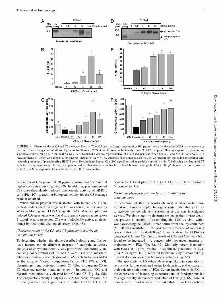
To identify the FXa-induced cleavage products on a molecularlevel, MALDI-TOF MS analyses were performed. C3 (100 mg/ml)or C5 (100 mg/ml) were incubated with FXa (10 mg/ml). Themolecular mass of the C3-cleavage product generated by FXaexactly matched the predicted molecular mass for native C3a of9090 Da (Fig. 3A). As an internal control, C3a expressed witha histidine tag (MRGSHHHHHHGS) from Escherichia coli re-vealed an identical molecular mass of 9090 Da after subtractingthe mass of the histidine tag (data not shown).Due to the N-linked glycosylation of C5a, deglycosylation of
C5a was necessary prior to MS analysis. Purified C5a from humanplasma (kindly provided by Complement Tech, Tyler, TX) wasused as a standard. The molecular mass of the deglycosylated C5astandard (Fig. 3B, upper panel) was identical to the FXa-inducedcleavage product C5a (Fig. 3B, bottom panel).
C3 and C5 cleavage by human plasmin
As the central factor of the fibrinolysis cascade, plasmin not onlycleaves fibrin, but also fibronectin, thrombospondin, laminin, andvon Willebrand factor (23). To determine whether the fibrinolysissystem also interacts with the complement cascade, human plas-
min was incubated with human C3 or C5 as a function of con-centration. Plasmin was found to cleave C3 into C3a fragments, asdetected by Western blot analysis using anti–C3a-IgG (Fig. 4A)and quantitative ELISA measurements (Fig. 4B). Plasmin-induced
FIGURE 2. FXa-mediated C3 and C5 cleavage with subsequent generation of C3a and C5a. Human C3 or C5 (5 mg; concentration 100 mg/ml) were
incubated in DPBS in the absence or presence of increasing concentrations of FXa for 90 min at 37˚C. A, Western blot detection of C3a following exposure
of C3 to FXa. Equal protein loading was ensured for all samples. Blots are representative of at least three independent experiments. B, ELISA evaluation for
C3a following coincubation of FXa and native C3. pp , 0.05 versus control (n = 3). C, Chemotaxis assay using HMC-1 cells to determine biological
activity of C3-cleavage product postexposure to FXa (n = 6). Recombinant human C3a (100 ng/ml) served as a positive control. D, Western blot analysis of
samples following exposure of C5 to FXa using anti-C5a IgG. Nonglycosylated, rC5a served as a positive control. Blot is representative of three in-
dependent experiments. E, ELISA assessment of the preparations described in D. pp , 0.05 versus control. F, Evaluation of C5 + FXa preparations for
chemotactic activity using human neutrophils. Recombinant human C5a (100 ng/ml) served as a positive control. For each experimental condition, n = 6.
FIGURE 3. MS analysis of FXa-induced cleavage products of C3 and
C5. MALDI-TOF MS analysis of the C3 and C5 cleavage products fol-
lowing FXa incubation. A, Mass sizes of C3a produced by FXa-induced C3
cleavage. B, Mass sizes of deglycosylated, recombinant human C5a (upper
panel) and C5a generated by incubation of C5 with FXa (lower panel).
generation of C3a peaked at 20 mg/ml plasmin and decreased athigher concentrations (Fig. 4A, 4B). In addition, plasmin-derivedC3a dose-dependently induced chemotactic activity of HMC-1cells (Fig. 4C), suggesting biological activity for the C3 cleavageproduct obtained.When human plasmin was incubated with human C5, a con-
centration-dependent cleavage of C5 was found, as assessed byWestern blotting and ELISA (Fig. 4D, 4E). Maximal plasmin-induced C5a-generation was found at plasmin concentrations above2 mg/ml. Again, generated C5a was biologically active, as deter-mined by neutrophil chemotaxis assays (Fig. 4F).
Characterization of the C3- and C5-proteolytic activity ofcoagulation factors
To determine whether the above-described clotting and fibrino-lysis factors exhibit different degrees of catalytic activities,analysis of enzymatic activity was performed. Accordingly, in-creasing concentrations of C3 or C5 were used as a substrate,whereas a constant concentration of 80 nM each factor was addedas the enzyme. Various coagulation factors (TF, FVIIa, FVII,plasminogen, and activated protein C) failed to generate C3 orC5 cleavage activity (data not shown). In contrast, FXa andplasmin most effectively cleaved both C3 and C5 (Fig. 5A, 5B).The enzymatic activity analyses (n = 5 for each) revealed thefollowing order: FXa . plasmin . thrombin . FIXa . FXIa .
control for C3 and plasmin . FXa . FIXa = FXIa . thrombin. control for C5.
Serum complement activation by Fxa: inhibition byanticoagulants
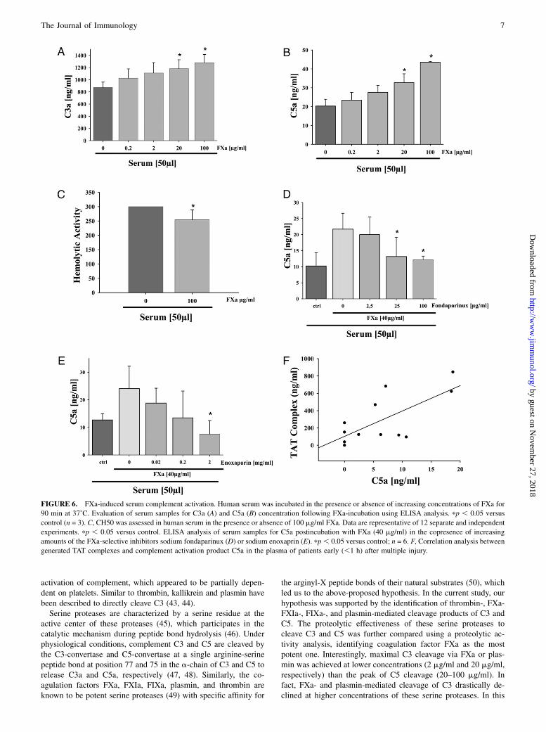
To determine whether the results obtained in vitro can be trans-ferred into a more complex biological system, the ability of FXato activate the complement system in serum was investigatedex vivo. We also sought to determine whether the in vitro cleav-age process is capable of assembling the TCC ex vivo, whichwas assessed by the CH50. Human serum from healthy volunteers(50 ml) was incubated in the absence or presence of increasingconcentrations of FXa (0–100 mg/ml) and analyzed by ELISA forgenerated C3a and C5a. Serum levels of C3a and C5a were bothfound to be increased in a concentration-dependent manner oninubation with FXa (Fig. 6A, 6B). Similarly, serum incubationwith FXa (100 mg/ml) resulted in significant generation of TCC(384 6 24 ng/ml TCC), reflected functionally by a small but sig-nificant decrease in serum hemolytic activity (Fig. 6C).The specificity of FXa-dependent anaphylatoxin generation in
serum was further evaluated using fondaparinux and enoxaparin,both selective inhibitors of FXa. Serum incubation with FXa inthe copresence of increasing concentrations of fondaparinux ledto a significantly suppressed production of C5a (Fig. 6D). Similarresults were found when a different inhibitor of FXa protease
FIGURE 4. Plasmin-induced C3 and C5 cleavage. Human C3 or C5 (each at 5 mg; concentration 100 mg /ml) were incubated in DPBS in the absence or
presence of increasing concentrations of plasmin for 90 min (37˚C). A and D, Western blot analysis of C3 or C5 samples following exposure to plasmin. As
a positive control, 50 ng of rC3a or rC5a was used. Depicted blots are representative of n = 3 independent experiments. B and E, C3a- or C5a-ELISA
measurements of C3 or C5 samples after plasmin incubation (n = 3). C, Analysis of chemotactic activity of C3 preparation following incubation with
increasing amounts of plasmin using HMC-1 cells. Recombinant human C3a (100 ng/ml) served as positive control (n = 6). F, Following incubation of C5
with increasing amounts of plasmin, samples served as chemotactic stimulus for isolated human neutrophils. C5a (100 ng/ml) was used as a positive
control. n = 6 per experimental condition. pp , 0.05 versus control.
activity, enoxaparin, was used (Fig. 6E). Additional experimentssubstituting human plasma for human serum demonstrated similarpatterns of C3a and C5a generation in the presence of FXa (datanot shown). Collectively, these data suggest functional ex vivocleavage of C3 and C5 by FXa.For a first transfer of the reported ex vivo findings to a relevant
clinical setting, plasma from 12 polytrauma patients within the firsthour after trauma was analyzed for activation of the coagulationand complement systems (for detailed patients’ demography, see Ref.24). In accordance with a previous study in multiple injured patients(25), very early activation (and dysfunction) of the coagulation sys-tem was found with significant formation of TAT complexes. A sub-sequent analysis determined whether these changes in TAT com-plexes were associated with complement activation. Early post-trauma (within 1 h postinjury), there was a positive correlationbetween the generation of the TAT complexes and the anaphy-latoxin C5a concentrations in plasma (correlation coefficient r =0.697; p = 0.012; n = 12; Fig. 6F). The present in vitro and in vivoresults are summarized in a simplified scheme (Fig. 7).
DiscussionIn the current study, we investigated the interaction between thecoagulation/fibrinolysis cascades and the complement system in vi-tro and ex vivo. Exposure of C3 to thrombin resulted in time- andconcentration-dependent generation of C3a in vitro. C5 was alsocleaved by thrombin to produce C5a. In parallel experiments, in-cubation of C3 and C5 with either FXa or plasmin resulted ingeneration of C3a and C5a. The resulting cleavage products ex-hibited intact chemotactic activity and were indistinguishable fromnative C3a or C5a when assessed by MS. Incubation of serum orplasma with FXa resulted in a concentration-dependent generationof C3a and C5a as well as decreased hemolytic serum activity.The coagulation/fibrinolysis cascades and the complement sys-
tem appear to be triggered simultaneously by severe tissue in-jury (26, 27), acute trauma (28), or during systemic inflammation(9). Pathophysiologically, the formation of thrombin at the site ofinjury following the activation of the coagulation cascade may notonly provide a physical barrier against invading micro-organisms(29), but also trigger the complement system. Locally producedanaphylatoxins help in activating cellular immune responses (30).Thus, the innate serine protease system, with its three major col-umns, coagulation, fibrinolysis, and complement, may be essentialfor both an effective protection against bleeding and invading patho-gens. However, when the injurious load is excessive, uncontrolledand simultaneous activation of the complement and coagulation/fibrinolysis systems can occur. In particular, excessive generation
of C5a is known to have adverse effects during systemic infection(31, 32). Therefore, it is tempting to speculate whether variousclotting factors might contribute locally and systemically to generateC3a and C5a, which, in turn act as chemoattractants for phago-cytic cells to the site of inflammation, where these cells release theirmajor arsenals of tissue-damaging proteases, reactive oxygen spe-cies, and cytokines/chemokines (15, 18, 33, 34). An important rolefor C5a/C5aR signaling has also been postulated in the fibrinolysissystem (35). Finally, many proinflammatory cytokines can causedecreased levels of several anticoagulant proteins including throm-bomodulin, the endothelial cell protein C receptor, and protein S(36), resulting in an inflammatory, procoagulant state. Mannan-binding lectin-associated serine protease 2, a protease that is char-acteristic of the lectin pathway of complement activation, can trig-ger coagulation by cleaving prothrombin into active thrombin (13).The procoagulant activities of complement are increased when an-ticoagulant mechanisms are inhibited; for example, the formation ofa complex between C4b-binding protein and protein S results ina decrease in the availability of protein S to act as a cofactor for theanticoagulant protein C pathway (37). In addition, the thrombin-prothrombin complex activates carboxypeptidase B, which, in turn,blocks C5a to counteract the inflammatory mediators generated atthe site of vascular injury (38). Profound effects of the comple-ment system on the coagulation system and vice versa have alsobeen found for some innate regulators, such as the C1-inhibitor orthe thrombin activatable fibrinolysis inhibitor. The latter is a potentand broadly reactive carboxypeptidase (39), which is generated bythe thrombomodulin–thrombin complex and reacts in an anticoag-ulant manner (40). It not only moderates fibrinolysis but also hassome anti-inflammatory effects due to its ability to inactivate C3aand C5a by removing carboxy-terminal arginine residues from thesecomponents (41). Thus, we are now beginning to understand that,rather than acting as separate, independent cascades, the coagulation/fibrinolysis system and the complement cascade cross talk exten-sively with each other and mutually fine-tune their activationstatus (16, 17).Previously, the presence of C3 was thought to be indispensable
for the assembly of the C5-convertase to generate C5a. Surpris-ingly, the presence of C5a was recently discovered in activated C3-deficient serum, where enhanced levels of thrombin, a primarycomponent of the activated coagulation system, induced C5 con-version to C5a (6). In the current study, we found evidence ofthrombin-mediated cleavage of C3 into C3a in a dose- and time-dependent manner in complement-sufficient human serum. In linewith these findings, Kalowski et al. (42) reported in 1975 thatthrombin and thromboplastin injected into rabbits somehow led to
FIGURE 5. C3-and C5-proteolytic activity of various coagulation factors (FXa, FIXa, FXIa, plasmin, thrombin). A and B, C3- and C5-proteolytic activity
of various coagulation/fibrinolysis factors (each at 80 nM) were assessed by ELISA measurements of C3a and C5a generated within 90 min in the presence
of increasing concentrations of the substrate (10, 54, 108, 270, 540, 810, and 1080 nM C3 or C5). Every proteolytic activity value represents the average of
duplicate measurements based on five independent experiments.
activation of complement, which appeared to be partially depen-dent on platelets. Similar to thrombin, kallikrein and plasmin havebeen described to directly cleave C3 (43, 44).Serine proteases are characterized by a serine residue at the
active center of these proteases (45), which participates in thecatalytic mechanism during peptide bond hydrolysis (46). Underphysiological conditions, complement C3 and C5 are cleaved bythe C3-convertase and C5-convertase at a single arginine-serinepeptide bond at position 77 and 75 in the a-chain of C3 and C5 torelease C3a and C5a, respectively (47, 48). Similarly, the co-agulation factors FXa, FXIa, FIXa, plasmin, and thrombin areknown to be potent serine proteases (49) with specific affinity for
the arginyl-X peptide bonds of their natural substrates (50), whichled us to the above-proposed hypothesis. In the current study, ourhypothesis was supported by the identification of thrombin-, FXa-FXIa-, FIXa-, and plasmin-mediated cleavage products of C3 andC5. The proteolytic effectiveness of these serine proteases tocleave C3 and C5 was further compared using a proteolytic ac-tivity analysis, identifying coagulation factor FXa as the mostpotent one. Interestingly, maximal C3 cleavage via FXa or plas-min was achieved at lower concentrations (2 mg/ml and 20 mg/ml,respectively) than the peak of C5 cleavage (20–100 mg/ml). Infact, FXa- and plasmin-mediated cleavage of C3 drastically de-clined at higher concentrations of these serine proteases. In this
FIGURE 6. FXa-induced serum complement activation. Human serum was incubated in the presence or absence of increasing concentrations of FXa for
90 min at 37˚C. Evaluation of serum samples for C3a (A) and C5a (B) concentration following FXa-incubation using ELISA analysis. pp , 0.05 versus
control (n = 3). C, CH50 was assessed in human serum in the presence or absence of 100 mg/ml FXa. Data are representative of 12 separate and independent
experiments. pp , 0.05 versus control. ELISA analysis of serum samples for C5a postincubation with FXa (40 mg/ml) in the copresence of increasing
amounts of the FXa-selective inhibitors sodium fondaparinux (D) or sodium enoxaprin (E). pp, 0.05 versus control; n = 6. F, Correlation analysis between
generated TAT complexes and complement activation product C5a in the plasma of patients early (,1 h) after multiple injury.
case, it is tempting to speculate that further cleavage on the Cterminus of C3a occurred because the C3a/C3a-des-arg neo-epitope Ab of the ELISA system failed to detect the cleavageproduct. This is supported by the significant loss of the chemo-tactic activity known to be specifically dependent on the C-ter-minal part of C3a. Thus, when components of the coagulation/fibrinolysis system are only moderately triggered, activation of thecomplement cascade may be mainly achieved through the cleav-age of C3, whereas, when coagulation/fibrinolysis is massivelyactivated, C5 may represent the primary target for cleavage.The concentrations of coagulation factors required to cleave
purified complement proteins appear to be supraphysiological,which might suggest that such interactions between complementand coagulation are less relevant to the in vivo situation. However,the data presented in this study do not support this conclusion.It has been clear from a number of studies that cofactors andbiological surfaces exert vital effects on the activity of plasma en-zymes (51, 52). The prothrombinase complex (consisting of FXa,FVa, and negatively charged surface) increases FXa activity tocleave prothrombin into thrombin 300,000-fold as compared withFXa and prothombin alone. Furthermore, little is known about thepathophysiological concentrations of activated clotting/fibrinolysisfactors produced locally and systemically upon excessive activationof the coagulation system (e.g., during hemorrhagic shock and dis-seminated coagulopathy after severe tissue trauma). In this regard,a C3a-like fragment has been found in blood clots, indicating localcomplement activation and deposition (53). It is striking that theclinically established anticoagulants enoxaparin and fondaparinux,targeting FXa, are highly capable of inhibiting complement acti-vation. Thus, selective FXa inhibitors, and theoretically other anti-coagulants, might also act as immune modulators, a barely under-stood new function.To our knowledge, the current study represents the first sys-
tematic assessment of the communication between the complementsystem and the coagulation/fibrinolysis cascades, providing novelinsights into their complex interplay. Limitations of our studyinclude the fact that it remains to be determined if our findings canbe extrapolated into a much more complex in vivo setting inhumans. As an initial evaluation of the potential transferability ofour in vitro findings, the present findings were assessed by a moreelaborate ex vivo system using serum and plasma samples. Based
on significant changes to the CH50 and generation of TCC in thepresence of FXa, it is likely that the activated coagulation systemclosely interacts with the complement cascade on various mo-lecular levels ex vivo, generating not only anaphylatoxins but alsothe opsonin C3b and C5b-9. This could be mirrored by the presentresults in multiple injured patients of a very early synchronizedappearance of coagulation (TAT complexes) and complementactivation (C5a) products. However, because correlation is notnecessary causative, these findings will have to be confirmed ina standardized experimental in vivo setting. In the current study,factors of the tissue factor pathway failed to affect the complementsystem, whereas serine proteases of the contact activation pathwaywere able to significantly activate the complement system. It is nowbecoming clear that a distinction between the intrinsic and extrinsiccoagulation pathways in the coagulation cascade represents anantiquated description of test tube coagulation and does not reflectphysiological in vivo coagulation (54). As a result, a cell-basedmodel of coagulation has been developed (55), which appearsto much better mimic the complex interactions of cellular and plas-ma components in vivo. The intricate interplay of the coagulationand fibrinolysis cascades with various cells further complicatestheir interactions with the complement system. Thus, unravel-ing the precise communications between coagulation/fibrinolysis,various cells, and the complement system will represent a majorfuture scientific challenge. In addition, therapeutic interventionstargeting one cascade might result in unfavorable, so far un-anticipated adverse effects on the other cascade and the cells in-volved.In conclusion, we provide the first evidence, to our knowledge,
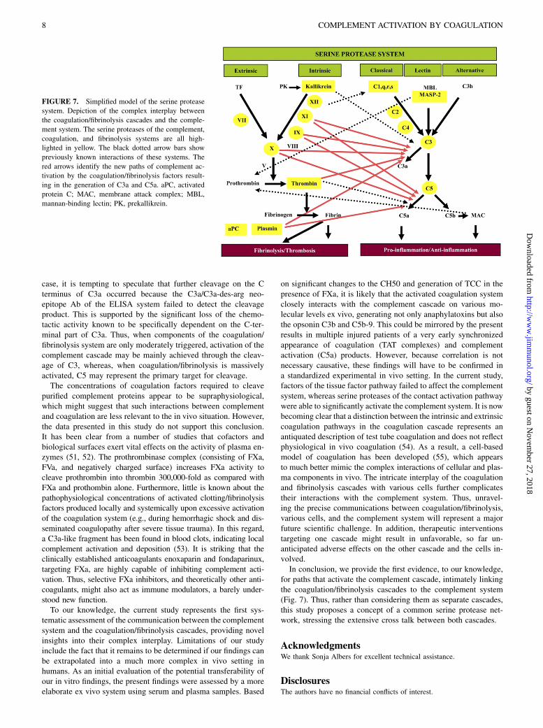
for paths that activate the complement cascade, intimately linkingthe coagulation/fibrinolysis cascades to the complement system(Fig. 7). Thus, rather than considering them as separate cascades,this study proposes a concept of a common serine protease net-work, stressing the extensive cross talk between both cascades.
AcknowledgmentsWe thank Sonja Albers for excellent technical assistance.
DisclosuresThe authors have no financial conflicts of interest.
FIGURE 7. Simplified model of the serine protease
system. Depiction of the complex interplay between
the coagulation/fibrinolysis cascades and the comple-
ment system. The serine proteases of the complement,
coagulation, and fibrinolysis systems are all high-
lighted in yellow. The black dotted arrow bars show
previously known interactions of these systems. The
red arrows identify the new paths of complement ac-
tivation by the coagulation/fibrinolysis factors result-
ing in the generation of C3a and C5a. aPC, activated
References1. Krem, M. M., and E. Di Cera. 2002. Evolution of enzyme cascades from em-
bryonic development to blood coagulation. Trends Biochem. Sci. 27: 67–74.2. Esmon, C. T. 2004. The impact of the inflammatory response on coagulation.
Thromb. Res. 114: 321–327.3. Rittirsch, D., M. A. Flierl, and P. A. Ward. 2008. Harmful molecular mechanisms
in sepsis. Nat. Rev. Immunol. 8: 776–787.4. Davis, A. E., III, P. Mejia, and F. Lu. 2008. Biological activities of C1 inhibitor.
Mol. Immunol. 45: 4057–4063.5. Ghebrehiwet, B., M. Silverberg, and A. P. Kaplan. 1981. Activation of the
classical pathway of complement by Hageman factor fragment. J. Exp. Med.153: 665–676.
6. Huber-Lang, M., J. V. Sarma, F. S. Zetoune, D. Rittirsch, T. A. Neff,S. R. McGuire, J. D. Lambris, R. L. Warner, M. A. Flierl, L. M. Hoesel, et al.2006. Generation of C5a in the absence of C3: a new complement activationpathway. Nat. Med. 12: 682–687.
7. Clark, A., A. Weymann, E. Hartman, Y. Turmelle, M. Carroll, J. M. Thurman,V. M. Holers, D. E. Hourcade, and D. A. Rudnick. 2008. Evidence for non-traditional activation of complement factor C3 during murine liver regeneration.Mol. Immunol. 45: 3125–3132.
8. Lidington, E. A., D. O. Haskard, and J. C. Mason. 2000. Induction of decay-accelerating factor by thrombin through a protease-activated receptor 1 andprotein kinase C-dependent pathway protects vascular endothelial cells fromcomplement-mediated injury. Blood 96: 2784–2792.
9. Levi, M., T. van der Poll, and H. R. Buller. 2004. Bidirectional relation betweeninflammation and coagulation. Circulation 109: 2698–2704.
10. Hecke, F., U. Schmidt, A. Kola, W. Bautsch, A. Klos, and J. Kohl. 1997. Cir-culating complement proteins in multiple trauma patients—correlation withinjury severity, development of sepsis, and outcome. Crit. Care Med. 25: 2015–2024.
11. Ikeda, K., K. Nagasawa, T. Horiuchi, T. Tsuru, H. Nishizaka, and Y. Niho. 1997.C5a induces tissue factor activity on endothelial cells. Thromb. Haemost. 77:394–398.
12. Ritis, K., M. Doumas, D. Mastellos, A. Micheli, S. Giaglis, P. Magotti, S. Rafail,G. Kartalis, P. Sideras, and J. D. Lambris. 2006. A novel C5a receptor-tissuefactor cross-talk in neutrophils links innate immunity to coagulation pathways.J. Immunol. 177: 4794–4802.
13. Krarup, A., R. Wallis, J. S. Presanis, P. Gal, and R. B. Sim. 2007. Simultaneousactivation of complement and coagulation by MBL-associated serine protease 2.PLoS ONE 2: e623.
14. Wiedmer, T., C. T. Esmon, and P. J. Sims. 1986. Complement proteins C5b-9stimulate procoagulant activity through platelet prothrombinase. Blood 68: 875–880.
15. Wojta, J., C. Kaun, G. Zorn, M. Ghannadan, A. W. Hauswirth, W. R. Sperr,G. Fritsch, D. Printz, B. R. Binder, G. Schatzl, et al. 2002. C5a stimulatesproduction of plasminogen activator inhibitor-1 in human mast cells and baso-phils. Blood 100: 517–523.
16. Esmon, C. T. 2004. Interactions between the innate immune and blood co-agulation systems. Trends Immunol. 25: 536–542.
17. Markiewski, M. M., B. Nilsson, K. N. Ekdahl, T. E. Mollnes, and J. D. Lambris.2007. Complement and coagulation: strangers or partners in crime? TrendsImmunol. 28: 184–192.
18. Hartmann, K., B. M. Henz, S. Kruger-Krasagakes, J. Kohl, R. Burger, S. Guhl,I. Haase, U. Lippert, and T. Zuberbier. 1997. C3a and C5a stimulate chemotaxisof human mast cells. Blood 89: 2863–2870.
19. Flierl, M. A., M. Perl, D. Rittirsch, C. Bartl, H. Schreiber, V. Fleig, G. Schlaf,U. Liener, U. B. Brueckner, F. Gebhard, and M. S. Huber-Lang. 2008. The roleof C5a in the innate immune response after experimental blunt chest trauma.Shock 29: 25–31.
20. Flierl, M. A., D. Rittirsch, B. A. Nadeau, D. E. Day, F. S. Zetoune, J. V. Sarma,M. S. Huber-Lang, and P. A. Ward. 2008. Functions of the complement com-ponents C3 and C5 during sepsis. FASEB J. 22: 3483–3490.
21. Poon, M. C., R. d’Oiron, I. Hann, C. Negrier, L. de Lumley, A. Thomas,A. Karafoulidou, C. Demers, A. Street, A. Huth-Kuhne, et al. 2001. Use ofrecombinant factor VIIa (NovoSeven) in patients with Glanzmann thrombas-thenia. Semin. Hematol. 38(4, Suppl 12): 21–25.
22. Bono, F., P. Schaeffer, J. P. Herault, C. Michaux, A. L. Nestor, J. C. Guillemot,and J. M. Herbert. 2000. Factor Xa activates endothelial cells by a receptorcascade between EPR-1 and PAR-2. Arterioscler. Thromb. Vasc. Biol. 20: E107–E112.
23. Bonnefoy, A., and C. Legrand. 2000. Proteolysis of subendothelial adhesiveglycoproteins (fibronectin, thrombospondin, and von Willebrand factor) byplasmin, leukocyte cathepsin G, and elastase. Thromb. Res. 98: 323–332.
24. Amara, U., M. Kalbitz, M. Perl, M. A. Flierl, D. Rittirsch, M. Weiss,M. Schneider, F. Gebhard, and M. Huber-Lang. 2010. Early expression changesof complement regulatory proteins (CRegs) and C5a receptor (CD88) on leu-kocytes after multiple injury in humans. Shock 33: 568–575.
25. Lampl, L., M. Helm, A. Specht, K. H. Bock, W. Hartel, and E. Seifried. 1994.[Blood coagulation parameters as prognostic factors in multiple trauma: canclinical values be an early diagnostic aid?]. Zentralbl. Chir. 119: 683–689.
26. Lasser, E. C., J. Slivka, J. H. Lang, W. P. Kolb, S. G. Lyon, A. E. Hamblin, andG. Nazareno. 1979. Complement and coagulation: causative considerations incontrast catastrophies. AJR Am. J. Roentgenol. 132: 171–176.
27. Bazargani, F., A. Albrektsson, N. Yahyapour, and M. Braide. 2005. Low mo-lecular weight heparin improves peritoneal ultrafiltration and blocks complementand coagulation. Perit. Dial. Int. 25: 394–404.
28. Brohi, K., M. J. Cohen, M. T. Ganter, M. J. Schultz, M. Levi, R. C. Mackersie, andJ. F. Pittet. 2008. Acute coagulopathy of trauma: hypoperfusion induces systemicanticoagulation and hyperfibrinolysis. J. Trauma 64: 1211–1217, discussion 1217.
29. Sun, H. 2006. The interaction between pathogens and the host coagulationsystem. Physiology (Bethesda) 21: 281–288.
30. Peng, Q., K. Li, K. Anderson, C. A. Farrar, B. Lu, R. A. Smith, S. H. Sacks, andW. Zhou. 2008. Local production and activation of complement up-regulates theallostimulatory function of dendritic cells through C3a-C3aR interaction. Blood111: 2452–2461.
31. Ward, P. A. 2004. The dark side of C5a in sepsis. Nat. Rev. Immunol. 4: 133–142.32. Laudes, I. J., J. C. Chu, S. Sikranth, M. Huber-Lang, R. F. Guo, N. Riedemann,
J. V. Sarma, A. H. Schmaier, and P. A. Ward. 2002. Anti-c5a amelioratescoagulation/fibrinolytic protein changes in a rat model of sepsis. Am. J. Pathol.160: 1867–1875.
33. Huber-Lang, M., E. M. Younkin, J. V. Sarma, N. Riedemann, S. R. McGuire,K. T. Lu, R. Kunkel, J. G. Younger, F. S. Zetoune, and P. A. Ward. 2002.Generation of C5a by phagocytic cells. Am. J. Pathol. 161: 1849–1859.
34. Czermak, B. J., V. Sarma, N. M. Bless, H. Schmal, H. P. Friedl, and P. A. Ward.1999. In vitro and in vivo dependency of chemokine generation on C5a andTNF-alpha. J. Immunol. 162: 2321–2325.
35. Shushakova, N., N. Tkachuk, M. Dangers, S. Tkachuk, J. K. Park, J. Zwirner,K. Hashimoto, H. Haller, and I. Dumler. 2005. Urokinase-induced activation ofthe gp130/Tyk2/Stat3 pathway mediates a pro-inflammatory effect in humanmesangial cells via expression of the anaphylatoxin C5a receptor. [Publishederratum appears in 2007 J. Cell. Sci. 120: 2137.] J. Cell Sci. 118: 2743–2753.
36. Shebuski, R. J., and K. S. Kilgore. 2002. Role of inflammatory mediators inthrombogenesis. J. Pharmacol. Exp. Ther. 300: 729–735.
37. Rezende, S. M., R. E. Simmonds, and D. A. Lane. 2004. Coagulation, in-flammation, and apoptosis: different roles for protein S and the protein S-C4bbinding protein complex. Blood 103: 1192–1201.
38. Nishimura, T., T. Myles, A. M. Piliponsky, A. M. Piliposky, P. N. Kao,G. J. Berry, and L. L. Leung. 2007. Thrombin-activatable procarboxypeptidase Bregulates activated complement C5a in vivo. [Published erratum appears in 2007Blood 109: 3632.] Blood 109: 1992–1997.
39. Bajzar, L. 2000. Thrombin activatable fibrinolysis inhibitor and an anti-fibrinolytic pathway. Arterioscler. Thromb. Vasc. Biol. 20: 2511–2518.
40. Esmon, C. T. 2005. The interactions between inflammation and coagulation.Br. J. Haematol. 131: 417–430.
41. Myles, T., T. Nishimura, T. H. Yun, M. Nagashima, J. Morser, A. J. Patterson,R. G. Pearl, and L. L. Leung. 2003. Thrombin activatable fibrinolysis inhibitor,a potential regulator of vascular inflammation. J. Biol. Chem. 278: 51059–51067.
42. Kalowski, S., E. L. Howes, Jr., W. Margaretten, and D. G. McKay. 1975. Effectsof intravascular clotting on the activation of the complement system: The role ofthe platelet. Am. J. Pathol. 78: 525–536.
43. Thoman, M. L., J. L. Meuth, E. L. Morgan, W. O. Weigle, and T. E. Hugli. 1984.C3d-K, a kallikrein cleavage fragment of iC3b is a potent inhibitor of cellularproliferation. J. Immunol. 133: 2629–2633.
44. Goldberger, G., M. L. Thomas, B. F. Tack, J. Williams, H. R. Colten, andG. N. Abraham. 1981. NH2-terminal structure and cleavage of guinea pig pro-C3,the precursor of the third complement component. J. Biol. Chem. 256: 12617–12619.
45. Yousef, G. M., A. D. Kopolovic, M. B. Elliott, and E. P. Diamandis. 2003.Genomic overview of serine proteases. Biochem. Biophys. Res. Commun. 305:28–36.
46. Polgar, L. 2005. The catalytic triad of serine peptidases. Cell. Mol. Life Sci. 62:2161–2172.
47. Sandoval, A., R. Ai, J. M. Ostresh, and R. T. Ogata. 2000. Distal recognition sitefor classical pathway convertase located in the C345C/netrin module of com-plement component C5. J. Immunol. 165: 1066–1073.
48. Sahu, A., and J. D. Lambris. 2001. Structure and biology of complement proteinC3, a connecting link between innate and acquired immunity. Immunol. Rev.180: 35–48.
49. Hayashi, K., T. Takehisa, N. Hamato, R. Takano, S. Hara, T. Miyata, andH. Kato. 1994. Inhibition of serine proteases of the blood coagulation system bysquash family protease inhibitors. J. Biochem. 116: 1013–1018.
50. Jackson, C. M., and Y. Nemerson. 1980. Blood coagulation. Annu. Rev. Biochem.49: 765–811.
51. Mann, K. G., S. Krishnaswamy, and J. H. Lawson. 1992. Surface-dependenthemostasis. Semin. Hematol. 29: 213–226.
52. Komiyama, Y., A. H. Pedersen, and W. Kisiel. 1990. Proteolytic activation ofhuman factors IX and X by recombinant human factor VIIa: effects of calcium,phospholipids, and tissue factor. Biochemistry 29: 9418–9425.
53. Sundsmo, J. S., and D. S. Fair. 1983. Relationships among the complement,kinin, coagulation, and fibrinolytic systems. Springer Semin. Immunopathol. 6:231–258.
54. Rossaint, R., V. Cerny, T. J. Coats, J. Duranteau, E. Fernandez-Mondejar,G. Gordini, P. F. Stahel, B. J. Hunt, E. Neugebauer, and D. R. Spahn. 2006. Keyissues in advanced bleeding care in trauma. Shock 26: 322–331.
55. Hoffman, M., and D. M. Monroe, III. 2001. A cell-based model of hemostasis.Thromb. Haemost. 85: 958–965.


![Molecular Basis of Complement Resistance of Human Melanoma ...cancerres.aacrjournals.org/content/canres/53/3/592.full.pdf · [CANCER RESEARCH 53. 592-599. February I. 1993] Molecular](https://static.documents.pub/doc/80x56/5d5beaea88c9936c6e8b85d6/molecular-basis-of-complement-resistance-of-human-melanoma-cancer-research.jpg)


