Page 1
Aus der Medizinischen Klinik und Poliklinik IV der Ludwig-Maximilians-
Universität München
Direktor: Prof. Dr. med. Martin Reincke
Molecular Mechanisms of
Crystal-Induced Neutrophil Cell Death
Dissertation
zum Erwerb des Doktorgrades der Humanbiologie
an der Medizinischen Fakultät der
Ludwig-Maximilians-Universität München
vorgelegt von
Jyaysi Bhagirath Desai
aus Ahmedabad, Indien
2017
Page 2
Mit Genehmigung der Medizinischen Fakultät
der Ludwig-Maximilians-Universität München
Berichterstatter : Prof. Dr. med. Hans-Joachim Anders
Mitberichterstatter : Prof. Dr. Kirsten Lauber
Mitberichterstatter : Priv.-Ooz. Dr. rer. nat. Gerald Schmid
Dekan : Prof. Dr. med. dent. Reinhard Hickel
Tag der mündlichen Prüfung : 25.04.2017
Page 3
, …
For my beloved parents and sister
Bhagirath Desai, Milka Desai & Bhuyasi Desai
Page 4
Index i
TABLE OF CONTENTS
Zusammenfassung iv
Summary vi
1. Introduction 1
1.1 Crystallopathies 1
1.2 Gouty arthritis 4
1.2.1 Onset and peak of acute gouty arthritis 4
1.2.2 Spontaneous resolution of gouty arthritis 4
1.2.3 Pathophysiological mechanisms underlying gout onset and resolution 5
1.3 Neutrophils 6
1.3.1 Neutrophil generation and circulation in blood stream 7
1.3.2 Neutrophil migration and chemotaxis 7
1.3.3 Neutrophil phagocytosis 8
1.3.4 Neutrophil extracellular traps (NETs) 8
1.3.5 Signaling components in NET release 9
1.3.6 NETs in host defense mechanism 10
1.3.7 Killing mechanisms by NETs 11
1.3.8 NETs in autoimmune diseases 12
1.3.9 NETs in gout 13
1.4 Regulated cell death 14
1.4.1 Apoptosis 15
1.4.2 Regulated necrosis 16
1.5 Neutrophil death 18
1.5.1 NETosis: NET formation in association with cell death 18
1.5.2 NET release without neutrophil death 21
1.6 The necroinflammation concept 23
2. Hypotheses/objectives 25
3. Material and Methods 26
3.1 Instruments and Chemicals 26
Page 5
Index ii
3.1.1 Instruments 26
3.1.2 Chemicals and reagents 27
3.2 Experimental procedures 32
3.2.1 Animals 32
3.3 Blood collection 33
3.3.1 Human blood sample collection 33
3.3.2 Mouse blood sample collection 33
3.4 Human and mouse neutrophil isolation 33
3.5 Induction of NETs 33
3.6 Live cell SYTOX imaging 34
3.6.1 Quantification of Sytox+ dead cells 34
3.7 Confocal imaging and immunostaining 34
3.8 Transmission and scanning electron microscopy 36
3.9 Quantitative analysis of NETs and cell death 36
3.9.1 Pico green assay 36
3.9.2 Reactive oxygen species assay 37
3.9.3 Lactate dehydrogenase cell death assay 37
3.9.4 Cell viability assay 37
3.10 Other In-vitro analysis 37
3.10.1 Cell culture 37
3.10.2 Cell freezing and thawing 38
3.10.3 Stimulation experiments 39
3.11 Protein isolation and western blotting 39
3.12 Cytokine ELISA 40
3.13 Flow cytometry for neutrophil population 40
3.14 Statistical analysis 40
4. Results 41
4.1Part I: Crystals induce neutrophil cell death and NET formation 41
4.2 Part II: Molecular mechanisms of MSU & PMA-induced NET formation & cell death
50
Page 6
Index iii
4.2.1 PMA and MSU induce release of IL-1β but not TNF-α from neutrophils 50
4.2.2 Anakinra, etanercept, anti-TLR4, FAS deficiency do not block PMA & MSU
induced NETs & cell death 50
4.2.3 Screening of different cell death inhibitors for PMA induced NETs and cell death
51
4.2.4 Nec-1 and NSA inhibit overall PMA-induced NET formation and cell death 52
4.2.5 RIPK3 and p-MLKL is upregulated during PMA induced cell death and NETs 56
4.2.6 Nec-1 and NSA inhibit both MSU-induced NET formation and cell death 57
4.2.7 p-MLKL is upregulated in MSU crystal-induced NET formation and cell death 60
4.2.8 ROS production is upstream of p-MLKL in PMA and MSU-induced NETs and
cell death 61
4.2.9 Ripk3-/- neutrophils do not undergo NET formation & cell death upon different
stimuli 63
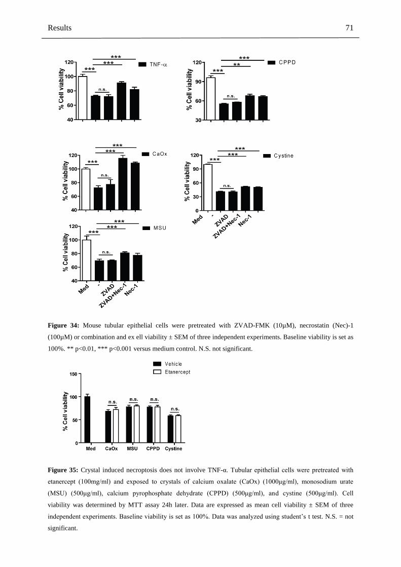
4.3 Part III: Crystal-induced necroptosis in non-immune cells 68
4.3.1 Crystal cytotoxicity involves necroptosis of non-immune cells 68
5. Discussion 76
6. References 85
7. Abbrevations 98
8. Appendix 101
9. Acknowledgement 105
Page 7
Declaration iv
Declaration
I hereby declare that all of the present work embodied in this thesis was carried out by me
from 06/2013 until 08/2016 under the supervision of Prof. Dr. Hans Joachim Anders,
Nephrologisches Zentrum, Medizinische Klinik und Poliklinik IV, Innenstadt Klinikum der
Universität München. This work has not been submitted in part or full to any other university
or institute for any degree or diploma.
Part of the work was done by others, as mentioned below:
1. Professor Helen Liapis, Department of Pathology and Immunology,Washington
University School of Medicine, Saint Louis, Missouri, USA has performed the
scanning electron microscopy. The data are presented in results part I.
2. Dr. Bastian Popper, Department of Anatomy and Cell Biology, Ludwig-Maximilians
Universität, Munich, Germany has performed the transmission electron microscopy.
The data are presented in results part I, part II and Part III.
Part of the work has been published in Eur J Immunol. 2016 Jan;46(1):223-9.
Part of the work is published as a review in Cell Mol Life Sci. 2016 Jun;73(11-12):2211-9.
Part of the work has been published for publication in Nat Commun. 2016 Jan 28;7:10274.
Part of the work is published as a review in Semin Nephrol. 2016 May;36(3):162-73.
Part of the work is submitted for a patent at the European patent office. EU prov. patent appl.
14192043.9-1412, 06.11.2014.
Date: …………… Signature: ……………..
Place: Munich, Germany (Jyaysi Bhagirath Desai)
Page 8
Zusammenfassung iv
Zusammenfassung
Sowohl verschiedene Kristalle als auch feste Nano- und Mikropartikel verursachen
viele akute und chronische physiologische Störungen (Kristallopathien) wie Gicht,
Pseudogicht, Atherosklerose, Silikose, Asbestose, Rhabdomyolyse, verschiedene Formen
kristalliner Nephropathie, sowie Nephro-/Urolithiasis. Die meisten Kristall-induzierten
Schädigungen gehen oft mit einer starken Entzündungsreaktion einher. Zusammen mit der
Entzündung verursachen Kristalle auch Zelltod; die genauen molekularen Mechanismen der
kristallinen Zelltoxizität sind aber bisher noch ungeklärt. Wir untersuchten den Kristall-
induzierten Zelltod von Neutrophilen Granulozyten. Dabei beobachteten wir das viele
verschiedene Kristalle wie z.B. Calcium Oxalat (CaOx), Mononatrium-Urat (MSU),
Calciumphosphat (CaP), Calciumpyrophsphat-Dihydrat (CPPD), Cystein, Cholesterol, Alaun,
Asbest, Siliziumdioxid und Titan-Dioxid (TiO2)-Nanopartikel Neutrophil Extracellular Trap
(NET) Formation und den damit verbundenen Zelltod der Neutrophilen, der NETose genannt
wird, induzieren. Der genaue molekulare Mechanismus, der die NET-Formation verursacht,
ist allerdings noch nicht bekannt.
Nekrose wurde traditionell immer als nicht programmierter Zelltod angesehen, dem
überhaupt keine biochemischen Mechanismen zugrunde liegen. Im Zusammenhang mit
nekrotischem Zelltod wurden jedoch mittlerweile viele verschiedene biochemische
Signalwege entdeckt die unter dem Begriff der regulierten Nekrose zusammengefasst sind.
Deswegen gingen wir in unserer Hypothese davon aus dass NETose nur eine andere Art
Zelltod mit bestimmten molekularen Mechansimen sein könnte. Wir fanden heraus dass
MSU-Kristall-induzierter Zelltod von Neutrophilen die Signalkaskade der receptor-interacting
protein kinase (RIPK) 1-RIPK3-MLKL vermittelten Nekroptose auslöst. Außerdem konnten
wir zeigen, dass die RIPK-1-Stabilisatoren Necrostatin-1 oder Necrostatin-1s und der MLKL-
Inhibitor Necrosulfamid die NET-Formation von menschlichen und murinen Neutrophilen bei
der MSU- oder PMA-induzierten Produktion von reaktiven Sauerstoffspezies (ROS)
verhindern konnten. Diese Präparate haben jedoch keinen Einfluss auf die MSU- oder PMA-
induzierte Produktion von reaktiven Sauerstoffspezies (ROS). Desweiteren konnte bei
Neutrophilen von Patienten mit septischer Granulomatose (CGD) ein Mangel an PMA-
induzierter MLKL-Phosphorylierung gezeigt werden. Ferner verhinderte der genetisch
bedingte Mangel von RIPK3 bei Mäusen die MSU-induzierte NET-Formation in vitro und in
vivo. Folglich könnten bei der NET-Formation und dem Zelltod der Neutrophilen der ROS-
induzierte Signalweg der Nekroptose beteiligt sein.
Page 9
Zusammenfassung v
Außerdem beobachteten wir dass die Kristalle von CaOx, MSU, CPPD und Cystein in
vitro bei verschiedenen Nicht-Immunzelltypen, wie z.B. murinen und humanen
Tubulusepithelzellen, humanen Nieren-Progenitorzellen, murinen embryonalen Fibroblasten
und humanen synovialen Fibroblasten, eine caspase-unabhängige Form des Zelltods
verursachen, die aber durch den RIPK1-Stabilisator Necrostatin-1 inhibiert wurde.
Zusammengefasst zeigen diese Daten, dass RIPK1, RIPK3 und MLKL neue therapeutische
Ziele bei der Therapie von Gicht und anderen Kristallopathien darstellen könnten.
Page 10
Summary vi
Summary
Various crystals as well as solid nano- and micro- particles cause injury in a wide
range of acute and chronic physiological disorders (crystallopathies) including gout,
pseudogout, atherosclerosis, silicosis, asbestosis, rhabdomyolysis, and diverse forms of
crystalline nephropathy or nephro-/urolithiasis. Most of the crystal-induced injuries are
associated with strong inflammatory responses. Together with inflammation, crystals also
induce cell death; however the molecular mechanisms of crystal cytotoxicity remain elusive
till date. We studied the crystal induced cell death in neutrophils. We observed that a wide
range of crystals e.g. calcium oxalate (CaOx), monosodium urate (MSU), calcium phosphate
(CaP), calcium pyrophosphate dihydrate (CPPD), cysteine, cholesterol, alum, asbestos, silica
and titanium dioxide (TiO2) nanoparticles (20nm and 80nm) induce Neutrophil Extracellular
Trap (NET) formation, and associated neutrophil cell death that is referred to as NETosis.
However, the outside-in signaling pathway triggering NET formation is yet unknown.
Traditionally, necrosis was always considered a non-programmed cell death, which
does not involve any biochemical signaling mechanism. However, meanwhile many
biochemical pathways have been discovered to be associated with necrotic cell death, known
as regulated necrosis. Therefore, we hypothesized that NETosis might be just another form of
cell death involving distinct molecular mechanism. We found that MSU crystal-induced
neutrophil cell death triggers the signaling cascade of regulated necrosis pathway of receptor-
interacting protein kinase (RIPK) 1-RIPK3- MLKL mediated necroptosis. We show that the
(RIPK)-1-stabilizers necrostatin-1 or necrostatin-1s and the MLKL-inhibitor
necrosulfonamide prevent MSU- or PMA-induced NET formation in human and mouse
neutrophils. These compounds do not affect MSU- or PMA-induced production of reactive
oxygen species (ROS). Moreover, neutrophils of chronic granulomatous disease (CGD)
patients are shown to lack PMA-induced MLKL phosphorylation. Further, genetic deficiency
of RIPK3 in mice prevented MSU-induced NET formation in vitro and in vivo. Thus, NET
formation and neutrophil death may involve the signaling pathway defining necroptosis
downstream of ROS production.
Furthermore, we found that crystals of CaOx, MSU, CPPD, and cystine trigger a
caspase-independent cell death in vitro in different non-immune cell types e.g. mouse and
human tubular epithelial cells, human renal progenitor cells types, mouse embryonic
fibroblasts (L929) as well as human synovial fibroblasts, which was inhibited by a RIPK1
stabilizer necrostatin-1. Taken together, these data imply that RIPK1, RIPK3, and MLKL
may represent novel molecular targets in gout or other crystallopathies.
Page 11
Introduction 1
1. Introduction
1.1 Crystallopathies
Crystals as well as crystal-like solid nano- and microparticles (for simplicity referred
to as crystals) cause diverse acute and chronic medical disorders, which have not been
considered a common disease entity (figure 1)1. These crystals interact with different cell
types in the physiological setting, leading to specific pathophysiological mechanisms that lead
to a disease condition. Crystals can form from intrinsic sources or enter the body from
outside. Crystallization inside the body mostly involves local supersaturation of minerals,
dietary metabolites or drug overdose. Such crystal deposits often affect excretory organs such
as the biliary and urinary tract where concentration and supersaturation is a common initiator
of the crystallization process and stone formation (Table 1). Also, endogenous proteins can
undergo self-assembly to polycrystalline structures. The process of beta-sheet fibrils self-
perpetuating fibrillation to plaque-like amyloid deposits in amyloidosis or Alzheimer disease
resembles mineral crystallization around a nucleus. Particulate matter that enters the body
from the outside mostly include occupational, environmental or cigarette smoking-related
dust, mainly affect the lungs. Other sources of extrinsic particles are metallic, plastic or
silicone implants, cosmetics or nanocarriers used for drugs. All of these can evoke similar
responses as crystals. Particle size is a critical determinant of the tissue response.
Macrophages and other phagocytes are usually the first to engulf particles for phagocytosis,
which is possible for nanoparticles and microparticles of few micrometers in diameter.
Phagosomes fuse with lysosomes that contain numerous lytic proteases. The inability of
phagolysosomes to digest the nano- or microparticle cargo induces cell stress, autophagy, and
eventually leakage of lysosomal content into the cytoplasm. Crystal needles and other larger
particles that exceed the size of macrophages may induce giant cell formation as a way to
internalize larger particles2. Calculi and implants of a much larger size are subjects of
frustrated phagocytosis. Various crystal or crystal-like particle related diseases are listed in
Table 1.
Page 12
Introduction 2
Table 1: Crystal- or crystal-like particulate-related diseases
Crystal/particle Disorder Major disease manifestation
Intrinsic inorganic crystals
Brushite Nephro-/urolithiasis Renal colic
Ca++
carbonate Cholecysto-/docholithiasis
Nephro-/urolithiasis
Biliary colic
Renal colic
Ca++
oxalate Nephro-/urolithiasis
Acute oxalate nephropathy
Polyethelene glycol poisoning
Dietary oxalosis: Black tea, star
fruit, rhubarb, vitamin C, nuts, …
Bariatric surgery-/short bowl-
related
Chronic oxalate nephropathy
Primary hyperoxaluria
Renal colic
Acute kidney injury
Acute kidney injury
Acute kidney injury, renal colic
Acute kidney injury, renal colic
Chronic kidney disease
CKD, organ oxalosis
Ca++
pyrophosphate
/Ca++
phosphate
Pseudogout, chondrocalcinosis,
hemochromatosis, hyperparathyroidism
Vascular calcification, calciphylaxis
Warfarin calcification
Dent`s disease, Nephrocalcinosis
Acute monarthritis, periarthritis, bursitis
Tissue ischemia, ischemic necrosis
Tissue ischemia, ischemic necrosis
Chronic kidney disease
Intrinsic organic crystals or microparticles
Adenine Adenine phosphoribosyl transferase
deficiency
Nephro-/urolithiasis, renal colic, chronic
kidney disease
Amyloid Amyloid-β in Alzheimer disease
Amylin in diabetes
Dementia
Hyperglycemia
Bile pigment Cholecysto-/docholithiasis
Bile cast nephropathy
Biliary colic, pancreatitis
Acute kidney injury
Cholesterol Atherosclerosis
Cholesterol embolism
Nonalcoholic steatohepatitis
Cholesteryl ester storage disease
Cholesterol granuloma
Cholecysto-/docholithiasis
Tissue ischemia, ischemic necrosis
Ischemic necrosis
Acute lipotoxic liver disease
Chronic lipotoxic live disease
Bone lesions
Biliary colic
Cystine Cystinosis CKD, urolithiasis, extrarenal
Page 13
Introduction 3
Light chains Myeloma cast nephropathy
Crystalloglobulinemia
Light chain Fanconi syndrome
Crystal-storing histiocytosis
Fibrillary glomerulonephritis
Immunotactoid glomerulopathy
Acute kidney injury
Thrombotic microangiopathy
Renal tubulopathy, CKD
Renal tubulopathy, CKD
Proteinuria, CKD
Proteinuria, CKD
Monosodium urate Gout
Nephro-/urolithiasis
Urate nephropathy
Acute monarthritis, bursitis
Chronic tophous gout
Renal colic
Acute kidney injury
Myoglobin Myoglobin cast nephropathy Acute kidney injury
Fibrillar α-synuclein Parkinson disease Motor symptoms (parkinsonism)
Uromodulin Cast nephropathies Acute kidney injury
Extrinsic crystals or particulates
Asbestos Lung asbestosis, malignancy Pulmonary fibrosis, mesothelioma
Drugs:
Aciclovir,MTX,
Indinavir,sulfadiazine
Drug-related kidney injury Acute kidney injury, renal colic
Hemozoin Malaria Hemolysis, SIRS
Implants, implant
debris particles
Implant-related injury
Monarthritis, aseptic osteolysis,
foreign body reactions
Occupational dusts:
silica, asbestos,
cotton, charcoal…
Acute dust-induced lung injury
Pneumoconiosis (silicosis, asbestosis,
anthracosis, …)
Dust-induced respiratory failure
Lung fibrosis
Tobacco smoke
particulates
Smoking-related COPD, emphysema Chronic respiratory distress
Air pollutants Smog-related asthma, pneumonitis,
COPD
Acute respiratory distress
Ca++
: calcium, CKD: chronic kidney disease, SIRS: systemic inflammatory response
syndrome, COPD: chronic obstructive pulmonary disease. Adapted from Mulay et. al. 20161
Page 14
Introduction 4
1.2 Gouty arthritis
Gout is one of the most relevant crystallopathies. It is one of the most severe and
frequent inflammatory rheumatic diseases. The pathology and symptoms visceral and chronic
gout are less well-defined3 as gout is most commonly presented as acute episodic arthritis
event. Gouty arthritis, like all other clinical manifestations of gout, is triggered by the
formation of needle-shaped MSU crystals. Due to the physiological conditions of uric acid
overload either from excess oral uptake or massive cellular release of uric acid or from an
impaired renal uric acid, MSU crystals are formed and deposited within joints. The final
product of purine metabolism, uric acid when in excess, circulates as urate anion and
combines with sodium ions to make MSU. Though hyperuricemia is one of the most
important factors of gout onset, other factors such as temperature, pH, mechanical stress,
cartilage components, and other synovial and serum factors may also contribute to the
pathophysiological mechanisms3,4
. MSU crystals are deposited in the form of tophi. Tophi
resemble granulomas and cause trafficking of immune cells such as neutrophils, consistent
with a role of inflammation in promoting tophus formation and remodeling5. These tophi
usually develop in osteoarthritic toes and hand joints, suggesting a role of trauma, altered
hydration (swelling), and connective tissue matrix structure and turnover.
1.2.1 Onset and peak of acute gouty arthritis
Gouty arthritis is characterized by a sudden onset within 2-4 hours that starts often
during nighttime so that the patient wakes up with a painful joint early in the morning. The
classical feature of gouty arthritis is an excruciating pain with articular and periarticular
swelling and heat. When smaller joints are affected also redness may occur. Formation of
MSU crystals is considered as an onset of gout that is followed by the massive infiltration of
immune cells. The infiltrated neutrophils and monocytes ingest the MSU crystals at the site of
tissue injury, which triggers a strong inflammatory response.
1.2.2 Spontaneous resolution of gouty arthritis
A characteristic feature of acute gout is its self-resolving nature after a few days. This
spontaneous resolution suggests that the body turns on effective mechanisms to prevent an
inflammatory response. Mechanisms that are involved in shutting down gouty arthritis, such
Page 15
Introduction 5
as negative regulators of toll-like receptor (TLR) and cytokine signaling, clearance of
apoptotic cells and aggregated NETs are described in more detail below.
1.2.3 Pathophysiological mechanisms underlying gout onset and resolution
MSU crystals act as a danger signal and trigger an inflammatory cascade including the
activation of the NLRP3 inflammasome and the subsequent release of active interleukin (IL)-
1β via cleavage of pro-IL-1β6. Signaling for pro-IL-1β production occurs through pattern
recognition receptors (PRRs) such as TLRs. These TLRs (e.g. lipopolysaccharide (LPS))
promote MyD88 signaling for the release of IL-1β by human mononuclear cells7 8 9. Evidence
suggests that negative regulators of TLRs, cytokines, clearance of apoptotic cells and
aggregated Neutrophil extracellular traps (NETs), contribute to the resolution of an acute gout
attack. Inflammation induced via TLRs is a systemic and protective response to microbial
pathogens or injury that needs to be fine-tuned and regulated, as uncontrolled inflammation
can cause morbidity and mortality10
. Negative regulators of TLR signaling have been
identified to shut down inflammation. Soluble decoy TLRs (sTLRs) including sTLR-4 have
the ability to extracellularly regulate TLR signaling by competing with TLR agonists, which
leads to prevention of acute inflammatory responses11
. A study reported that sTLR-4 inhibits
nuclear factor (NF)-κB activation following LPS stimulation12
. Pro-inflammatory cytokines
are controlled by a number of other mechanisms including signal transducers and activators of
transcription (STATs), cytokine inducible SH2-containing protein (CIS) and suppressors of
cytokine signaling (SOCS)13,14
.
Data from an air pouch model of MSU crystal-induced inflammation indicated that
both TLR2/4 and the ligand CD14 are involved in driving inflammation by mediating crystal
uptake and pro-IL-1β priming15
. IL-1β production and IL-1R signaling are crucial factors in
driving the inflammatory response since mice deficient in the IL-1β or IL-1 receptor (IL-1R)
showed MSU crystal-induced inflammation6.
One classical mechanism involved in the resolution of acute gouty inflammation is the
non-inflammatory phagocytosis of apoptotic neutrophils by macrophages16,17
. The mechanism
of apoptotic cell clearance by both macrophages and neutrophils has been linked to the
production of transforming growth factor (TGF)β116,18
. In patients suffering from gout,
TGFβ1 has been found in high levels in the synovial fluid during the resolution phase of acute
inflammation19
20
. Furthermore, in vitro studies have shown that TGFβ1 can down regulate
the expression of IL-1R on hemopoietic cells21,22
suggesting a contribution of TGFβ1 during
Page 16
Introduction 6
gout resolution by limiting IL-1β signaling. IL-1R antagonist (IL-1Ra) as an endogenous
mediator can function as a competitive inhibitor of IL-1R23,24
. This is one of the mechanisms
that the immune system uses to control IL-1β signaling. In vitro as well as in vivo data
demonstrated that IL-1Ra has the ability to block the pro-inflammatory activities of IL-1
cytokines, indicating a regulatory role for IL-1Ra in acute inflammation25
26
27
. Elevated
levels of IL-1Ra have been found in the synovial fluid from acute gout patients with resolving
inflammation, suggesting a link between the release of IL-1Ra and the shutdown of IL-1β
signaling13
. Numerous reports have demonstrated that TGFβ1 can induce the secretion of IL-
1Ra by human circulating monocytes28
29
as well as neutrophils30
highlighting a role for IL-
1Ra in the resolution of gouty arthritis that was further confirmed by a number of clinical
trials showing that the recombinant IL-1Ra Anakinra is effective at relieving acute gout
attacks31,32
33
.
Furthermore, to limit the tumor necrosis factor (TNF) α induced inflammatory
response, gout patients produce a higher level of the soluble TNF receptors (sTNFR)-I/II as
well as IL-1013
. Indeed, in vitro and in vivo studies show that the extracellular release of
soluble sTNFR-I/II can act to inhibit TNFα signaling by sequestering TNFα, whereas
retrovirally transfected IL-10 blocks MSU crystal-induced inflammation, including
suppression of TNFα production in a murine air pouch model34
. Serum sIL-6R levels are also
increased in gout patient. However, it is not clear whether sIL-6R is pro-inflammatory due to
the ability to activate cells that do not express IL-6R via trans-signaling35
or abrogates bone
damage in chronic gout36
.
NETs released by activated neutrophils during the gout episode and MSU crystal-
NETs aggregate play an important role in the resolution of a gout attack37
. The biology of
generation, biochemical nature and importance of neutrophils and NETs in gout and other
diseases is discussed in detail in the following section.
1.3 Neutrophils
Neutrophils are important effector cells of the immune system38
. They are also known
as polymorphonuclear leukocytes (PMNs). Neutrophils are the first responders to any kind of
bacterial or fungal infection and were historically considered the nonspecific pus forming
white blood cells. They have a short lifespan of around 5.4 days during which they continue a
process of maturation and senescence39
. Until the late 20th century short lived neutrophils
were often ignored as immune cells compared to other long-lived immune cells like
Page 17
Introduction 7
monocytes. It was believed that neutrophils were incapable of the ‘more’ important functions
of the immune system like antigen presentation, significant protein synthesis that contributes
to the production of important immune modulating factors40
. However, the neutrophil
research over the past two decades changed these classical views and neutrophils are now
considered as important immune cells involved in host defense mechanisms with their unique
characteristics of chemotaxis, phagocytosis and lastly forming NETs.
1.3.1 Neutrophil generation and circulation in blood stream
In the bone marrow, neutrophils are generated from granulocyte-committed
progenitors called myeloblasts41
. Myeloblasts differentiate into promyelocytes and
myelocytes following the neutrophil lineage. The cell division is restricted following the
differentiation to metamyelocyte state from myelocytes. Neutrophils mature after this stage
and their nuclei become segmented. After 5-6 days of maturation of the last myelocytic
division, neutrophils enter the blood circulation42
. The migration of neutrophils into the blood
stream is a chemokine-regulated process. The chemokine receptor CXCR2 is upregulated in
young neutrophils. The higher amounts of CXCL1 and CXCL2 (ligands for CXCR2) in bone
marrow direct the neutrophils towards the blood circulation. Under physiological
circumstances, the life span of neutrophils in the circulation is 6-8 hours. Neutrophil
senescence involves upregulation of CXCR4 facilitating homing back to the bone marrow and
other organs, where neutrophils undergo apoptosis and phagocytic clearance by
macrophages43
.
1.3.2 Neutrophil migration and chemotaxis
The neutrophil infiltration rate is increased in cases of infection or inflammation.
Under inflammatory condition, cytokines such as G-CSF, IL-8, TNFα etc. or danger-
associated molecular patterns (DAMPs) endorse the migration of neutrophils towards the site
of infection44
. Neutrophil recruitment follows four steps known as a) tethering; initiated by
binding of P-selectin and E-selectin present on endothelial surface to their ligands like P-
selectin glycoprotein ligand 1 (PSGL1) on neutrophil surface, followed by, b) rolling which is
mediated by selectins followed by, c) crawling and d) transmigration, which depend on
integrins38
. Chemotaxis is a special mobility property of neutrophils. It is a process in which
neutrophils get activated by specific chemoattractants that are released from the site of
infection. These activated neutrophils sense a gradient of chemoattractants and migrate
towards them45
.
Page 18
Introduction 8
1.3.3 Neutrophil phagocytosis
Neutrophils are the first immune cells reaching the site of infection and are the
primary executors of rapid response against most of the fungal and bacterial infections. The
recruited neutrophils engage with opsonized microbes by means of special receptors such as
FcγRs and C-type lectin receptors46
. This leads to the formation of a phagosome; a vacuole
trapping the pathogens47
. Apart from these receptors, neutrophils express wide ranges of other
PRRs like TLRs, Nod-like receptors (NLRs) etc. that can recognize and interact with
pathogens directly. This process of uptaking the pathogen is known as phagocytosis.
Neutrophils are very efficient and rapid (less than 20s) phagocytes48
. The phagocytosis
process of neutrophils differs from that of macrophages, the well-known phagocytes. The
phagosomes in neutrophils get rapidly fused with the neutrophil granules present in the
cytoplasm. This process is regulated by cytosolic-free calcium in neutrophils to ensure
efficient targeting and fusion of granules with phagosomes49
. These granules contain
hydrolytic enzymes as well as NADPH oxidases that initiate pathogen-killing mechanisms.
The activation of NADPH oxidases leads to the oxidative burst and generation of reactive
oxygen species (ROS) that can directly or indirectly kill the pathogens50
. Patients of chronic
granulomatous disease (CGD) are prone to infectious diseases as they lack one of the subunits
of NADPH oxidase40
. Thus, NADPH oxidase is a crucial player in host defense mechanisms
of neutrophils. Uncontrolled release of neutrophil granular and oxidative products leads to
tissue injury in a variety of infectious and inflammatory diseases51
.
1.3.4 Neutrophil extracellular traps (NETs)
Neutrophil phagocytosis was considered as one of the major functional ability of
neutrophils until 2004. The laboratory of Professor A. Zychlinsky discovered a novel property
of neutrophils in 2004 that opened a new horizon in neutrophil biology research52
. Upon
bacterial infections, neutrophils release their genetic material and form web like structures
made up of chromatin fibers (15-17nm diameter) decorated with granular enzymes like
neutrophil elastase (NE), myeloperoxidase (MPO), cathepsin G etc. (50nm). These structures
were called NETs52
.
Page 19
Introduction 9
Figure 1: Induction of neutrophil extracellular traps. Neutrophils exclude their genetic material together with
granular enzymes upon bacterial infection or in response to stimuli like micoorganisms, LPS, PMA, MSU
crystals, cytokines or Chemokines. Adapted and modified from Desai, et. al 201653
.
Over last decade, many laboratories have reported NETs in various bacterial, fungal, viral
infections. In fact, extracellular trap formation is also reported in other cell types like
eosinophils54
, monocytes/macrophages55
, mast cells56
as well as in the wide range of
organisms from plants to animals like mice, cattle, horses, fish, cats, rabbits, invertebrates and
humans57
. Thus, the chromatin extracellular traps on a general note are also called
‘extracellular traps’ (ETs)58
.
Various stimuli like phorbol myristate acetate (PMA), a wide range of bacterial
pathogens (e.g. E.coli, S.aureus etc.), fungal pathogens (e.g. C albicans, Aspergillus
fumigatus), LPS, complement c5a, different cytokines and chemokines like TNF, IL-6 can
induce NET formation in vitro. Schauer et.al recently showed that NETs can also be formed
during non-infectious disease conditions like gout37
. In this case, neutrophils rapidly form
NETs because of MSU crystals accumulation within the joints. The mechanisms by which
NETs are formed in response to various stimuli could be different. However, the literature is
still in its primitive stage and the exact molecular pathways involved in NET formation
processes remain unclear till date.
1.3.5 Signaling components in NET release
Morphologically, once activated neutrophils undergo several changes to release NETs.
Neutrophils become flat and multiple lobes of the nucleus are lost within one hour after
Page 20
Introduction 10
activation. This is followed by chromatin decondensation together with the simultaneous
disintegration of granules. Around two hours after stimulation in vitro, the plasma membrane
has ruptured leading to the release of NETs in extra-cellular space52
. Thus, NET-forming
neutrophils die at the end of this process. This process of neutrophil cell death during NET
formation is called ‘NETosis’59
. However, recent reports also suggest the possibility of
neutrophils forming NETs without cell death60
. Neutrophils in such cases release chromatin
by forming specialized vacuoles, without plasma membrane rupture. Such NETs are named
‘vital NETs’ (which will be discussed later). Indeed, the literature is quite confusing when it
comes to the signaling pathways that could be involved in NET formation procedures.
ROS generated during the phagocytic process plays a crucial role in NET formation.
The process of NET release requires the activation of NADPH oxidase via the Raf-MEK-
ERK pathway, ROS production, and upregulation of anti-apoptotic proteins59
61
. Accordingly,
neutrophils from humans or mice deficient in NADPH-oxidase cannot execute NET release59
62 37
. But how do ROS exactly mediate NET formation? Several theories emerged to describe
the involvement of ROS in “NETosis”, e.g. through NE, MPO, and histone deimination etc.
Upon activation of neutrophils, ROS triggers the MPO-dependent proteolytic activity of NE63
.
In the cytosol, NE degrades F-actin to arrest the actin dynamics of neutrophils before
translocating to the nucleus where it degrades core histones e.g. H1 and then H4 and promotes
chromatin decondensation63
64
. MPO further synergizes with NE to induce chromatin
decondensation independent of its enzymatic activity64
. Accordingly, humans deficient in
MPO as well as mice deficient in NE cannot form NETs64
65
. However, the molecular
mechanisms downstream to ROS production and upstream of NET formation are not clear.
The reasons for the limited knowledge of NETs related biochemical pathways is the short life
span of neutrophils, difficulties in studying NETosis in vivo as well as the inability to do
genetic manipulations, and the lack of a neutrophil cell line that represent the physiology of
primary neutrophils.
1.3.6 NETs in host defense mechanism
Entrapment by NETs. NETs can physically adhere to microbes to trap them. One possible
mechanism of pathogen attachment to NETs could be the sticky nature of extracellular DNA
molecules as well as electrostatic interactions. Pathogens like bacteria get trapped in NETs
due to the electrostatic interactions between positively charged bacterial surfaces and
negatively charged extracellular DNA present in NETs. Apart from these general
Page 21
Introduction 11
mechanisms, specifically surfactant protein D, a C-type lectin-receptor (CLR), is known to
form an intermediary bridge between neutrophil and pathogen that is crucial for their binding.
Trapping bacteria. A wide range of bacteria including Streptococcus pneumoniae,
Staphylococcus aureus, Escherichia coli are able to bind to extracellular DNA coming from
NETs in vitro66
60
. There are several in vivo pieces of evidence of bacterial capture by NETs.
For example entrapment of Klebsiella pneumonia in NETs during lung infection in mice is
reported using an imaging approach64
. Furthermore, intravital microscopy of liver revealed
E.coli captured by NETs67
. Several other laboratories have produced images of pathogen
trapped in NETs using electron microscopy, immunofluorescence, spinning disc confocal
microscopy as well as live-cell imaging approaches. In fact, the bacteria capture rate is
increased by three to four folds after NET release independent of macrophage activities60
.
Several pathogens like Streptococcus pyogenes, Pneumococcus species, group A
Streptococcus (GAS) and Staphylococcus aureus can synthesize endonucleases that are
attached to their surfaces. These nucleases (e.g. DNase I) can cleave NETs made up of
chromatin fibers and pathogens get released from the NETs68
. Inhibiting DNase activity in
infections from group A Streptococcus (GAS) significantly increases neutrophil-mediated
pathogen clearance and reduces tissue necrosis. This could be a potential therapy for chronic
diseases like sepsis.
Trapping Fungi. NETs together with phagocytosis play an important role in host defense
mechanisms against fungal infections. Using similar imaging approaches of
immunofluorescence, scanning electron microscopy (SEM) techniques as well as 2-photon
microscopy, several laboratories have provided visual evidence of the entrapment of different
fungal species like Candida albicans, Aspergillus fumigatus etc69
70
.
Trapping Virus. Recent evidence suggests that apart from bacterial and fungal infections, viral
infections can also be potent stimuli to release NETs from neutrophils71
. The human
immunodeficiency virus 1 (HIV-1) induces NETs in vitro via TLR7 and TLR8. Such studies
have raised a tremendous interest in studying NETs, as they seem to have the capacity to
capture a huge range of pathogens.
1.3.7 Killing mechanisms by NETs
After trapping and immobilizing pathogens, whether NETs can carry out direct
pathogen killing functions or not is one of the most intriguing research question. The
composition of NETs e.g. several granular proteases like NE, MPO, Cathepsin-G etc.,
antimicrobial peptides (the BPI protein and defensins), ion chelators (calgranulin), as well as
Page 22
Introduction 12
histones as a crucial component of the chromatin structure are well-known antimicrobial
proteins57
. Thus, it is likely that NETs can potentially carry out killing mechanisms by
themselves in cases of different infections.
Microbial killing by NETs. Histones from NETs are shown to be one of the strongest
bactericidal components that can kill a range of different bacteria72
. Apart from histones, the
granular enzyme NE, one of the important NET components, can inactivate virulence factors
by catalyzing their splitting in different bacterial species like Shigella flexneri, Salmonella
typhimurium, and Yersinia enterocolitica. NETs can also potentially kill Pseudomonas in
suspension during cystic fibrosis disease73
. Some of the NET components like MPO have the
potential to carry out independent killing processes e.g. in S. aureus infection. However, other
groups also have shown less or no significant killing by NETs upon S. aureus infection74
.
Several in vitro studies showed that PMA induced NETs could stop C. albicans and
Aspergillus growth through calprotectin mediated zinc chelation75
. However, the role of
histone-mediated NET killing is debated during fungal infections76
. Interestingly, HIV-1
virus-induced NETs could completely inactivate entrapped virions and this process was
restricted by the addition of DNase leading to degradation of NETs71
.
Endothelium killing by NETs. Apart from pathogen killing mechanisms, several researchers
have shown that NETs are involved in tissue injury in cases of sterile as well as non-sterile
infections. LPS-activated platelets derived NETs can kill endothelial cells67
. Especially
extracellular histones coming from NETs can be cytotoxic for endothelial cells in several
diseases like sepsis77
. NETs in such cases can induce injury of the vasculature. Moreover,
NETs are also important contributors to induce thrombosis as well as are associated with
injury and inflammation in various autoimmune diseases like systemic lupus erythematosus
(SLE) and anti-neutrophil cytoplasmic antibodies (ANCA) vasculitis. Various NET-
associated diseases are discussed in detail in the following section.
1.3.8 NETs in autoimmune diseases
ANCA vasculitis and Systemic Lupus Erythematosus
Patients with an autoimmune disease or SLE possess increased levels of ANCAs, anti-
nuclear antibodies against e.g. histones, DNA, and ribonucleoproteins60
. NETs play a crucial
role in the disease progression in these disorders. Infection-induced NETs are major resources
Page 23
Introduction 13
of extracellular histones and DNA, neutrophil enzymes and several antimicrobial peptides as
primary antigenic components in SLE78
. Furthermore, it is recently observed that the blood of
SLE patients contains a population of low-density granulocytes, which can undergo rapid
NET formation ex vivo79
. Components like DNA that release from NETs activate
plasmacytoid dendritic cells to secrete interferon (IFN)-α). IFN-α promotes auto reactivity
and expands the SLE progression. As a positive loop, IFN-α primes neutrophils to release
NETs, which can activate DCs80
. This positive feedback loop plays an important role in the
disease progression of SLE and ANCA vasculitis. Moroever, the SLE patient serum possesses
elevated levels of DNase I activities, lower expression levels of DNase I inhibitors and high
levels of autoantibodies that protect NETs from DNase degradation78
. These patients may
later develop kidney disorders like lupus nephritis or severe glomerulonephritis. Apart from
therapies involving anti-IFN-α strategies, NETs are the potential therapeutic targets for the
treatment of SLE and ANCA vasculitis60
.
Rheumatoid arthritis
Rheumatoid arthritis (RA) is a systematic autoimmune disease that primarily occurs in
the synovial joints. NETs are a major inflammatory component of the disease and are
primarily found in synovial fluids (SF) of joints, under the skin and in rheumatoid nodules
during RA episodes60
. NETs contain high levels of citrullinated proteins that can be adverse in
RA because RA patients also have elevated levels of anti-citrullinated peptide antibodies
(ACPAs) in SF81
. Furthermore, ACPA containing SF can prime neutrophils from RA patients
to induce NETs81
. Thus, NETs play a crucial role in disease progression of RA. It is possible
that within NET components, histones are the important molecules to undergo citrullination
process and hence can be represented as an important source of citrullinated proteins in the
joint of RA patients. In fact, RA patients exhibit increased levels of anti-PAD4 antibodies and
PAD4 expression82
. Thus, citrullinated histones may be a good diagnostic marker to study
NETs in RA.
1.3.9 NETs in gout
Recent studies demonstrated that in vitro neutrophils form NETs following stimulation
with MSU crystals, that was further confirmed after analysis of synovial fluid and tissue
sections from patients suffering gout in vivo83
84
. Neutrophils ingest MSU crystals leading to
NETosis, whereby DNA is rapidly released into the extracellular space. The released DNA
fragments form traps, which in turn cluster around MSU crystals to generate aggregated NETs
Page 24
Introduction 14
(aggNETs)37
. These large DNA/MSU crystal structures, also known as gout tophi, can only
form, when neutrophils are present in high numbers, usually at the later phase of a gout
attack85
. Functional studies have shown that aggNETs can degrade and inactivate pro-
inflammatory cytokines, such as IL-1β, TNFα, IL-6, and the chemokine monocyte
chemoattractant protein (MCP)-1 via neutrophil serine proteases37
. It is known that NETosis
and aggNETs are dependent on the oxidative burst in promoting inflammation and tissue
damage37
. In contrast, studies now suggest an anti-inflammatory function of ROS in
facilitating the formation of aggNETs to trap and degrade inflammatory mediators in MSU
crystal-induced inflammation37
as well as other autoimmune diseases such as collagen-
induced arthritis86,87
. Moreover, human monocyte-derived macrophages from healthy
volunteers efficiently ingest NETs via an immunologically silent process88
. Together, these
data underline the molecular process of aggNETs and the phagocytosis of NETs by
macrophages as possible mechanisms in shutting down inflammation in gouty arthritis.
To summarize, NETs play a crucial role in most of these diseases as mentioned above.
Whether neutrophils that form NETs also undergo cell death during this process is still largely
unexplored.
1.4 Regulated cell death
Cell death is a crucial phenomenon in physiology to maintain the homeostasis and
functionality of all tissues. To understand the mechanisms of cell death pathways has been
one of the strong interests of molecular biologists and biochemists for a long time. Several
biochemical pathways are now known to regulate cell death modes in different cell types in
different diseases as well as under normal conditions. Typically, the cell death modes are
classified in two categories: 1) programmed form of cell death, 2) non-programmed form of
cell death89
. According to the Nomenclature Committee for Cell Death, the adjective
‘programmed’ is thought to be involved in maintaining physiological processes such as
developmental processes as well as maintenance of tissue homeostasis. Whereas the adjective
‘regulated’ is used to identify cell death events that rely on definite cellular and molecular
mechanisms. Traditionally, apoptosis is an example of such programmed regulated form of
cell death. The non-programmed form of cell death can also be called ‘accidental cell death’.
Typically, this mode of cell death is thought to not be dependent on specific molecular
pathways like programmed cell death pathways. Necrosis was believed to be such form of cell
death, which was always conceived as pharmacologically incontrollable. Necrosis was
Page 25
Introduction 15
thought to be mostly resulting due to harsh environmental conditions like severe changes in
temperature, pH, osmotic pressure etc. As necrosis was always considered as an accidental
cell death without involving specific molecular mechanisms, it was considered as ‘non-
regulated’ form of cell death.
1.4.1 Apoptosis
Apoptosis is a caspase-dependent mode of programmed cell death that is regulated by
two distinct biochemical pathways: i) intrinsic pathway ii) extrinsic pathway (figure 2). A
wide range of injury and intracellular stress signals like DNA damage, oxidative stress,
cytosolic calcium overload, endoplasmic reticulum (ER) stress promote mitochondrial outer
membrane permeabilization (MOMP) that leads to the release of mitochondrial components
e.g. cytochrome C, Smac, Omi/HtrA2 etc90
91
. This leads to activation of caspase 3 dependent
intrinsic pathways of apoptosis. Whereas, various extracellular stress signals e.g. different
injury associated cytokine release, DAMPs, pathogen-associated molecular patterns (PAMPs)
induce the extrinsic pathway of apoptosis. These danger signals associate with different
transmembrane receptors viz. death receptors (DRs), TLRs or NLRs and further activate
caspase 8. The proteolytic activation of caspases during apoptosis cause morphological
changes in apoptotic cells that include chromatin condensation, nuclear fragmentation
(karyorrhexis), plasma membrane blebbing, cellular shrinkage (pyknosis) leading to the
formation of apoptotic bodies 92
, which are cleared by local phagocytes without triggering
immune responses due to the leakage of intracellular components.
Page 26
Introduction 16
Figure 2: Different modes of regulated cell death pathways (RCD). RCD can be further divided in two
categories. A) Caspase dependent RCD that includes cell death modes of apoptosis and caspase-1/caspase-11
dependent pyroptosis. B) Caspase independent RCD including necroptosis, mitochondrial permeability transport
(MPT)-regulated necrosis (RN), Pathanatos, Ferroptosis and NETosis. Adapted from Linkermann et. al 201493
.
1.4.2 Regulated necrosis
In contrast to apoptosis, necrosis was traditionally considered to be a non-programmed
mode of cell death occurring as a result of some physicochemical insult. However, in the last
decade several genetic evidences94
as well as pharmacological inhibitors95
have identified the
involvement of defined biochemical signaling pathways that leads to plasma membrane
rupture and cellular leakage as well as cellular swelling and termed them regulated necrosis
(RN)89
. Depending on the kind of signaling pathway involved, regulated necrosis is
categorized into necroptosis, ferroptosis, mitochondria permeability transition (MPT) RN
pyroptosis and parthenatos (figure 2). They are described in detail in the following section.
Page 27
Introduction 17
Necroptosis
Necroptosis is a regulated mode of cell death that is driven by the protein cascade of
receptor interacting protein kinase 1 (RIPK1) and RIPK3, and MLKL96
. This cascade gets
activated in response to various DAMPs, virus, DRs, and TLRs96,97
. It is one of the most
studied pathways of regulated necrosis. Upon injury, RIPK1 gets phosphorylated, which
further leads to downstream events of RIPK3 and MLKL phosphorylation. The
phosphorylated MLKL is translocated to the plasma membrane, where it is thought to be
associated with pore formation within the plasma membrane and thus executing cell death98
.
Various chemical inhibitors like necrostatin-195
and necrosulfonamide (NSA)99
are known to
inhibit necroptosis in certain injury and diseases.
Ferroptosis
Ferroptosis is a form of RCD that is dependent on intracellular iron metabolism100
. It
was identified in RAS-transformed tumour cells, when treated with a lethal molecule called
erastin. The small molecule erastin induces ferroptosis by inhibiting XC− Cystine/Glutamate
antiporter that allows the exchange of extracellular L-Cystine and intracellular L-Glutamate
across the plasma membrane100
. This antiporter is required for glutathione biosynthesis. Upon
depletion of intracellular glutathione, glutathione peroxidase 4 (GPX4) cannot degrade
accumulating lipid peroxides, resulting in ROS-mediated lipid peroxidation and cell death101
.
Ferrostatin 1 is a chemical inhibitor of ferroptosis, however, the mechanism of action of this
compound is yet unknown100
.
Mitochondrial permeability transition-related necrosis
MPT means a condition in which the permeability of the inner mitochondrial
membrane is increased, which drives a massive influx of water molecules due to osmotic
forces102
. This can 1) disrupt biosynthetic and bioenergetic mitochondrial functions that
depend on the transmembrane potential (Δψm), 2) release various mitochondrial proteins,
which can contribute to activation of both apoptotic cascade and regulated necrosis cascade
(MPT-RN)102
. MPT together with mitochondrial outer membrane permeabilization (MOMP)
represents one of the crucial gateways to initiate apoptotic process in several
pathophysiological conditions. During MPT-RN, the mitochondrial matrix protein called
cyclophilin D (CYPD) control the MPT pore103
. Immunosuppressive compounds, such as
cyclosporine A or sanglifehrin A inhibit CYPD and thus, inhibit MPT-RN, independently of
their immunosuppressive activities104
. This mode of RCD is involved in different types of
ischemia-reperfusion injury as well as acute organ failure105
.
Page 28
Introduction 18
Pyroptosis
The term pyroptosis was given for a special way of macrophage cell death upon
Salmonella enterica infections106
. Pyroptosis is regulated cell death that involves caspases that
are different than caspases involved in apoptotic cascades. Pyroptosis depends on caspase 1
and caspase 11. It involves activation of inflammasomes and is followed by caspase-11
activation107
. This mode of immune cell death results in a large amount of IL-1β and IL-18
secretion. In this way, pyroptotic cells are usually associated with higher inflammation rates
and a highly immunogenic mode of cell death108
. Immune cells like activated macrophages as
well as dendritic cells die by switching on pyroptosis. This form of RCD was demonstrated to
be associated with various bacterial and viral infections108
109
.
Parthanatos
Ted and Valina Dawson demonstrated a regulated necrosis pathway called
‘parthanatos’ that depends on the hyper-activation of poly (ADP-ribose) (PAR) polymerase 1
(PARP1)110
. PARPs like PARP1 get activated upon DNA breaks induced by ultraviolet light,
ROS, Ca2+
signalling pathway110
. PARP1, when over-activated, can lead to parthanatos.
Several preclinical pieces of evidence suggest that PARP inhibition may serve as a potential
target for the treating various cancers, vascular or neurodegenerative diseases111
.
According to the new categorization, NETosis, cell deaths a result of NET formation,
was also considered to be part of this RCD. However, unlike the above RCD, the molecular
mechanisms involved in NETosis remain elusive.
1.5 Neutrophil death
1.5.1 NETosis: NET formation in association with cell death
NETs were first described using extensive cell imaging techniques after stimulation of
human neutrophils with phorbol 12-myristate 13-acetate (PMA) or IL-852
. Three years later,
the same group reconfirmed an observation made in 1996 that neutrophils undergo a distinct
form of cell death following PMA stimulation, which is neither apoptosis nor necrosis, and
named it “NETosis”112
. “NETosis”, unlike apoptosis or necrosis, was described to involve
expansion of the nuclear material, chromatin decondensation, nuclear envelope disintegration,
subsequent mixing of cytoplasmic and nuclear components followed by plasma membrane
rupture and release of NETs112
113
. Since then, NET release was considered to imply
neutrophil death and the term “NETosis” was established in the literature. In the last decade,
researchers extensively studied “NETosis” mostly using conventional bioassays, which do not
Page 29
Introduction 19
distinguish the two phenomena NET release and neutrophil death (Table 1). For example, the
most widely used assays for NET release, Picogreen and Sytox assays, involve detection of
cell-free DNA as the main principle. However, since these methods also detect necrosis-
related passive release of chromatin, it is difficult to distinguish this process from a proactive
chromatin expulsion114
115
. Some researchers used lactose dehydrogenase (LDH) assay to
measure NET formation in vitro116
. However, cells and tissues release LDH upon toxic or
injury-related damage117
, making this assay highly unspecific for the distinguishing NET
formation and cell death.
Furthermore, detection of histone deimination (citrullination of histones) by
immunoblots or immunohistology was considered as an indicator for NET formation, since
histone deimination induces chromatin decondensation, which is an essential step during NET
release59
. Several studies implicated the involvement of peptidyl-arginine deiminase 4
(PAD4) in NET formation59
118
. PAD4 is the enzyme required for citrullination of histones
and chromatin decondensation during NET formation59
. Accordingly, chemical inhibition of
PAD4 using Cl-amidine impaired NETosis in animal models of anti-GBM disease or lupus
nephritis77
119
. However, the requirement of PAD4 in NET formation is a debated question
owing to the unspecific effects of Cl-amidine for PAD4120,121
, as well as the inconsistencies
observed in NET formation in Pad4-deficient mice. For example, Pad4-deficient mice
displayed impaired NET formation during necrotizing fasciitis117
but succumbed to influenza
pneumonitis, which involves influenza virus-induced NETs in the lung122
. These disparities
suggest that the involvement of PAD4 in NET formation depends on the stimulus. Indeed, it
is shown that some stimuli e.g. calcium ionophores activate PKCζ, and thus PAD4, as well as
PMA activate PKCα and, thus, inhibit PAD4123
, while both stimuli still induce NET release.
Another widely used technique for assaying NET formation is ‘microscopy’.
Researchers have used immunofluorescence (IF), confocal, as well as electron microscopy
techniques to characterize either the presence of NETs, by detecting the co-localization of
neutrophil-specific proteins and DNA59
, or the morphological appearances of NETs52
59
.
However, the main drawback of using these techniques is the need for cell fixation
prior to microscopic examinations. Therefore, this technique cannot really distinguish the
process of NET formation and cell death. Researchers also used enzyme-linked
immunosorbent assay (ELISA), a technique to detect complexes of neutrophil-specific
proteins and DNA, e.g. MPO-DNA or NE-DNA complexes etc114
115
116
as an indicator of the
Page 30
Introduction 20
NET formation. However, although these assays confirm the presence of NETs, they fail to
distinguish the NET formation and cell death.
Table 2. NET evaluation and bioassay characteristics
LDH: Lactate dehydrogenase, MPO: Myeloperoxidase, NE: Neutrophil elastase, WB: Western Blot, IF:
Immunofluorescence, DAPI: 4', 6-diamidino-2-phenylindole, CitH3: Citrullinated histones etc. Table adapted
from Desai et. al 201653
In contrast, time-lapse video microscopy allowed observing NET formation59
.
Neutrophils are imaged using a combination of nuclear (Sytox, Hoechst, Pico), cytoplasmic
(calcien, granular dyes e.g. NE) and cell death dyes (propidium iodide, annexin V) making it
feasible to identify different components of NET formation process and cell death, in a
manner dependent on each stimulus and time course59
. Moreover, Zhao W. et. al. reported the
Method
Target Identify NETs Distinguish NET
release and Cell
death
Ref.
PicoGreen/
Sytox assay
(Spectrofluorometry)
Extracellular and dead
cell DNA
No No 112,124
LDH assay
(Spectrometry)
LDH release No No 113
MPO-DNA complexes
(Capture ELISA)
MPO and DNA Yes No 114,115
NE-DNA complexes
(Capture ELISA)
NE and DNA Yes No 125
Histone deamination
(IF Microscopy, WB)
Citrullinated histones Yes No 117,126
Morphology
(IF and Confocal
Microscopy)
DNA (DAPI, Sytox)
Granule protein (MPO,
NE, CitH3)
Yes
Yes
No
No
52,59,12
3
Micromorphology
(Electron Microscopy)
Ultrastructure of nuclei /
cytoplasm
Yes No 52,59
Live cell imaging
(Time Lapse
Microscopy)
DNA (Sytox, Hoechst,
etc), Cytoplasm (Cell
tracking dye)
Yes Yes 59
Combination of
Microscopy and Flow
Cytometry
Subcellular morphology Yes Yes 127
Page 31
Introduction 21
use of a combination of microscopy and flow cytometry for simultaneous detection and
quantification of NET formation127
. Interestingly, this technique also claimed to distinguish
between NET formation with and without cell death127
. Together, as few methods are suitable
to clearly distinguish NET release from neutrophil death, these two phenomena often seem
connected and are referred to as “NETosis”. However, as neutrophils surviving NET release
have been documented and when NET release upon certain stimuli can be inhibited with
`conventional` cell death inhibitors it seems obvious that the term “NETosis” is no longer
universally appropriate.
1.5.2 NET release without neutrophil death
Neutrophils can form NETs upon certain kind of bacterial infections in vivo without dying 60
.
Pilsczeck et. al. reported that upon infection with Staphylococcus aureus, neutrophils formed
NETs within 5-60 minutes without dying and were independent of ROS production. These
early NETs were also observed in vivo using spinning disk microscopy within 10 min after
subcutaneous injection of S. aureus 66
. Obviously, during this process, the neutrophil`s plasma
membrane remains intact and the chromatin was released from the nucleus via intracellular
vesicles that fused with the outer membrane to release NETs in the extracellular space 128
.
NET release without neutrophil death was also observed within 30 minutes after stimulation
of neutrophils with bacteria, fungi or LPS 66,67,128,129
. This rapid NET formation is mediated
by the complement system, TLR2 or fibronectin 128,129
. Importantly, neutrophils releasing
such NETs rapidly remained motile in-vivo, retaining the possibility to multitask during the
early infection phase 128
. This rapid NET release indicates the dynamic functions of neutrophil
to trap bacteria in NETs, while the anuclear neutrophils are still able to contribute to bacterial
killing by phagocytosis128,130
.
Page 32
Introduction 22
Figure 3. Neutrophil death and NET formation. Aging neutrophils die by apoptosis whereas stimuli like
crystals, PMA, LPS, and TNF induce NET release associated with neutrophil death, referred to as necroptosis.
Neutrophils are also known to release NETs without death, referred to as vital NET formation, upon certain
bacterial stimuli, e.g. S. aureus. Inhibitors of necroptosis, e.g. RIPK1 and MLKL inhibitors as well as inhibitors
of ROS, proteases and PAD can block NET release associated with neutrophil death. Figure adapted from Desai
et. al 201653
.
Furthermore, S. aureus-induced rapid NETs are composed of histones, confirming that
the NETs are originated from the nuclei, without involving mitochondrial DNA 66
. While
other stimuli like LPS, complement factor C5a induce NETs release from mitochondrial DNA
together with granular enzymes after GM-CSF priming in a ROS-dependent manner 131
.
Interestingly, S. aureus rapidly induced NET release even before ROS were generated,
whereas Aspergillus-induced NETs independent of ROS 66,129
. Moreover, statins (cholesterol-
lowering drugs) have been reported to block the oxidative burst of PMNs, still enhancing
NET formation against S. aureus 55
. Growing evidence demonstrated that NET formation with
cell death involves oxidant generation, whereas rapid NET formation without cell death may
or may not involve oxidant generation (figure 3). For example, stimuli like PMA or bacteria-
induced ROS-dependent NET formation and cell death, while stimuli like ionomycin or
certain bacterial/fungal products induced ROS-independent rapid NET formation without cell
death132
.
Page 33
Introduction 23
Table 3: Definition of important NET-related terms
RIPK = receptor interacting protein kinase, MLKL = mixed lineage kinase domain-like. Adapted from Desai et.
al 201653
.
1.6 The necroinflammation concept
At the end of cellular necrosis, various DAMPs are released that in turn can initiate
inflammatory tissue responses in neighboring healthy cells. Furthermore, the same
inflammatory responses within tissue cells lead to necrotic cell death of the same, leading to
release of more DAMPs. This auto-amplification look between cell necrosis and
inflammatory responses is known as necroinflammation93
. The auto-amplification loop of
necroinflammation consists of two mechanisms that enforce each other, i.e. DAMP-mediated
Term Definition
NET Neutrophil extracellular traps are chromatin expulsed from neutrophils
decorated with nuclear and cytosolic components such as proteolytic enzymes
NETting The process of NET formation by groups of neutrophils, e.g. in pus, tophus or
thrombus formation
NETosing The neutrophils/PMNs capable for forming NETs
NETosis NET formation in association with death of the neutrophil, common in pus,
tophi or thrombosis, but the term does not specify the mode of cell death
Suicidal
NETosis
NET formation in association with the death of the neutrophil, but the term
does not specify the mode of cell death. “Suicidal” implies that the trigger for
death is intrinsic, which is usually not the case. Imprecise term that is to be
avoided
Lytic NETosis NET formation by pathogen-induced lysis of neutrophils e.g. S. aureus
Vital NETosis NET formation without the death of the neutrophil. As “Osis” implies death
and “Vital” implies alive the term is a contradiction in itself and should be
avoided
Neutrophil
necroptosis
Neutrophil death that can be blocked by inhibitors of the necroptosis pathway
(RIPK3-MLKL)
Neutrophil
apoptosis
Neutrophil death that can be blocked by inhibitors of caspase 3, 8, and 9
Page 34
Introduction 24
activation of innate immunity and cytokine-induced regulated cell necrosis. Together, the
auto-amplification loop of necrosis and inflammation drives the crescendo from local
inflammatory tissue injury to acute organ dysfunction, organ failure or eventually up to a
systemic inflammatory response syndrome and multiorgan failure133
.
Gouty arthritis is a strong inflammatory disease initiated by the accumulation of MSU
crystals in synovial fluid. This leads to a strong inflammatory response and massive
infiltration of immune cells like neutrophils and macrophages. These immune cells especially
neutrophils upon the contact with MSU crystals may undergo necrotic cell death (NET-
associated cell death) releasing DAMPs in extracellular space like DNA and histones. This
may further trigger inflammation, setting up the loop of necroinflammation in the synovium
and leading to further tissue damage.
NETosis was first described as form of neutrophil death that was different from
classical apoptosis and necrosis59
. In the last decade, cell biology domain re-classified the
classical cell death categories of apoptosis and necrosis into a myriad of novel categories
defined by distinct causative signaling pathways134
. Whether NETosis is one of those or a
distinct category has remained unclear. The immunology domain observed that leukocyte-
death-upon-activation is a common phenomenon not only for effector T cells but also for
inflammasome-activated macrophages and dendritic cells135
126
. Furthermore, NET formation
was described to occur also without immediate neutrophil death60
, which was quickly referred
to as “vital NETosis”60
127
(figure 3), although this term is an obvious contradiction in itself
(Table 2). Such nomenclatures in the part mirror and potentially account for the general
confusion about the evolving spectrum of evidence on NET formation. Therefore, the scope
of this thesis is to decipher the molecular mechanisms involved in NET formation and
neutrophil death, especially in crystallopathies.
Page 35
Hypotheses 25
2. Hypotheses/objectives
Based on above literature we hypothesized that-
1. Different crystals, irrespective of their nature, induce NET formation and neutrophil
death.
Various crystallopathies are associated with strong inflammatory responses.
2. Crystal-induced NET formation and neutrophil death involve regulated necrosis
pathways.
3. Molecules involved in the regulated necrosis pathways will serve as potential
therapeutic targets for the treatment of crystallopathies e.g. gout.
Page 36
Material and Methods 26
3. Material and Methods
3.1 Instruments and Chemicals
3.1.1 Instruments
Balance:
Analytic Balance, BP 110 S Sartorius, Göttingen, Germany
Mettler PJ 3000 Mettler-Toledo, Greifensee, Switzerland
Cell Incubators:
Type B5060 EC-CO2 Heraeus Sepatech, München, Germany
Centrifuges:
Heraeus, Minifuge T VWR International, Darmstadt, Germany
Heraeus, Biofuge primo Kendro Laboratory Products GmbH, Hanau,
Germany
Heraeus, Sepatech Biofuge A Heraeus Sepatech, München, Germany
ELISA-Reader:
Tecan, GENios Plus Tecan, Crailsheim, Germany
Fluorescence Microscopes:
Leica DMi8 Leica Microsystems, Cambridge, UK
Olympus BX50
Zeiss observer microscope
Olympus Microscopy, Hamburg, Germany
Zeiss, Germany
Zeiss Libra 120 TEM Zeiss, Germany
Page 37
Material and Methods 27
Spectrophotometer:
Beckman DU® 530 Beckman Coulter, Fullerton, CA, USA
TaqMan Sequence Detection
System:
ABI prism ™ 7700 sequence
detector
PE Biosystems, Weiterstadt, Germany
Other Equipments:
Nanodrop PEQLAB Biotechnology GMBH, Erlangen, Germany
Cryostat RM2155 Leica Microsystems, Bensheim, Germany
Cryostat CM 3000 Leica Microsystems, Bensheim, Germany
Homogenizer ULTRA-TURRAX IKA GmbH, Staufen, Germany
Microtome HM 340E Microm, Heidelberg, Germany
pH meter WTW WTW GmbH, Weilheim, Germany
Thermomixer 5436 Eppendorf, Hamburg, Germany
Vortex Genie 2™ Bender & Hobein AG, Zürich, Switzerland
Water bath HI 1210 Leica Microsystems, Bensheim, Germany
3.1.2 Chemicals and reagents
RNeasy Mini Kit Qiagen GmbH, Hilden, Germany
RT-PCR primers PE Biosystems, Weiterstadt, Germany
SYTOX green assay Life Technologies, Eugene, OR, USA
Pico green dsDNA assay
ROS assay: DCFDA dye
LDH cytotoxicity assay
Life Technologies, Eugene, OR, USA
Sigma lifesciences, Germany
Roche, Germany
Page 38
Material and Methods 28
MTT viability assay
TO-PRO®-3Iodide
Sigma-Aldrich, Steinheim, Germany
Life Technologies, Eugene, OR, USA
Cell culture:
DMEM-medium Biochrom KG, Berlin, Germany
RPMI-1640 medium GIBCO/Invitrogen, Paisley, Scotland, UK
FSC Biochrom KG, Berlin, Germany
Dulbecco’s PBS (1×) PAA Laboratories GmbH, Cölbe, Germany
Trypsine/EDTA (1×) PAA Laboratories GmbH, Cölbe, Germany
Penicillin/Streptomycin (100×) PAA Laboratories GmbH, Cölbe, Germany
Antibodies:
Ripk3 Abcam, Cambridge, UK
Ripk1 Abcam, Cambridge, UK
p-MLKL(human) Merck Millipore, Germany
Histone
Histone IgG
Cell signaling, Danvers, MA
clone BWA3, Immunomedics, NJ, USA
Neutrophil elastase Santa Cruz Biotechnology, Santa Cruz, CA
Myeloperoxidase Cell signaling, Danvers, MA
Cit-H3 Cell signaling, Danvers, MA
DNA antibody Roche, Mannheim, Germany
HRP linked Anti-Rabbit secondary
Ab
Cell signaling, Danvers, MA
HRP linked Anti-Mouse secondary
Ab
Cell signaling, Danvers, MA
HRP linked Anti-Goat secondary Ab Dianova, Hamburg, Germany
Page 39
Material and Methods 29
β-Actin Cell signaling, Danvers, MA
Antihuman CD15 BD Bioscience, Germany
Antihuman CD11B BD Bioscience, Germany
Antihuman CD66b Biolegends, Germany
Elisa Kits:
mouse IL-1ß R &D Systems, Minneapolis, MN, USA
mouse TNF-α Biolegend, San Diego, CA
Inhibitors
Necrostatin-1
Necrostatin-1s
Necrosulfanamide
Ferrostatin-1
Pan caspase inhibitor zVAD
PAD inhibitor-Cl amidine
Crystals
Calcium oxalate
Monosodium urate
Calcium phsphate
Calcium pyrophosphate (CPP)
Cystine
Myoglobin
Cholesterol
Crocidolite Asbestos
Enzo Life Sciences, Lörrach, Germany
Biovision, Milpitas, CA
Millipore, Schwalbach, Germany
CalBiochem, Germany
Enzo Life Sciences, Lörrach, Germany
Merck Millipore, Germany
Alfa Aesar, Germany
Invivogen, Toulouse, France
Chem Cruz
Invivogen, Toulouse, France
Sigma life sciences, Germany
Sigma life sciences, Germany
Invivogen, Toulouse, France
SPI-CHEM
Page 40
Material and Methods 30
Silica
Alum
TiO2 20nm
TiO2 80nm
Chemicals:
Alfa Aesar
Invivogen, Toulouse, France
Io-li-tec nanomaterials, Heilbronn, Germany
Io-li-tec nanomaterials, Heilbronn, Germany
PMA Sigma–Aldrich, Steinheim, Germany
LPS InvivoGen, San Diego, CA
TNFa Immunotools, Germany
Histone Roche, Germany
Acetone Merck, Darmstadt, Germany
AEC Substrate Packing Biogenex, San Ramon, USA
Bovines Serum Albumin Roche Diagnostics, Mannheim, Germany
Skim milk powder Merck, Darmstadt, Germany
DEPC Fluka, Buchs, Switzerland
DMSO Merck, Darmstadt, Germany
EDTA Calbiochem, SanDiego, USA
30% Acrylamide Carl Roth GmbH, Karlsruhe, Germany
TEMED Santa Cruz Biotechnology, Santa Cruz, CA
Eosin Sigma, Deisenhofen, Germany
Ethanol Merck, Darmstadt, Germany
Formalin Merck, Darmstadt, Germany
Hydroxyethyl cellulose Sigma-Aldrich, Steinheim, Germany
HCl (5N) Merck, Darmstadt, Germany
Isopropanol Merck, Darmstadt, Germany
Calcium chloride Merck, Darmstadt, Germany
Page 41
Material and Methods 31
Calcium dihydrogenphosphate Merck, Darmstadt, Germany
Calcium hydroxide Merck, Darmstadt, Germany
MACS-Buffer Miltenyl Biotec, Bergisch Gladbach, Germany
Beta mercaptoethanol Roth, Karlsruhe, Germany
Sodium acetate Merck, Darmstadt, Germany
Sodium chloride Merck, Darmstadt, Germany
Sodium citrate Merck, Darmstadt, Germany
Sodium dihydrogenphosphate Merck, Darmstadt, Germany
Potassium chloride Merck, Darmstadt, Germany
Dextran
Lymphoprep (density of 0,177g/mL)
Sigma Aldrich, St.Louis, USA
Stemcell, Germany
RIPA buffer Sigma, Deisenhofen, Germany
Chemiluminescence system Amersham, Buckinghamshire, UK
Penicillin Sigma, Deisenhofen, Germany
Roti-Aqua-Phenol Carl Roth GmbH, Karlsruhe, Germany
Streptomycin Sigma, Deisenhofen, Germany
Tissue Freezing Medium Leica, Nussloch, Germany
Trypan Blue Sigma, Deisenhofen, Germany
Oxygenated water DAKO, Hamburg, Germany
Xylol Merck, Darmstadt, Germany
Miscellaneous:
PVDF membrane Millipore, Schwalbach, Germany
Microbeads Miltenyl Biotech, Germany
Preseparation Filters Miltenyl Biotec, Bergish Gladbach, Germany
Super Frost® Plus microscope slides Menzel-Gläser, Braunschweig, Germany
Page 42
Material and Methods 32
Needles BD Drogheda, Ireland
Pipette’s tip 1-1000μL Eppendorf, Hamburg, Germany
Syringes Becton Dickinson GmbH, Heidelberg, Germany
Heparin tubes S-monovette, Germany
Plastic histocasettes NeoLab, Heidelberg, Germany
8 well chamber slides Ibidi, Martinsreid, Germany
Lab Tek II chamber slides Nunc international, USA
Tissue culture dishes Ø 100x20mm TPP, Trasadingen, Switzerland
Tissue culture dishes Ø 150x20mm TPP, Trasadingen, Switzerland
Tissue culture dishes Ø 35x10mm Becton Dickinson, Franklin Lakes, NJ, USA
Tissue culture flasks 150 cm2 TPP, Trasadingen, Switzerland
Tubes 15 and 50 mL TPP, Trasadingen, Switzerland
Tubes 1.5 and 2 mL TPP, Trasadingen, Switzerland
All other reagents were of analytical grade and are commercially available from Invitrogen,
SIGMA or ROTH.
3.2 Experimental procedures
3.2.1 Animals
C57BL/6 wild-type mice and Ripk3-deficient C57BL/6 mice were obtained from
Charles River (Sulzfeld, Germany) and Genentech (Viswa Dixit) respectively. All mice were
kept under normal housing conditions under a 12-h light and dark cycle. Water and standard
chow (Ssniff, Soest, Germany) were available ad libitum for the complete duration of the
study. Cages, bedding, nestles, food, and water were sterilized by autoclaving before use.
Page 43
Material and Methods 33
3.3 Blood collection
3.3.1 Human blood sample collection
All blood donors provided written informed consent forms approved by the local
ethical committee for human neutrophil isolation. Blood samples from a healthy donor for
most of the studies, as well as blood from CGD patients in some studies, were used. Whole
blood was collected into heparin tubes from the facial vein and was processed for neutrophil
isolation.
3.3.2 Mouse blood sample collection
Mouse blood samples were collected by retro-orbital bleeding technique, under
isoflurane anesthesia, in microcentrifuge tubes containing heparin. Mice were carefully bled
to harvest the maximum amount of peripheral blood.
3.4 Human and mouse neutrophil isolation
As mentioned above, whole blood from either mice or human subjects was collected
into heparin tubes in a very careful manner to avoid the activation of neutrophils by physical
means. The blood sample was mixed with 1,25% of high molecular weight dextran (molecular
weight 450K-650K), and RBCs were allowed to settle at 40C. The upper clear yellowish
leucocyte-rich layer was separated and lysed for the remaining RBCs using hypnotic lysis
(ddH2O) and tonicity was maintained by adding 0,15M KCl. Resultant leucocytes were
enriched for neutrophils by gradient centrifugation with Biocoll solution (density of 0,177).
As the neutrophils were very sensitive to the external stimuli, care was taken not to activate
them by aggressive shaking and all the procedure were performed at 40C. No glass material
was used as neutrophils sticks to the glass surface. Once the pure neutrophils were isolated,
suspended in plain RPMI media and then incubated at 370C in CO2 incubator for 30 minutes.
3.5 Induction of NETs
Isolated mouse or human neutrophils were suspended (0.5x10⁶ or 5x10
6) in 200µl to
500µl plain RPMI. They were seeded into 8 well µ slides or 12-well plates (Ibidi, Martinsried,
Germany) to settle for 30 min in a 5% carbon dioxide atmosphere at 37°C. These neutrophils
were exposed to different doses of different cell death inhibitors Nec-1 (Enzo Life Sciences,
Lörrach, Germany), Nec-1s (Biovision, Milpitas, CA), Ferrostatin (Calbiochem, Germany) ,
zVAD (Enzo Life Sciences, Lörrach, Germany), NSA (Millipore, Schwalbach, Germany),
Page 44
Material and Methods 34
PAD inhibitor-Cl-amidine (Merck Millipore,Germany) for 30 min pretreatment before adding
stimulators of NETs.
To induce NETs various stimuli were used. Neutrophils were incubated for 2-6 hours
after stimulations with stimuli like PMA (Sigma–Aldrich, Steinheim, Germany), LPS
(InvivoGen, San Diego, CA), TNF (Immunotools, Germany) or various crystals like CaOx
(Alfa Aesar, Germany), MSU (Invivogen, Toulouse, France), CaP (Sigma life sciences,
Germany), Cystine (Chem Cruz), CPPD (Invivogen, Toulouse, France), Cholesterol
(Invivogen, Toulouse, France), alum (Invivogen, Toulouse, France), silica (Alfa Aesar,
Germany), asbestos (SPI-CHEM), and TiO2 (Io-li-tec nanomaterials, Heilbronn,
Germany).The NET soups were harvested for different biochemical assays, whereas the
neutrophils in culture plates were preceded for different imaging approaches.
3.6 Live cell SYTOX imaging
In order to study the neutrophil cell death after different treatments and stimulations
with different stimuli, 0.1 μM Sytox green dye (Life Technologies, Eugene, OR, USA) was
added post stimulation. The cells were incubated in dark for 10min at RT to allow the cells to
take up the dye. Sytox green is a DNA staining dye, which is a cell membrane impermeable
dye. Thus, the cell death, which involves plasma membrane and the release of extracellular
DNA, can be detected. Neutrophil cell death was detected by performing live cell imaging
using Zeiss observer microscope.
3.6.1 Quantification of Sytox+ dead cells
Multiple images per stimulation group were taken to perform the quantitative analysis
of Sytox+ dead cells. In the case of mouse neutrophils, the sytox+ cells were counted
manually using the imageJ software. Total %cell death was compared within the different
groups. Due to the higher cell number for human neutrophils, the mean green area/field was
calculated using ImageJ software to indicate the cell death.
3.7 Confocal imaging and immunostaining
For Confocal imaging, neutrophils were fixed using 4% formaldehyde for 10 min at
RT after removing the supernatant. The cells were washed with 1X PBS for 3 times and were
stored in PBS at -20 °C. The cells prepared as described above, were incubated with the
Page 45
Material and Methods 35
following primary antibodies: antihistone IgG (clone BWA3, Immunomedics, Morris Plains,
NJ, USA) Neutrophil elastase (Santa Cruz Biotechnology, Santa Cruz, CA), citH3 (Cell
signaling, Danvers, MA) and TO-PROR�-3 Iodide (Life Technologies) for 1 h in PBS or
0.1% milk solution in room temperature. After washing, the sections were incubated with
secondary antibodies guinea pig Alexa Fluor 488 (1:100, Invitrogen, Carlsbad, CA) or rabbit
Cy3 (1:200, Jackson ImmunoResearch Laboratories, West Grove, PA) for 30 minutes at room
temperature. Stainings were evaluated using confocal microscopy with LSM 510 microscope
and LSM software (Carl Zeiss AG).
For immunohistological studies, tophus containing air pouch skin sections from each
mouse were fixed in formalin (10 % in PBS or Saline) overnight and processed using tissue
processors (Leica) and paraffin blocks were prepared. 2µm thick paraffin-embedded sections
were cut. De-paraffinization was carried out using xylene (3 * 5 min) followed by re-
hydration, which was carried out by incubating the sections in 100% absolute ethanol (3 * 3
min), 95% ethanol (2 * 3 min) and 70% ethanol (1 * 3 min) followed by washing with PBS (2
* 5 min). Blocking endogenous peroxidase was carried out by incubating sections in H2O2 and
methanol mixture (20 ml of 30% H202 in 180ml of methanol) for 20 min in the dark followed
by washing in PBS (2* 5min). For unmasking of antigen, sections were dipped in antigen
unmasking solution (3 ml of antigen unmasking solution + 300 ml of distilled water) and
cooked in the microwave for a total of 10 min (4*2.5 min, every 2.5 min water level was
checked and made up to the initial levels with distilled water every time). After microwave
cooking sections were cooled to room temperature for 20 min and washed with PBS.
Blocking endogenous biotin was carried out by incubating sections with one drop of Avidin
(Vector) for 15 min followed by incubation with Biotin (Vector) for further 15 min. After the
incubation was over sections were washed with PBS (2* 5 min).
For several antibodies, the immunofluorescent studies were performed on five
micrometer-thick cryo/frozen kidney or bladder sections without fixation with 4%
formaldehyde.
Sections were incubated with different primary antibodies either for 1 hour at room
temperature or overnight at 40C in a wet chamber followed by wash with PBS (2* 5 min).
After washing sections were incubated with biotinylated secondary antibodies (1:300, dilution
in PBS) for 30 min followed by a wash with PBS (2* 5 min). Substrate solution (ABC
solution, Vector) was added and sections were incubated for 30 min at room temperature in a
wet chamber followed by a wash with PBS (1* 5 min). Tris (1* 5 min) and sections were
Page 46
Material and Methods 36
stained for DAB followed by counterstaining with methyl green (Fluka). Then sections were
washed with alcohol (96 %) to remove excess stain and xylene. Sections were dried and
mounted with VectaMount (Vector).
The primary antibodies used in the study are mentioned above. For each
immunostaining, negative controls staining was performed by incubation with a respective
isotype antibody instead of primary antibody.
3.8 Transmission and scanning electron microscopy
Primary neutrophils with/without different stimuli were fixed in 2.0 %
paraformaldehyde/ 2.0 % glutaraldehyde, in 0.1 M sodium phosphate buffer, pH 7.4 for 24
hours, followed by 3 washes x15 min in 0.1 M sodium phosphate buffer, pH 7.4 and distilled
water. For transmission EM kidneys were post-fixed, in phosphate cacodylate-buffered 2 %
OsO4 for 1h, dehydrated in graded ethanols with a final dehydration in propylene oxide and
embedded in Embed-812 (Electron Microscopy Sciences, Hatfield, PA). Ultrathin sections
(~90-nm thick) were stained with uranyl acetate and Venable's lead citrate. Specimens were
viewed with a Zeiss Libra 120 Transmission electron microscope.
For scanning electron microscopy, kidney tissue was rinsed with PBS and postfixed
with 1% aqueous osmium tetroxide for a total of 2.5 h. Subsequently, tissues were rinsed and
dehydrated through a graded series of ethanol to absolute ethanol and critical point dried
using liquid CO2. After mounting on stubs the specimens were sputter coated with a gold-
palladium alloy for 1.5 min. Images were viewed with a Hitachi 2600 electron microscope.
3.9 Quantitative analysis of NETs and cell death
3.9.1 Pico green assay
The NETs include extracellular chromatin. The supernatant was harvested to quantify
the DNA release from NET forming neutrophils post various NET inducing stimuli. After
removing floating free nuclei (centrifugation at 1500 rpm for 5 min), DNA release in
supernatant was determined by adding 50 μL of Pico green dsDNA assay kit reagent (Life
Technologies; 1:250 in TE buffer) to 50 μLof cell culture supernatant, and fluorescence was
read after 5 min with an excitation and emission wave lengths of 485 and 535 nm.
Page 47
Material and Methods 37
3.9.2 Reactive oxygen species assay
Primary neutrophils (0.5x10⁶ /well) were seeded in 96 well plates. The cells were pre-
incubated with DCFDA dye followed by the treatment with different cell death inhibitors and
stimuli like PMA or MSU crystals for 2 hours. DCFDA is deacetylated by cellular esterases to
a non-fluorescent compound, which is later oxidized by reactive oxygen species (ROS) into
2’, 7’ –dichlorofluorescein (DCF) after diffusion in the cells. The amount of the fluorescent
compound was measured using the photospectrometer at excitation and emission wave
lengths of 485 and 535 nm.
3.9.3 Lactate dehydrogenase cell death assay
Primary neutrophils (0.5x10⁶ /well) were seeded in 96 well plates in plain RPMI
medium. Post the treatment with different inhibitors and crystal stimulations for 2h, the
supernatant was collected and transferred to a new 96 well plate. The cell-free supernatant
was incubated with substrates of lactose dehydrogenase (LDH) cytotoxicity detection kit
(Roche, Germany). LDH activity is higher in the case of cell death. During this assay, LDH
activity is determined by a coupled enzymatic reaction between LDH and substrate dye. The
absorbance of the dye was read at 492nm, which directly correlates, to the amount of LDH
present in the supernatant.
3.9.4 Cell viability assay
To check the cell viability, cells were seeded (0.1x10⁶ /well) in 96 well plates. Post the
treatment with different inhibitors and crystal stimulations for 24h, the 15µl 3-(4,5-
Dimethylthiazol-2-yl)-2,5-Diphenyltetrazolium Bromide (MTT) dye (Sigma-alderich,
Germany) was added to the wells. The yellow coloured MTT dye interacts with mitochondrial
dehydrogenases of viable cells and produces purple coloured crystals soluble in acidified
isopropanol. The absorbance was read at 570nm using photospectrometer. The absorbance
was directly proportional to the number of viable cells in the well.
3.10 Other In-vitro analysis
3.10.1 Cell culture
MTCs, human synovial fibroblasts (K4IM cells) and L929 cells were maintained in
DMEM/F12 (GIBCO, Invitrogen, CA, USA) containing 10% fetal calf serum, 1% penicillin–
streptomycin. Human kidney cells (HK-2) were maintained in RPMI (GIBCO, Invitrogen,
CA, USA) containing 10% fetal calf serum and 1% penicillin–streptomycin. MTCs are a cell
line immortalized with SV-40 virus41, and K4IM cells are a cell line immortalized with SV-
Page 48
Material and Methods 38
40 T large antigen42, 43. MTCs and K4IM cells were generously provided by EG Neilson
and PJ Nelson, respectively. L929 cells and HK-2 cells were purchased from ATCC. Human
renal progenitor cells were prepared from human kidney tissues using standard sieving
technique through graded mesh screens (60, 80, 150 and 200 mesh)24. All cells were
stimulated with different doses of crystals of CaOx (1–2 mm size; Alfa aesar, Germany),
MSU (25–125 nm size; Invivogen, Germany), CPPD (25–125 nm size; Invivogen, Germany)
and cystine (1–2 mm size; Sigma Aldrich, Germany) in different experiments. Cells were
pretreated with necrostatin-1 (50 or 100 mM) or ZVAD-FMK (10 mM) or combination 30
min before crystals stimulations whenever required.
Primary tubular epithelial cells were isolated from the kidneys and were maintained in
DMEM/F12 containing 10% fetal calf serum, 1% penicillin–streptomycin, 125 ng ml�1
prostaglandin E1 (Calbiochem, Germany), 25 ng ml�1 EGF, 1.8 mgml�1 l-thyroxine, 3.38
ng ml�1 hydrocortisone and 2.5 mg ml�1 of insulin–transferrin—sodium selenite
supplement (all from Sigma Aldrich, Germany unless mentioned). Cells were grown to
confluence before use in experiments. Primary renal human progenitor cells were plated in
EGM-MV (Lonza Ltd., Basel, Switzerland) with 20% FBS (Hyclone, Logan, Utah).
Generation of clones was achieved by limiting dilution in 96-well plates and in four-chamber
glass slides (VWR International, West Chester, Pennsylvania). All the cells were cultured in
an incubator at 370C, 5% CO2. CellTiter 96 non-radioactive cell proliferation assay (MTT) kit
(Promega, Germany) was used to evaluate cell survival after 24 h following the
manufacturer’s instructions. Results were expressed as a percentage of the vehicle control.
3.10.2 Cell freezing and thawing
At earlier passages, large amounts of cells were grown under standard culture
conditions and were frozen for future use. Cells to be frozen were detached from the culture
plates and were spun down under sterile conditions for 3 min at 1000 RPM. The cell pellet
was maintained on ice and carefully re-suspended in cold freezing medium (90 % respective
culture medium and 10 % DMSO) by pipetting the suspension repeatedly up and down. 1.5
ml aliquots were quickly dispensed into freezing vials (40C). The cells were slowly frozen at –
200C for 1 h and then at –80
0C overnight. The next day, all aliquots were transferred to liquid
nitrogen.
Page 49
Material and Methods 39
In order to thaw cells, a frozen vial was removed from liquid nitrogen and put in a
water bath at 370C. The cells were then dispensed in 5 ml of warm complete growth medium
and spun down at 1000 RPM for 5-7 min. Then the old medium was removed and the cells
were re-suspended in fresh medium and transferred to new culture plate. The medium was
changed once more after 24 h.
3.10.3 Stimulation experiments
For cell stimulation experiments, the cells were seeded at a density of 5X10⁵ cells/well
in six-well plates in DMEM and grown overnight to the confluence. Cells were treated with 5
µg/ml ultrapure LPS (InvivoGen, San Diego, CA) and incubated at 370C, 5% CO2 for 0.5–18
h. Total cell protein was extracted for western blot analysis and supernatants were collected
for IL-6 or TNF-α ELISA. Nuclear proteins were isolated by using high-salt extraction.
3.11 Protein isolation and western blotting
Proteins from primary neutrophils from cell culture were extracted using RIPA buffer
(Sigma, Germany) containing protease inhibitors (Roche, Germany). In brief, after neutrophil
stimulations to induce NETosis, the supernatant was harvested carefully without disturbing
the NETs on the surface. The cells were then lysed in the RIPA buffer containing protease
inhibitors. The lysates were then maintained at constant agitation for two hours at 40C. The
samples were then centrifuged for 5 min at 5000 rpm at 40C. Then the supernatant (proteins)
was separated in a new tube and the pellet was discarded. Protein estimation was done using
Barford´s assay.
After determination of protein concentrations, 50µg of the protein was mixed with 5x
SDS loading buffer (100 mM Tris-HCl, 4% SDS, 20% glycerol, and 0.2% bromophenol blue)
for Western blot analysis. Samples were heated at 95°C for 5 min. Proteins were separated by
SDS PAGE and then transferred to a polyvinylidene difluoride (PVDF) membrane.
Nonspecific binding to the membrane was blocked for 1hr at room temperature with 5% milk
in Tris-buffered saline buffer (20 mM Tris-HCl, 150 mM NaCl, and 0.1% Tween 20). The
membranes were then incubated overnight at 4°C with primary antibodies. After washing, the
membrane was incubated with respective secondary antibodies in Tris buffered saline buffer.
The signals were visualized by an enhanced chemiluminescence system (Amersham,
Buckinghamshire, UK).
Page 50
Material and Methods 40
3.12 Cytokine ELISA
All cytokine levels in supernatant collected from in-vitro cells stimulations were
estimated using ELISA kits following the manufacturer’s instructions. In brief, The NUNC
ELISA plate wells were captured overnight at 4°C with the capture antibody in coating buffer.
Next day the plates were washed 3 times with the washing buffer as given in protocol for 3
times and blocked with the blocking solution or assay diluent for 1 hour or as specified. Again
the washings were repeated 3 times followed by addition of standards; samples and sample
diluent (blank) into the wells of tap dried plate and incubated at RT for 2 hours. Washings
followed this for 5 times or as specified. Then HRP/AP conjugated secondary antibody
diluted in assay diluent was added. Incubate the plate as specified for the primary Ab. The
wells were washed again for 5-7 times or as specified and incubated with the 100 μl of
substrate A and B (1:1 mixture) for 25-30 min in the dark to develop colour. The reaction was
stopped by addition of 100 μl 1 M H2SO4. The reading of the absorbance was taken at 450
nm and the reference wavelength was 620 nm using a spectrophotometer (TECAN-Genios
Plus).
3.13 Flow cytometry for neutrophil population
Flow cytometric analysis of neutrophils after neutrophil isolation was performed on a
FACS Calibur flow cytometer (BD) as described. Every isolate was incubated with binding
buffer containing either anti-human CD15 (BD bioscience), CD11B (BD bioscience) and
CD66b (Biolegend) for 45 min at 40C in the dark were used to detect neutrophils.
3.14 Statistical analysis
Data are presented as mean ±SEM. For multiple comparisons of groups one-way
ANOVA was used followed by post-hoc Bonferroni`s test, using SigmaStat (Jandel Scientific,
Erkarath, Germany). Paired Student`s t-test was used for the comparison of single groups. A
value of p < 0.05 was considered to indicate statistical significance.
Page 51
Results 41
4. Results
4.1 Part I: Crystals induce neutrophil cell death and NET formation
Crystals are deposits of various sizes and shapes composed of atoms, ions or
biomolecules. Crystal formation under physiological conditions can cause tissue injury and
inflammation in diseases like gouty arthritis, pulmonary silicosis or asbestosis, cholesterol
crystals driving atherogenesis and in oxalate, cystine or urate nephropathy. These crystals are
known to induce direct cytotoxicity during injury. However, the mechanisms are not yet fully
known. To understand the role of various crystals on cytotoxicity, we first screened a wide
range of crystals like CaOx, MSU, CPPD, CaP, cholesterol, myoglobin, asbestos, silica, alum
crystals and titanium dioxide nanoparticles. To understand the shape and size of these
crystals, we performed transmission electron microscopy (figure 4).
Figure 4: Transmission electron microscopy images of different crystals. Crystals were fixed with EM
fixative and were subjected to perform TEM imaging. (Scale bar: 0.5 μm). CaOX: calcium oxalate, MSU:
monosodium urate, CPPD: calcium pyrophosphate dihidrate, TiO2: Titanium dioxide.
Page 52
Results 42
Figure 5: Flow cytometry of isolated human neutrophils. Human neutrophils were isolated using Ficoll.
Density gradient and the obtained neutrophil pellet was stained for the surface markers, CD15, CD11b and
CD66b. More than 95% purity was obtained for this isolation method.
LD
H r
ele
as
e (
O.D
. 4
92
nm
)
Med
ium
CaO
XM
SU
Ca
phosphat
e
Cys
tine
CPP
D
Myo
globin
Chole
ster
ol
0.0
0.1
0.2
0.3
0.4
*** *** ***
**
**
*****
Figure 6: Various crystals induce necrotic cell death of neutrophils. Human primary neutrophils were
exposed to different concentrations of CaOx, MSU, Calcium phosphate, CPPD, cysteine, Myoglobin and
cholesterol crystals (500µg/ml). Cell death was assessed by LDH assay 2h later. Data are expressed as mean cell
viability±s.e.m. of three independent experiments. Data were analyzed using Student’s t-test. **P<0.01 and
***P<0.001 versus respective medium control. CaOX: calcium oxalate, MSU: monosodium urate, CPPD:
calcium pyrophosphate dihidrate, TiO2: Titanium dioxide.
It is known that crystals also exert direct cytotoxic effects leading to necrotic rather
than apoptotic cell death. Since neutrophils are the first responder cells to reach the site of
injury and infection, we were interested in understanding the interactions between crystals and
neutrophils. Neutrophils were isolated from human peripheral blood. More than 95% pure
Page 53
Results 43
neutrophil population was obtained using Ficoll density gradient centrifugation (figure 5).
Isolated neutrophils were then exposed to various crystals for 2 hours at 37oC. The
supernatants were harvested and tested for LDH release. Almost all crystals led to necrotic
cell death of neutrophils (figure 6).
To understand the morphology of neutrophil cell death upon crystal stimulation, we
exposed primary neutrophils to various crystals for 2 hours and processed them for scanning
electron microscopy and transmission electron microscopy. The SEM images (figure 7)
showed that upon crystal stimulation, neutrophils undergo cell death that resembles NET
formation and we observed NET-crystal aggregates upon stimulation of most of the crystals.
Since NET formation can occur with or without cell death, we were interested in investigating
the morphology of these crystal-induced NET like structures.
Page 57
Results 47
Figure 7: SEM images of neutrophils cell death and NET formation. Primary human neutrophils were fixed with
EM fixative after 2h of various crystal stimulations. Images from each group are represented in both higher and
lower magnifications. CaOX: calcium oxalate, MSU: monosodium urate, CPPD: calcium pyrophosphate
dihidrate, TiO2: Titanium dioxide.
For TEM, the primary neutrophils were stimulated with different crystals for 2 hours
at 37oC in 1.5 ml eppendorf tubes and were fixed with EM fixative for 10 min at RT. The
samples were processed further for TEM. We saw that upon stimulation with different
crystals, most of the neutrophils appeared necrotic with plasma membrane ruptures. The
nuclear material looked less condensed, leading to the assumption of chromatin being
expelled during cell death. The TEM images (figure 8) showed that indeed the plasma
membrane rupture was associated with neutrophil cell death upon crystal exposure, indicating
NET related necrotic form of cell death. Furthermore, to study the crystal-NET aggregates,
we performed immunofluorescence analysis of neutrophils stimulated with different crystals.
Neutrophils were fixed with 4% paraformaldehyde post crystal stimulation. The cells were
stained for NET markers: citH3, neutrophil elastase and DNA (figure 9). We observed that
most of the crystals induced strong crystal-NET aggregates. These aggregates were not
present, when neutrophils were treated with histones or myoglobin solutions. Thus, NET
aggregates looked a very specific feature only associated with neutrophil-micro/nano particle
interactions.
Page 58
Results 48
Figure 8: TEM images of neutrophil cell death upon crystal stimulation. Primary human neutrophils were
fixed with EM fixative after 2h of various crystal stimulations. The neutrophil cell death appears to be
associated with plasma membrane rupture and chromatin decondensation (Scale bar: 5µm) CaOX: calcium
oxalate, MSU: monosodium urate, CPPD: calcium pyrophosphate dihidrate.
Page 59
Results 49
Figure 9: Immunofluorescence imaging of NET-crystal aggregates. Post 2h stimulation of human primary
neutrophils with different crystals, cells were fixed and were stained for NET markers. (DNA: Blue, Neutrophil
elastase: Green, citrunilated histone: Red). CaOX: calcium oxalate, MSU: monosodium urate, CaP: calcium
phosphate, CPPD: calcium pyrophosphate dihidrate, TiO2: Titanium dioxide.
Page 60
Results 50
Time (hr)
TN
F-
(p
g/m
l)
0 1 2 30
50
100
150PMA
MSU
*
Time (hr)
IL-1
ß (
pg
/ml)
0 1 2 30
20
40
60
80PMA
MSU
+
*
***+++
A B
Figure 10: Human neutrophils were stimulated with either PMA or MSU for different time (0-3h). Release of
IL-1β (A) and TNF-α (B) release in the supernatant was detected by ELISA. *p<0.05, +p<0.05, *** p<0.01,
+++p<0.001 versus medium control. PMA: phorbol myristate acetate, MSU: monosodium urate.
4.2 Part II: Molecular mechanisms of MSU & PMA-induced NET formation & cell
death
We specifically studied neutrophils since neutrophils are the first responder cells that
reach at the site of crystal injury. We were particularly interested in dissecting the molecular
mechanisms involved in MSU crystal-induced neutrophil NET formation and neutrophil cell
death.
4.2.1 PMA and MSU induce release of IL-1β but not TNF-α from neutrophils
To analyze the chemokines and cytokines released during the PMA and MSU-induced
NET formation as well as cell death, the supernatant of neutrophils was collected in a time-
dependent manner (1-3 hours) after stimulations with PMA (25nM) and MSU crystals
(20pg/cell). We observed that IL-1B was released in a time-dependent manner upon both
PMA and MSU stimulations (figure 10A) for neutrophils isolated from healthy individuals.
Whereas the cytokine TNFa was not released in a time-dependent manner during this process
(figure 10 B).
4.2.2 Anakinra, etanercept, anti-TLR4, FAS deficiency do not block PMA & MSU induced
NETs & cell death
To further check if the release of IL-1 or TNFa plays a role in the process of NET
formation or cell death upon PMA and MSU crystal stimulations, the healthy control human
neutrophils were pre-treated with the IL-1 receptor antagonist anakinra (dose) and TNFa
Page 61
Results 51
antagonist Etanercept followed by PMA or MSU crystal stimulations for 2 hours. The
supernatant was collected and the DNA release into supernatant was checked using the Pico
green assay to measure the cell death as well as NET release. Our data suggests that both the
pre-treatment with neither anakinra nor etanercept had any effect on the DNA release upon
PMA and MSU crystal stimulations (figure 11A). In fact, pre-treatment with TLR4 antibody
did not affect the DNA release ruling out the role of TLR4 in this process (figure 11B).
Moreover, the neutrophils isolated from Faslpr
mice were able to form NETs and undergo cell
death upon PMA and MSU which showed that the lack of Fas did not affect the neutrophil
NETs and cell death (figure 11C).
4.2.3 Screening of different cell death inhibitors for PMA induced NETs and cell death
According to our hypothesis, we wanted to check if different cell death pathways are involved
in the process of NETosis. We screened the chemical inhibitors of different cell death
signaling pathways like pan-caspase inhibitor zVAD-FMK (10µM) for apoptosis, Stabilizer
of RIPK1 necrostatin-1 and more specific necroptosis inhibitor necrostatin-1s for necroptosis
or ferrostatin for the pathway of Ferroptosis (figure 12). The isolated human neutrophils from
healthy controls were pre-treated with this cell death inhibitor for 30 min prior to stimulations
with 25nM PMA, a standard NET and cell death inducer for 2h. The supernatant was
harvested and the DNA release was measured using the Pico green assay. As shown in figure
13, neither zVAD-FMK nor ferrostatin-1 (Fer-1) had an effect, excluding caspase-mediated
extrinsic apoptosis or pyroptosis, or ferroptosis respectively. In contrast, first-generation
necrostatin Nec-1 and Nec-1s decreased overall cell death and NET formation as assessed by
the release of DNA using Pico green dye. This indicated that RIP1-RIP3-
MLKL mediated necroptosis cascade might be involved in the the process of neutrophil cell
death.
Page 62
Results 52
Figure 11: DNA release into the supernatant 2 hours after stimulation with either PMA or MSU crystals was
quantified by PicoGreen. Human neutrophils were prestimulated with either Anakinra (IL-1R1 antagonist)
(100ng/ml) or Etanercept (TNF-α antagonist) (100ng/ml) (D), Isotype IgG control antibody or anti-TLR4
neutralizing antibody (100ng/ml) (E) before adding either PMA or MSU crystals. F: Neutrophils were isolated
from wild type and C57-BL6lpr mice, and were stimulated with either PMA or NSA. DNA release into the
supernatant 2 hours after stimulation was quantified by PicoGreen. Data are means ± standard error of mean
from three independent experiments. *p<0.05, N.S: not significant versus medium control. PMA: phorbol
myristate acetate, MSU: monosodium urate.
4.2.4 Nec-1 and NSA inhibit overall PMA-induced NET formation and cell death
To address the involvement of the necroptosis cascade in PMA-induced NETs and
neutrophil cell death, neutrophils isolated from healthy individuals were pre-treated with
different doses of Nec-1 and Nec-1s. Nec-1 inhibits necroptosis via modulating RIPK1 and
preventing downstream events of RIPK3 and MLKL phosphorylation and necrosome
formation. Furthermore, we also pre-treated neutrophils with different doses of human species
specific MLKL inhibitor necrosulfanamide (NSA).
n.s. n.s.
n.s.
n.s.
Page 63
Results 53
Figure 12: Human neutrophils were exposed to PMA in the presence of several cell death inhibitors Nec-1 (50
μM), Nec-1s (50 μM), zVAD-FMK (10 μM), and Ferrostatin-1 (Fer-1, 2 μM) as indicated. DNA release into the
supernatant was quantified by PicoGreen detection after 2 h as a marker of NET-related chromatin release.
*p<0.05 versus PMA treatment. PMA: phorbol myristate acetate, Nec-1: necrostatin-1, zVAD: pan-caspase
inhibitor, Ferro: ferrostatin-1.
MLKL is a downstream protein of the necrosome complex. We observed that
treatment with first-generation Nec)-1, Nec-1s, and the NSA decreased overall cell death and
NET formation as assessed by nuclear SYTOX uptake, the release of DNA using Pico green
dye, and chromatin release to induce NET structures (figures 13-16). Ultrastructurally, Nec-1
or NSA inhibited the rupture of nuclear and plasma membranes of neutrophils upon PMA
stimulation (figure 17).
Figure 13: DNA release into the supernatant after exposure to PMA was quantified by PicoGreen detection after
2h as a marker of NET-related chromatin release. All data are expressed as means ± SE of the mean from three
independent experiments. *p < 0.05 versus PMA. PMA: phorbol myristate acetate, nec-1: necrostatin-1, NSA:
necrosulfanamide.
.
Page 64
Results 54
Figure 14: SYTOX green immunofluorescence 2h after 25nm PMA exposure to human neutrophils in after
pretreatment with either necrostatin-1 (Nec-1) or necrosulfonamide (NSA) as indicated. SYTOX green detects
permeability of plasma and nuclear membranes. Representative images are shown at an original magnification of
200x. PMA: phorbol myristate acetate, nec-1: necrostatin-1, NSA: necrosulfanamide.
Page 65
Results 55
Figure 15: A quantitative analysis of Sytox+ve dead cells, NETs after 2h of PMA exposure is shown as means
± SE of the mean from three independent experiments. All data are expressed as means ± SE of the mean from
three independent experiments. *p < 0.05, **p < 0.01, ***p < 0.001 versus PMA. PMA: phorbol myristate
acetate, nec-1: necrostatin-1, NSA: necrosulfanamide.
Figure 16: Confocal microscopy of human neutrophils post stimulating with 25nm PMA for 2h after
pretreatment with either necrostatin-1 (Nec-1) or necrosulfonamide (NSA) as indicated stained with FITC-
antihistones (green) and TOPRO (blue) to detect histones and DNA in extracellular traps. Representative images
are shown at an original magnification of 400x. PMA: phorbol myristate acetate, nec-1: necrostatin-1, NSA:
necrosulfanamide.
Page 66
Results 56
Media PMA
Nec-1+PMA NSA+PMA
Figure 17: Transmission electron microscopy images of human neutrophils exposed to PMA in the presence or
absence of necrostatin-1 (Nec-1) or NSA (scale bar: 5 μm). PMA: phorbol myristate acetate, nec-1: necrostatin-
1, NSA: necrosulfanamide.
4.2.5 RIPK3 and p-MLKL is upregulated during PMA induced cell death and NETs
RIPK3 and MLKL proteins are central players of the necrosome complex that initiate
and carry out necroptosis. During activation of this pathway, RIPK3 is upregulated and
eventually phosphorylated. RIPK3 further phosphorylates the downstream MLKL protein.
Phosphorylation of MLKL is a crucial event of the necroptosis cascade. Since RIPK3 is
known to be involved in other signaling pathways, phosphorylation of MLKL is considered to
be the best hallmark of necroptosis on-set. We found that both RIPK3 and phosphorylation of
MLKL are upregulated in a time-dependent manner (0-3h) during PMA induced NET
formation and neutrophil cell death (figure 18).
Page 67
Results 57
Overall, the data indicate that PMA, which is a very common stimulous for NET
induction is activating the RIPK1-RIPK3-MLKL mediated necroptosis, cell death cascade.
This is followed by plasma membrane rupture and release of the NETs in extracellular space.
A
B C
C
Figure 18: Western blot for RIPK3 and phosphorylated MLKL at different time points after PMA stimulation of
human neutrophils. β-Actin serves as loading control (n=3). B), C) Quantitative analysis of Western blots at
different time points for RIPK3 and P-MLKL after PMA stimulation of human neutrophils. ** p<0.01, ***
p<0.001 versus baseline. PMA: phorbol myristate acetate, Ripk3: receptor interacting kinase 3, p-MLKL:
phosphor mixed lineage kinase like.
4.2.6 Nec-1 and NSA inhibit both MSU-induced NET formation and cell death
PMA is a commonly used NET inducer in-vitro. However, it is not a physiologically
relevant stimulous that is associated with infectious or inflammatory diseases, in-vivo. To
understand the involvement of necroptosis in NET related neutrophil cell death, we focused
on another stimulous MSU crystals, associated with the pathophysiology of gout. MSU
crystals trigger NET formation, which first drives massive inflammation and subsequently
fosters the resolution of inflammation, explaining both gouty arthritis and tophus related
immune energy. We exposed human neutrophils isolated from healthy individuals to MSU
Time (hr)PMA stimulation
p-M
LK
L /
-acti
n
0 1 2 30.0
0.5
1.0
1.5
**
**
**
Page 68
Results 58
crystals for 2h in-vitro. MSU crystals are known to form NETs. We observed that pre-
treatment with different doses of Nec-1 and NSA followed by MSU crystal stimulations
decreased overall cell death and NET formation as assessed by nuclear SYTOX uptake, the
release of DNA using Pico green dye, and chromatin release to induce NET structures (figures
19-21).
Figure 19: SYTOX green immunofluorescence 2h after 20pg/cell MSU exposure to human neutrophils in after
pretreatment with either necrostatin-1 (Nec-1) or necrosulfonamide (NSA) as indicated. SYTOX green detects
permeability of plasma and nuclear membranes. Representative images are shown at an original magnification of
200x.
Page 69
Results 59
A B
Figure 20: A) A quantitative analysis of Sytox+ve dead cells, NETs is shown as means ± SE of the mean from
three independent experiments. B) DNA release into the supernatant after exposure to MSU crystals was
quantified by PicoGreen detection after 2h as a marker of NET-related chromatin release. All data are expressed
as means ± SE of the mean from three independent experiments. *p < 0.05, ***p < 0.001 versus MSU. MSU:
monosodium urate, nec-1: necrostatin-1, NSA: necrosulfanamide.
Figure 21: Confocal microscopy of human neutrophils post stimulating with 20pg/cell MSU crystals for 2h after
pretreatment with either necrostatin-1 (Nec-1) or necrosulfonamide (NSA) as indicated stained with FITC-
antihistones (green) and TOPRO (blue) to detect histones and DNA in extracellular traps. Representative images
are shown at an original magnification of 400x. MSU: monosodium urate, nec-1: necrostatin-1, NSA:
necrosulfanamide.
Moreover, Nec-1 or NSA inhibited the rupture of nuclear and plasma membranes of
neutrophils upon PMA stimulation as observed in their ultrastructural appearances using TEM
(figure 22).
Page 70
Results 60
Figure 22: Transmission electron microscopy images of human neutrophils exposed to MSU in the presence or
absence of necrostatin-1 (Nec-1) or NSA (scale bar: 5 μm). . MSU: monosodium urate, nec-1: necrostatin-1,
NSA: necrosulfanamide.
4.2.7 p-MLKL is upregulated in MSU crystal-induced NET formation and cell death
In addition to the pharmacological inhibition of the RIP1-RIP3-MLKL mediated
necroptosis pathway in MSU crystal-treated human neutrophils, we checked if the necroptosis
cascade was activated during the MSU crystal-induced NET formation and neutrophil cell
death. Human neutrophils isolated from the healthy controls were subjected to MSU crystals
(20pg/cell) exposure for 0-2h. The cells were harvested and the expression of phosphor-
MLKL was checked. As mentioned earlier, phosphorylation of MLKL protein is considered
to be the most important and exclusive event of the onset of necroptosis cascade. We
observed that indeed, the MLKL phosphorylation increased in a time-dependent manner upon
MSU crystal stimulations, confirming the activation of necroptosis pathway during MSU
crystal-induced neutrophil NET formation and cell death (figure 23).
Page 71
Results 61
Taken together, our data show the importance of necroptosis pathway during MSU crystal-
induced neutrophil cell death. This to our knowledge is the first evidence of the involvement
of necroptosis during the event of NETosis.
A B
Figure 23: (A) Phospho-MLKL western blot after MSU stimulation of human neutrophils. (B) Phospho-MLKL
quantification after MSU stimulation of human neutrophils. β-actin was used as loading control. (n=3). *p<0.05,
** p<0.01, *** p<0.001 versus baseline. . MSU: monosodium urate, p-MLKL: phosphor mixed lineage kinase
like.
4.2.8 ROS production is upstream of p-MLKL in PMA and MSU-induced NETs and cell
death
Generation of ROS is one of the key features of the NET formation event. We were
further interested in checking if ROS are generated before or after the activation of
necroptosis upon PMA or MSU crystal-induced NET formation and neutrophil cell death. We
pre-treated the human neutrophils with Nec-1 or NSA before PMA or MSU crystal
stimulations for 2h. After collecting the supernatant, we measured the quantity of ROS in the
supernatant by using DCFDA dye (figure 24A and 24B). We observed no significant
difference in ROS generation upon Nec-1 and NSA treatment suggesting that ROS might be
produced upstream of the RIP1-RIP3-MLKL mediated cell death upon PMA and MSU
stimulations. Moreover, we observed that MLKL phosphorylation was abrogated in
neutrophils of patients with CGD upon PMA stimulation (figure 25). All in all, our
experiments indicate the possibility of ROS being generated upstream of necroptosis.
MSU
p-MLKL
β-actin
Time
(hr)
Time (hr)MSU stimulation
p-M
LK
L /
-act
in
0 1 2 30
1
2
3
4
* *
Page 72
Results 62
F
luo
rescen
ce in
ten
sit
y
Med - Nec1 NSA H2O2
80
100
120
140
*
N.S.
N.S.
*
MSU
Flu
ore
scen
ce in
ten
sit
y
Med - Nec1 NSA H2O2
80
100
120
140
**N.S.
N.S.
*
PMA
A B
Figure 24: Human neutrophils were pre-stimulated with either Nec-1 or NSA before adding PMA (A) or MSU
(B). ROS were quantified using DCFDA dye. H2O2 was used as a positive control. Data are means ± standard
error of mean. *p<0.05, **p<0.01, *** p<0.01 versus medium control.
A
B
Figure 25: Representative western blot for phosphorylated MLKL, totals MLKL (A) and quantification (B) at
different time points after PMA stimulation. The experiment was performed in neutrophils isolated from 2
healthy control or 2 chronic granulomatous disease (CGD) patients with NADPH-oxidase-deficiency (n=2 in
each group). β-actin was used as loading control. *p<0.05, **p<0.01, *** p<0.01 versus medium control.
CGD Patient Healthy
0h 1h 2h 3h
Phospho- MLKL
Total MLKL
β-actin
PMA PMA
Time (hr)
p-M
LK
L /
-act
in
0 1 2 30.0
0.2
0.4
0.6
0.8
CGD patient
Healthy
**
***
Page 73
Results 63
4.2.9 Ripk3-/- neutrophils do not undergo NET formation & cell death upon different
stimuli
To understand the functional role of necroptosis during the NET formation and
neutrophil cell death events, we switched to mouse neutrophils as our model system to take
advantages from molecular and genetic tools developed over the last decade. We isolated
neutrophils from peripheral blood collected from Ripk3+/+ and Ripk3−/− mice and
performed the in-vitro NETosis experiments like human neutrophils. Ripk3−/− mice are
viable and physiologically normal. We exposed neutrophils from Ripk3+/+ and Ripk3−/−
mice to different stimuli like MSU crystals, LPS and PMA for 2h like human neutrophils to
induce NET formation and cell death. Considering the fact that mice neutrophils might take
longer time to undergo NET formation and cell death, we also performed MSU crystals, LPS
and PMA stimulations for 6h time point. We found that for both the time points, the Ripk3−/−
neutrophils lacked the Sytox uptake upon the MSU, LPS and PMA stimulations. Overall
neutrophil cell death decreased significantly compared to control Ripk3+/+ mice (figure 26,
figure 27). The total number of NETs formed in Ripk3−/− mice was significantly decreased
(figure 28, figure 29). Furthermore, through confocal imaging we observed that while the
Ripk3+/+ neutrophils formed spectacular NETs made up of DNA (blue) and histones (green)
decorated with neutrophil elastase (red), the Ripk3+/+ neutrophils failed to make these NETs
(figure 30).
Together, the in-vitro studies with mice neutrophils showed that RIPK3 is a central protein
involved in PMA or MSU-induced NET formation. This observation is in line with our results
with the treatment with pharmacological antagonists in human neutrophils.
Page 74
Results 64
Figure 26: Neutrophils were isolated from the blood of WT and Ripk3- deficient mice and exposed to MSU
crystal, LPS, and PMA for 6h. Sytox green was used to identify dying cells in culture. Representative images are
shown at an original magnification of 200x.
Page 75
Results 65
Sytox green
Ripk-3 +/+ PMA Ripk-3 -/- PMA
Ripk-3 +/+ Control Ripk-3 -/- Control
Ripk-3 +/+ PMA Ripk-3 -/- PMA
Magnification 200X
Ripk-3 +/+ MSU Ripk-3 -/- MSU
Ripk-3 +/+ LPS Ripk-3 -/- LPS
Figure 27: Neutrophils were isolated from the blood of WT and Ripk3- deficient mice and exposed to MSU
crystal, LPS, and PMA for 2h. Sytox green was used to identify dying cells in culture. Representative images are
shown at an original magnification of 200x
Page 76
Results 66
A B
Figure 28: A quantitative analysis of number of NETs MSU crystals (20pg/cell) LPS (100nM) and PMA
(25nM) stimulations for A)6h and B) for 2h is shown as means ± SE of the mean from three independent
experiments. *p<0.05, **p<0.01 versus medium control, t-test. N.S: not significant.
Figure 29: A quantitative analysis of Sytox+ dead cells upon MSU crystals (20pg/cell) LPS(100nM) and
PMA(25nM) stimulations for 2h is shown as means ± SE of the mean from three independent experiments.
*p<0.05, **p<0.01 versus medium control, t-test. N.S: not significant
No
. o
f N
ET
s/ h
pf
MSU LPS PMA0
10
20
30
40
Ripk3+/+
Ripk3-/-
*
**
**
Page 77
Results 67
Figure 30: Neutrophils were isolated from the blood of wild type and Ripk3-deficient mice and exposed to
MSU, LPS, and PMA for 6hrs. A: Confocal microscopy for these cells stained with FITC-anti-histones (green)
and TOPRO (blue) to identify NETs. Representative images are shown at an original magnification of 400x
Page 78
Results 68
4.3 Part III: Crystal-induced necroptosis in non-immune cells
4.3.1 Crystal cytotoxicity involves necroptosis of non-immune cells
The involvement of necroptosis cascade upon MSU crystal stimulations in neutrophils
led us to our next interest of understanding the crystal induced non-immune cell death. What
is the mode of crystal-induced non-immune cell death? To address this question we first
studied the cytotoxic effects of calcium oxalate (CaOx), monosodium urate (MSU), calcium
pyrophosphate dehydrate (CPPD) and cystine crystals on kidney epithelial cells in vitro.
Transmission electron microscopy images and rhodamine-sytox stains to identify living and
dead cells, respectively, showed that kidney tubular epithelial cells die by primary necrosis
after crystal exposure (figure 31).
Figure 31: TEM images show that these crystals induce necrosis of tubular epithelial cells as indicated by
ruptured plasma membranes (middle, �2,000), scale bar, 2 mm. The images on the right show the same
Page 79
Results 69
rhodamine-labelled monolayers (red) 24 h later. When Sytox green is added to the medium cells with permeable
plasma membranes turn green indicating cell death (x 200), scale bar, 40 mm.
A B
Figure 32: (A): Mouse tubular epithelial cell viability on crystal exposure by 3-(4,5- dimethylthiazol-2-yl)-2,5-
diphenyltetrazolium bromide assay with and without the pan-caspase inhibitor ZVAD–FMK–FMK. All data are
s.e.m.±means of at least three independent experiments. CaOx, MSU, CPPD, NS, not significant. *P< 0.05
versus medium control, ***P< 0.001 versus respective control. (B). Mouse tubular epithelial cells were
pretreated with zVAD-FMK (10µM) and exposed to calcium oxalate (CaOx) crystals (1000µg/ml). Cell viability
was determined by MTT assay.
We found that different crystal induced cytotoxicity was not affected by pan-caspase
inhibition (figure 32A and 32B). Having excluded the contribution of caspase-dependent
forms of cell death, we focussed on necroptosis, a known regulated form of necrosis of non-
immune cells. The RIPK1 stabilisator necrostatin-1 partially (MSU, CPPD and cystine) or
entirely (CaOx) prevented crystal-induced tubular epithelial cell death also in the absence of
ZVAD–FMK (figure 33 and figure 34) suggesting a role for RIPK1 independent of caspases.
When crystals were replaced by recombinant TNF- as a stimulus, necrostatin-1 had the same
effect. However, the TNF- blocker etanercept had no protective effect on crystal-induced
necroptosis (figure 35). This showed that TNF- does not mediate the crystal induced
necroptosis in tubular epithelial cells.
We further validated our finding in different cell types. We found that necrostatin-1
also suppressed crystal cytotoxicity in L929 cells, primary human synovial fibroblasts,
primary renal progenitor cells and HK-2 cells in a dose dependent manner (figures 36-39).
Page 80
Results 70
Figure 33: Mouse tubular epithelial cells were exposed to different concentrations of CaOx, MSU, CPPD or
cystine crystals as indicated in the presence or absence of necrostatin (Nec)-1 (100 mM) together with the pan-
caspase inhibitor ZVAD–FMK–FMK posed to recombinant TNF-α (300ng/ml) as well as crystals of calcium
oxalate (CaOx) (1000µg/ml), monosodium urate (MSU) (500µg/ml), calcium pyrophosphate dehydrate (CPPD)
(500µg/ml) and cystine (500µg/ml). Cell viability was determined by MTT assay 24h later. Data are expressed
as mean c(10 mM). Cell viability was assessed by MTT assay 24 h later. Data are expressed as mean cell
viability±s.e.m. of three independent experiments. Baseline viability is set as 100%. Data were analysed using
Student’s t-test. *Po0.05, **Po0.01 and ***Po0.001 versus respective medium control. NS, not significant.
MTT, 3-(4,5-dimethylthiazol-2-yl)-2,5-diphenyltetrazoliumbromide)
Page 81
Results 71
Figure 34: Mouse tubular epithelial cells were pretreated with ZVAD-FMK (10µM), necrostatin (Nec)-1
(100µM) or combination and ex ell viability ± SEM of three independent experiments. Baseline viability is set as
100%. ** p<0.01, *** p<0.001 versus medium control. N.S. not significant.
Figure 35: Crystal induced necroptosis does not involve TNF-α. Tubular epithelial cells were pretreated with
etanercept (100mg/ml) and exposed to crystals of calcium oxalate (CaOx) (1000μg/ml), monosodium urate
(MSU) (500μg/ml), calcium pyrophosphate dehydrate (CPPD) (500μg/ml), and cystine (500μg/ml). Cell
viability was determined by MTT assay 24h later. Data are expressed as mean cell viability ± SEM of three
independent experiments. Baseline viability is set as 100%. Data was analyzed using student’s t test. N.S. = not
significant.
Page 82
Results 72
Figure 36: Crystals induce necroptosis in human synovial fibroblasts. Human synovial fibroblasts cells were
exposed to crystals of calcium oxalate (CaOx), monosodium urate (MSU), calcium pyrophosphate dehydrate
(CPPD) and cystine at different concentrations as indicated in the presence or absence of ZVAD-FMK-FMK
(10μM) and necrostatin (Nec)-1 (100μM). Cell viability was determined by MTT assay 24h later. Data are
expressed as mean cell viability ± SEM of three independent experiments. Baseline viability is set as 100%. Data
was analyzed using student’s t test. * p<0.05, ** p<0.01, *** p<0.001 either versus vehicle control.
Page 83
Results 73
Figure 37: Crystals induce necroptosis in L929. L929 cells were exposed to crystals of calcium oxalate (CaOx),
monosodium urate (MSU), calcium pyrophosphate dehydrate (CPPD) and cystine at different concentrations as
indicated in the presence or absence of ZVAD-FMK-FMK (10μM) and necrostatin (Nec)-1 (100μM). Cell
viability was determined by MTT assay 24h later. Data are expressed as mean cell viability ± SEM of three
independent experiments. Baseline viability is set as 100%. Data was analyzed using student’s t-test. * p<0.05,
** p<0.01, *** p<0.001 either versus vehicle control.
Page 84
Results 74
Figure 38: Crystals induce necroptosis in HK-2 cells. HK-2 cells were exposed to crystals of calcium oxalate
(CaOx), monosodium urate (MSU), calcium pyrophosphate dehydrate (CPPD) and cystine at different
concentrations as indicated in the presence or absence of ZVAD-FMK-FMK (10μM) and necrostatin (Nec)-1
(100μM). Cell viability was determined by MTT assay 24h later. Data are expressed as mean cell viability ±
SEM of three independent experiments. Baseline viability is set as 100%. Data was analyzed using student’s t
test. * p<0.05, ** p<0.01, *** p<0.001 either versus vehicle control.
Page 85
Results 75
Figure 39: Crystal-induced necroptosis in human renal progenitor cells. Primary human renal progenitor cells
were exposed to calcium oxalate (CaOx), monosodium urate (MSU), calcium pyrophosphate dehydrate (CPPD)
and cystine at different concentrations as indicated in the presence or absence of ZVAD-FMKFMK (10μM) and
necrostatin (Nec)-1 (100μM). Cell viability was determined by MTT assay 24h later. Data are expressed as
percentage cell viability ± SEM of three independent experiments. Baseline viability is set as 100%. Data was
analyzed using student’s t-test. * p<0.05, ** p<0.01, *** p<0.001 all versus vehicle control.
Page 86
Discussion 76
5. Discussion
Various crystallopathies share certain common pathomechanisms, i.e. mechanical
obstruction due to crystal deposits, cytotoxicity of cells that interact with crystals, and crystal
induced inflammatory responses and activation of immune system. Various examples of
crystallopathies include kidney stones that are formed as a result of hyperoxaluria or
hypercalcuria leading to nephrocalcinosis. In the urinary tract, crystallization usually starts in
the renal tubules where supersaturation is a consequence of stepwise concentrating the
glomerular filtrate and of the active secretion of calcium, uric acid, oxalate, phosphate or drug
metabolites (Table 1)136,137
and this eventually hinder normal kidney functions. Furthermore,
crystal masses may also cause vascular obstruction but through mechanisms other than stone
formation. For example, atherosclerosis is a consequence of an accumulation of cholesterol
crystals in the tunica intima of the arteria wall, the atheroma 138
. In gouty arthritis, non-
aggregating crystal masses of MSU form creamy tophi in subcutaneous layer of skin. Particles
like silica and asbestos are known to induce silicosis and asbestosis, diseases associated with
lung inflammation.
Crystals induce direct inflammation, cytotoxicity and inflammation-driven necrosis, an
auto-amplifying process referred to as “necroinflammation”.Upon interactions with crystals,
cells experience significant cell stress, actin depolymerization, ROS production, and enhanced
autophagy and cell death 139
In various crystallopathies, associated crystals act as DAMPs that
activate the innate immune system.. A wide range of sterile particulates like crystals formed
as a result of aberrant metabolism (calcium oxalate, monosodium urate, calcium
pyrophosphate dehydrate, cystine, adenine, cholesterol etc.), external toxins (asbestos, silica),
ischemia-reperfusion injury, and chemotherapy or irradiation to target depletion of specific
cell population can elicit inflammation140
141
.
Neutrophils are the first immune responder cells which are largely recruited at the site
of injury caused by various crystals. Our data suggest that beyond their capacity to trigger
inflammation, crystals of calcium oxalate, MSU, calcium phosphate. CPPD, cystine,
cholesterol, silica, asbestos and different sizes of titanium dioxide nanoparticles induce direct
cytotoxic effects in neutrophils. We observed that upon exposure to a large variety of crystals,
primary human neutrophils undergo cell death in vitro. From various imaging approaches
using TEM, SEM and confocal microscopy methods, we observed that the neutrophils often
make crystal-NET aggregates. Thus, upon different crystal stimulation, neutrophils often form
NETs which are associated with neutrophil cell death named NETosis.
Page 87
Discussion 77
Cell death categories are no longer defined by morphological features but by the
executing biochemical pathway, vice versa, the involved route of cell death is proven by
preventing death via specific inhibition of the causative pathway134
. Alike apoptosis, necrosis
can be a programmed form of cell death, referred to regulated cell necrosis96
. Based on the
molecular mechanisms involved, these regulated necrosis pathways are named as RIPK-
mediated necroptosis, iron-mediated ferroptosis, PARP-mediated parthanatos, mitochondrial
permeability transition-related regulated necrosis, caspase-1/11-mediated pyroptosis etc134
.
However, it remained unclear if NETosis is at all a unique cell death category also because
none of the other cell death categories is cell type-specific. In contrast, NETosis is mode of
cell death distinguished just based on morphological appearance. Moreover, as none of the
aforementioned signaling elements is specific for neutrophils, the underlying route of
“NETosis” remained elusive. In addition, NET release has been reported in cells other than
neutrophils, referred to as “ETosis”58
. We were interested in dissecting the exact mode of cell
death beyond the morphological appearance of ‘NET structures’ upon crystal stimulations. Is
NETosis a unique mode of cell death? Are these NET-like structures consequence of mere
another known regulated necrosis pathways that involve plasma membrane rupture?
Since we were interested in the pathophysiological mechanisms of gout, we focused
on MSU crystals induced neutrophil cell death. We observed that only chemical inhibitors of
the necroptosis pathway such as RIPK1 stabilizers necrostatin (Nec)-1 and Nec-1s or the
MLKL inhibitor NSA inhibited NET release and neutrophil necrosis upon 2 hours of PMA or
MSU crystals stimulation of human neutrophils142
. These stimuli also increased the
expression of RIPK3 and pMLKL, two core proteins of necroptosis signaling, in a time-
dependent manner, suggesting the involvement of the necroptosis signaling pathway in PMA-
and MSU crystal-induced NET release142
(figure 40).
These findings were corroborated by similar observations in Ripk3-deficient murine
neutrophils. Furthermore, Nec-1 or NSA did not affect MSU crystals- or PMA-induced ROS
production in neutrophils and neutrophils of patients with chronic granulomatous disease
(non-functional NADPH oxidase) did not express pMLKL after PMA stimulation142
,
suggesting that pMLKL is a downstream event to ROS. Obviously, PMA and MSU crystals
trigger NET-like chromatin release in the context of neutrophil necroptosis. Recently, Schauer
et. al. demonstrated that MSU crystals can induce aggregates of NETs, and the tophi,
pathognomonic structures of chronic gout, share characteristics of aggregated NETs37
.
Page 88
Discussion 78
Figure 40: Chromatin release as a consequence of neutrophil necroptosis. Stimuli like MS) crystals and
PMA induce activation of a receptor interacting protein kinase (RIPK)3- and mixed lineage kinase domain-like
(MLKL)-dependent signaling pathway downstream of ROS. This leads to plasma membrane rupture and NET-
like chromatin release together with granular enzymes as a consequence at around 2 hours of stimulation.
Page 89
Discussion 79
We observed that the deficiency of Ripk3, as well as inhibitors of the necroptosis pathway,
inhibited MSU crystal-induced NET formation and gout-like tophus formation in vivo142
,
confirming the involvement of neutrophil necroptosis along NET release (figure 40).
Unlike RIPK3/MLKL-mediated neutrophil necroptosis143
, the rapid NET formation
without cell death does not involve perforation of the plasma membrane66
. Accordingly, the
independency of RIPK3/MLKL pathway for this rapid NET formation (45 min after
neutrophil stimulation) was also recently demonstrated by Amini et. al. using stimuli like E.
coli, GM-CSF-primed LPS or complement factor C5a 144
. On first view, these observations
seem contradictory to the involvement of necroptosis in PMA- and MSU crystal-induced NET
release during neutrophil necrosis143
. But obviously the nature of NET formation can differ in
terms of timing, type of stimuli or context of disease. The term ‘NETosis’ is inappropriate not
only to describe a vital process but also when NET release comes as a passive process
secondary to plasma membrane rupture of neutrophils undergoing necroptosis or even other
forms of regulated cell death. On the other hand NET formation from neutrophils that remain
vital is a primary event and a particular feature of host defense unique to neutrophils. In
addition, it becomes important to carefully design future studies related to NETs and carefully
distinguish the process of NET formation from that of neutrophil cell death.
In an independent study, we investigated crystal induced cell death mechanisms in
different non-immune cell types37
. Crystals in the micrometer range of calcium oxalate,
calcium pyrophosphate, cystine or monosodium urate induce direct cell necrosis in a variety
of different epithelial and mesenchymal cell types 145
. Crystal-induced cell death involve
RIPK-3 and MLKL hence, crystals kill cells via a regulated form of necrosis classified as
necroptosis (Figure 41) 146
. This finding could be validated in vivo as Ripk3- or Mlkl-deficient
mice are protected from calcium oxalate crystal-induced tubular necrosis as acute kidney
injury147
. RIPK1 is an upstream inhibitor of necroptosis and small molecule modulators of
RIPK1, the necrostatins, enhance this inhibitory effect and can suppress crystal-induced
necroptosis145,148
.
Page 90
Discussion 80
Figure 41: Schematic illustration of crystal-induced necroptosis and inflammation (necroinflammation).
During crystallophathies, crystals are formed and deposited inside the organ, for example, kidney stone disease
or joint, for example, gouty arthritis. On crystallization, crystals are phagocytized by parenchymal cells where
they activate the RIPK1, RIPK3 and MLKL pathway of necroptosis, a prototype form of regulated necrosis, by
inducing a series of phosphorylation events.
Page 91
Discussion 81
How exactly crystals trigger RIPK1 activation in both immune and non-immune cells
remains uncertain. It was shown that MSU crystals can switch on the Syk signalling in
dendritic cells via binding to lipid rafts149
. Crystals activate the NLRP3 inflammasome via
lysosomal leakage of cathepsins into the cytosol150
. Cathepsin B was reported to inhibit
necroptosis by cleaving RIPK1151
. This could also be a possibility of upstream events that
lead to crystal-induced necroptosis. Our data also suggest that crystal-induced cytotoxicity
might not involve TNFsince TNFblocker etanercept did not block necroptosis in-vitro.
However, it is certain that secondary TNF-driven necroptosis further contributes to
cytotoxicity in in-vivo conditions (figure 41). Consequently, the therapeutic blockade of this
pathway, for example, with the soluble TNFR2-IgG fusion protein or the RIPK1 stabilisator
necrostatin-1 prevented crystal-induced tissue necrosis and organ dysfunction152
. These data
indicate that crystal-induced necroptosis can be one of the initial responses upon onset of
various crystallopathies.
Apart from cytotoxic effects, crystals can directly activate immune system and
promote inflammation. Martinon & Tschopp showed that MSU and CPPD crystals activate
macrophages for caspase-1-dependent release of mature IL-1β by activating the NLRP3
inflammasome6. Subsequently, this finding was confirmed in large range of crystals and
microparticles153
. These include crystals or particulates of cholesterol 154
, calcium oxalate 155
,
calcium phosphate 156
, calcium pyrophosphate 157
, cystine 158
, silica 159,160
, asbestos 160
,
cigarette smoke-related microparticles 161
, and nanoparticles, e.g. formed by titanium dioxide
162,163, carbon
164, polysterene
165,. Whether crystal cytotoxicity is a consequence of NLRP3
activation or an inflammasome-independent event with distinct signalling pathways are the
important questions. Crystal-induced NLRP3 activation could imply NLRP3-caspase-1/11-
mediated pyroptosis as a mode of crystal-induced regulated cell death, as it has been
described to occur in infected macrophages166
107
167
. However, we excluded this option since
caspase blockade did not affect crystal cytotoxicity of neutrophils as well as other non-
immune cells. Moreover, caspase blockade also excluded apoptosis as another form of
regulated cell death.
Together with direct interaction with immune system, crystals also trigger
inflammation by killing cells because necrotic cells release numerous proinflammatory
elements such as cytokines like IL1-ß, IL-6, TNFproinflammatory alarmins, proteases, and
DAMPs that have the capacity to activate Toll-like receptors (e.g. HMGB1, histones,
mitochondrial DNA, demethylated DNA/RNA, etc.) or inflammasomes (e.g. ATP, histones,
Page 92
Discussion 82
uric acid, dsDNA, etc.) 168,169
(Figure 42). Activation of these receptors ultimately leads to the
release of proinflammatory cytokines, kinins, and lipid mediators that install a local
inflammatory response, i.e. vasodilation endothelial dysfunction with increased vascular
permeability (swelling) and leukocyte infiltration. Crystal induced neutrophil necroptosis lead
to release of chromatin made up of histones and DNA. Histones are known to have direct
cytotoxic affects and can further act as DAMPs. Also local complement activation can be
involved170,171
. Thus, crystal induced necroptosis of immune cells (neutrophils) and non-
immune cells leads to an inflammatory response and pro-inflammatory cytokines like
TNFcan also further activate the RIPK1, RIPK3 and MLKL pathway of necroptosis via
TNFR1 in neighbouring cells. This auto-amplification loop between cell death and
inflammation is known as necroinflammation (figure 42).
Necroinflammation eventually leads to aggravation of tissue injury, and if remain
uncontrolled then to organ failure51
. Thus, blocking necroptosis during the early stage of
tissue injury may help to prevent subsequent auto-amplification loop of inflammation,
immune-mediated pathology and organ failure. Thus, Necroinflammation contribute to the
typical clinical presentations of acute crystallopathies such as gouty arthritis 3.
However, there are some limitations to the conclusions drawn from the present study.
1. Most of the conclusions of the study are based on in vitro observation. Activity of
neutrophils in physiological condition is highly influenced by surrounding environment
and presence of various factors.
2. Neutrophils are highly sensitive cells and can be activated by mere errors in physical
handling. This leads to different observations in different settings (different laboratories).
3. One of the limitations of the study is methods that are used to study neutrophil NETs and
cell death. We have combined approaches from cell death fields to use chemical inhibitors
of cell death mechanisms and approaches of imaging to study the morphology of NETs.
However, various imaging approach cannot distinguish between NETs and cell death.
Whether chromatin release is a secondary event to neutrophil necroptosis after plasma
membrane rupture or it involves another specific biochemical pathway cannot be fully be
answered with present approach.
Page 93
Discussion 83
Figure 42: Schematic representation of necroinflammation. The regulated necrosis in immune and non-
immune cells like necroptosis or NETs lead to the release of various DAMPs like mitochondrial protein,
chromatin or cytosolic proteins. These DAMPs can induce strong inflammatory responses by activating
inflammatory signaling pathways like inflammosomes or NET formation. Furthermore, these inflammatory
responses lead to induction of more DAMPs, inducing the vicious cycle of necroinflammation.
Page 94
Discussion 84
4. The source of neutrophils may affect the NET formation process in case of mouse
neutrophils. Different stimuli may also lead to NET formation without involving cell
death. Amini et. al showed ‘vital NETs’ in mouse neutrophils derived from Ripk3-
deficient mice upon stimulations with E. coli, GM-CSF-primed LPS or complement factor
C5a but not with crystals 144
. This look contradictory to our observation. However, the
source of neutrophils was different in both the studies. Amini et. al showed rapid NETs in
bone-marrow derived mouse neutrophils whereas our study used peripheral blood
neutrophils. More detail studies are thus needed to understand NET formation procedures
and their interaction with neutrophil cell death.
5. To study and image NETs in in vivo models is extremely difficult. Imaging NETs and cell
death in vivo may help understand neutrophil necroptosis in a better manner.
6. Whether the variety of the involved forms of regulated cell death depend on the crystal
type, crystal size or the affected cell type remains to be further characterized in detail.
In conclusion, crystals are potent inducers of necroptosis in immune and non-immune
cells, and NET-like chromatin release is a consequence or a secondary event following
necroptotic neutrophil death. These findings imply TNFR1, RIPK1, RIPK3 and MLKL being
potential therapeutic targets to limit tissue injury in crystallopathies i.e. gouty arthritis.
Page 95
References 85
6. References
1. Mulay, S.R. & Anders, H.J. Crystallopathies. N Engl J Med 374, 2465-2476 (2016).
2. Harris, B.J. & Dalhaimer, P. Particle shape effects in vitro and in vivo. Front Biosci
(Schol Ed) 4, 1344-1353 (2012).
3. Neogi, T. Clinical practice. Gout. N Engl J Med 364, 443-452 (2011).
4. Martillo, M.A., Nazzal, L. & Crittenden, D.B. The crystallization of monosodium
urate. Curr Rheumatol Rep 16, 400 (2014).
5. Dalbeth, N., et al. Cellular characterization of the gouty tophus: a quantitative
analysis. Arthritis Rheum 62, 1549-1556 (2010).
6. Martinon, F., Petrilli, V., Mayor, A., Tardivel, A. & Tschopp, J. Gout-associated uric
acid crystals activate the NALP3 inflammasome. Nature 440, 237-241 (2006).
7. Giamarellos-Bourboulis, E.J., et al. Crystals of monosodium urate monohydrate
enhance lipopolysaccharide-induced release of interleukin 1 beta by mononuclear cells
through a caspase 1-mediated process. Ann Rheum Dis 68, 273-278 (2009).
8. Liu-Bryan, R., Pritzker, K., Firestein, G.S. & Terkeltaub, R. TLR2 signaling in
chondrocytes drives calcium pyrophosphate dihydrate and monosodium urate crystal-
induced nitric oxide generation. J Immunol 174, 5016-5023 (2005).
9. Liu-Bryan, R., Scott, P., Sydlaske, A., Rose, D.M. & Terkeltaub, R. Innate immunity
conferred by Toll-like receptors 2 and 4 and myeloid differentiation factor 88
expression is pivotal to monosodium urate monohydrate crystal-induced inflammation.
Arthritis Rheum 52, 2936-2946 (2005).
10. Nathan, C. Points of control in inflammation. Nature 420, 846-852 (2002).
11. Colotta, F., Dower, S.K., Sims, J.E. & Mantovani, A. The type II 'decoy' receptor: a
novel regulatory pathway for interleukin 1. Immunol Today 15, 562-566 (1994).
12. Iwami, K.I., et al. Cutting edge: naturally occurring soluble form of mouse Toll-like
receptor 4 inhibits lipopolysaccharide signaling. J Immunol 165, 6682-6686 (2000).
13. Chen, Y.H., et al. Spontaneous resolution of acute gouty arthritis is associated with
rapid induction of the anti-inflammatory factors TGFbeta1, IL-10 and soluble TNF
receptors and the intracellular cytokine negative regulators CIS and SOCS3. Ann
Rheum Dis 70, 1655-1663 (2011).
14. Yoshimura, A., Nishinakamura, H., Matsumura, Y. & Hanada, T. Negative regulation
of cytokine signaling and immune responses by SOCS proteins. Arthritis Res Ther 7,
100-110 (2005).
Page 96
References 86
15. Liote, F., et al. Inhibition and prevention of monosodium urate monohydrate crystal-
induced acute inflammation in vivo by transforming growth factor beta1. Arthritis
Rheum 39, 1192-1198 (1996).
16. Savill, J.S., et al. Macrophage phagocytosis of aging neutrophils in inflammation.
Programmed cell death in the neutrophil leads to its recognition by macrophages. J
Clin Invest 83, 865-875 (1989).
17. Fadok, V.A., et al. Macrophages that have ingested apoptotic cells in vitro inhibit
proinflammatory cytokine production through autocrine/paracrine mechanisms
involving TGF-beta, PGE2, and PAF. J Clin Invest 101, 890-898 (1998).
18. Steiger, S. & Harper, J.L. Neutrophil cannibalism triggers transforming growth factor
beta1 production and self regulation of neutrophil inflammatory function in
monosodium urate monohydrate crystal-induced inflammation in mice. Arthritis
Rheum 65, 815-823 (2013).
19. Fava, R., Olsen, N., Keski-Oja, J., Moses, H. & Pincus, T. Active and latent forms of
transforming growth factor beta activity in synovial effusions. J Exp Med 169, 291-
296 (1989).
20. Scanu, A., et al. Cytokine levels in human synovial fluid during the different stages of
acute gout: role of transforming growth factor beta1 in the resolution phase. Ann
Rheum Dis 71, 621-624 (2012).
21. Dubois, C.M., et al. Transforming growth factor beta is a potent inhibitor of
interleukin 1 (IL-1) receptor expression: proposed mechanism of inhibition of IL-1
action. J Exp Med 172, 737-744 (1990).
22. Redini, F., Mauviel, A., Pronost, S., Loyau, G. & Pujol, J.P. Transforming growth
factor beta exerts opposite effects from interleukin-1 beta on cultured rabbit articular
chondrocytes through reduction of interleukin-1 receptor expression. Arthritis Rheum
36, 44-50 (1993).
23. Furst, D.E. Anakinra: review of recombinant human interleukin-I receptor antagonist
in the treatment of rheumatoid arthritis. Clin Ther 26, 1960-1975 (2004).
24. Schiff, M.H. Role of interleukin 1 and interleukin 1 receptor antagonist in the
mediation of rheumatoid arthritis. Ann Rheum Dis 59 Suppl 1, i103-108 (2000).
25. Seckinger, P., et al. Natural and recombinant human IL-1 receptor antagonists block
the effects of IL-1 on bone resorption and prostaglandin production. J Immunol 145,
4181-4184 (1990).
Page 97
References 87
26. McColl, S.R., Paquin, R., Menard, C. & Beaulieu, A.D. Human neutrophils produce
high levels of the interleukin 1 receptor antagonist in response to
granulocyte/macrophage colony-stimulating factor and tumor necrosis factor alpha. J
Exp Med 176, 593-598 (1992).
27. Ulich, T.R., et al. The intratracheal administration of endotoxin and cytokines. III. The
interleukin-1 (IL-1) receptor antagonist inhibits endotoxin- and IL-1-induced acute
inflammation. Am J Pathol 138, 521-524 (1991).
28. Turner, M., et al. Induction of the interleukin 1 receptor antagonist protein by
transforming growth factor-beta. Eur J Immunol 21, 1635-1639 (1991).
29. Wahl, S.M., Costa, G.L., Corcoran, M., Wahl, L.M. & Berger, A.E. Transforming
growth factor-beta mediates IL-1-dependent induction of IL-1 receptor antagonist. J
Immunol 150, 3553-3560 (1993).
30. Roberge, C.J., et al. Crystal-induced neutrophil activation. V. Differential production
of biologically active IL-1 and IL-1 receptor antagonist. J Immunol 152, 5485-5494
(1994).
31. So, A., De Smedt, T., Revaz, S. & Tschopp, J. A pilot study of IL-1 inhibition by
anakinra in acute gout. Arthritis Res Ther 9, R28 (2007).
32. Terkeltaub, R., et al. The interleukin 1 inhibitor rilonacept in treatment of chronic
gouty arthritis: results of a placebo-controlled, monosequence crossover, non-
randomised, single-blind pilot study. Ann Rheum Dis 68, 1613-1617 (2009).
33. Gratton, S.B., Scalapino, K.J. & Fye, K.H. Case of anakinra as a steroid-sparing agent
for gout inflammation. Arthritis Rheum 61, 1268-1270 (2009).
34. Murakami, Y., Akahoshi, T., Kawai, S., Inoue, M. & Kitasato, H. Antiinflammatory
effect of retrovirally transfected interleukin-10 on monosodium urate monohydrate
crystal-induced acute inflammation in murine air pouches. Arthritis Rheum 46, 2504-
2513 (2002).
35. Rose-John, S. IL-6 trans-signaling via the soluble IL-6 receptor: importance for the
pro-inflammatory activities of IL-6. Int J Biol Sci 8, 1237-1247 (2012).
36. Choe, J.Y., Lee, G.H. & Kim, S.K. Radiographic bone damage in chronic gout is
negatively associated with the inflammatory cytokines soluble interleukin 6 receptor
and osteoprotegerin. J Rheumatol 38, 485-491 (2011).
37. Schauer, C., et al. Aggregated neutrophil extracellular traps limit inflammation by
degrading cytokines and chemokines. Nat Med 20, 511-517 (2014).
Page 98
References 88
38. Kolaczkowska, E. & Kubes, P. Neutrophil recruitment and function in health and
inflammation. Nat Rev Immunol 13, 159-175 (2013).
39. Pillay, J., et al. In vivo labeling with 2H2O reveals a human neutrophil lifespan of 5.4
days. Blood 116, 625-627 (2010).
40. Quinn, M.T., DeLeo, F.R. & Bokoch, G.M. Neutrophil methods and protocols.
Preface. Methods Mol Biol 412, vii-viii (2007).
41. Mary, J.Y. Normal human granulopoiesis revisited. II. Bone marrow data. Biomed
Pharmacother 39, 66-77 (1985).
42. Fliedner, T.M., Cronkite, E.P., Killmann, S.A. & Bond, V.P. Granulocytopoiesis. Ii.
Emergence and Pattern of Labeling of Neutrophilic Granulocytes in Humans. Blood
24, 683-700 (1964).
43. Martin, C., et al. Chemokines acting via CXCR2 and CXCR4 control the release of
neutrophils from the bone marrow and their return following senescence. Immunity 19,
583-593 (2003).
44. Mukaida, N., Harada, A. & Matsushima, K. Interleukin-8 (IL-8) and monocyte
chemotactic and activating factor (MCAF/MCP-1), chemokines essentially involved in
inflammatory and immune reactions. Cytokine Growth Factor Rev 9, 9-23 (1998).
45. Gambardella, L. & Vermeren, S. Molecular players in neutrophil chemotaxis--focus
on PI3K and small GTPases. J Leukoc Biol 94, 603-612 (2013).
46. Allen, L.A. & Aderem, A. Molecular definition of distinct cytoskeletal structures
involved in complement- and Fc receptor-mediated phagocytosis in macrophages. J
Exp Med 184, 627-637 (1996).
47. Nordenfelt, P. & Tapper, H. Phagosome dynamics during phagocytosis by neutrophils.
J Leukoc Biol 90, 271-284 (2011).
48. Mayadas, T.N., Cullere, X. & Lowell, C.A. The multifaceted functions of neutrophils.
Annu Rev Pathol 9, 181-218 (2014).
49. Jaconi, M.E., et al. Cytosolic free calcium elevation mediates the phagosome-
lysosome fusion during phagocytosis in human neutrophils. J Cell Biol 110, 1555-
1564 (1990).
50. Williams, R. Killing controversy. J Exp Med 203, 2404 (2006).
51. Mulay, S.R., Linkermann, A. & Anders, H.J. Necroinflammation in Kidney Disease. J
Am Soc Nephrol 27, 27-39 (2016).
52. Brinkmann, V., et al. Neutrophil extracellular traps kill bacteria. Science 303, 1532-
1535 (2004).
Page 99
References 89
53. Desai, J., Mulay, S.R., Nakazawa, D. & Anders, H.J. Matters of life and death. How
neutrophils die or survive along NET release and is "NETosis" = necroptosis? Cell
Mol Life Sci 73, 2211-2219 (2016).
54. Yousefi, S., et al. Catapult-like release of mitochondrial DNA by eosinophils
contributes to antibacterial defense. Nat Med 14, 949-953 (2008).
55. Chow, O.A., et al. Statins enhance formation of phagocyte extracellular traps. Cell
Host Microbe 8, 445-454 (2010).
56. von Kockritz-Blickwede, M., et al. Phagocytosis-independent antimicrobial activity of
mast cells by means of extracellular trap formation. Blood 111, 3070-3080 (2008).
57. Vorobjeva, N.V. & Pinegin, B.V. Neutrophil extracellular traps: mechanisms of
formation and role in health and disease. Biochemistry (Mosc) 79, 1286-1296 (2014).
58. Wartha, F. & Henriques-Normark, B. ETosis: a novel cell death pathway. Sci Signal 1,
pe25 (2008).
59. Fuchs, T.A., et al. Novel cell death program leads to neutrophil extracellular traps. J
Cell Biol 176, 231-241 (2007).
60. Yipp, B.G. & Kubes, P. NETosis: how vital is it? Blood 122, 2784-2794 (2013).
61. Hakkim, A., et al. Activation of the Raf-MEK-ERK pathway is required for neutrophil
extracellular trap formation. Nat Chem Biol 7, 75-77 (2011).
62. Rohm, M., et al. NADPH oxidase promotes neutrophil extracellular trap formation in
pulmonary aspergillosis. Infect Immun 82, 1766-1777 (2014).
63. Metzler, K.D., Goosmann, C., Lubojemska, A., Zychlinsky, A. & Papayannopoulos,
V. A myeloperoxidase-containing complex regulates neutrophil elastase release and
actin dynamics during NETosis. Cell Rep 8, 883-896 (2014).
64. Papayannopoulos, V., Metzler, K.D., Hakkim, A. & Zychlinsky, A. Neutrophil
elastase and myeloperoxidase regulate the formation of neutrophil extracellular traps.
J Cell Biol 191, 677-691 (2010).
65. Metzler, K.D., et al. Myeloperoxidase is required for neutrophil extracellular trap
formation: implications for innate immunity. Blood 117, 953-959 (2011).
66. Pilsczek, F.H., et al. A novel mechanism of rapid nuclear neutrophil extracellular trap
formation in response to Staphylococcus aureus. J Immunol 185, 7413-7425 (2010).
67. Clark, S.R., et al. Platelet TLR4 activates neutrophil extracellular traps to ensnare
bacteria in septic blood. Nat Med 13, 463-469 (2007).
68. Buchanan, J.T., et al. DNase expression allows the pathogen group A Streptococcus to
escape killing in neutrophil extracellular traps. Curr Biol 16, 396-400 (2006).
Page 100
References 90
69. Urban, C.F., Reichard, U., Brinkmann, V. & Zychlinsky, A. Neutrophil extracellular
traps capture and kill Candida albicans yeast and hyphal forms. Cell Microbiol 8, 668-
676 (2006).
70. McCormick, A., et al. NETs formed by human neutrophils inhibit growth of the
pathogenic mold Aspergillus fumigatus. Microbes Infect 12, 928-936 (2010).
71. Saitoh, T., et al. Neutrophil extracellular traps mediate a host defense response to
human immunodeficiency virus-1. Cell Host Microbe 12, 109-116 (2012).
72. Xu, J., Zhang, X., Monestier, M., Esmon, N.L. & Esmon, C.T. Extracellular histones
are mediators of death through TLR2 and TLR4 in mouse fatal liver injury. J Immunol
187, 2626-2631 (2011).
73. Young, R.L., et al. Neutrophil extracellular trap (NET)-mediated killing of
Pseudomonas aeruginosa: evidence of acquired resistance within the CF airway,
independent of CFTR. PLoS One 6, e23637 (2011).
74. Parker, H., Albrett, A.M., Kettle, A.J. & Winterbourn, C.C. Myeloperoxidase
associated with neutrophil extracellular traps is active and mediates bacterial killing in
the presence of hydrogen peroxide. J Leukoc Biol 91, 369-376 (2012).
75. Jenne, C.N., et al. Neutrophils recruited to sites of infection protect from virus
challenge by releasing neutrophil extracellular traps. Cell Host Microbe 13, 169-180
(2013).
76. Urban, C.F., et al. Neutrophil extracellular traps contain calprotectin, a cytosolic
protein complex involved in host defense against Candida albicans. PLoS Pathog 5,
e1000639 (2009).
77. Kumar, S.V., et al. Neutrophil Extracellular Trap-Related Extracellular Histones
Cause Vascular Necrosis in Severe GN. J Am Soc Nephrol 26, 2399-2413 (2015).
78. Hakkim, A., et al. Impairment of neutrophil extracellular trap degradation is
associated with lupus nephritis. Proc Natl Acad Sci U S A 107, 9813-9818 (2010).
79. Villanueva, E., et al. Netting neutrophils induce endothelial damage, infiltrate tissues,
and expose immunostimulatory molecules in systemic lupus erythematosus. J
Immunol 187, 538-552 (2011).
80. Mahajan, A., Herrmann, M. & Munoz, L.E. Clearance Deficiency and Cell Death
Pathways: A Model for the Pathogenesis of SLE. Front Immunol 7, 35 (2016).
81. Khandpur, R., et al. NETs are a source of citrullinated autoantigens and stimulate
inflammatory responses in rheumatoid arthritis. Sci Transl Med 5, 178ra140 (2013).
Page 101
References 91
82. Dwivedi, N. & Radic, M. Citrullination of autoantigens implicates NETosis in the
induction of autoimmunity. Ann Rheum Dis 73, 483-491 (2014).
83. Schorn, C., et al. Bonding the foe - NETting neutrophils immobilize the pro-
inflammatory monosodium urate crystals. Front Immunol 3, 376 (2012).
84. Schorn, C., et al. Monosodium urate crystals induce extracellular DNA traps in
neutrophils, eosinophils, and basophils but not in mononuclear cells. Front Immunol 3,
277 (2012).
85. Schett, G., Schauer, C., Hoffmann, M. & Herrmann, M. Why does the gout attack
stop? A roadmap for the immune pathogenesis of gout. RMD Open 1, e000046 (2015).
86. Hultqvist, M., et al. Enhanced autoimmunity, arthritis, and encephalomyelitis in mice
with a reduced oxidative burst due to a mutation in the Ncf1 gene. Proc Natl Acad Sci
U S A 101, 12646-12651 (2004).
87. Olofsson, P., et al. Positional identification of Ncf1 as a gene that regulates arthritis
severity in rats. Nat Genet 33, 25-32 (2003).
88. Farrera, C. & Fadeel, B. Macrophage clearance of neutrophil extracellular traps is a
silent process. J Immunol 191, 2647-2656 (2013).
89. Galluzzi, L., Kepp, O., Krautwald, S., Kroemer, G. & Linkermann, A. Molecular
mechanisms of regulated necrosis. Semin Cell Dev Biol 35, 24-32 (2014).
90. van Loo, G., et al. The serine protease Omi/HtrA2 is released from mitochondria
during apoptosis. Omi interacts with caspase-inhibitor XIAP and induces enhanced
caspase activity. Cell Death Differ 9, 20-26 (2002).
91. Van Loo, G., et al. A matrix-assisted laser desorption ionization post-source decay
(MALDI-PSD) analysis of proteins released from isolated liver mitochondria treated
with recombinant truncated Bid. Cell Death Differ 9, 301-308 (2002).
92. Vanden Berghe, T., Kaiser, W.J., Bertrand, M.J. & Vandenabeele, P. Molecular
crosstalk between apoptosis, necroptosis, and survival signaling. Mol Cell Oncol 2,
e975093 (2015).
93. Linkermann, A., Stockwell, B.R., Krautwald, S. & Anders, H.J. Regulated cell death
and inflammation: an auto-amplification loop causes organ failure. Nat Rev Immunol
14, 759-767 (2014).
94. Holler, N., et al. Fas triggers an alternative, caspase-8-independent cell death pathway
using the kinase RIP as effector molecule. Nat Immunol 1, 489-495 (2000).
95. Degterev, A., et al. Chemical inhibitor of nonapoptotic cell death with therapeutic
potential for ischemic brain injury. Nat Chem Biol 1, 112-119 (2005).
Page 102
References 92
96. Linkermann, A. & Green, D.R. Necroptosis. N Engl J Med 370, 455-465 (2014).
97. Zhou, W. & Yuan, J. SnapShot: Necroptosis. Cell 158, 464-464 e461 (2014).
98. Su, L., et al. A plug release mechanism for membrane permeation by MLKL.
Structure 22, 1489-1500 (2014).
99. Sun, L., et al. Mixed lineage kinase domain-like protein mediates necrosis signaling
downstream of RIP3 kinase. Cell 148, 213-227 (2012).
100. Dixon, S.J., et al. Ferroptosis: an iron-dependent form of nonapoptotic cell death. Cell
149, 1060-1072 (2012).
101. Yang, W.S., et al. Regulation of ferroptotic cancer cell death by GPX4. Cell 156, 317-
331 (2014).
102. Izzo, V., Bravo-San Pedro, J.M., Sica, V., Kroemer, G. & Galluzzi, L. Mitochondrial
Permeability Transition: New Findings and Persisting Uncertainties. Trends Cell Biol
(2016).
103. Elrod, J.W. & Molkentin, J.D. Physiologic functions of cyclophilin D and the
mitochondrial permeability transition pore. Circ J 77, 1111-1122 (2013).
104. Baines, C.P., et al. Loss of cyclophilin D reveals a critical role for mitochondrial
permeability transition in cell death. Nature 434, 658-662 (2005).
105. Vanden Berghe, T., Linkermann, A., Jouan-Lanhouet, S., Walczak, H. &
Vandenabeele, P. Regulated necrosis: the expanding network of non-apoptotic cell
death pathways. Nat Rev Mol Cell Biol 15, 135-147 (2014).
106. Brennan, M.A. & Cookson, B.T. Salmonella induces macrophage death by caspase-1-
dependent necrosis. Mol Microbiol 38, 31-40 (2000).
107. Case, C.L., et al. Caspase-11 stimulates rapid flagellin-independent pyroptosis in
response to Legionella pneumophila. Proc Natl Acad Sci U S A 110, 1851-1856
(2013).
108. Lamkanfi, M. & Dixit, V.M. Mechanisms and functions of inflammasomes. Cell 157,
1013-1022 (2014).
109. Doitsh, G., et al. Cell death by pyroptosis drives CD4 T-cell depletion in HIV-1
infection. Nature 505, 509-514 (2014).
110. Andrabi, S.A., Dawson, T.M. & Dawson, V.L. Mitochondrial and nuclear cross talk in
cell death: parthanatos. Ann N Y Acad Sci 1147, 233-241 (2008).
111. Curtin, N.J. & Szabo, C. Therapeutic applications of PARP inhibitors: anticancer
therapy and beyond. Mol Aspects Med 34, 1217-1256 (2013).
Page 103
References 93
112. Arai, Y., et al. Serum neutrophil extracellular trap levels predict thrombotic
microangiopathy after allogeneic stem cell transplantation. Biol Blood Marrow
Transplant 19, 1683-1689 (2013).
113. Mori, Y., et al. alpha-Enolase of Streptococcus pneumoniae induces formation of
neutrophil extracellular traps. J Biol Chem 287, 10472-10481 (2012).
114. Kessenbrock, K., et al. Netting neutrophils in autoimmune small-vessel vasculitis. Nat
Med 15, 623-625 (2009).
115. Nakazawa, D., et al. Enhanced formation and disordered regulation of NETs in
myeloperoxidase-ANCA-associated microscopic polyangiitis. J Am Soc Nephrol 25,
990-997 (2014).
116. Sayah, D.M., et al. Neutrophil extracellular traps are pathogenic in primary graft
dysfunction after lung transplantation. Am J Respir Crit Care Med 191, 455-463
(2015).
117. Li, P., et al. PAD4 is essential for antibacterial innate immunity mediated by
neutrophil extracellular traps. J Exp Med 207, 1853-1862 (2010).
118. Huang, H., et al. Damage-associated molecular pattern-activated neutrophil
extracellular trap exacerbates sterile inflammatory liver injury. Hepatology 62, 600-
614 (2015).
119. Knight, J.S., et al. Peptidylarginine deiminase inhibition is immunomodulatory and
vasculoprotective in murine lupus. J Clin Invest 123, 2981-2993 (2013).
120. Knuckley, B., et al. Substrate specificity and kinetic studies of PADs 1, 3, and 4
identify potent and selective inhibitors of protein arginine deiminase 3. Biochemistry
49, 4852-4863 (2010).
121. Causey, C.P., et al. The development of N-alpha-(2-carboxyl)benzoyl-N(5)-(2-fluoro-
1-iminoethyl)-l-ornithine amide (o-F-amidine) and N-alpha-(2-carboxyl)benzoyl-
N(5)-(2-chloro-1-iminoethyl)-l-ornithine amide (o-Cl-amidine) as second generation
protein arginine deiminase (PAD) inhibitors. J Med Chem 54, 6919-6935 (2011).
122. Narasaraju, T., et al. Excessive neutrophils and neutrophil extracellular traps
contribute to acute lung injury of influenza pneumonitis. Am J Pathol 179, 199-210
(2011).
123. Neeli, I. & Radic, M. Opposition between PKC isoforms regulates histone deimination
and neutrophil extracellular chromatin release. Front Immunol 4, 38 (2013).
Page 104
References 94
124. Köcktritz-Blickwede, M.V., Chow, O., Ghochani, M. & Nizet, V. Visualization and
Functional Evaluation of Phagocyte Extracellular Traps. in Methods in Microbiology,
Vol. 37 (eds. Kabelitz, D. & Kaufmann, S.H.E.) I39-I60 (Elsevier Ltd., 2010).
125. Sayah, D.M., et al. Neutrophil extracellular traps are pathogenic in primary graft
dysfunction after lung transplantation. American journal of respiratory and critical
care medicine 191, 455-463 (2015).
126. Shi, J., et al. Cleavage of GSDMD by inflammatory caspases determines pyroptotic
cell death. Nature 526, 660-665 (2015).
127. Zhao, W., Fogg, D.K. & Kaplan, M.J. A novel image-based quantitative method for
the characterization of NETosis. J Immunol Methods 423, 104-110 (2015).
128. Yipp, B.G., et al. Infection-induced NETosis is a dynamic process involving
neutrophil multitasking in vivo. Nature medicine 18, 1386-1393 (2012).
129. Byrd, A.S., O'Brien, X.M., Johnson, C.M., Lavigne, L.M. & Reichner, J.S. An
extracellular matrix-based mechanism of rapid neutrophil extracellular trap formation
in response to Candida albicans. J Immunol 190, 4136-4148 (2013).
130. Peschel, A. & Hartl, D. Anuclear neutrophils keep hunting. Nature medicine 18, 1336-
1338 (2012).
131. Yousefi, S., Mihalache, C., Kozlowski, E., Schmid, I. & Simon, H.U. Viable
neutrophils release mitochondrial DNA to form neutrophil extracellular traps. Cell
death and differentiation 16, 1438-1444 (2009).
132. Parker, H., Dragunow, M., Hampton, M.B., Kettle, A.J. & Winterbourn, C.C.
Requirements for NADPH oxidase and myeloperoxidase in neutrophil extracellular
trap formation differ depending on the stimulus. Journal of leukocyte biology 92, 841-
849 (2012).
133. Mulay, S.R., Kumar, S.V., Lech, M., Desai, J. & Anders, H.J. How Kidney Cell Death
Induces Renal Necroinflammation. Semin Nephrol 36, 162-173 (2016).
134. Galluzzi, L., et al. Molecular definitions of cell death subroutines: recommendations
of the Nomenclature Committee on Cell Death 2012. Cell Death Differ 19, 107-120
(2012).
135. Jorgensen, I. & Miao, E.A. Pyroptotic cell death defends against intracellular
pathogens. Immunol Rev 265, 130-142 (2015).
136. Syed, F., Mena-Gutierrez, A. & Ghaffar, U. A case of iced-tea nephropathy. N Engl J
Med 372, 1377-1378 (2015).
Page 105
References 95
137. Karaolanis, G., Lionaki, S., Moris, D., Palla, V.V. & Vernadakis, S. Secondary
hyperoxaluria: a risk factor for kidney stone formation and renal failure in native
kidneys and renal grafts. Transplant Rev (Orlando) 28, 182-187 (2014).
138. Janoudi, A., Shamoun, F.E., Kalavakunta, J.K. & Abela, G.S. Cholesterol crystal
induced arterial inflammation and destabilization of atherosclerotic plaque. Eur Heart
J (2015).
139. Huang, D., Zhou, H. & Gao, J. Nanoparticles modulate autophagic effect in a
dispersity-dependent manner. Sci Rep 5, 14361 (2015).
140. Shen, H., Kreisel, D. & Goldstein, D.R. Processes of sterile inflammation. J Immunol
191, 2857-2863 (2013).
141. Chen, G.Y. & Nunez, G. Sterile inflammation: sensing and reacting to damage. Nat
Rev Immunol 10, 826-837 (2010).
142. Desai, J., et al. PMA and crystal-induced neutrophil extracellular trap formation
involves RIPK1-RIPK3-MLKL signaling. Eur J Immunol 46, 223-229 (2016).
143. Desai, J., et al. Neutrophil extracellular trap formation can involve RIPK1-RIPK3-
MLKL signalling. European journal of immunology (2015).
144. Amini, P., et al. NET formation can occur independently of RIPK3 and MLKL
signaling. European journal of immunology (2015).
145. Mulay, S.R., et al. Cytotoxicity of crystals involves RIPK3-MLKL-mediated
necroptosis Nat Commun in press(2016).
146. Linkermann, A. & Green, D. Mechanisms of disease: Necroptosis. N Engl J Med 370,
in press (2014).
147. Linkermann, A., et al. Synchronized renal tubular cell death involves ferroptosis. Proc
Natl Acad Sci U S A 111, 16836-16841 (2014).
148. Kearney, C.J., Cullen, S.P., Clancy, D. & Martin, S.J. RIPK1 can function as an
inhibitor rather than an initiator of RIPK3-dependent necroptosis. FEBS J 281, 4921-
4934 (2014).
149. Ng, G., et al. Receptor-independent, direct membrane binding leads to cell-surface
lipid sorting and Syk kinase activation in dendritic cells. Immunity 29, 807-818 (2008).
150. Orlowski, G.M., et al. Correction: Multiple Cathepsins Promote Pro-IL-1beta
Synthesis and NLRP3-Mediated IL-1beta Activation. J Immunol 196, 503 (2016).
151. McComb, S., et al. Cathepsins limit macrophage necroptosis through cleavage of Rip1
kinase. J Immunol 192, 5671-5678 (2014).
Page 106
References 96
152. Mulay, S.R., et al. Cytotoxicity of crystals involves RIPK3-MLKL-mediated
necroptosis. Nat Commun 7, 10274 (2016).
153. Strowig, T., Henao-Mejia, J., Elinav, E. & Flavell, R. Inflammasomes in health and
disease. Nature 481, 278-286 (2012).
154. Duewell, P., et al. NLRP3 inflammasomes are required for atherogenesis and activated
by cholesterol crystals. Nature 464, 1357-1361 (2010).
155. Mulay, S.R., et al. Calcium oxalate crystals induce renal inflammation by NLRP3-
mediated IL-1beta secretion. J Clin Invest 123, 236-246 (2013).
156. Ea, H.K., So, A., Liote, F. & Busso, N. Basic calcium phosphate crystals induce
NLRP3 inflammasome activation: the in vitro and in vivo face to face. Proc Natl Acad
Sci U S A 108, E1361; author reply E1362 (2011).
157. Diamantopoulos, A.P., Brodin, C., Hetland, H. & Haugeberg, G. Interleukin 1beta
blockade improves signs and symptoms of chronic calcium pyrophosphate crystal
arthritis resistant to treatment. J Clin Rheumatol 18, 310-311 (2012).
158. Prencipe, G., et al. Inflammasome activation by cystine crystals: implications for the
pathogenesis of cystinosis. J Am Soc Nephrol 25, 1163-1169 (2014).
159. Hornung, V., et al. Silica crystals and aluminum salts activate the NALP3
inflammasome through phagosomal destabilization. Nat Immunol 9, 847-856 (2008).
160. Dostert, C., et al. Innate immune activation through Nalp3 inflammasome sensing of
asbestos and silica. Science 320, 674-677 (2008).
161. Pauwels, N.S., et al. Role of IL-1alpha and the Nlrp3/caspase-1/IL-1beta axis in
cigarette smoke-induced pulmonary inflammation and COPD. Eur Respir J 38, 1019-
1028 (2011).
162. Baron, L., et al. The NLRP3 inflammasome is activated by nanoparticles through
ATP, ADP and adenosine. Cell Death Dis 6, e1629 (2015).
163. Winter, M., et al. Activation of the inflammasome by amorphous silica and TiO2
nanoparticles in murine dendritic cells. Nanotoxicology 5, 326-340 (2011).
164. Palomaki, J., et al. Long, needle-like carbon nanotubes and asbestos activate the
NLRP3 inflammasome through a similar mechanism. ACS Nano 5, 6861-6870 (2011).
165. Lunov, O., et al. Amino-functionalized polystyrene nanoparticles activate the NLRP3
inflammasome in human macrophages. ACS Nano 5, 9648-9657 (2011).
166. Fink, S.L. & Cookson, B.T. Caspase-1-dependent pore formation during pyroptosis
leads to osmotic lysis of infected host macrophages. Cell Microbiol 8, 1812-1825
(2006).
Page 107
References 97
167. von Moltke, J., Ayres, J.S., Kofoed, E.M., Chavarria-Smith, J. & Vance, R.E.
Recognition of bacteria by inflammasomes. Annu Rev Immunol 31, 73-106 (2013).
168. Rock, K.L., Latz, E., Ontiveros, F. & Kono, H. The Sterile Inflammatory Response.
Annual Review of Immunology, Vol 28 28, 321-342 (2010).
169. Franklin, B.S., Mangan, M.S. & Latz, E. Crystal Formation in Inflammation. Annu
Rev Immunol (2016).
170. An, L.L., et al. Complement C5a potentiates uric acid crystal-induced IL-1beta
production. Eur J Immunol 44, 3669-3679 (2014).
171. Samstad, E.O., et al. Cholesterol crystals induce complement-dependent
inflammasome activation and cytokine release. J Immunol 192, 2837-2845 (2014).
Page 108
Abbrevations 98
7. Abbrevations
A
ACPAs anticitrullinated peptide antibodies
ANAs antibodies against histones
ANCAs anti-neutrophil cytoplasmic antibodies
C
CGD chronic granulomatous disease
COPD chronic obstructive pulmonary disease
CPP calcium pyrophosphate dihydrate
CYPD cyclophilin D
D
DAMPs danger associated molecular patterns
DC dendritic cells
E
ELISA enzyme-linked immunosorbent assay
G
GAS group A Streptococcus
G-CSF Granulocyte-colony stimulating factor
GPX4 glutathione peroxidase 4
H
HIV-1 human immunodeficiency virus 1
I
IF immunofluorescence
IFN-α interferon α
IL interleukin
Page 109
Abbrevations 99
N
NADPH
Nicotinamide adenine dinucleotide
phosphate
NET Neutrophil extracellular trap
NE neutrophil elastase
P
PAD4 peptidyl-arginine deiminase 4
PARP1 poly ADP-ribose plymerase 1
PMA phorbol 12-myristate 13-acetate
PMN polymorphonuclear leukocytes
PRR pattern recognition receptors
PSGL1 P-selectin glycoprotein ligand 1
R
RA Rheumatoid arthritis
RCD regulated cell death
RIPK receptor-interacting protein kinase
RN regulated necrosis
RNP ribonucleoproteins
L
LPS lipopolysaccharide
M
MCP-1 monocyte chemoattractant protein
MLKL mixed lineage kinase domain-like
MOMP mitochondrial outer membrane permeabilization
MPO myeloperoxidase
MPT Mitochondrial permeability transition
Page 110
Abbrevations 100
ROS reactive oxygen species
S
SEM scanning electron microscopy
SF synovial fluids
SLE Systemic lupus erythematosus
SOCS suppressors of cytokine signaling
STATs
signal transducers and activators of
transcription
T
TGF transforming growth factor
TLR toll-like receptor
TNF Tumour necrosis factor
Page 111
Appendix 101
8. Appendix
Composition of buffers used
FACS buffer :
Sterile DPBS 500 ml
Na Azide 500 mg (0.1 % )
BSA 1 g (0.2 %)
10X HBSS (Hank’s Balanced Saline Solution) with Ca, Mg:
For 1000 ml
KCl 4 g
KH2PO
4 0.6 g
NaCl 80 g
Na2HPO
4.2H
2O 0.621 g
NaHCO3
3.5 g
CaCl2
1.4 g (or CaCl2.2H
2O 1.854 g)
MgCl2.6H
2O 1 g
MgSO4.7H
2O 1 g
D-Glucose 10 g
Dissolve in 900 ml of distilled water and adjust to pH 7.4 with 1N HCl or 1N NaOH.
Make up the volume with distilled water to 1000 ml.
10X HBSS (Hank’s Balanced Saline Solution) without Ca, Mg:
For 1000 ml
KCl 4 g
KH2PO4 0.6 g
Page 112
Appendix 102
NaCl 80 g
Na2HPO
4.2H
2O 0.621 g
Dissolve in 1000 ml and autoclave.
50 % Glycerol in 20 mM Tris-HCl (pH 7.5), 1 mM MgCl2:
a. 0.48 g of Tris-HCl in 100 ml of distilled water, adjust pH to 7.4 (= 40 mM)
b. 50 ml of Glycerol 100 % + 50 ml of 40 mM Tris-HCl (20 mM)
c. Add 100 ul of 1M MgCl2 solution.
EDTA 2 mM:
EDTA 7.44 mg in 10 ml HBSS (without Ca, Mg)
To be preheated in 37 0C water bath before use.
Citrate buffer 10X:
110 mM Sodiumcitrate in ddH2O
with 2N NaOH to pH 6
PBS:
2.74 M NaCl
54 mM KCl
30 mM KH2PO4
130 mM Na2HPO4
in ddH2O
Adjust pH to 7.5 with HCl
Page 113
Appendix 103
Gel Running Buffer (10x):
Tris 30g
Glycine 144g
SDS 5g
Make up volume to 1000ml (pH 8.3)
Transfer Buffer (1x):
Tris 1.5g
Glycine 7.2g
Make up volume to 500ml
TBS (10x):
Tris 24.23g
NaCl 80.06g
Conc. HCL around 17.5ml
Make up volume to 1000ml (pH 7.6)
TBS-T (1x):
TBS (1x) 1000ml
Tween 20 1ml
Sample buffer:
Milipore water 3,8 ml
0,5 M Tris HCl, pH 6,8 1 ml
Glycerol 0,8 ml
10% SDS 1,6 ml
Page 114
Appendix 104
2- mercaptoehanol 0,4 ml
1% (w/v) bromophenol blue 0,4 ml
Total 8 ml
Separating Buffer (1x):
Tris (1.5mM) 18,2g
SDS 400mg
Make up volume to 100ml (pH 8.8)
Stacking Buffer (1x):
Tris (0.5mM) 6,05g
SDS 400mg
Make up volume to 100ml (pH 6.8)
Staining solution:
Methanol 500ml
Acetic acid 100ml
Water 400ml
Coomasie Brilliant blue R : 2.5 g (0.25%)
Destaining solution:
Methanol 150ml
Acetic acid 100ml
Water 750ml
Page 115
Acknowledgement 105
9. Acknowledgement
I would like to acknowledge some important people, who have helped, cared, taught and inspired me
to keep going on. My sincere gratitude to all of you with the deepest of my conscious being!
I would like to thank my PhD mentor Prof. Hans-Joachim Anders. He taught, encouraged,
supported, guided with patience and inspired me. Thank you for accepting me as your student and
instilling the confidence in me and assistance with the transition to a new professional perspective. I
would also like to thank my immediate supervisor and dearest friend Dr. Shrikant Ramesh Mulay.
Your expertise, advice, support and care have uplifted my spirits and have inspired me a lot. As a dear
one, you are the one that has made everything possible so far in Munich. Thanks for being by my side,
always. I would sincerely like to acknowledge and thank Dr.Andreas Linkermann for his support and
enthusiastic zeal towards my scientific work. I also thank Volker Vielhauer, Dr. Bruno Luckow and
Dr. Peter Nelson for constructive discussions and our collaborators Dr. Bastian Popper and Professor
Helen Liapis for their valuable support for this thesis.
My sincere gratitude to my lab mates Santhosh, Julian, Sathish, Daigo, Mohsen, Maceij, Lukas R.,
Lukas K., Steffi, Simone, Marc, Jonny, Melissa, Nuru, Orestes, Dana, Tomo, Xie, Bea, Anais, Heny,
Martrez, Jana, Dan, Kathy, Jenny, Yutian, Que yu, Shanqing & Ewa for the wonderful time and
memories. It would be unfinished without Onkar ‘bhai’ and Supriya ‘didi’ who have helped me in
every situation. Thank you for everything bhai and didi. I am also indebted to my violin teacher
Anwar and other friends, Hari raj, Nilay, Rupa, Anar, Munjal, Krishna, Rupak, Jaini, Preethi didi,
Sneha, Prajakta, Priyanka, Jaydeep, Itika, Dhawal, Urjita & others for the great time in Munich. My
sincere thanks to Pavan for making it possible to dance on my PhD project. I also say thank you to my
friends Shishir, Jui, Sneha, Astha, Gayatri ma’am, Aditi, Palak, Roby, Harsh, Devanshi, Bhoomi,
Dhruvi and Aashini for their forever love and care.
There are no words to express my feeling, love and affectionate gratitude to my family, my beloved
fathers: Bhagirath Desai, Rajiv Gandhi and Mayur Vashee ; my mothers: Milka Desai, Beena Gandhi
and Ila Vashee and all my beloved grandparents for their love, care and unconditional sacrifices. My
gratitude to my sister Bhuyasi and my brothers Saurabh and Anshumaan for their constant support.
Lastly, it would have not been possible without one ever smiling man who has been there for me and
continues to support and encourage me in my endeavours. Ashutosh, thanks a lot for giving me not
only the wings but also the whole sky to wonder and a limitless horizon to dream for. Thank you very
much!
It is my duty to express my tearful acknowledgement to the animals, which have been sacrificed
for the betterment of human kind.
Date: …………….. Jyaysi Bhagirath Desai
Page 116
Curriculum Vitae 106
Curriculum Vitae
Educational Qualification:
2013-till date : Doctoral Research Fellow, Ludwig Maximillian University,AG
Prof. Dr. HJ Anders, Nephrology centre, Munich, Germany
2010-2011: M.Sc. Neuroscience (Neuro-developmental Biology), Institute of
Psychiatry, King’s College London (grade: Merit)
2007-2010: B.Sc. Biochemistry (vocational Biotechnology), St. Xavier’s college,
Ahmedabad, India (grade: First)
2005-2007 Higher Secondary School, Gujarat higher secondary board, India (grade:
First)
Research Experience:
Doctoral research: ´Molecular mechanisms of crystal induced neutrophil cell death’
under guidance of Prof. Dr. HJ Anders at LMU klinikum Munich.
A combined biochemical and various imaging approach to dissect the exact molecular
mechanism of a unique neutrophil cell death called NETosis (neutrophil extracellular traps) to
define the precise role of neutrophils in immunity. Model system: Mouse and Human
neutrophils.
Research assistant: ‘Tissue specific chromatin adaptation upon the influence of
environment’ at the faculty of medicine, LMU Munich.
A combined Biochemical and Genomic approach to develop cell type specific tools to study
chromatin plasticity in order to study the changes in transcription process upon outer
environmental stimuli. Model system: Drosophila melanogaster
Master’s Project: ‘Transcriptional and epigenetic signatures defining neural stem
cells and astrocyte potential’ under guidance of Dr. Angela Bithell at Centre for
Cellular Basis of Behaviour (CCBB), King’s College London.
Jyaysi Desai (MSc Neuroscience)
Doctoral Research Fellow Nephrologisches Zentrum,
Page 117
Curriculum Vitae 107
Research publications:
Mulay SR, Kumar VRS, Lech M, Desai J, Anders HJ. Semin Nephrol. 2016
May;36(3):162-73. How kidney cell death induces renal necroinflammation.
Mulay SR, Romoli S, Desai J, Kumar VRS, Anders HJ, Thomasova D, Am J Pathol.
2016 Jun;186(6):1442-53. Murine double minute-2 inhibition ameliorates established
crescentic glomerulonephritis.
Desai J, Mulay SR, Nakazawa D, Anders HJ: Matters of life and death. Cell Mol Life
Sci. 2016 Jun;73(11-12):2211-9. How neutrophils die or survive along NET release
and is “NETosis” = necroptosis?
Mulay SR, Desai J, Kumar SVR, Eberhard JN, Thomasova D, Romoli S, Grigorescu
M, Kulkarni OP, Popper B, Vielhauer V, Zuchtriegel G, Reichel C, Bräsen JH,
Romagnani P, Bilyy R, Munoz LE, Herrmann M, Liapis H, Krautwald S, Linkermann
A, Anders HJ. Nat Commun. 2016 Jan 28;7:10274. Cytotoxicity of crystals involves
RIPK3-MLKL-mediated necroptosis.
Mulay SR, Eberhard JN, Pfann V, Marschner J, Romoli S, Desai J, Grigorescu M,
Kumar SVR, Rathkolb B, Wolfgang H, Herrmann M, Bilyy R, Bäuerle T, Asplin J,
Aronson PS, Knauf F, Anders HJ. Am J Physiol Renal Physiol. 2016 Jan
13:ajprenal.00488.2015. Oxalate-induced chronic kidney disease with its uremic and
cardiovascular complications in C57BL/6 mice.
Desai J*, Kumar VRS*, Mulay SR, Konrad L, Romoli S, Schauer C, Herrmann M,
Bilyy R, Müller S, Popper B, Thomasova D, Krautwald S, Linkermann A, Anders HJ.
Eur J Immunol. 2016 Jan;46(1):223-9. Monosodium urate crystal-induced cell death
and neutrophil extracellular trap formation involve RIPK1-RIPK3-MLKL signaling.
Michelucci A, Bithell A, Burney MJ, Johnston CE, Wong KY, Teng SW, Desai J,
Gumbleton N, Anderson G, Stanton LW, Williams BP, Buckley NJ. Mol Neurobiol.
2015 Jul 4. The Neurogenic Potential of Astrocytes Is Regulated by Inflammatory
Signals.
Lorenz G, Desai J, Anders HJ. Curr Opin Nephrol Hypertens. 2014 May;23(3):211-7.
Lupus nephritis: update on mechanisms of systemic autoimmunity and kidney
immunopathology.
Oral Presentations:
‘DAMP-induced NET formation is mediated by RIPK3-MLKL pathway of
necroptosis’ at 7th
DAMPs HMGB1 meeting at Bonn, Germany (September 2015)
‘The role of H3.3 in cell type specific gene expression’ at Elite Network Bavaria
summer retreat (July 2012)
Poster Presentations:
The RIPK3-MLKL pathway of necroptosis mediates formation of neutrophil
extracellular traps’ at Cell death meeting at Cold Spring Harbor Laboratory, Long
Islands, New York, USA (September 2015)
Page 118
Curriculum Vitae 108
‘DAMP-induced NET formation is mediated by RIPK3-MLKL pathway of
necroptosis’ at 7th
DAMPs HMGB1 meeting at Bonn, Germany (September 2015)
‘Transcriptional and epigenetic signatures defining neural stem cells and astrocyte
potential’ at Institute of Psychiatry (IoP), King’s College London (September 2011)
Co-curricular activities:
Participated in the ‘Dance you PhD’ competition by ‘science’ journal to explain my
PhD research project through performing art video:https://www.youtube.com/watch?v=SeX0YW1TxP4
My scientific drawings are published in below research articles (2014):
i) Trends Mol Med. 2014 Mar;20(3):154-65. doi: 10.1016/j.molmed.2013.12.002.
Epub 2014 Jan 14
ii) Semin Immunopathol. 2014 Jan 9. PMID:24402709
Organized a Nobel Laureate talk of late Sir Herald Kroto at Gene centre Munich
(July 2013)
Network: Graduate program of Protein dynamics in Health and disease, Elite Network
of Bavaria, Germany (2012-13)
Honors and Awards:
Cold Spring Harbor Laboratory travel award for cell death meeting (2015)
‘Dance your PhD 2015’: Best dance video in Chemistry category and Audience choice
award (2015).
Biological research studentship and membership, Elite Network Bavaria (ENB),
Germany (2012-13)
Biological Sciences Undergraduate Research Award, St. Xavier’s College(2009-10)
M. Kamut and R. Froehlich fellowship for Biochemistry and Biotechnology (2009-10)
Principal’s prize for all round excellence, St. Xavier’s College. (2010)
References:
Professor Hans-Joachim Anders 2. Dr. Angela Bithell
Nephrologisches Zentrum Lecturer in Stem Cell Biology
Medizinische Klinik und Poliklinik IV and Regenerative medicine
Ludwig Maximilians Universität München School of Pharmacy
Ziemssenstr. 1, D-80336 München University of Reading
Reading RG5 6AP UK
[email protected] [email protected]