University of South Carolina University of South Carolina Scholar Commons Scholar Commons Theses and Dissertations Fall 2019 Molecular Modeling of Tethered Polyelectrolytes for Novel Molecular Modeling of Tethered Polyelectrolytes for Novel Biomedical Applications Biomedical Applications Merina Jahan Follow this and additional works at: https://scholarcommons.sc.edu/etd Part of the Chemical Engineering Commons Recommended Citation Recommended Citation Jahan, M.(2019). Molecular Modeling of Tethered Polyelectrolytes for Novel Biomedical Applications. (Doctoral dissertation). Retrieved from https://scholarcommons.sc.edu/etd/5574 This Open Access Dissertation is brought to you by Scholar Commons. It has been accepted for inclusion in Theses and Dissertations by an authorized administrator of Scholar Commons. For more information, please contact [email protected].
Transcript
University of South Carolina University of South Carolina
Scholar Commons Scholar Commons
Theses and Dissertations
Fall 2019
Molecular Modeling of Tethered Polyelectrolytes for Novel Molecular Modeling of Tethered Polyelectrolytes for Novel
Biomedical Applications Biomedical Applications
Merina Jahan
Follow this and additional works at: https://scholarcommons.sc.edu/etd
Part of the Chemical Engineering Commons
Recommended Citation Recommended Citation Jahan, M.(2019). Molecular Modeling of Tethered Polyelectrolytes for Novel Biomedical Applications. (Doctoral dissertation). Retrieved from https://scholarcommons.sc.edu/etd/5574
This Open Access Dissertation is brought to you by Scholar Commons. It has been accepted for inclusion in Theses and Dissertations by an authorized administrator of Scholar Commons. For more information, please contact [email protected].
Figure 1.1 Schematic representation of a tethered polymer brush. Thefigure is adopted from Szleifer and Carignano 1996. . . . . . . . . 2
Figure 1.2 Rotational Isomeric State Model for a hydrocarbon chain. θ isthe bond angle and φ is the rotation angle. l0 is the bond length. 10
Figure 1.3 Schematic representation of a Wormlike Chain with space curve r(s). 11
Figure 2.1 (A) Schematic representation of aptamer binding to a targetprotein depending on structure formation. After the adjust-ment of the binding conditions, the aptamer folds into a 3Dstructure, upon which it interacts with the target molecule (e.g.,a protein), resulting in a stable target-aptamer complex. (B)The crystallographic structure of the G protein-coupled recep-tor kinase 2 (GRK2)-C13 complex is depicted as an examplefor a target-aptamer complex (Wolter and Günter Mayer 2017). . 14
Figure 2.3 Targeted delivery of the anticancer drug docetaxel (Dxtl) en-capsulated by the nanoparticle functionalized with an anti-prostatespecific membrane antigen (anti-PSMA) aptamer. The nanopar-ticle aptamer bioconjugate selectively delivers the drug to prostatecancer cells expressing the PSMA on their surface and not tonormal cells, which do not have the PSMA (Khati 2010). . . . . . 20
Figure 2.4 Schematic representation of the end-grafted polymer in the saltsolution environment. The circles on the polyelectrolyte seg-ments represent acid groups; the red segments are negativelycharged, and the black segments are protonated and thereforecharge neutral. The cations are colored blue to denote positivecharge and are either monovalent in the case of NaCl or diva-lent in the case ofMgCl2. The negative ions are shown as smallgreen circles. . . . . . . . . . . . . . . . . . . . . . . . . . . . . . 22
Figure 2.12 Aptamer protonation profile in MgCl2 salt at high surface cov-erage (0.005 molecules/nm2). . . . . . . . . . . . . . . . . . . . . 33
Figure 2.13 T and C Aptamer volume fraction profile in NaCl salt at lowersurface coverage (0.0001 molecules/nm2). . . . . . . . . . . . . . 34
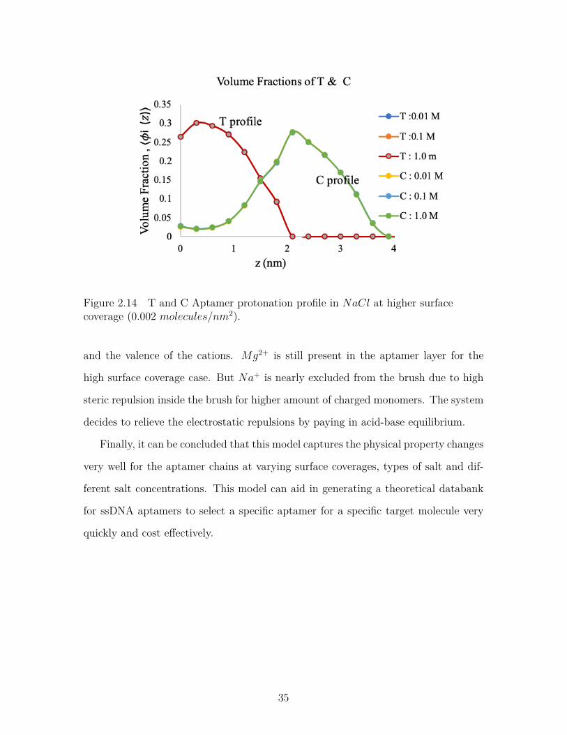
Figure 2.14 T and C Aptamer protonation profile in NaCl at higher surfacecoverage (0.002 molecules/nm2). . . . . . . . . . . . . . . . . . . 35
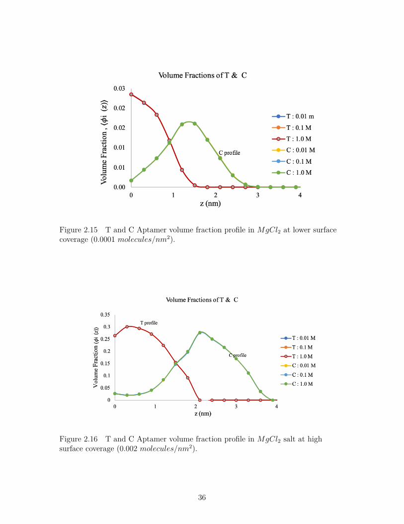
Figure 2.15 T and C Aptamer volume fraction profile in MgCl2 at lowersurface coverage (0.0001 molecules/nm2). . . . . . . . . . . . . . 36
Figure 2.16 T and C Aptamer volume fraction profile inMgCl2 salt at highsurface coverage (0.002 molecules/nm2). . . . . . . . . . . . . . . 36
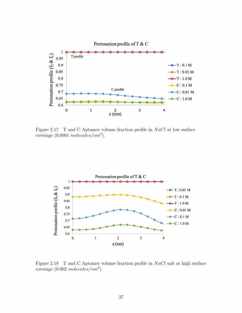
Figure 2.17 T and C Aptamer volume fraction profile inNaCl at low surfacecoverage (0.0001 molecules/nm2). . . . . . . . . . . . . . . . . . . 37
Figure 2.18 T and C Aptamer volume fraction profile in NaCl salt at highsurface coverage (0.002 molecules/nm2). . . . . . . . . . . . . . . 37
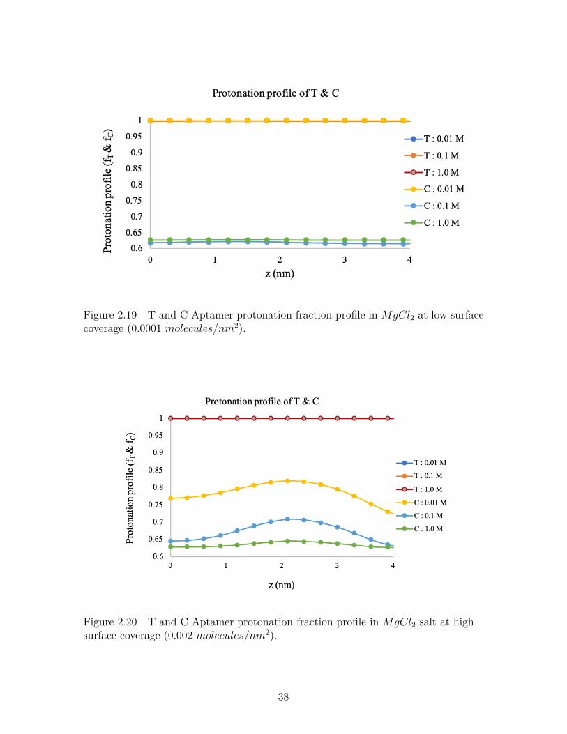
Figure 2.19 T and C Aptamer protonation fraction profile in MgCl2 at lowsurface coverage (0.0001 molecules/nm2). . . . . . . . . . . . . . 38
Figure 2.20 T and C Aptamer protonation fraction profile in MgCl2 salt athigh surface coverage (0.002 molecules/nm2). . . . . . . . . . . . 38
xi
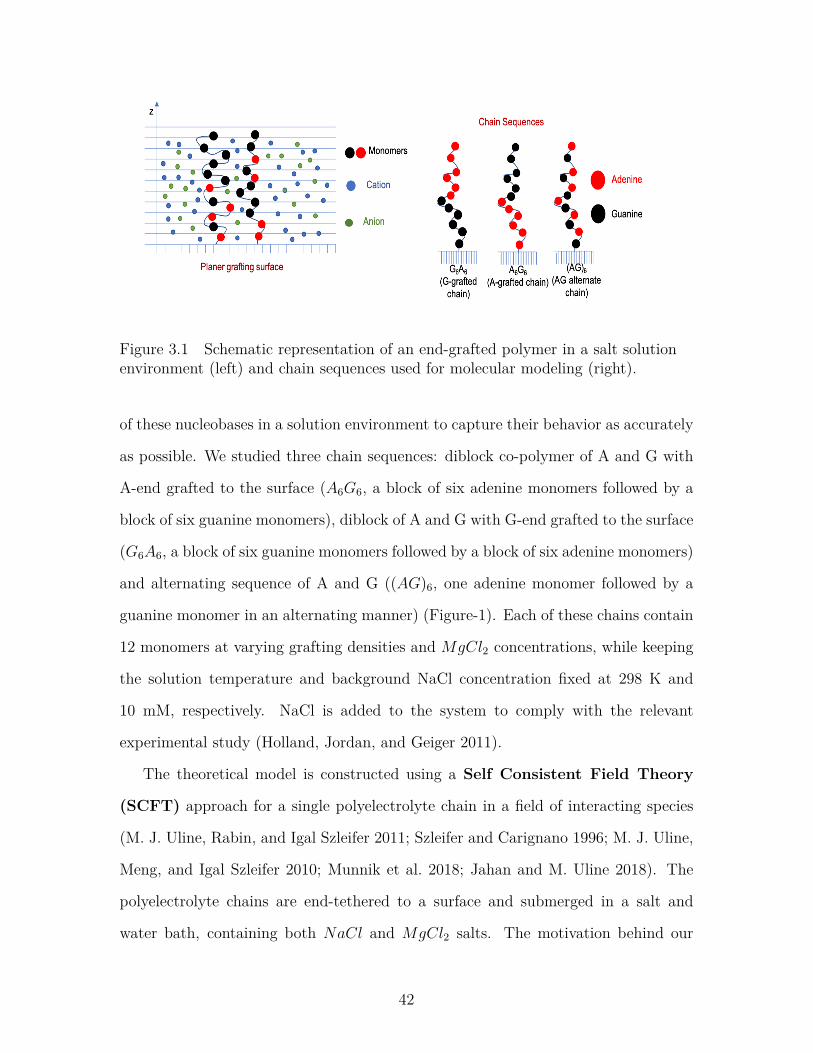
Figure 3.1 Schematic representation of an end-grafted polymer in a saltsolution environment (left) and chain sequences used for molec-ular modeling (right). . . . . . . . . . . . . . . . . . . . . . . . . . 42
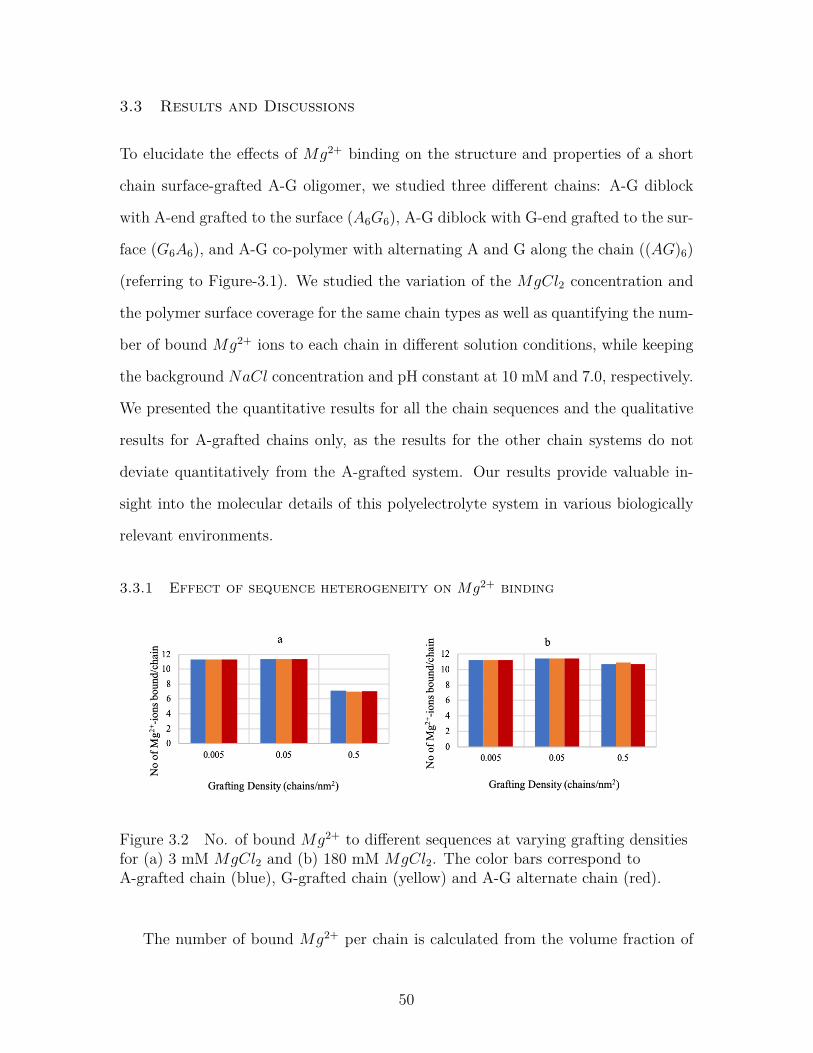
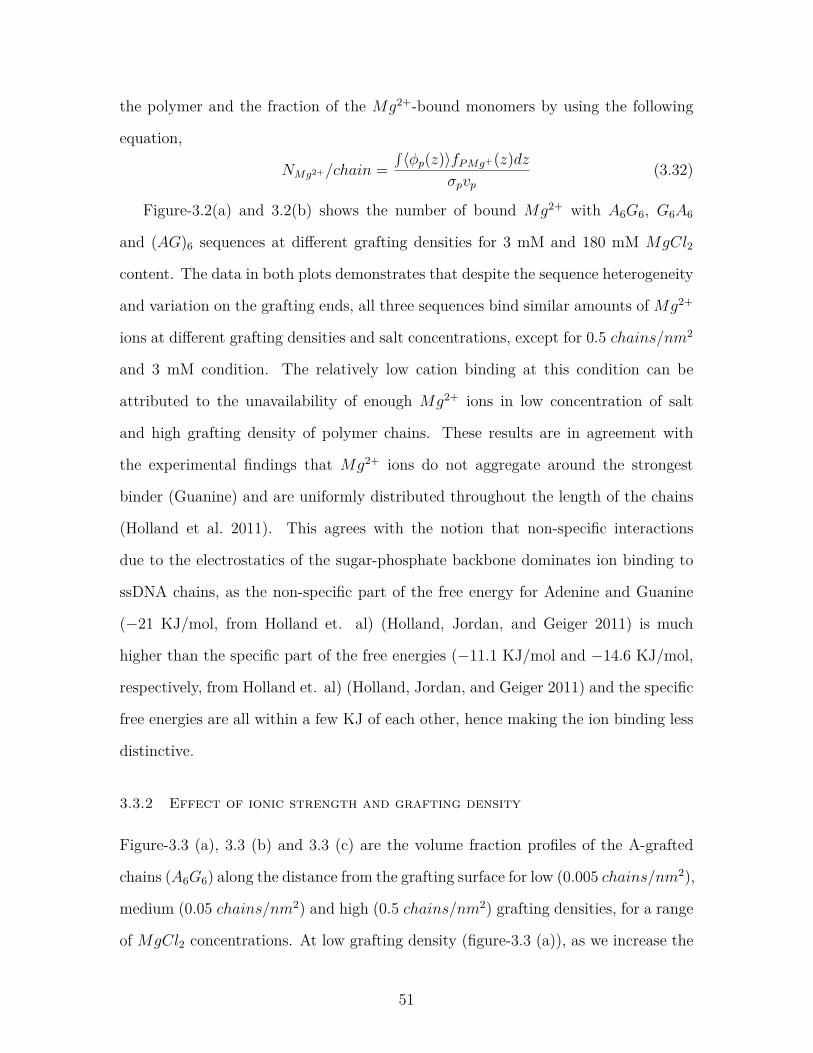
Figure 3.2 No. of bound Mg2+ to different sequences at varying graftingdensities for (a) 3 mM MgCl2 and (b) 180 mM MgCl2. Thecolor bars correspond to A-grafted chain (blue), G-grafted chain(yellow) and A-G alternate chain (red). . . . . . . . . . . . . . . . 50
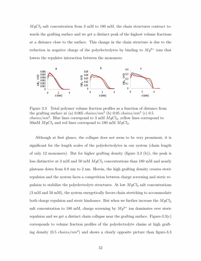
Figure 3.3 Total polymer volume fraction profiles as a function of distancefrom the grafting surface at (a) 0.005 chains/nm2 (b) 0.05chains/nm2 (c) 0.5 chains/nm2. Blue lines correspond to 3mM MgCl2, yellow lines correspond to 50mM MgCl2 and redlines correspond to 180 mM MgCl2. . . . . . . . . . . . . . . . . 52
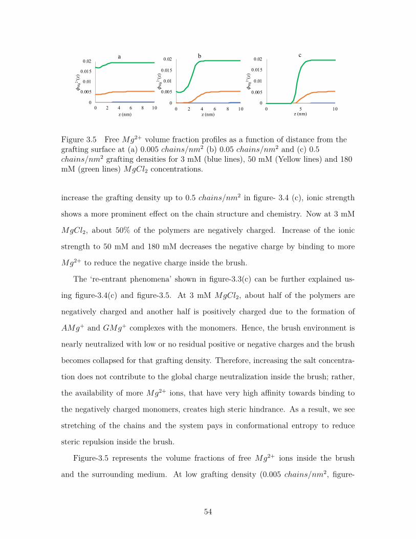
Figure 3.5 Free Mg2+ volume fraction profiles as a function of distancefrom the grafting surface at (a) 0.005 chains/nm2 (b) 0.05chains/nm2 and (c) 0.5 chains/nm2 grafting densities for 3mM (blue lines), 50 mM (Yellow lines) and 180 mM (greenlines) MgCl2 concentrations. . . . . . . . . . . . . . . . . . . . . . 54
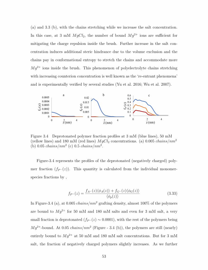
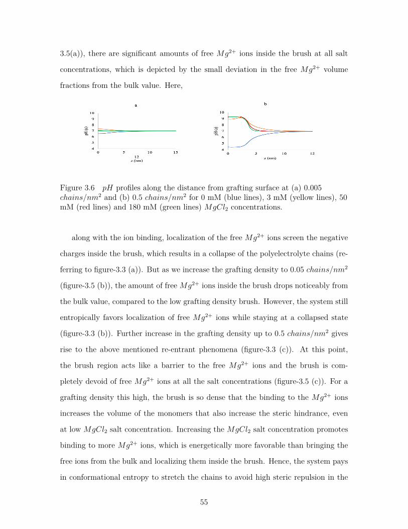
Figure 3.6 pH profiles along the distance from grafting surface at (a) 0.005chains/nm2 and (b) 0.5 chains/nm2 for 0 mM (blue lines), 3mM (yellow lines), 50 mM (red lines) and 180 mM (green lines)MgCl2 concentrations. . . . . . . . . . . . . . . . . . . . . . . . . 55
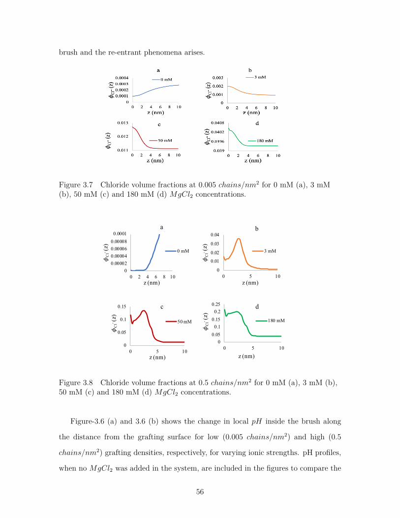
Figure 3.7 Chloride volume fractions at 0.005 chains/nm2 for 0 mM (a),3 mM (b), 50 mM (c) and 180 mM (d) MgCl2 concentrations. . . 56
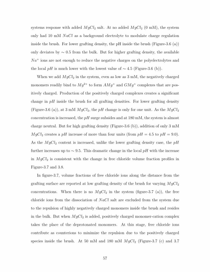
Figure 3.8 Chloride volume fractions at 0.5 chains/nm2 for 0 mM (a), 3mM (b), 50 mM (c) and 180 mM (d) MgCl2 concentrations. . . . 56
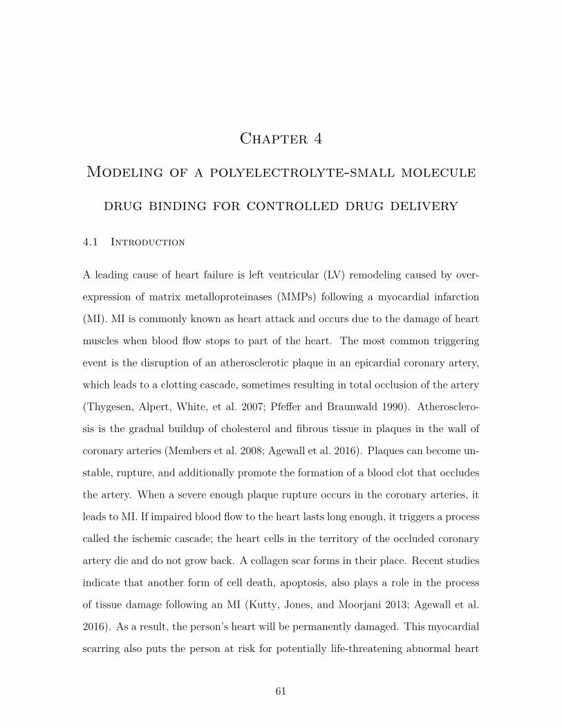
Figure 4.1 Schematic diagram of a human heart in normal condition andafter Myocardial Infarction (MI). Figure adopted from Compli-cations of myocardial infarction Kernel Description n.d. . . . . . . 62
xii
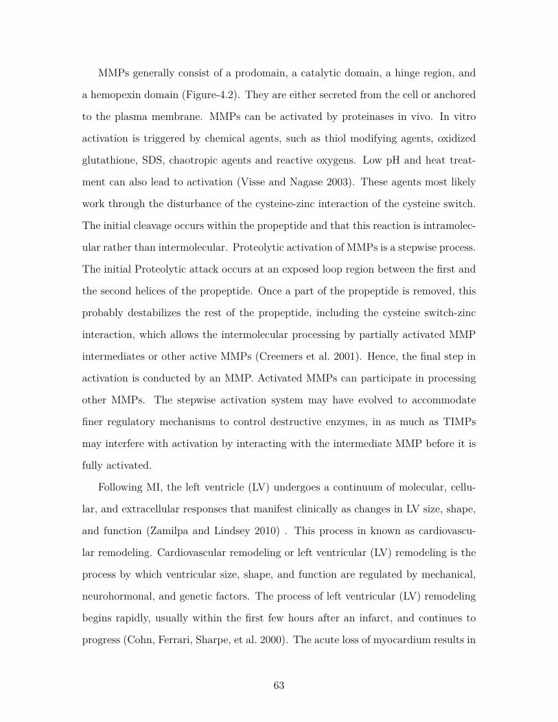
Figure 4.2 Domain structure of MMPs. The domain organization of MMPsis as indicated: S, signal peptide; Pro, propeptide; Cat, cat-alytic domain; Zn, active-site zinc; Hpx, hemopexin domain;Fn, fibronectin domain; V, vitronectin insert; I, type I trans-membrane domain; II, type II transmembrane domain; G, GPIanchor; Cp, cytoplasmic domain; Ca, cysteine array region; andIg, IgG-like domain. Figure adopted from Visse and Nagase 2003. 64
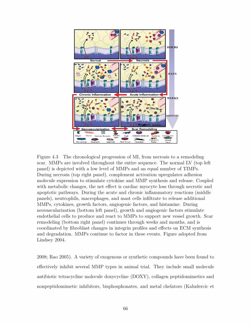
Figure 4.3 The chronological progression of MI, from necrosis to a remod-eling scar. MMPs are involved throughout the entire sequence.The normal LV (top left panel) is depicted with a low levelof MMPs and an equal number of TIMPs. During necrosis(top right panel), complement activation upregulates adhesionmolecule expression to stimulate cytokine and MMP synthesisand release. Coupled with metabolic changes, the net effect iscardiac myocyte loss through necrotic and apoptotic pathways.During the acute and chronic inflammatory reactions (middlepanels), neutrophils, macrophages, and mast cells infiltrate torelease additional MMPs, cytokines, growth factors, angiogenicfactors, and histamine. During neovascularization (bottom leftpanel), growth and angiogenic factors stimulate endothelial cellsto produce and react to MMPs to support new vessel growth.Scar remodeling (bottom right panel) continues through weeksand months, and is coordinated by fibroblast changes in integrinprofiles and effects on ECM synthesis and degradation. MMPscontinue to factor in these events. Figure adopted from Lindsey2004. . . . . . . . . . . . . . . . . . . . . . . . . . . . . . . . . . . 66
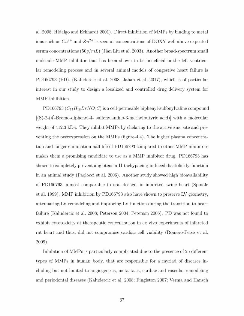
Figure 4.4 Structure of PD166793 (panel A). The tight binding of theinhibitor in the catalytic site of the enzyme is due to car-boxylic acid-zinc ligation, the carboxylate hydrogen bondingwith Glu202 and hydrogen bonding between the sulfonamidemoiety and Leu164 and Ala165. In addition, S1’ pocket presentin MMP-3 is occupied by 4’-bromo- substituted biphenyl ringsystem resulting in a more potent inhibition (panel B). Figureadopted from Kaludercic et al. 2008 . . . . . . . . . . . . . . . . . 68
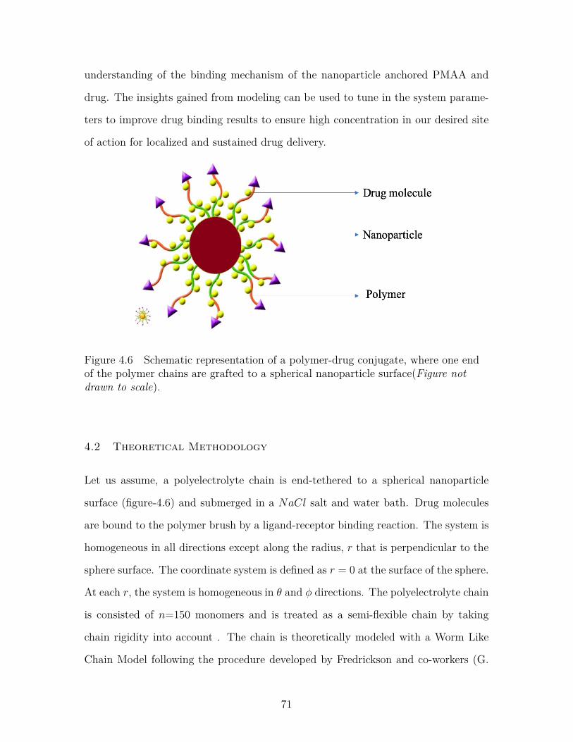
Figure 4.6 Schematic representation of a polymer-drug conjugate, whereone end of the polymer chains are grafted to a spherical nanopar-ticle surface(Figure not drawn to scale). . . . . . . . . . . . . . . . 71
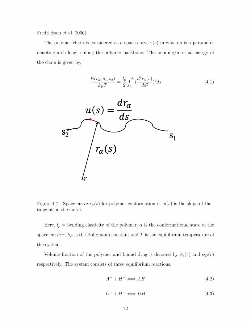
Figure 4.7 Space curve rα(s) for polymer conformation α. u(s) is the slopeof the tangent on the curve. . . . . . . . . . . . . . . . . . . . . . 72
xiii
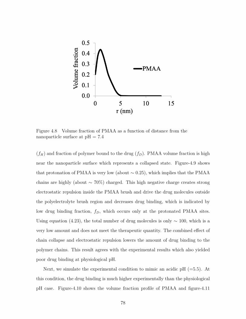
Figure 4.8 Volume fraction of PMAA as a function of distance from thenanoparticle surface at pH = 7.4 . . . . . . . . . . . . . . . . . . 78
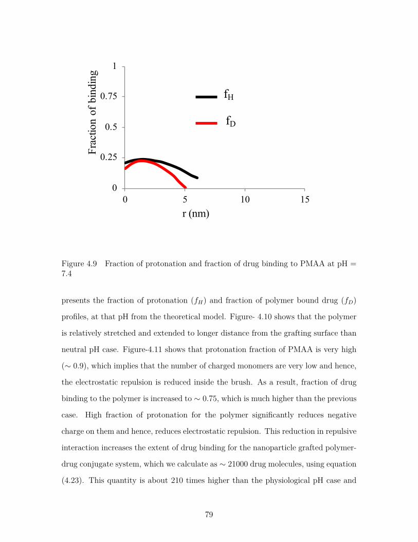
Figure 4.9 Fraction of protonation and fraction of drug binding to PMAAat pH = 7.4 . . . . . . . . . . . . . . . . . . . . . . . . . . . . . . 79
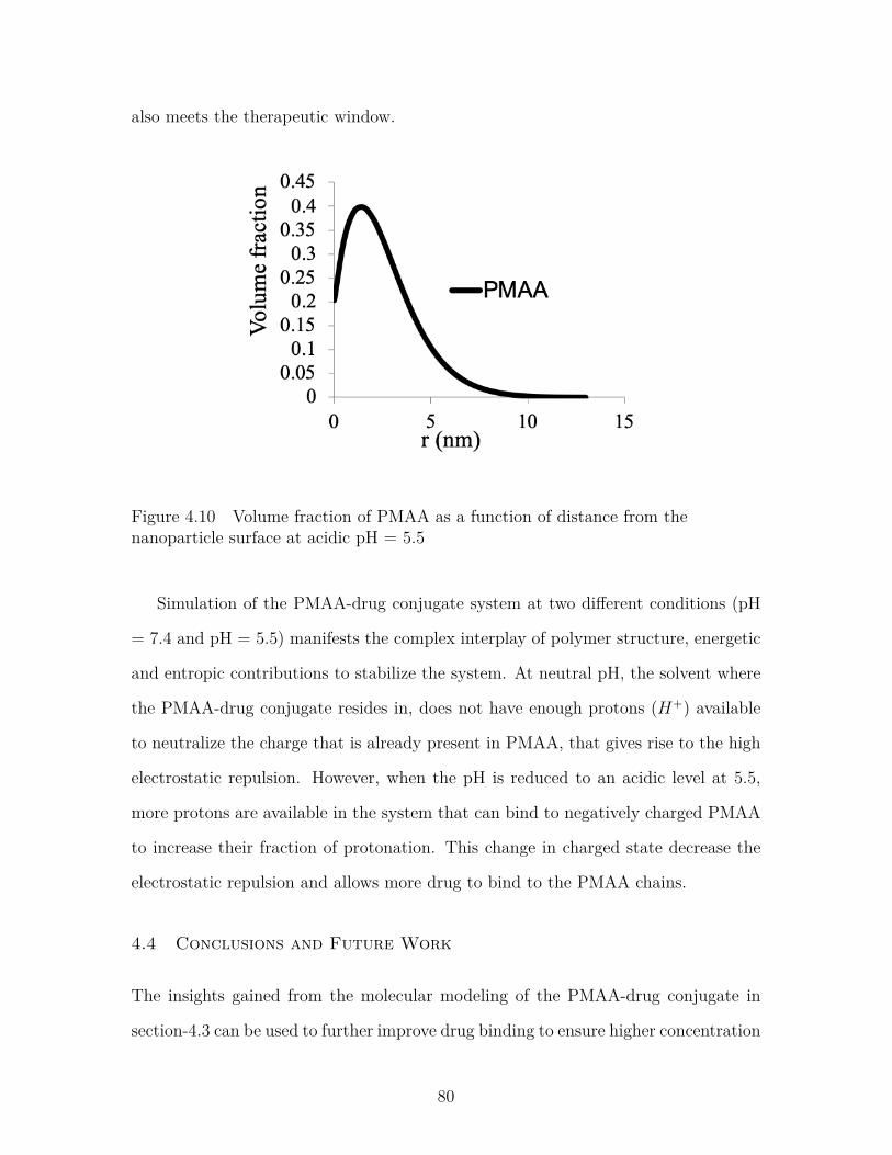
Figure 4.10 Volume fraction of PMAA as a function of distance from thenanoparticle surface at acidic pH = 5.5 . . . . . . . . . . . . . . . 80
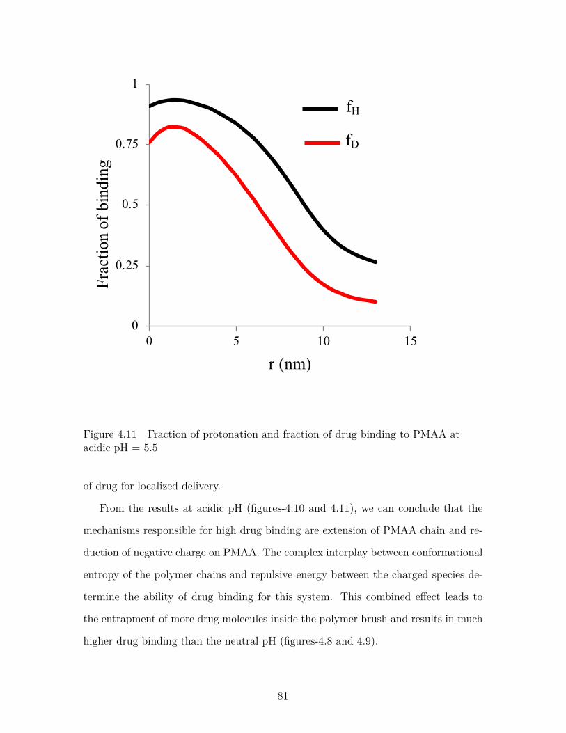
Figure 4.11 Fraction of protonation and fraction of drug binding to PMAAat acidic pH = 5.5 . . . . . . . . . . . . . . . . . . . . . . . . . . 81
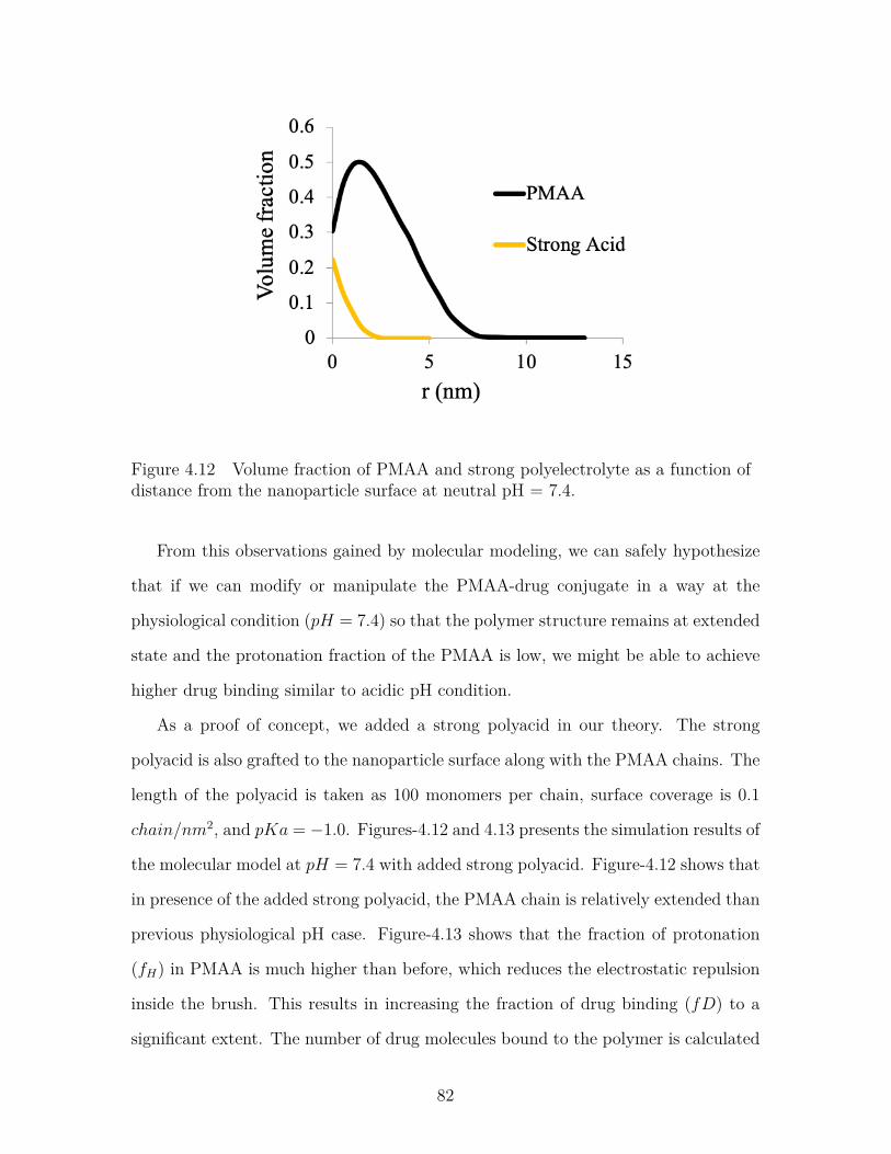
Figure 4.12 Volume fraction of PMAA and strong polyelectrolyte as a func-tion of distance from the nanoparticle surface at neutral pH =7.4. . . . . . . . . . . . . . . . . . . . . . . . . . . . . . . . . . . . 82
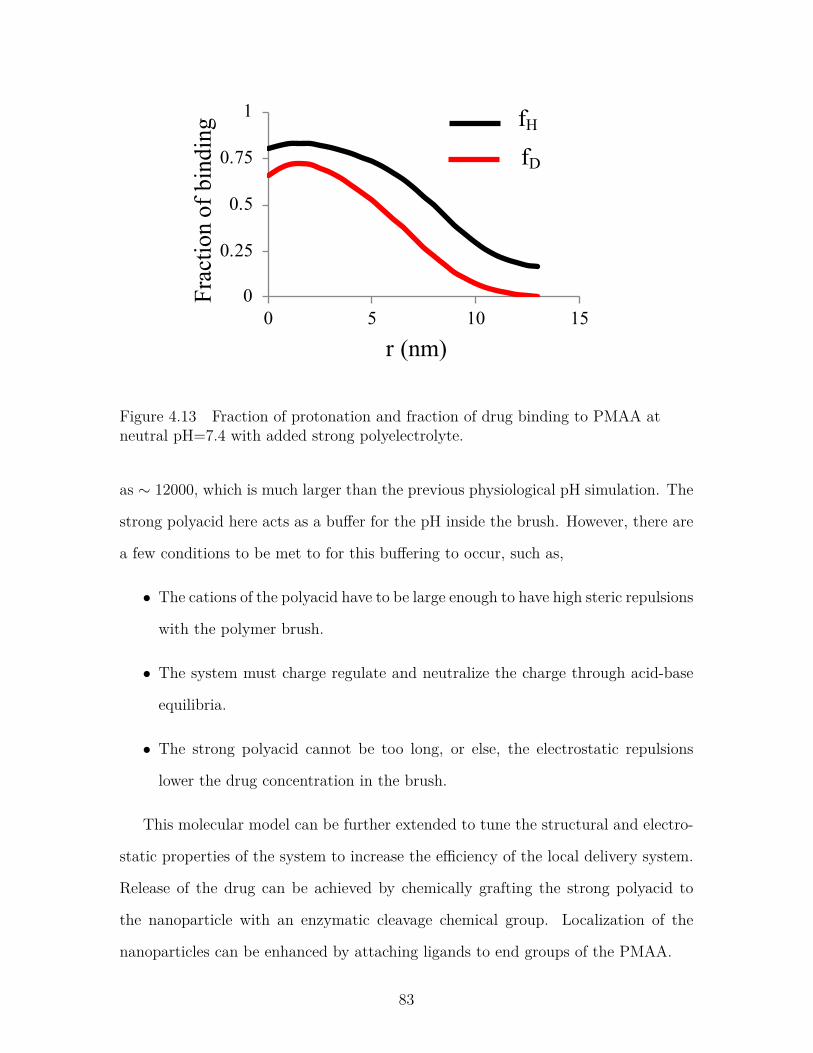
Figure 4.13 Fraction of protonation and fraction of drug binding to PMAAat neutral pH=7.4 with added strong polyelectrolyte. . . . . . . . 83
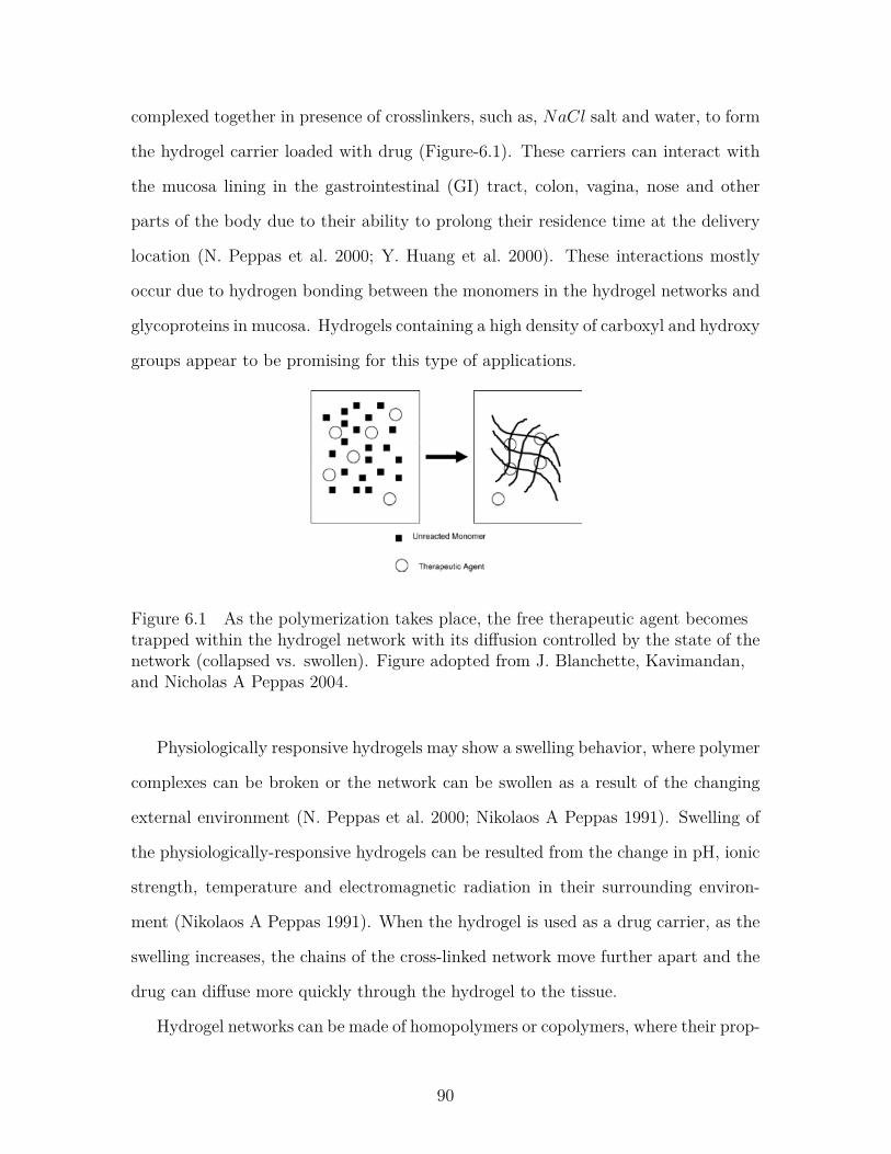
Figure 6.1 As the polymerization takes place, the free therapeutic agentbecomes trapped within the hydrogel network with its diffusioncontrolled by the state of the network (collapsed vs. swollen).Figure adopted from J. Blanchette, Kavimandan, and NicholasA Peppas 2004. . . . . . . . . . . . . . . . . . . . . . . . . . . . . 90
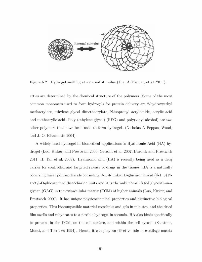
Figure 6.2 Hydrogel swelling at external stimulus (Jha, A. Kumar, et al. 2011). 91
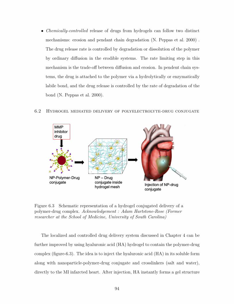
Figure 6.3 Schematic representation of a hydrogel conjugated delivery of apolymer-drug complex. Acknowledgement : Adam Hartstone-Rose (Former researcher at the School of Medicine, Universityof South Carolina) . . . . . . . . . . . . . . . . . . . . . . . . . . 94
xiv
Chapter 1
Introduction
Macromolecules, generally known as polymers, are large molecules made up of single
units called monomers. The major classes of molecules that are necessary for life on
earth are biological macromolecules or biopolymers, such as, proteins, lipids, carbo-
hydrates and nucleic acids. From lipid bilayers in our cell membrane to DNA in our
hereditary genes, all are examples of polymers. All of these polymers show different
level and nature of interactions with one another and also with other organic or inor-
ganic substances in their surrounding environment. All the functions in a living body
are governed by these interactions. Hence, it is of utmost importance to have a fun-
damental understanding of the interactions of different polymers with other organic
or inorganic substances to leverage their capabilities in novel biomedical applications.
Polyelectrolytes are a class of polymers that are capable of protonating and depro-
tonating in aqueous solution environment. Their monomer units bear an electrolyte
group that dissociates and makes the polymers charged in suitable polar solvent
(mostly water). Polyelectrolytes can be positively charged, which are called polyca-
tions, or negatively charged, called polyanions. Polyelectrolytes are of strong interest
in polymer science due to their wide range of applications and because most biological
macromolecules, such as, DNA, some proteins, fatty acids, etc., are polyelectrolytes.
Polyelectrolytes in solutions exhibit significantly different behaviors than uncharged
macromolecules and low molecular weight electrolytes (Hara 1992). The presence
of charges on the polyelectrolyte chains leads to intra and intermolecular interac-
tions that are stronger and of much larger range than uncharged polymers. These
1
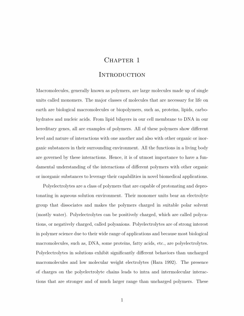
Figure 1.1 Schematic representation of a tethered polymer brush. The figure isadopted from Szleifer and Carignano 1996.
interactions give rise to distinctive conformational, thermodynamic, electrostatic and
chemical properties of the polyelectrolytes. These properties can be tuned to employ
the polyelectrolytes in a variety of biomedical applications ranging from biosensing
to controlled drug delivery (Scranton, Rangarajan, and Klier 1995).
When one end of a polymer chain is grafted or anchored to a surface, they are
called ‘tethered polymers’. The properties of tethered polymers in solution environ-
ments are qualitatively different than that of polymers in bulk. This difference stems
from the presence of the grafting surface that limits their configurational space and
the two-dimensional anchoring gives the repulsive interaction between neighboring
chains a different nature than their bulk counterparts (Szleifer and Carignano 1996).
Tethered polymers change the interaction of the tethering surface with their surround-
ing environments which makes them promising candidates for surface modification of
a variety of materials. They are found to be useful in a wide range of applications,
including but not limited to, colloid stabilization (Napper 1983), biocompatible mate-
rials (Brannon-Peppas 2000), controlled drug delivery vehicles (Torchilin et al. 1994;
2
Ji et al. 2019; J. S. Kim et al. 2019), biosensors (Badoux, Billing, and Klok 2019;
Hu et al. 2019; Andersson and Knoll 2019), etc. When the tethered polymers have
charge on them, they are called ‘tethered polyelectrolytes’, which are the subject of
interest for this study.
Along with experimental studies, theory played a vital role since the very begin-
ning of polymer science. The work of first generation of polymer theorists tackled
fundamental problems of polymer chain conformations, colligative properties and
phase behavior from the perspective of physical chemistry. The second generation
of polymer scientists combined the concepts of theoretical physics with polymer sta-
tistical mechanics to analyze important problems, such as, excluded volume effect in
polymers. The third generation of polymer research developed the foundation of the
most prominent polymer theory named as Self Consistent Field Theory (SCFT) to
study equilibrium properties of inhomogeneous polymers. Various analytical tech-
niques were employed to solve the SCFT equations and apply the theory in broad
areas of applications, such as, polymer alloys, block copolymers, graft copolymers and
tethered polymer layers. With the advance in computing technologies, the current
generation of polymer science employs various numerical and computer simulation
techniques, such as, Monte Carlo (MC), Molecular Dynamics (MD) simulations (G.
Fredrickson et al. 2006).
This research endeavor focuses on understanding the structural, physicochemical
and thermodynamic property changes of tethered polyelectrolytes in solution envi-
ronments when they interact with other neighboring molecules and incorporate that
into a molecular theory to facilitate their applications in areas of unmet biomedical
needs. This molecular theory follows a single-chain mean field approach that is based
on the Self Consistent Field Theory (SCFT) and takes into account the structural,
thermodynamic and electrostatic properties of all the molecules involved in a system.
Each of these properties shapes the nature of interactions among the biomolecules
3
and their environments. The molecular theory aims to explain the thermodynamic
and physicochemical property changes of tethered polyelectrolytes for biosensing and
drug delivery applications and leverage their tunability to achieve better performance
of these systems.
1.1 Self-Consistent Field Theory (SCFT) modeling of tethered
polyelectrolyte chains in solution
Self Consistent Field Theory was originally developed to treat bulk polymer systems
containing freely jointed chains (Sam F Edwards 1965). It was later modified to study
inhomogeneous systems where the polymers are tethered to hard surfaces (Dolan and
Samuel Frederick Edwards 1974).
The basic concept of SCFT is that the polymer chains are considered to be af-
fected by a position r dependent single field, w(r), which is the average or mean of
all the attractive and repulsive interactions between the polymer segments and their
surrounding environments. It is called ‘self-consistent’, because the mean field is de-
rived self consistently by assuming that the field variables are stationary with respect
to the mean field w(r) and then solving the equations defining the field variables
simultaneously that also gives mean field w(r) (G. Fredrickson et al. 2006).
SCFT has been employed extensively to study tethered polyelectrolytes for nu-
merous applications and nicely captured their physicochemical behavior in solution
environments (Pincus 1991; Zhulina and Borisov 1997). Polyelectrolytes can be cat-
egorized as ‘strong’ or ‘weak’ polyelectrolytes depending on their degree of dissoci-
ation. Strong polyelectrolytes completely dissociate in solution, such as Polystyrene
Sulfonate (PSS), whereas weak polyelectrolytes are partially dissociated, such as Poly-
acrylic acid (PAA), nucleic acids, etc. The tethered polyelectrolyte systems behave
differently in presence and absence of salt in the solution, which was captured ac-
curately with SCFT (Pincus 1991; Borisov and Zhulina 1998; Borisov, Birshtein,
4
and Zhulina 1991; Zhulina, Borisov, and Birshtein 1992). In absence of salt, densely
grafted strong polyelectrolytes form an ‘osmotic’ regime where all the counterions are
trapped inside the brush and osmotic pressure creates swelling effect inside the brush.
However, for sparsely grafted weak polyelectrolytes, the electrostatic attraction be-
tween the polyions and the counterions is not enough to trap the counterions inside
the brush, which results in dispersing them in outer solution environment, breaking
the local electroneutrality and creating a charged brush.
When salt is added to the tethered polyelectrolyte system, the cations and anions
from the dissociated salt creates screening effect and diminishes electrostatic swelling
that results in collapse of the brush (Brettmann et al. 2016; M. J. Uline, Rabin, and
Igal Szleifer 2011). The presence of salt also ensures global electroneutrality of the
polyelectrolyte system by the mobile counterions (Zhulina and Borisov 1997; Rikkert
Nap, Gong, and Igal Szleifer 2006). All of the molecular theories reported in this dis-
sertation includes salt as an integral part, because most polyelectrolytic formulations
require addition of salt to control the ionic strength and charge regulation inside and
outside the brush (G. Fredrickson et al. 2006).
With all these scopes of variability, tethered polyelectrolytic systems in various
biomedical applications possess a wide range of parameter space. Furthermore, rapid
development of new intelligent polymer-based materials makes the scenario more com-
plicated. While combinatorial discovery chemistry provides powerful tools to study
these materials, the process is often very expensive and time consuming. Theoretical
study on this wide parameter space for new biomedical applications renders to be
very useful in this regard, to scan the properties of these systems and map them into
a generalized theory. The insights gained from the theory would enable the experi-
mental researchers to optimize the number of experiments conducted and accelerate
the materials discovery process.
Apart from SCFT, Molecular Dynamics (MD) and Monte Carlo (MC) simulations
5
are powerful tools to study polymeric systems with wide design space. Both of the MD
and MC techniques track the motion of particles or molecules through Lagrangian or
Hamiltonian dynamics. In MD simulation, the temporal evolution or trajectory of the
coordinates and the momenta of a given macromolecular structure is studied (Paquet
and Viktor 2015). The trajectory is important to access valuable time-dependent
information about the system, such as, the accessibility of a given molecular surface,
the intermolecular interaction, etc. (Lindorff-Larsen et al. 2012; Harris et al. 2013).
MC simulation generates an ensemble of representative configurations under specific
thermodynamic conditions for a complex polymeric system through sampling of most
probable conformations (Fichthorn and Weinberg 1991; Paquet and Viktor 2015).
MC simulations are not time dependent and provide an ensemble of representative
configurations and conformations, which consequently gives probabilities and relevant
thermodynamic observables, such as the free energy.
To most accurately model complicated many-body systems as tethered polyelec-
trolytes, one would intuitively think of MD or MC, as these models use particle
co-ordinates for exact solution of the equation of motion of the molecules (MD) or
sample the configuration space (MC) (Szleifer and Carignano 1996). However, track-
ing each molecule in a system and solving the equations of motion requires defining the
interactions between different units, polymer segments, and solvent molecules. While
using these approaches provides valuable information on the underlying physics of
tethered polyelectrolytes, the computational cost of conducting these calculations is
intractable and resources are often unavailable to most researchers (G. Fredrickson et
al. 2006). To tackle this difficulty, coarse-grained field based models as SCFT, where
the fundamental degrees of freedom is a mean-field of all the available interactions
instead of particle co-ordinates, proves to be more useful to provide crucial thermo-
dynamic and structural information at a good degree of accuracy. That is why, we
choose SCFT to model tethered polyelectrolytes for various biomedical applications.
6
In this work, we have studied polyelectrolytic biomolecule Aptamer, which is a
type of ssDNA capable of binding to a specific target molecule with high affinity and
specificity. Molecular modeling of aptamers would enable us to have fundamental
understanding on their property changes in various biological environments and use
that for high-throughput experiments to design new aptamers with increased func-
tionality. We have also studied a nanoparticle-polyelectrolyte mediated drug delivery
system for enhanced repair in case of a cardiovascular disease. These studies can pro-
vide valuable support to experimental researchers to design new polymeric materials
for highly efficient biosensing and drug delivery technologies.
1.2 Physical Significance of Molecular Modeling
Molecular modeling of tethered polymeric systems provides valuable information
about the underlying mechanisms of physicochemical property changes and allows
us to predict the system behavior. A major contributor in defining the structure of
polyelectrolyte brush is the ionic strength of the salt present in the system and va-
lence of the cations. Previous studies have reported that addition of a small amount
of salt resulted in collapse of the polymer chains due to the decrease in charge on
the polymer chains, but high concentration of salt creates high steric repulsion that
results in chain stretching (Rikkert J Nap, S. H. Park, and Igal Szleifer 2018; Gong
et al. 2007). The results of these theoretical studies were in qualitative agreements
with collaborative experimental research (Y. Park et al. 2012).
The major property changes of the polymeric systems discussed in this dissertation
are: volume fractions and fraction of protonation of all the species (polymer, water,
anions and cations) and pH in different layers of the brush. The position dependent
volume fraction profiles of the polymers correspond to the layer by layer assembly of
the polymer brush and carry the information about whether the chains are in collapsed
or extended state. This information about structure and length of the chains can
7
complement experimental X-ray Scattering data to understand the underpinnings of
a system behavior (Pütz, Curro, and Grest 2001). The Volume fraction profiles of
water, anions and cations at different positions in the system shows the inclusion or
exclusion of the respective species from the brush layer, which can be experimentally
measured by Atomic force Microscopy (AFM) (Holland et al. 2011; Holland, Jordan,
and Geiger 2011). The volume fraction data can be used to calculate the height of
the polymer brush and the number of other molecules or ions trapped in the brush,
which can correspond to UV-spectroscopy data through distinct absorption peaks
(Dunlap et al. 2018). The protonation profiles presents the distribution of charged
monomers throughout the polymer layer. High fraction of protonation means less
charged monomers and vice versa. Presence of charge in a polymer chain can also be
detected by AFM, which can be accompanied by the protonation fraction from the
molecular model to comprehend the charge scenario.
1.3 Assumptions of the Molecular Theory
The assumptions that we used to construct the molecular theories in this dissertation
are:
• Single chain mean field approximation: To construct the molecular theory
based on SCFT, we follow a single chain approach developed by Szleifer and his
group (Szleifer and Carignano 1996; Rikkert Nap, Gong, and Igal Szleifer 2006).
In this approach, for a many chain system, instead of looking at all the chain
molecules, we look at a central chain with all its intramolecular interactions
taken exactly, while taking the intermolecular interaction with a mean field
approximation. This approach enables us to understand the conformational
changes of the tethered polyelectrolytes depending on thermodynamic control
variables, that is, surface coverage and temperature. This provides valuable
8
insights on the coupling that exists between the chain conformations and the
thermodynamic behavior of the layer.
• Incompressibility Constraint or Volumetric Constraint: It is assumed
that the polyelectrolyte system is incompressible, meaning all the available vol-
ume is filled with either of the species (polymer, solvent, salt and water) present
in the system. Hence, the summation of volume fractions of all the species at a
certain location equals to unity. Additionally, the incompressibilty constraint is
a way to take into account the repulsive interactions between polymer segments
that ensures self-avoidance of a chain (Szleifer and Carignano 1996).
• We neglect any volume change of the monomers due to protonation and metal
binding reactions and assume that the segments have the same volume whether
they are protonated and bound to other ions or not. This assumption is made
due to the lack of volume change data and used only while carrying out the
numerical calculations.
• The dielectric coefficient, ε, is assumed to be invariant with position and taken
as twice as the dielectric coefficient of water, following the argument of Uline
et al (M. J. Uline, Rabin, and Igal Szleifer 2011).
1.4 Chain Models
The solutions of the molecular models require conformational statistics of the polymer
in that particular application. Based on the length scale of the system and chain
length, we use two chain models listed below in our molecular theories:
1.4.1 Rotational Isomeric State Model
Rotational isomeric State (RIS) model was developed by P. J. Flory to treat the con-
figuration dependant properties of chain molecules and to establish the connections
9
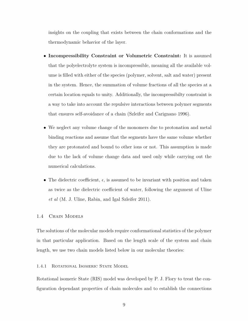
Figure 1.2 Rotational Isomeric State Model for a hydrocarbon chain. θ is the bondangle and φ is the rotation angle. l0 is the bond length.
between conformational energy and the properties of the macromolecules (P. Flory
1974; Paul J Flory and Volkenstein 1969). In this model , each bond can have three
possible states: trans, gauche+, and gauche− with angles φ=0, +120o and −120o,
respectively and the angle between bonds is θ=68o. The continuous rotational de-
grees of freedom about the backbone single bonds in the polymer are replaced by a
finite number of trans, gauche+, and gauche− states. RIS is generally used to treat
flexible chains with intermediate chain length. For details on calculations regarding
RIS, the above mentioned references can be consulted.
1.4.2 Wormlike Chain Model
Many polymers in biological systems exhibit rod-like rigidity in their structure that
makes them semiflexible. A more appropriate chain model for the semi-flexible chains
is Kratky-Porod model, which is generally known as Wormlike Chain model (WLC)
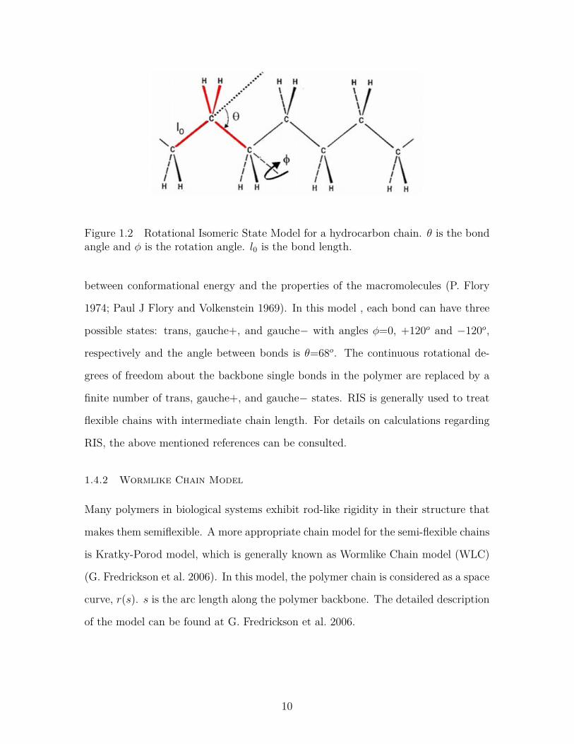
(G. Fredrickson et al. 2006). In this model, the polymer chain is considered as a space
curve, r(s). s is the arc length along the polymer backbone. The detailed description
of the model can be found at G. Fredrickson et al. 2006.
10
Figure 1.3 Schematic representation of a Wormlike Chain with space curve r(s).
1.5 Thesis statement
This dissertation investigates the following statement:
Molecular level understanding of thermodynamic and physicochemical property changes
of tethered polyelectrolytes can be leveraged to design new systems for biosensing and
drug delivery applications.
1.6 Organization of the Dissertation
The rest of this dissertation is organized as follows-
• In Chapter 2, we developed a molecular theory with a biological polyelectrolyte
called Aptamer to understand its thermodynamic and physicochemical property
changes in an aqueous solution environment. The understanding gained from
this study can aid in the selection of specific aptamers against specific target
molecules of biological interest.
• In Chapter 3, aptamer behavior is studied in presence of divalent metal cations
(Mg2+) with a molecular theory and the cation binding is quantified to de-
termine the nature of the ion cloud. This field theoric model helps to set up
the foundation for future studies involving secondary and tertiary structures of
aptamers interacting with multivalent metal ions.
• In Chapter 4, we theoretically design a localized and controlled drug delivery
system for prolonged release of a drug for enhanced cardiovascular repair by
11
using a nanoparticle grafted polyelectrolyte as the drug carrier. The insights
gained from modeling can be used to tune in the system parameters to improve
drug binding results to ensure high concentration in our desired site of action
for localized and sustained drug delivery.
• Chapter 5 draws conclusion of this dissertation.
• Chapter 6 discusses the possible future directions that this research can take.
12
Chapter 2
Modeling of Aptamers
Aptamers are a very promising class of biomolecules that have multifunctional us-
age for various Biomedical applications. Aptamers are single stranded DNA/ RNA
oligonucleotides or peptide molecules which can bind to proteins, small molecules,
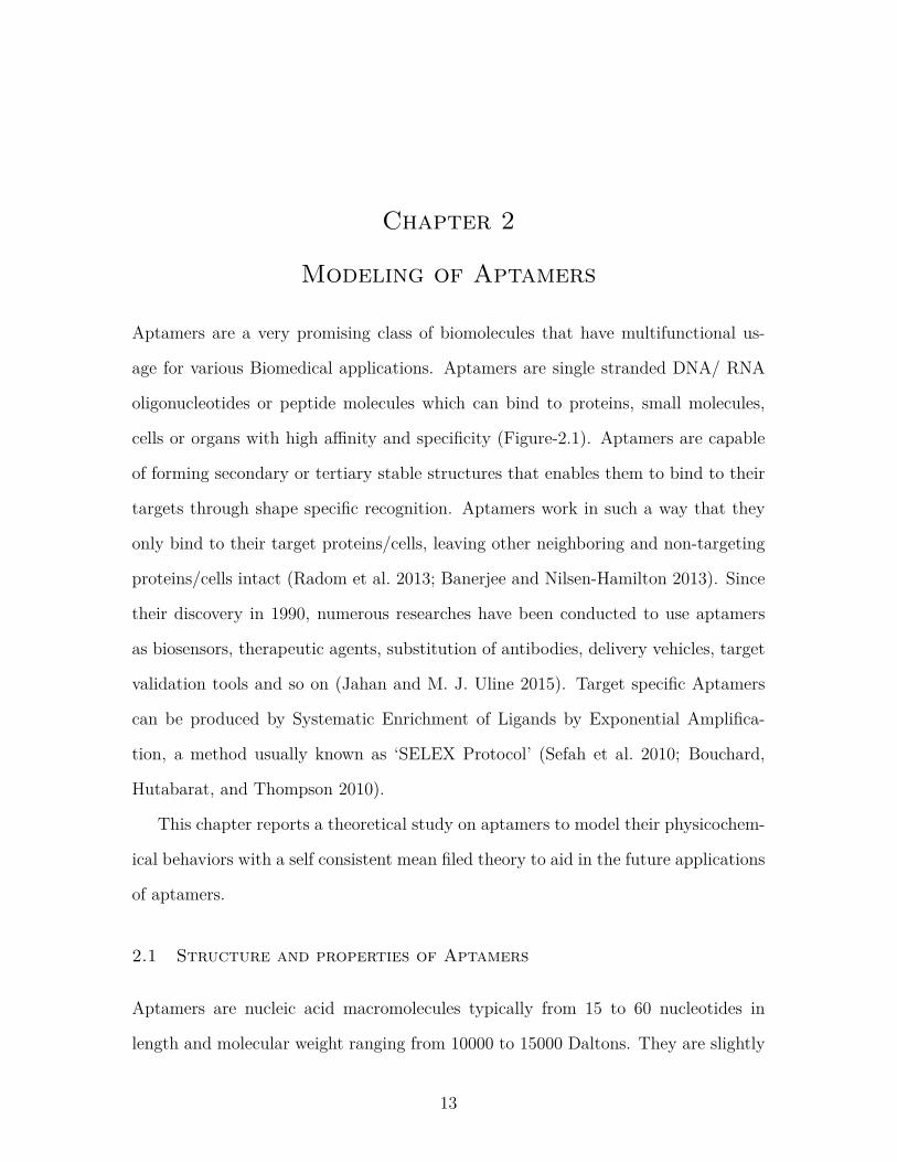
cells or organs with high affinity and specificity (Figure-2.1). Aptamers are capable
of forming secondary or tertiary stable structures that enables them to bind to their
targets through shape specific recognition. Aptamers work in such a way that they
only bind to their target proteins/cells, leaving other neighboring and non-targeting
proteins/cells intact (Radom et al. 2013; Banerjee and Nilsen-Hamilton 2013). Since
their discovery in 1990, numerous researches have been conducted to use aptamers
as biosensors, therapeutic agents, substitution of antibodies, delivery vehicles, target
validation tools and so on (Jahan and M. J. Uline 2015). Target specific Aptamers
can be produced by Systematic Enrichment of Ligands by Exponential Amplifica-
tion, a method usually known as ‘SELEX Protocol’ (Sefah et al. 2010; Bouchard,
Hutabarat, and Thompson 2010).
This chapter reports a theoretical study on aptamers to model their physicochem-
ical behaviors with a self consistent mean filed theory to aid in the future applications
of aptamers.
2.1 Structure and properties of Aptamers
Aptamers are nucleic acid macromolecules typically from 15 to 60 nucleotides in
length and molecular weight ranging from 10000 to 15000 Daltons. They are slightly
13
Figure 2.1 (A) Schematic representation of aptamer binding to a target proteindepending on structure formation. After the adjustment of the binding conditions,the aptamer folds into a 3D structure, upon which it interacts with the targetmolecule (e.g., a protein), resulting in a stable target-aptamer complex. (B) Thecrystallographic structure of the G protein-coupled receptor kinase 2 (GRK2)-C13complex is depicted as an example for a target-aptamer complex (Wolter andGünter Mayer 2017).
larger than small molecules but smaller than antibodies in size. Aptamers can be
composed of a modified sugar backbone in 2′ end (i.e., 2′- fluoro, 2′-O- methyl, phos-
phorothioate). Secondary structure of aptamers, either short helical arms or single
stranded loops, are defined from their complementary base pairing. Tertiary or sta-
ble structures resulting from these secondary structures enables aptamers to bind
to their targets through shape specific recognition. Van der Waals, hydrogen bond-
ing, electrostatic interaction and hydrophobic interactions are responsible for strong
aptamer-target binding (Pendergrast et al. 2005). They are hydrophilic and polyan-
ionic in nature. KD values of aptamers can range from 10 pM to 10 nM for proteins.
Here, KD = [L][R]/[LR] , where L denotes ligand or aptamers in our case, R is
14
receptor (protein) and LR stands for the ligand-receptor complex.
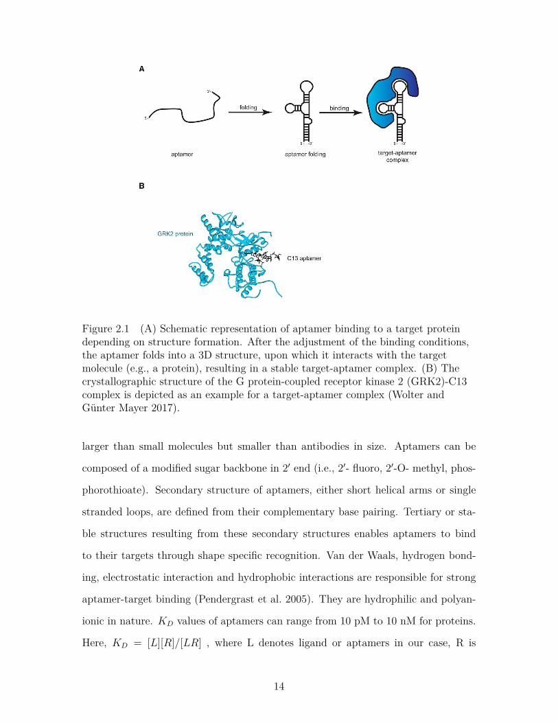
2.2 SELEX Protocol
Figure 2.2 SELEX Protocol (Sefah et al. 2010).
‘SELEX Protocol’ was first proposed by two independent groups (Ellington and
Szostak; Turek and Gold) in 1990. SELEX process requires a large library of single
stranded oligonucleotide templates derived from a chemical synthesis on a standard
DNA synthesizer. The library generally includes ∼ 1014 unique sequences (Bouchard,
Hutabarat, and Thompson 2010). The target of interest is incubated with the library,
followed by several washing steps to remove non-functional or unbound sequences.
Then the aptamer-target complex undergoes an elution process where the target is
separated from the binding sequences. These binding sequences then go through
a negative selection where they are allowed to mix with healthy cells, preferably
denoted as negative cells. The aptamers that bind to the negative cells are then
separated. The unbound aptamers after this step are the desired aptamers for that
target. The sequences are amplified by polymerase chain reaction (PCR) to yield a
practical amount. After several rounds of selection, the enriched library is cloned,
sequenced and characterized to isolate aptamers with desired characteristics (Sefah
15
et al. 2010; Bouchard, Hutabarat, and Thompson 2010).
2.3 Uses of Aptamers in Modern Biomedical Engineering
Aptamers can be used in various biomedical applications listed below:
2.3.1 Biosensors
Biosensors are a bimolecular probe that can measure the existence or concentration of
a specific biomolecule or biological structure (Sefah et al. 2009). The most immediate
commercial application of aptamers was as biosensors due to their compatibility with
various analytical technologies. They can specifically detect a large variety of targets
such as proteins, small molecules, nucleotides, metabolites, amino acids etc (Rimmele
2003). The interaction of aptamers with their targets can be converted to electrical
signals very conveniently using different transduction processes. The compatibility of
aptamers with various detection schemes like electrochemical, fluorescence, chemilu-
minescence, field effect transistors, potentiometry etc. surged the area of aptasensor
research (Sefah et al. 2009).
In aptamer biosensing, a ‘recognition aptamer’ for the specific target is coupled
with a ‘signaling aptamer’ by direct fusion of their nucleic acid sequences. For biosens-
ing applications, SELEX can be conducted in such a way that sequences for both
recognition and signaling can be selected for a target in the same round. This simple
system has the major advantage of ensuring that the recognition domain does not face
any adverse effect in their specificity upon binding to the signaling domain (Bunka
and Stockley 2006).
Quantum-dot aptamer beacons are a recent advancement of aptamers as biosen-
sors. Quantum-dots are flouropores having a distinct sharp emission profile. Aptamer
beacons consist of multiple aptamers bound to a single quantum-dot. Each aptamer
has a complimentary base pair carrying a quencher. Upon binding to the target, the
16
complement is displaced resulting in a large increase of fluorescence emission. These
highly specific aptamer beacons have great potential to be used for early detection
of diseases by binding to cell surface epitopes. Until now, aptamers have been se-
lected toward a broad range of targets, including metal ions (e.g., K+, Hg2+ and
Pb2+ ), small organic molecules (e.g., amino acids, ATP, antibiotics, vitamins and
cocaine) organic dyes, peptides and proteins (e.g., thrombin, growth factors and HIV-
associated peptides) and even whole cells or microorganisms (e.g., bacteria) (Günter
Mayer 2009).Another interesting development of biosensing aptamers is their conju-
gation with gold nanoparticles. Target binding causes a conformational change in the
aptamer leading to disassembly of the aggregated nanoparticles resulting in a visible
color change (Bunka and Stockley 2006).
2.3.2 Substitution of Antibodies
Antibodies are naturally occurring proteins found in the body and used by the im-
mune system to identify and neutralize antigens. Artificially produced antibodies
are also used in biomedical research for detection, identification and imaging of tar-
get molecules. In recent years, aptamers have shown very good compatibility as a
substitute of antibodies.
As compared to antibodies, aptamers are more stable in blood serum (W. Tan
et al. 2011). Chemically modified aptamers have better nuclease resistance than an-
tibodies. They are easily producible in commercial basis with a cost much lower than
antibodies. Antibodies only work extracellularly, but aptamers show both intracellu-
lar and extracellular functionality (Banerjee 2010). The most important advantage
of aptamers over antibodies is that the body does not show any immune response
against aptamers (Famulok and Mayer 1999; Foy et al. 2007). Prescribing antibody
always has a chance for immune response in the patient and once administered is
difficult to have control over the drug effect. With aptamer, however, the patients so
17
far treated did not show any kind of toxicity. For all these reasons, aptamers can suc-
cessfully replace antibodies for the treatment of macular degeneration, non-small cell
lung cancer, and thrombotic thrombocytopenic purpura, acute coronary syndrome,
von Willebrand factor-related disorder, angiomas, acute myeloid leukemia, and renal
cell carcinoma etc (Sundaram et al. 2013; Ng et al. 2006).
2.3.3 Therapeutic agents
Aptamers can modulate protein function which enables them to be used as therapeu-
tic agents. Chemical modifications of aptamers lead their increased half-life, nuclease
resistance and improved pharmacokinetics allowing their rigorous use in clinical ap-
plications. Even in unmodified form, aptamers can be used for treatment of transient
conditions like blood clotting with their low half-life and rapid clearance by the kid-
neys (Banerjee and Nilsen-Hamilton 2013). Conjugation of aptamers with PEG lead
to their increased half-life in blood. All kinds of modifications in aptamer structure
led to a significant enrichment of the aptamer in kidneys, liver, spleen, heart, and me-
diastinal lymph nodes, representing modulation in their pharmacokinetic properties
(W. Tan et al. 2011).
Superior targeting performance of aptamers raised the interest of using them for
cancer therapy. An aptamer named AS1411 is undergoing clinical trials, which can
specifically target a bcl-2 binding protein, nucleolin, responsible for cell proliferation.
Upon binding, AS1411 can enter the cancer cell and causes its death by apoptosis (W.
Tan et al. 2011). Anti-thrombin aptamers have been developed to increase clotting
time of human plasma. These aptamers are undergoing clinical trials for the treatment
of Acute Coronary Syndrome (ACS) (Günter Mayer 2009). Aptamers can also be used
as antiviral drug agents by preventing replication of escape mutants (Banerjee 2010).
18
2.3.4 Target validation tools
Target validation is the determination of whether a drug target is involved in disease
pathology. Aptamers can inhibit target function by blocking or knocking out gene
expression (Pendergrast et al. 2005). This makes them particularly important as
target validation tools . They bind with high affinity and specificity with target
molecules. Protein level function of aptamers can provide information complementary
to that obtained from gene-level validation approach. Aptamers can validate both
intracellular and extracellular targets. They can be easily delivered to the intracellular
target by using standard transfection techniques (Pendergrast et al. 2005).
2.3.5 Drug carriers
Aptamers can be assembled with different functional groups which provide the means
to use them as delivery vehicles that specifically address certain malignant cell sub-
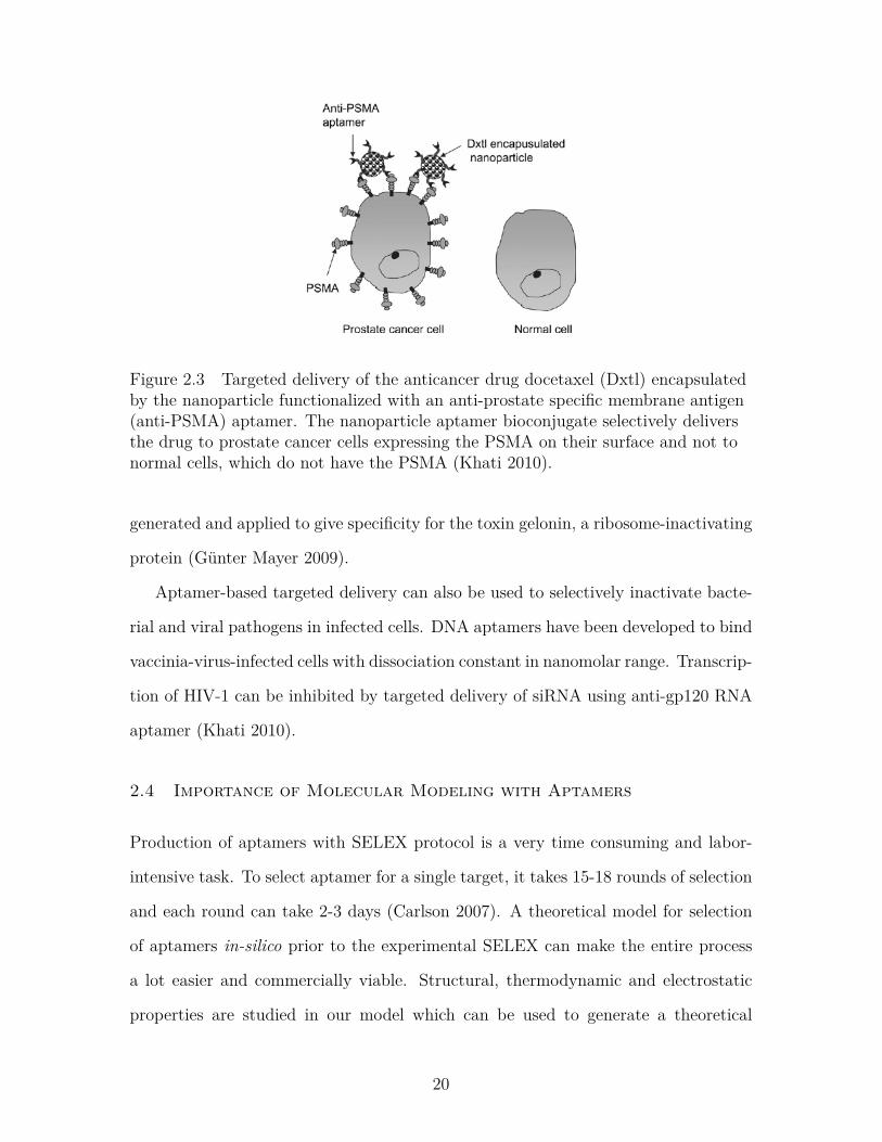
type (Song et al. 2008). They can be conjugated with nanoparticles for targeted
delivery of chemotherapeutic agents to cancer cells (Figure-2.3). The best charac-
terized aptamer in this regard is A10 which binds to the prostate specific membrane
antigen (PSMA) responsible for the onset and progression of cancer. This aptamer
can be conjugated with polymer coated nanoparticle encapsulated with chemothera-
peutics (i.e., Docetaxel) to its 5′-amino end. This chemotherapeutic treatment aided
by aptamers can reduce the size of cancer upto a total remission.
Other than chemotherapeutics, aptamers can be used for delivery of cell specific
small interfering RNA (siRNA) molecules. In this regard, siRNA molecules have
been coupled to aptamer A10 either directly by nucleotidic extensions or indirectly
through the assembly of tetrameric streptavidin-biotin complexes consisting of two
biotinylated aptamers and two biotinylated siRNA molecules per streptavidin moiety.
Both approaches were successful in the cell specific siRNA-mediated reduction of
the corresponding mRNA and protein levels. Aptamer-toxin conjugates were also
19
Figure 2.3 Targeted delivery of the anticancer drug docetaxel (Dxtl) encapsulatedby the nanoparticle functionalized with an anti-prostate specific membrane antigen(anti-PSMA) aptamer. The nanoparticle aptamer bioconjugate selectively deliversthe drug to prostate cancer cells expressing the PSMA on their surface and not tonormal cells, which do not have the PSMA (Khati 2010).
generated and applied to give specificity for the toxin gelonin, a ribosome-inactivating
protein (Günter Mayer 2009).
Aptamer-based targeted delivery can also be used to selectively inactivate bacte-
rial and viral pathogens in infected cells. DNA aptamers have been developed to bind
vaccinia-virus-infected cells with dissociation constant in nanomolar range. Transcrip-
tion of HIV-1 can be inhibited by targeted delivery of siRNA using anti-gp120 RNA
aptamer (Khati 2010).
2.4 Importance of Molecular Modeling with Aptamers
Production of aptamers with SELEX protocol is a very time consuming and labor-
intensive task. To select aptamer for a single target, it takes 15-18 rounds of selection
and each round can take 2-3 days (Carlson 2007). A theoretical model for selection
of aptamers in-silico prior to the experimental SELEX can make the entire process
a lot easier and commercially viable. Structural, thermodynamic and electrostatic
properties are studied in our model which can be used to generate a theoretical
20
databank containing a large number of aptamer conformations. This model takes
into account the size, shape, electrical properties and physical conformations of the
aptamers to study their structural and thermodynamic changes with varying biolog-
ical environments. This model lays the foundation to develop a predictive approach
to select the specific aptamer for a specific target that shows the most stable and
strongest binding. This approach will make the use of aptamers in biosensing, target
validation and other drug delivery applications more efficient. For example, using
this model will enable us to select an aptamer as a biosensing probe that will detect
a specific pathogen more accurately in human body. The accuracy will be ensured
through the understanding of all the chemical and thermodynamic aspects related
to the aptamer-target binding. Structural modification of aptamers with different
functional groups can enhance their stability and binding efficacy. Our model can
lead to the addition of a specific functional group to the aptamer chain in the most
accurate position for a specific application. To choose aptamer as a drug agent, this
model can ensure strongest binding with the pathogens and will work most effectively
to stop the signaling pathway for the progression of a certain disease.
2.5 Molecular Modeling of Aptamers
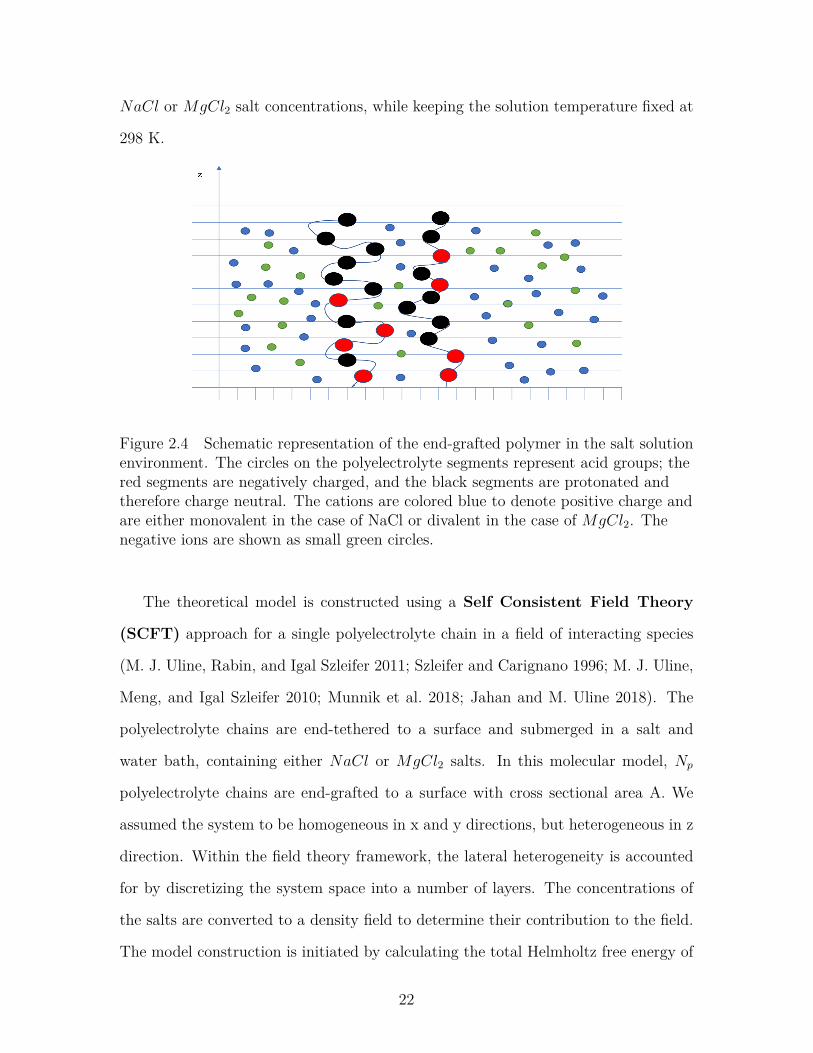
The molecular model in this work is developed to represent surface-grafted ssDNA
aptamers as a co-polymer with a combination of four monomer units- adenine (A),
guanine (G), Cytosine (C) and Thymine (T) in a coarse-grained level. We explicitly
considered the physical and chemical properties of these nucleobases in a solution
environment to capture their behavior as accurately as possible. We studied two chain
sequences: diblock co-polymer of A and G (A6G6, a block of six adenine monomers
followed by a block of six guanine monomers) and diblock of T and C (T6C6, a block
of six thymine monomers followed by a block of six cytosine monomers) (Figure-1).
Each of these chains contain 12 monomers at varying grafting densities and either
21
NaCl or MgCl2 salt concentrations, while keeping the solution temperature fixed at
298 K.
Figure 2.4 Schematic representation of the end-grafted polymer in the salt solutionenvironment. The circles on the polyelectrolyte segments represent acid groups; thered segments are negatively charged, and the black segments are protonated andtherefore charge neutral. The cations are colored blue to denote positive charge andare either monovalent in the case of NaCl or divalent in the case of MgCl2. Thenegative ions are shown as small green circles.
The theoretical model is constructed using a Self Consistent Field Theory
(SCFT) approach for a single polyelectrolyte chain in a field of interacting species
(M. J. Uline, Rabin, and Igal Szleifer 2011; Szleifer and Carignano 1996; M. J. Uline,
Meng, and Igal Szleifer 2010; Munnik et al. 2018; Jahan and M. Uline 2018). The
polyelectrolyte chains are end-tethered to a surface and submerged in a salt and
water bath, containing either NaCl or MgCl2 salts. In this molecular model, Np
polyelectrolyte chains are end-grafted to a surface with cross sectional area A. We
assumed the system to be homogeneous in x and y directions, but heterogeneous in z
direction. Within the field theory framework, the lateral heterogeneity is accounted
for by discretizing the system space into a number of layers. The concentrations of
the salts are converted to a density field to determine their contribution to the field.
The model construction is initiated by calculating the total Helmholtz free energy of
22
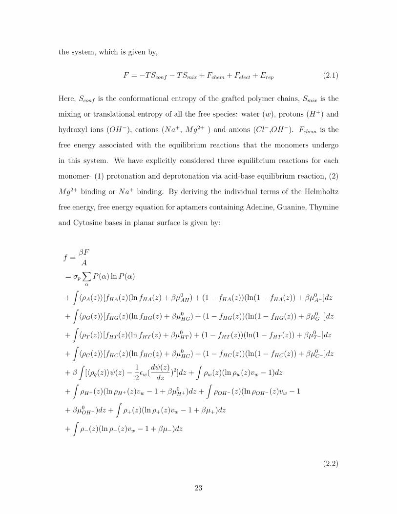
the system, which is given by,
F = −TSconf − TSmix + Fchem + Felect + Erep (2.1)
Here, Sconf is the conformational entropy of the grafted polymer chains, Smix is the
mixing or translational entropy of all the free species: water (w), protons (H+) and
hydroxyl ions (OH−), cations (Na+, Mg2+ ) and anions (Cl−,OH−). Fchem is the
free energy associated with the equilibrium reactions that the monomers undergo
in this system. We have explicitly considered three equilibrium reactions for each
monomer- (1) protonation and deprotonation via acid-base equilibrium reaction, (2)
Mg2+ binding or Na+ binding. By deriving the individual terms of the Helmholtz
free energy, free energy equation for aptamers containing Adenine, Guanine, Thymine
and Cytosine bases in planar surface is given by:
f = βF
A
= σp∑α
P (α) lnP (α)
+∫〈ρA(z)〉[fHA(z)(ln fHA(z) + βµ0
AH) + (1− fHA(z))(ln(1− fHA(z)) + βµ0A− ]dz
+∫〈ρG(z)〉[fHG(z)(ln fHG(z) + βµ0
HG) + (1− fHG(z))(ln(1− fHG(z)) + βµ0G− ]dz
+∫〈ρT (z)〉[fHT (z)(ln fHT (z) + βµ0
HT ) + (1− fHT (z))(ln(1− fHT (z)) + βµ0T− ]dz
+∫〈ρC(z)〉[fHC(z)(ln fHC(z) + βµ0
HC) + (1− fHC(z))(ln(1− fHC(z)) + βµ0C− ]dz
+ β∫
[〈ρq(z)〉ψ(z)− 12εw(dψ(z)
dz)2]dz +
∫ρw(z)(ln ρw(z)vw − 1)dz
+∫ρH+(z)(ln ρH+(z)vw − 1 + βµ0
H+)dz +∫ρOH−(z)(ln ρOH−(z)vw − 1
+ βµ0OH−)dz +
∫ρ+(z)(ln ρ+(z)vw − 1 + βµ+)dz
+∫ρ−(z)(ln ρ−(z)vw − 1 + βµ−)dz
(2.2)
23
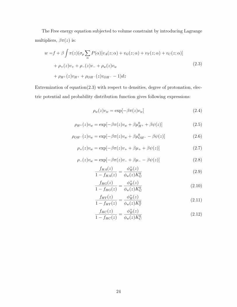
The Free energy equation subjected to volume constraint by introducing Lagrange
multipliers, βπ(z) is:
w =f + β∫π(z)(σp
∑α
P (α)[vA(z;α) + vG(z;α) + vT (z;α) + vC(z;α)]
+ ρ+(z)v+ + ρ−(z)v− + ρw(z)vw
+ ρH+(z)vH+ + ρOH−(z)vOH− − 1)dz
(2.3)
Extremization of equation(2.3) with respect to densities, degree of protonation, elec-
tric potential and probability distribution function gives following expressions:
ρw(z)vw = exp[−βπ(z)vw] (2.4)
ρH+(z)vw = exp[−βπ(z)vw + βµ0H+ + βψ(z)] (2.5)
ρOH−(z)vw = exp[−βπ(z)vw + βµ0OH− − βψ(z)] (2.6)
ρ+(z)vw = exp[−βπ(z)v+ + βµ+ + βψ(z)] (2.7)
ρ−(z)vw = exp[−βπ(z)v− + βµ− − βψ(z)] (2.8)
fHA(z)1− fHA(z) = φ+
H(z)φw(z)K0
G
(2.9)
fHG(z)1− fHG(z) = φ+
H(z)φw(z)K0
G
(2.10)
fHT (z)1− fHT (z) = φ+
H(z)φw(z)K0
T
(2.11)
fHC(z)1− fHC(z) = φ+
H(z)φw(z)K0
C
(2.12)
24
The probability distribution function (pdf) is derived from the functional minimiza-
tion with P (α),
P (α) = 1Q
exp[−∫nA(α; z)vA ln(1− fHA(z))dz
−∫nG(α; z)vG ln(1− fHG(z))dz −
∫nT (α; z)vT ln(1− fHT (z))dz
−∫nC(α; z)vC ln(1− fHC(z))dz + β
∫ψ(z)nA(α; z)dz
+ β∫ψ(z)nG(α; z)dz + β
∫ψ(z)nT (α; z)dz
+ β∫ψ(z)nC(α; z)dz − β
∫π(z)nA(α; z)vAdz
− β∫π(z)nG(α; z)vGdz − β
∫π(z)nT (α; z)vTdz − β
∫π(z)nC(α; z)vCdz]
(2.13)
Extremization of the free energy with respect to the electrostatic potential yields
Poisson equation,
εwd2ψ(z)dz2 = −〈ρq(z)〉 (2.14)
εwdψ(z)dz|z=0 = 0, lim
r→∞ψ(z) = 0 (2.15)
Equations (2.4) through (2.15) are solved simultaneously following the procedure
described in previous publications using this general approach (M. J. Uline, Rabin,
and Igal Szleifer 2011; Szleifer and Carignano 1996; Rikkert J Nap, S. H. Park, and
Igal Szleifer 2018). These integro-differential equations are solved numerically by
discretizing the space for a discretization length of 0.3 nm for 100 discrete layers.
Solution of these sets of non-linear coupled equations yields the unknowns of the
model, that are the Lagrange multiplier π(z) and the electrostatic potential ψ(z).
The inputs necessary to solve the system of equations are the bulk concentrations
of the salts, bulk pH, grafting density, volumes of different species, a set of polymer
conformations and the equilibrium reaction constants. The pKa values for A, G, T
and C are 3.5, 1.6, 9.7 and 4.2, respectively (Bloomfield and Crothers 2000). The
volume of Mg2+ is 0.18nm3, and the volumes for Na+ and Cl− are 0.05nm3. The set
25
of polymer conformations are derived using a Rotational Isomeric State (RIS) model
(Paul J Flory and Volkenstein 1969).
2.6 Results and Discussions
In the molecular model, the aptamer chains are grafted in a planar surface and is
assumed to be inhomogeneous in the direction perpendicular to the grafting surface.
The aptamer chains are submerged in a bath of anions, cations, H+, OH− and water
molecules. The number of polymer molecules is fixed, but the grafting surface is in
contact with a bath of ions and water. Therefore, we choose our system to be in grand
canonical ensemble. It has been assumed that the system is in a good solvent bath,
i.e.; there is no attractive interactions between the monomers. So, Chi parameter (χ)
is considered to be zero and therefore not included in the free energy equation.
Aptamers may contain four nucleotides: adenine, guanine, cytosine and thymine.
We took two aptamer chains, one containing Adenine and Guanine; another one
containing Thymine and Cytosine. Bulk pH of the system is chosen to be 4.0 due
to acidic nature of the aptamer chains. Each chain contains n=12 monomers, having
six of each nucleobase.
We varied the bulk salt concentration and surface coverage of aptamer chains
along with the type of salt to observe the changes in the structural and chemical
properties. We calculated the change in volume fraction and protonation profile with
the change in distance from grafting surface. Two different salts, NaCl and MgCl2
are used to observe the effect of cation size on polymer volume distribution.
26
2.6.1 Effect of salt concentration and surface coverage on aptamer
volume fractions: aptamer containing Adenine and Guanine
NaCl Salt Solution
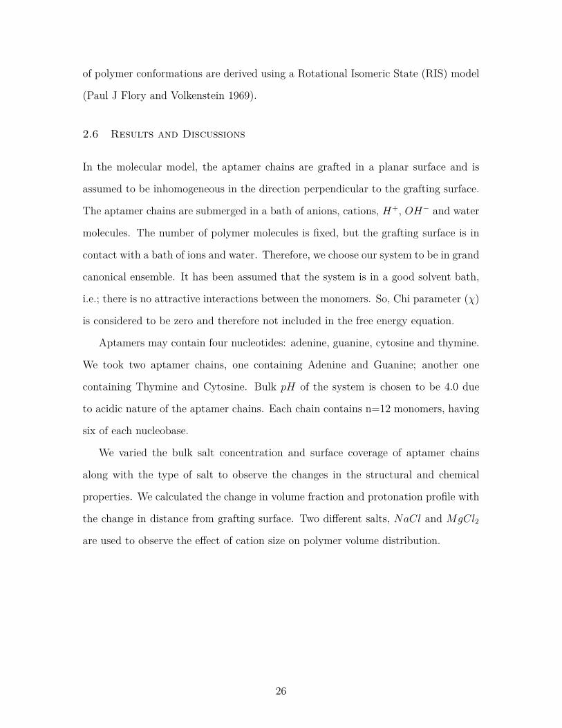
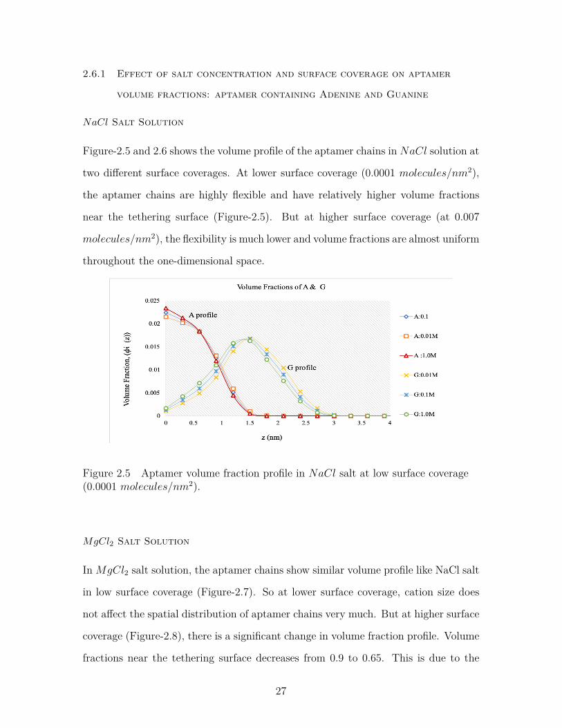
Figure-2.5 and 2.6 shows the volume profile of the aptamer chains in NaCl solution at
two different surface coverages. At lower surface coverage (0.0001 molecules/nm2),
the aptamer chains are highly flexible and have relatively higher volume fractions
near the tethering surface (Figure-2.5). But at higher surface coverage (at 0.007
molecules/nm2), the flexibility is much lower and volume fractions are almost uniform
throughout the one-dimensional space.
Figure 2.5 Aptamer volume fraction profile in NaCl salt at low surface coverage(0.0001 molecules/nm2).
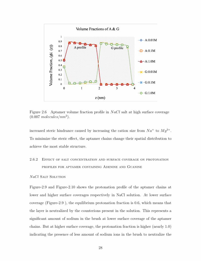
MgCl2 Salt Solution
InMgCl2 salt solution, the aptamer chains show similar volume profile like NaCl salt
in low surface coverage (Figure-2.7). So at lower surface coverage, cation size does
not affect the spatial distribution of aptamer chains very much. But at higher surface
coverage (Figure-2.8), there is a significant change in volume fraction profile. Volume
fractions near the tethering surface decreases from 0.9 to 0.65. This is due to the
27
Figure 2.6 Aptamer volume fraction profile in NaCl salt at high surface coverage(0.007 molecules/nm2).
increased steric hindrance caused by increasing the cation size from Na+ to Mg2+.
To minimize the steric effect, the aptamer chains change their spatial distribution to
achieve the most stable structure.
2.6.2 Effect of salt concentration and surface coverage on protonation
profiles for aptamer containing Adenine and Guanine
NaCl Salt Solution
Figure-2.9 and Figure-2.10 shows the protonation profile of the aptamer chains at
lower and higher surface coverages respectively in NaCl solution. At lower surface
coverage (Figure-2.9 ), the equilibrium protonation fraction is 0.6, which means that
the layer is neutralized by the counterions present in the solution. This represents a
significant amount of sodium in the brush at lower surface coverage of the aptamer
chains. But at higher surface coverage, the protonation fraction is higher (nearly 1.0)
indicating the presence of less amount of sodium ions in the brush to neutralize the
28
Figure 2.7 Aptamer volume fraction profile in MgCl2 at lower surface coverage(0.0001 molecules/nm2).
Figure 2.8 Aptamer volume fraction profile in MgCl2 salt at high surface coverage(0.002 molecules/nm2).
charged polymers.
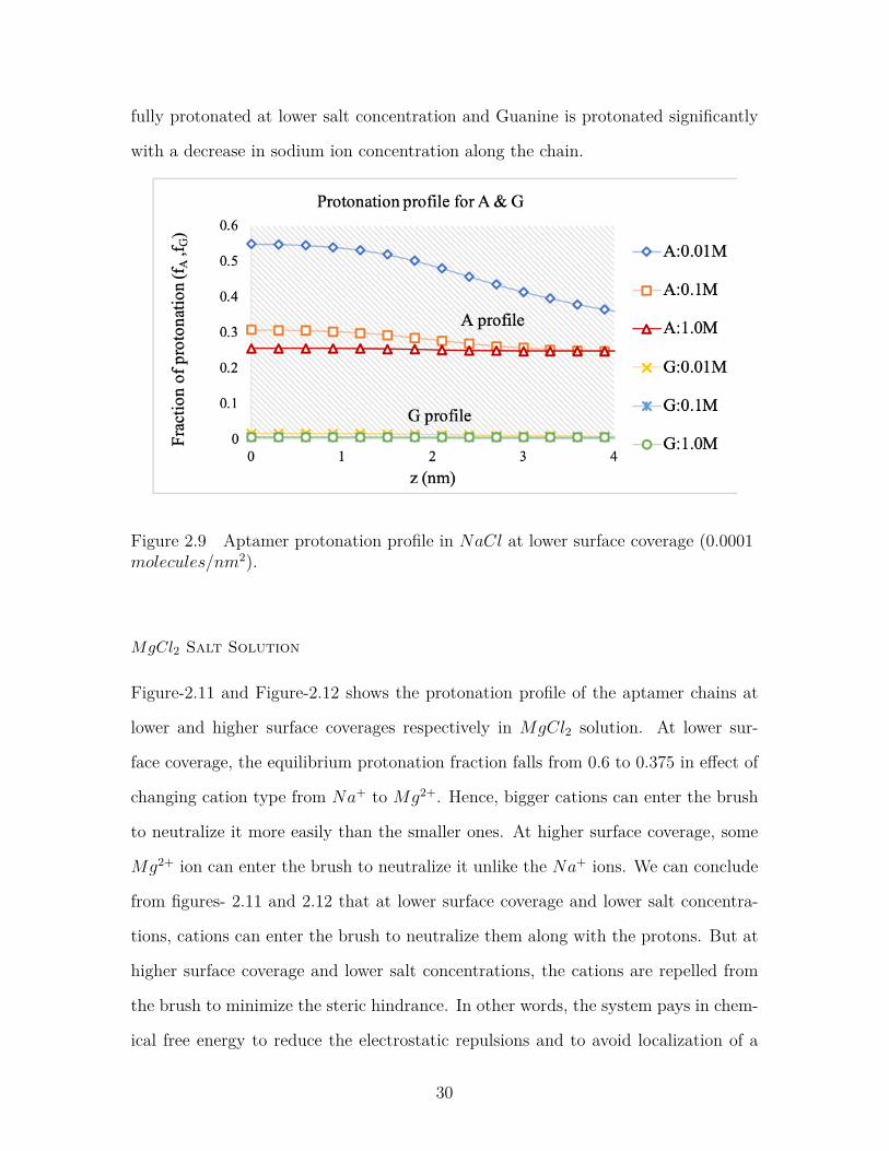
We can also see that protonation profiles of two different bases Adenine and Gua-
nine are different at different surface coverages. At lower surface coverage, Adenine
is protonated, but Guanine remains nearly unprotonated indicating the presence of
a large volume fraction of sodium ions. But at higher surface coverage, Adenine is
29
fully protonated at lower salt concentration and Guanine is protonated significantly
with a decrease in sodium ion concentration along the chain.
Figure 2.9 Aptamer protonation profile in NaCl at lower surface coverage (0.0001molecules/nm2).
MgCl2 Salt Solution
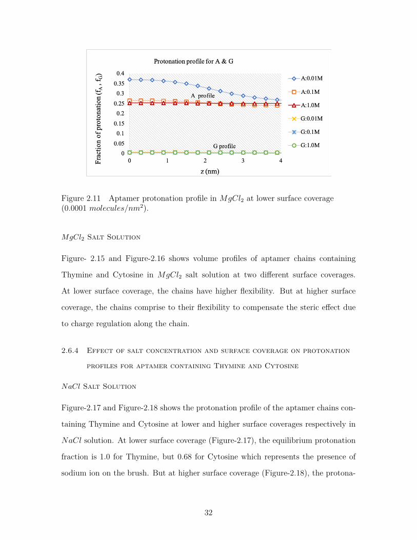
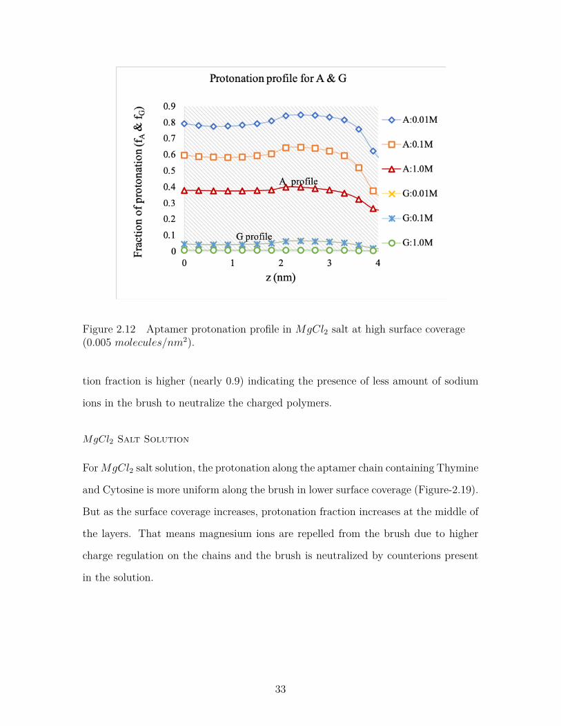
Figure-2.11 and Figure-2.12 shows the protonation profile of the aptamer chains at
lower and higher surface coverages respectively in MgCl2 solution. At lower sur-
face coverage, the equilibrium protonation fraction falls from 0.6 to 0.375 in effect of
changing cation type from Na+ to Mg2+. Hence, bigger cations can enter the brush
to neutralize it more easily than the smaller ones. At higher surface coverage, some
Mg2+ ion can enter the brush to neutralize it unlike the Na+ ions. We can conclude
from figures- 2.11 and 2.12 that at lower surface coverage and lower salt concentra-
tions, cations can enter the brush to neutralize them along with the protons. But at
higher surface coverage and lower salt concentrations, the cations are repelled from
the brush to minimize the steric hindrance. In other words, the system pays in chem-
ical free energy to reduce the electrostatic repulsions and to avoid localization of a
30
Figure 2.10 Aptamer protonation profile in NaCl salt at high surface coverage(0.007 molecules/nm2).
very high concentrations of ions, i.e.; counterion confinement (M. J. Uline, Rabin,
and Igal Szleifer 2011).
2.6.3 Effect of salt concentration and surface coverage on aptamer
volume fractions: aptamer containing Thymine and Cytosine
NaCl Salt Solution
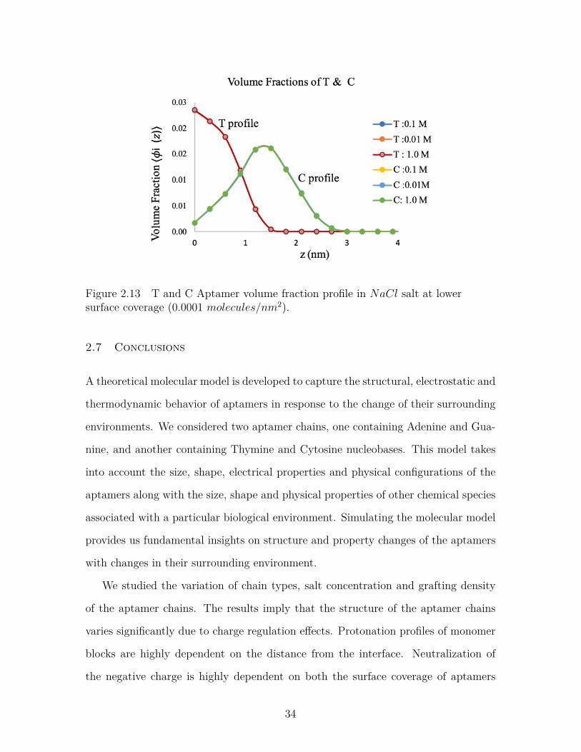
Figure-2.13 and Figure-2.14 shows volume profiles of aptamer chains containing
Thymine and Cytosine in NaCl salt solution at two different surface coverages. At
lower surface coverage (Figure-2.13), the chains are more flexible and have relatively
higher volume fraction near the tethering surface than the bulk. But at higher surface
coverage (Figure-2.14), the chains are more stretched and have higher volume fraction
on the mid-layer.
31
Figure 2.11 Aptamer protonation profile in MgCl2 at lower surface coverage(0.0001 molecules/nm2).
MgCl2 Salt Solution
Figure- 2.15 and Figure-2.16 shows volume profiles of aptamer chains containing
Thymine and Cytosine in MgCl2 salt solution at two different surface coverages.
At lower surface coverage, the chains have higher flexibility. But at higher surface
coverage, the chains comprise to their flexibility to compensate the steric effect due
to charge regulation along the chain.
2.6.4 Effect of salt concentration and surface coverage on protonation
profiles for aptamer containing Thymine and Cytosine
NaCl Salt Solution
Figure-2.17 and Figure-2.18 shows the protonation profile of the aptamer chains con-
taining Thymine and Cytosine at lower and higher surface coverages respectively in
NaCl solution. At lower surface coverage (Figure-2.17), the equilibrium protonation
fraction is 1.0 for Thymine, but 0.68 for Cytosine which represents the presence of
sodium ion on the brush. But at higher surface coverage (Figure-2.18), the protona-
32
Figure 2.12 Aptamer protonation profile in MgCl2 salt at high surface coverage(0.005 molecules/nm2).
tion fraction is higher (nearly 0.9) indicating the presence of less amount of sodium
ions in the brush to neutralize the charged polymers.
MgCl2 Salt Solution
ForMgCl2 salt solution, the protonation along the aptamer chain containing Thymine
and Cytosine is more uniform along the brush in lower surface coverage (Figure-2.19).
But as the surface coverage increases, protonation fraction increases at the middle of
the layers. That means magnesium ions are repelled from the brush due to higher
charge regulation on the chains and the brush is neutralized by counterions present
in the solution.
33
Figure 2.13 T and C Aptamer volume fraction profile in NaCl salt at lowersurface coverage (0.0001 molecules/nm2).
2.7 Conclusions
A theoretical molecular model is developed to capture the structural, electrostatic and
thermodynamic behavior of aptamers in response to the change of their surrounding
environments. We considered two aptamer chains, one containing Adenine and Gua-
nine, and another containing Thymine and Cytosine nucleobases. This model takes
into account the size, shape, electrical properties and physical configurations of the
aptamers along with the size, shape and physical properties of other chemical species
associated with a particular biological environment. Simulating the molecular model
provides us fundamental insights on structure and property changes of the aptamers
with changes in their surrounding environment.
We studied the variation of chain types, salt concentration and grafting density
of the aptamer chains. The results imply that the structure of the aptamer chains
varies significantly due to charge regulation effects. Protonation profiles of monomer
blocks are highly dependent on the distance from the interface. Neutralization of
the negative charge is highly dependent on both the surface coverage of aptamers
34
Figure 2.14 T and C Aptamer protonation profile in NaCl at higher surfacecoverage (0.002 molecules/nm2).
and the valence of the cations. Mg2+ is still present in the aptamer layer for the
high surface coverage case. But Na+ is nearly excluded from the brush due to high
steric repulsion inside the brush for higher amount of charged monomers. The system
decides to relieve the electrostatic repulsions by paying in acid-base equilibrium.
Finally, it can be concluded that this model captures the physical property changes
very well for the aptamer chains at varying surface coverages, types of salt and dif-
ferent salt concentrations. This model can aid in generating a theoretical databank
for ssDNA aptamers to select a specific aptamer for a specific target molecule very
quickly and cost effectively.
35
Figure 2.15 T and C Aptamer volume fraction profile in MgCl2 at lower surfacecoverage (0.0001 molecules/nm2).
Figure 2.16 T and C Aptamer volume fraction profile in MgCl2 salt at highsurface coverage (0.002 molecules/nm2).
36
Figure 2.17 T and C Aptamer volume fraction profile in NaCl at low surfacecoverage (0.0001 molecules/nm2).
Figure 2.18 T and C Aptamer volume fraction profile in NaCl salt at high surfacecoverage (0.002 molecules/nm2).
37
Figure 2.19 T and C Aptamer protonation fraction profile in MgCl2 at low surfacecoverage (0.0001 molecules/nm2).
Figure 2.20 T and C Aptamer protonation fraction profile in MgCl2 salt at highsurface coverage (0.002 molecules/nm2).
38
Chapter 3
Quantifying Divalent Cation Binding To ssDNA
Aptamers
3.1 Introduction
Aptamers are an important class of biomolecules consisting of single stranded DNA
(ssDNA), RNA, or peptides that can fold into unique secondary and tertiary struc-
tures for shape-specific target recognition(Keefe, Pai, and A. Ellington 2010). Due to
the highly specific and selective nature of their target binding, aptamers are widely
studied for a range of applications from biosensing (R. Liu et al. 2018; Cho, J.-W.
Lee, and A. D. Ellington 2009) to drug design (Dua et al. 2018; G. Zhou et al. 2018;
Foster and DeRosa 2014). A recent work reported a major breakthrough in biosensor
research by using aptamers with field-effect transistors to overcome the ‘Debye length
limitations’ (Nakatsuka et al. 2018). Aptamers are polyelectrolytic in nature with
their monomer units (nucleobases) participating in acid-base equilibrium and counte-
rion binding reactions with the surrounding solution environments. Charge regulation
and counterion binding in aptamers, or polyelectrolytes in general, are modulated by
the metal ions present in the system that can non-trivially alter their chemical and
structural properties (M. J. Uline, Rabin, and Igal Szleifer 2011; Rikkert J Nap and
Igal Szleifer 2018; Rikkert J Nap, Solveyra, and Igal Szleifer 2018; Zwanikken et al.
2011; R. Kumar, Sumpter, and Kilbey 2012; Lewis et al. 2013). Presence of metal ions
affects the performance of the aptamers as biosensing probes or therapeutics (Juewen
Liu, Cao, and Lu 2009; W. Zhou et al. 2014; J.-S. Lee, Han, and Mirkin 2007) due to
39
the electrostatic screening of the charges on their surface that changes their structure
and chemistry. These interactions of aptamers with metal ions are complicated in
nature owing to the fact that multiple binding sites on the nucleobases are capable
of such interactions, following different binding pathways and thus having varying
energy landscapes (Saenger 1984; Reshetnikov et al. 2011). In this work, we have
particularly addressed magnesium ion (Mg2+) binding because of its relevance to al-
most all nucleic acid related biological processes in the intracellular environment (Ono
et al. 2011; Anastassopoulou and Theophanides 2002; Pascal, Grover, and Westhof
2011).
A myriad of computational studies has been conducted with Molecular Dynamics
(MD) (Mocci and Laaksonen 2012) and Monte Carlo (MC)(Mills, Anderson, and
Record Jr 1985) simulations to elucidate the nature of metal ion binding to nucleic
acids. Most of such theoretical studies are based on double stranded DNA (dsDNA)
- monovalent cation (such as Na+, K+) interactions (Savelyev and MacKerell Jr
2015; Howard, Lynch, and Pettitt 2010; Gebala et al. 2016; Gebala et al. 2015).
Among the few that included multivalent cations, Hayes et al (Hayes et al. 2014)
employed a hybrid structure based MD model to explicitly count the number of
excess Mg2+ ions bound to RNA sequences in the presence of background potassium
chloride with a Manning condensation estimated by a Non-linear Poisson Boltzmann
equation. Li et al (Li, Nordenskiöld, and Mu 2011) used implicit Mg2+ binding
to dsDNA sequences with classical MD simulation to study the effect of counterion
condensation on DNA structure and conformational dynamics. While atomistic MD
simulations give a full distribution of the ion atmosphere around the nucleic acids,
they suffer from drawbacks due to the enormous computational cost and the choice of
force fields that might lead to over or underestimation of the same ion cloud (Savelyev
and MacKerell Jr 2015; Jacobson and Saleh 2016). These studies also rely heavily
on parameterization to match experimental studies, which imposes unrestrained bias
40
toward their agreement with experimental results (Jacobson and Saleh 2016). On
the other hand, almost all theoretical studies consider nucleic acid chains in bulk
conditions; therefore, characteristics of nucleic acid strands end-tethered to a surface
remain elusive.
Along this line, this study addresses metal ion binding to surface-anchored nucleic
acid oligomers with a Self Consistent Field Theory (SCFT) approach to construct a
comprehensive and statistically robust model for quantifying the number of Mg2+
ions bound to each chain, while capturing the ion-binding effect on their structure
and properties. The molecular model analyzesMg2+ binding to nucleic acid oligomers
containing adenine (A) and guanine (G) nucleobases, while trying to capture, as much
as possible, the details of experimental studies for a similar system. Metal ion binding
to the monomers is explicitly included with equilibrium binding reactions by using
experimentally derived (Holland et al. 2011) binding free energies for relevant binding
modes (Holland, Jordan, and Geiger 2011). The molecular model characterizes the
spatial variation of the structure and properties of the oligonucleotide chains along the
distance from the grafting surface, at varying ionic strength and grafting densities,
and quantifies the number of bound ions at thermodynamic equilibrium with the
oligonucleotides. The model explicitly accounts for the thermodynamic, structural
and electrostatic properties of all the species involved in the system, while remaining
free of adjustable parameters. This field theoric model helps to set up the foundation
for future studies involving secondary and tertiary structures of aptamers interacting
with multivalent metal ions.
3.2 Theoretical Methodology
The theoretical model in this work is developed to represent surface-grafted ssDNA
oligomers as a co-polymer with two monomer units- adenine (A) and guanine (G), in
a coarse-grained level. We explicitly considered the physical and chemical properties
41
Figure 3.1 Schematic representation of an end-grafted polymer in a salt solutionenvironment (left) and chain sequences used for molecular modeling (right).
of these nucleobases in a solution environment to capture their behavior as accurately
as possible. We studied three chain sequences: diblock co-polymer of A and G with
A-end grafted to the surface (A6G6, a block of six adenine monomers followed by a
block of six guanine monomers), diblock of A and G with G-end grafted to the surface
(G6A6, a block of six guanine monomers followed by a block of six adenine monomers)
and alternating sequence of A and G ((AG)6, one adenine monomer followed by a
guanine monomer in an alternating manner) (Figure-1). Each of these chains contain
12 monomers at varying grafting densities and MgCl2 concentrations, while keeping
the solution temperature and background NaCl concentration fixed at 298 K and
10 mM, respectively. NaCl is added to the system to comply with the relevant
experimental study (Holland, Jordan, and Geiger 2011).
The theoretical model is constructed using a Self Consistent Field Theory
(SCFT) approach for a single polyelectrolyte chain in a field of interacting species
(M. J. Uline, Rabin, and Igal Szleifer 2011; Szleifer and Carignano 1996; M. J. Uline,
Meng, and Igal Szleifer 2010; Munnik et al. 2018; Jahan and M. Uline 2018). The
polyelectrolyte chains are end-tethered to a surface and submerged in a salt and
water bath, containing both NaCl and MgCl2 salts. The motivation behind our
42
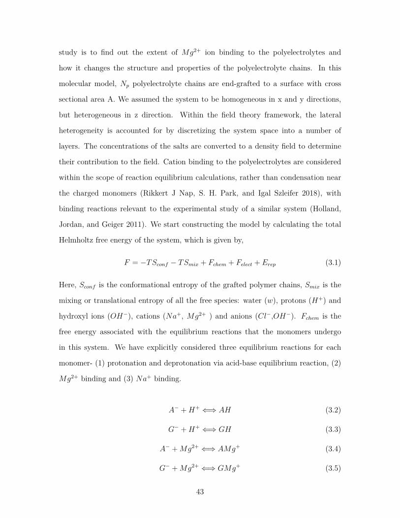
study is to find out the extent of Mg2+ ion binding to the polyelectrolytes and
how it changes the structure and properties of the polyelectrolyte chains. In this
molecular model, Np polyelectrolyte chains are end-grafted to a surface with cross
sectional area A. We assumed the system to be homogeneous in x and y directions,
but heterogeneous in z direction. Within the field theory framework, the lateral
heterogeneity is accounted for by discretizing the system space into a number of
layers. The concentrations of the salts are converted to a density field to determine
their contribution to the field. Cation binding to the polyelectrolytes are considered
within the scope of reaction equilibrium calculations, rather than condensation near
the charged monomers (Rikkert J Nap, S. H. Park, and Igal Szleifer 2018), with
binding reactions relevant to the experimental study of a similar system (Holland,
Jordan, and Geiger 2011). We start constructing the model by calculating the total
Helmholtz free energy of the system, which is given by,
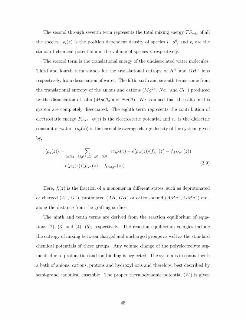
F = −TSconf − TSmix + Fchem + Felect + Erep (3.1)
Here, Sconf is the conformational entropy of the grafted polymer chains, Smix is the
mixing or translational entropy of all the free species: water (w), protons (H+) and
hydroxyl ions (OH−), cations (Na+, Mg2+ ) and anions (Cl−,OH−). Fchem is the
free energy associated with the equilibrium reactions that the monomers undergo
in this system. We have explicitly considered three equilibrium reactions for each
monomer- (1) protonation and deprotonation via acid-base equilibrium reaction, (2)
Mg2+ binding and (3) Na+ binding.
A− +H+ ⇐⇒ AH (3.2)
G− +H+ ⇐⇒ GH (3.3)
A− +Mg2+ ⇐⇒ AMg+ (3.4)
G− +Mg2+ ⇐⇒ GMg+ (3.5)
43
A− +Na+ ⇐⇒ ANa (3.6)
G− +Na+ ⇐⇒ GNa (3.7)
Felect is the total electrostatic energy due to the charged species and Erep is the
repulsive interactions between all the species due to steric hindrance. T is the tem-
perature of the system which is held constant at 298 K. Expansion of all the energy
and entropy terms gives the total Helmholtz free energy of the system,
Functional extremization with respect to the monomer fractions fi(z) yields the
governing equations for chemical equilibrium reactions for both monomers A and G,
fA−(z)fAH(z) = K0
AH
exp (−βπ(z)∆vAH)ρH+(z)vw
(3.23)
fA−(z)fANa(z) = K0
ANa
exp (−βπ(z)∆vANa)ρNa+(z)vw
(3.24)
fA−(z)fAMg+(z) = K0
AMg+exp (−βπ(z)∆vAMg+)
ρMg2+(z)vw(3.25)
fG−(z)fGH(z) = K0
GH
exp (−βπ(z)∆vGH)ρH+(z)vw
(3.26)
fG−(z)fGNa(z) = K0
GNa
exp (−βπ(z)∆vGNa)ρNa+(z)vw
(3.27)
fG−(z)fGMg+(z) = K0
GMg+exp (−βπ(z)∆vGMg+)
ρMg2+(z)vw(3.28)
The quantity K0i = exp (−β∆G0
i ) corresponds to the chemical equilibrium con-
stant that is derived from the standard chemical free energy ∆G0i of the respective
formation reactions for AH, GH, ANa, GNa, AMg+ or GMg+. ∆vi denotes the
volume change due to the reactions. The change in the standard free energy for the
reaction A−+Mg2+ ⇐⇒ AMg+ is given by ∆G0AMg+ = µ0
AMg+−µ0A−−µ0
Mg2+ and the
volume change of reaction is ∆vAMg+ = vAMg+−vA−−vMg2+ . The reaction constants
and change in volumes for other reactions can be derived in a similar manner.
Extremization of the free energy with respect to the electrostatic potential yields
Poisson equation,
εwd2ψ(z)dz2 = −〈ρq(z)〉 (3.29)
εwdψ(z)dz|z=0 = 0, lim
r→∞ψ(z) = 0 (3.30)
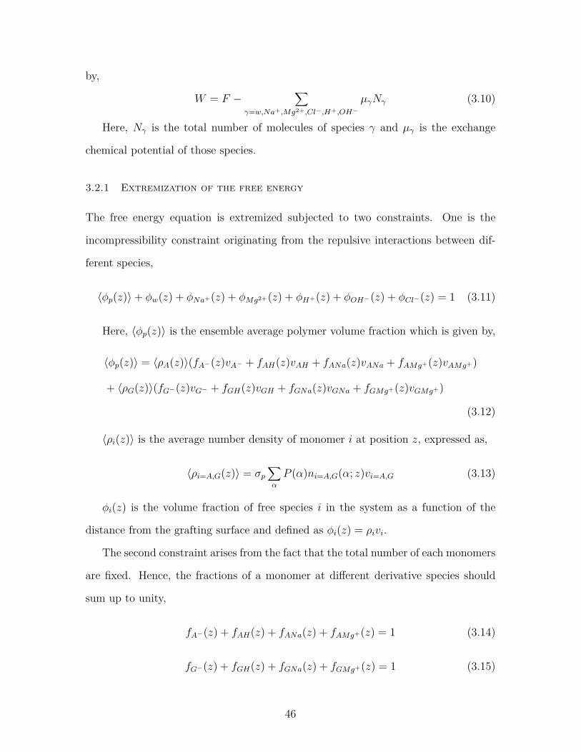
48
The probability distribution function (pdf) is derived from the functional mini-
mization with P (α),
P (α) = 1e
exp[−∫nA(α; z)vA(ln fA−(z) + βµ0
A− + βπ(z)vA− − βeψ(z))dz
−∫nG(α; z)vG(ln fG−(z) + βµ0
G− + βπ(z)vG− − βeψ(z))](3.31)
Equations (3.17) through (3.31) are solved simultaneously following the procedure
described in previous publications using this general approach (M. J. Uline, Rabin,
and Igal Szleifer 2011; Szleifer and Carignano 1996; Rikkert J Nap, S. H. Park, and
Igal Szleifer 2018). These integro-differential equations are solved numerically by
discretizing the space for a discretization length of 0.3 nm for 100 discrete layers.
Solution of these sets of non-linear coupled equations yields the unknowns of the
model, that are the Lagrange multiplier π(z) and the electrostatic potential ψ(z).
The inputs necessary to solve the system of equations are the bulk concentrations
of the salts, bulk pH, grafting density, volumes of different species, a set of polymer
conformations and the equilibrium reaction constants. The equilibrium constants for
the binding reactions between the monomers (Adenine and Guanine) and the Mg2+
cations are obtained from the binding free energies of the second harmonic generation
(SHG) and atomic force microscopy (AFM) studies of Holland et al (Holland, Jordan,
and Geiger 2011) by using the previously mentioned equation for equilibrium constant
K0i . The binding free energies are −32.1 KJ/mol and −35.6 KJ/mol for Adenine
and Guanine, respectively. The pKa values for G and A are 1.6 and 3.5, respectively
(Bloomfield and Crothers 2000). The volume of Mg2+aq is 0.18nm3, and the volumes
for Na+aq and Cl−aq are 0.05nm3. We did not include a change in volume upon binding
in this analysis. The set of polymer conformations are derived using a Rotational
Isomeric State (RIS) model (Paul J Flory and Volkenstein 1969).
49
3.3 Results and Discussions
To elucidate the effects of Mg2+ binding on the structure and properties of a short
chain surface-grafted A-G oligomer, we studied three different chains: A-G diblock
with A-end grafted to the surface (A6G6), A-G diblock with G-end grafted to the sur-
face (G6A6), and A-G co-polymer with alternating A and G along the chain ((AG)6)
(referring to Figure-3.1). We studied the variation of the MgCl2 concentration and
the polymer surface coverage for the same chain types as well as quantifying the num-
ber of bound Mg2+ ions to each chain in different solution conditions, while keeping
the background NaCl concentration and pH constant at 10 mM and 7.0, respectively.
We presented the quantitative results for all the chain sequences and the qualitative
results for A-grafted chains only, as the results for the other chain systems do not
deviate quantitatively from the A-grafted system. Our results provide valuable in-
sight into the molecular details of this polyelectrolyte system in various biologically
relevant environments.
3.3.1 Effect of sequence heterogeneity on Mg2+ binding
Figure 3.2 No. of bound Mg2+ to different sequences at varying grafting densitiesfor (a) 3 mM MgCl2 and (b) 180 mM MgCl2. The color bars correspond toA-grafted chain (blue), G-grafted chain (yellow) and A-G alternate chain (red).
The number of bound Mg2+ per chain is calculated from the volume fraction of
50
the polymer and the fraction of the Mg2+-bound monomers by using the following
equation,
NMg2+/chain =∫〈φp(z)〉fPMg+(z)dz
σpvp(3.32)
Figure-3.2(a) and 3.2(b) shows the number of bound Mg2+ with A6G6, G6A6
and (AG)6 sequences at different grafting densities for 3 mM and 180 mM MgCl2
content. The data in both plots demonstrates that despite the sequence heterogeneity
and variation on the grafting ends, all three sequences bind similar amounts of Mg2+
ions at different grafting densities and salt concentrations, except for 0.5 chains/nm2
and 3 mM condition. The relatively low cation binding at this condition can be
attributed to the unavailability of enough Mg2+ ions in low concentration of salt
and high grafting density of polymer chains. These results are in agreement with
the experimental findings that Mg2+ ions do not aggregate around the strongest
binder (Guanine) and are uniformly distributed throughout the length of the chains
(Holland et al. 2011). This agrees with the notion that non-specific interactions
due to the electrostatics of the sugar-phosphate backbone dominates ion binding to
ssDNA chains, as the non-specific part of the free energy for Adenine and Guanine
(−21 KJ/mol, from Holland et. al) (Holland, Jordan, and Geiger 2011) is much
higher than the specific part of the free energies (−11.1 KJ/mol and −14.6 KJ/mol,
respectively, from Holland et. al) (Holland, Jordan, and Geiger 2011) and the specific
free energies are all within a few KJ of each other, hence making the ion binding less
distinctive.
3.3.2 Effect of ionic strength and grafting density
Figure-3.3 (a), 3.3 (b) and 3.3 (c) are the volume fraction profiles of the A-grafted
chains (A6G6) along the distance from the grafting surface for low (0.005 chains/nm2),
medium (0.05 chains/nm2) and high (0.5 chains/nm2) grafting densities, for a range
of MgCl2 concentrations. At low grafting density (figure-3.3 (a)), as we increase the
51
MgCl2 salt concentration from 3 mM to 180 mM, the chain structures contract to-
wards the grafting surface and we get a distinct peak of the highest volume fractions
at a distance close to the surface. This change in the chain structure is due to the
reduction in negative charge of the polyelectrolytes by binding to Mg2+ ions that
lowers the repulsive interaction between the monomers.
Figure 3.3 Total polymer volume fraction profiles as a function of distance fromthe grafting surface at (a) 0.005 chains/nm2 (b) 0.05 chains/nm2 (c) 0.5chains/nm2. Blue lines correspond to 3 mM MgCl2, yellow lines correspond to50mM MgCl2 and red lines correspond to 180 mM MgCl2.
Although at first glance, the collapse does not seem to be very prominent, it is
significant for the length scales of the polyelectrolytes in our system (chain length
of only 12 monomers). But for higher grafting density (figure 3.3 (b)), the peak is
less distinctive at 3 mM and 50 mM MgCl2 concentrations than 180 mM and nearly
plateaus down from 0.8 nm to 2 nm. Herein, the high grafting density creates steric
repulsion and the system faces a competition between charge screening and steric re-
pulsion to stabilize the polyelectrolyte structures. At low MgCl2 salt concentrations
(3 mM and 50 mM), the system energetically favors chain stretching to accommodate
both charge repulsion and steric hindrance. But when we further increase theMgCl2
salt concentration to 180 mM, charge screening by Mg2+ ion dominates over steric
repulsion and we get a distinct chain collapse near the grafting surface. Figure-3.3(c)
corresponds to volume fraction profiles of the polyelectrolyte chains at high graft-
ing density (0.5 chains/nm2) and shows a clearly opposite picture than figure-3.3
52
(a) and 3.3 (b), with the chains stretching while we increase the salt concentration.
In this case, at 3 mM MgCl2, the number of bound Mg2+ ions are sufficient for
mitigating the charge repulsion inside the brush. Further increase in the salt con-
centration induces additional steric hindrance due to the volume exclusion and the
chains pay in conformational entropy to stretch the chains and accommodate more
Mg2+ ions inside the brush. This phenomenon of polyelectrolyte chains stretching
with increasing counterion concentration is well known as the ‘re-entrant phenomena’
and is experimentally verified by several studies (Yu et al. 2016; Wu et al. 2007).
00.00010.00020.00030.00040.0005
0 2 4
f P- (z
)
z (nm)
a
0
0.0050.01
0.0150.02
0 2 4
f P- (z
)
z (nm)
b
00.10.20.30.40.50.6
0 2 4
f P- (z
)z (nm)
c
Figure 3.4 Deprotonated polymer fraction profiles at 3 mM (blue lines), 50 mM(yellow lines) and 180 mM (red lines) MgCl2 concentrations. (a) 0.005 chains/nm2
(b) 0.05 chains/nm2 (c) 0.5 chains/nm2.
Figure-3.4 represents the profiles of the deprotonated (negatively charged) poly-
mer fraction (fP−(z)). This quantity is calculated from the individual monomer-
In Figure-3.4 (a), at 0.005 chains/nm2 grafting density, almost 100% of the polymers
are bound to Mg2+ for 50 mM and 180 mM salts and even for 3 mM salt, a very
small fraction is deprotonated (fP−(z) ∼ 0.0001), with the rest of the polymers being
Mg2+-bound. At 0.05 chains/nm2 (Figure - 3.4 (b)), the polymers are still (nearly)
entirely bound to Mg2+ at 50 mM and 180 mM salt concentrations. But for 3 mM
salt, the fraction of negatively charged polymers slightly increases. As we further
53
0
0.005
0.01
0.015
0.02
0 2 4 6 8 10
𝜙 Mg2+
(z)
z (nm)
a
0
0.005
0.01
0.015
0.02
0 2 4 6 8 10
𝜙 Mg2+
(z)
z (nm)
b
0
0.005
0.01
0.015
0.02
0 5 10
𝜙 Mg2+
(z)
z (nm)
c
Figure 3.5 Free Mg2+ volume fraction profiles as a function of distance from thegrafting surface at (a) 0.005 chains/nm2 (b) 0.05 chains/nm2 and (c) 0.5chains/nm2 grafting densities for 3 mM (blue lines), 50 mM (Yellow lines) and 180mM (green lines) MgCl2 concentrations.
increase the grafting density up to 0.5 chains/nm2 in figure- 3.4 (c), ionic strength
shows a more prominent effect on the chain structure and chemistry. Now at 3 mM
MgCl2, about 50% of the polymers are negatively charged. Increase of the ionic
strength to 50 mM and 180 mM decreases the negative charge by binding to more
Mg2+ to reduce the negative charge inside the brush.
The ‘re-entrant phenomena’ shown in figure-3.3(c) can be further explained us-
ing figure-3.4(c) and figure-3.5. At 3 mM MgCl2, about half of the polymers are
negatively charged and another half is positively charged due to the formation of
AMg+ and GMg+ complexes with the monomers. Hence, the brush environment is
nearly neutralized with low or no residual positive or negative charges and the brush
becomes collapsed for that grafting density. Therefore, increasing the salt concentra-
tion does not contribute to the global charge neutralization inside the brush; rather,
the availability of more Mg2+ ions, that have very high affinity towards binding to
the negatively charged monomers, creates high steric hindrance. As a result, we see
stretching of the chains and the system pays in conformational entropy to reduce
steric repulsion inside the brush.
Figure-3.5 represents the volume fractions of free Mg2+ ions inside the brush
and the surrounding medium. At low grafting density (0.005 chains/nm2, figure-
54
3.5(a)), there are significant amounts of free Mg2+ ions inside the brush at all salt
concentrations, which is depicted by the small deviation in the free Mg2+ volume
fractions from the bulk value. Here,
Figure 3.6 pH profiles along the distance from grafting surface at (a) 0.005chains/nm2 and (b) 0.5 chains/nm2 for 0 mM (blue lines), 3 mM (yellow lines), 50mM (red lines) and 180 mM (green lines) MgCl2 concentrations.
along with the ion binding, localization of the free Mg2+ ions screen the negative
charges inside the brush, which results in a collapse of the polyelectrolyte chains (re-
ferring to figure-3.3 (a)). But as we increase the grafting density to 0.05 chains/nm2
(figure-3.5 (b)), the amount of free Mg2+ ions inside the brush drops noticeably from
the bulk value, compared to the low grafting density brush. However, the system still
entropically favors localization of free Mg2+ ions while staying at a collapsed state
(figure-3.3 (b)). Further increase in the grafting density up to 0.5 chains/nm2 gives
rise to the above mentioned re-entrant phenomena (figure-3.3 (c)). At this point,
the brush region acts like a barrier to the free Mg2+ ions and the brush is com-
pletely devoid of free Mg2+ ions at all the salt concentrations (figure-3.5 (c)). For a
grafting density this high, the brush is so dense that the binding to the Mg2+ ions
increases the volume of the monomers that also increase the steric hindrance, even
at low MgCl2 salt concentration. Increasing the MgCl2 salt concentration promotes
binding to more Mg2+ ions, which is energetically more favorable than bringing the
free ions from the bulk and localizing them inside the brush. Hence, the system pays
in conformational entropy to stretch the chains to avoid high steric repulsion in the
55
brush and the re-entrant phenomena arises.
Figure 3.7 Chloride volume fractions at 0.005 chains/nm2 for 0 mM (a), 3 mM(b), 50 mM (c) and 180 mM (d) MgCl2 concentrations.
00.000020.000040.000060.00008
0.0001
0 2 4 6 8 10
𝜙C
l- (𝑧)
z (nm)
0 mM
0
0.01
0.02
0.03
0.04
0 5 10
𝜙C
l- (𝑧)
z (nm)
3 mM
0
0.05
0.1
0.15
0 5 10
𝜙C
l- (𝑧)
z (nm)
50 mM
00.05
0.10.15
0.20.25
0 5 10
𝜙C
l- (𝑧)
z (nm)
180 mM
dc
ba
Figure 3.8 Chloride volume fractions at 0.5 chains/nm2 for 0 mM (a), 3 mM (b),50 mM (c) and 180 mM (d) MgCl2 concentrations.
Figure-3.6 (a) and 3.6 (b) shows the change in local pH inside the brush along
the distance from the grafting surface for low (0.005 chains/nm2) and high (0.5
chains/nm2) grafting densities, respectively, for varying ionic strengths. pH profiles,
when no MgCl2 was added in the system, are included in the figures to compare the
56
systems response with added MgCl2 salt. At no added MgCl2 (0 mM), the system
only had 10 mM NaCl as a background electrolyte to modulate charge regulation
inside the brush. For lower grafting density, the pH inside the brush (Figure-3.6 (a))
only deviates by ∼ 0.5 from the bulk. But for higher grafting density, the available
Na+ ions are not enough to reduce the negative charges on the polyelectrolytes and
the local pH is much lower with the lowest value of ∼ 4.5 (Figure-3.6 (b)).
When we add MgCl2 in the system, even as low as 3 mM, the negatively charged
monomers readily bind to Mg2+ to form AMg+ and GMg+ complexes that are pos-
itively charged. Production of the positively charged complexes creates a significant
change in pH inside the brush for all grafting densities. For lower grafting density
(Figure-3.6 (a)), at 3 mM MgCl2, the pH change is only for one unit. As the MgCl2
concentration is increased, the pH surge subsides and at 180 mM, the system is almost
charge neutral. But for high grafting density (Figure-3.6 (b)), addition of only 3 mM
MgCl2 creates a pH increase of more than four units (from pH = 4.5 to pH = 9.0).
As the MgCl2 content is increased, unlike the lower grafting density case, the pH
further increases up to ∼ 9.5. This dramatic change in the local pH with the increase
in MgCl2 is consistent with the change in free chloride volume fraction profiles in
Figure-3.7 and 3.8.
In figure-3.7, volume fractions of free chloride ions along the distance from the
grafting surface are reported at low grafting density of the brush for varying MgCl2
concentrations. When there is no MgCl2 in the system (figure-3.7 (a)), the free
chloride ions from the dissociation of NaCl salt are excluded from the system due
to the repulsion of highly negatively charged monomers inside the brush and resides
in the bulk. But when MgCl2 is added, positively charged monomer-cation complex
takes the place of the deprotonated monomers. At this stage, free chloride ions
contribute as counterions to minimize the repulsion due to the positively charged
species inside the brush. At 50 mM and 180 mM MgCl2 (Figure-3.7 (c) and 3.7
57
(d), respectively), the free chloride ion concentration inside the brush is significantly
higher than the bulk and the local pH is close to neutral. But for MgCl2 salt
concentration as low as 3 mM, the available chloride ions are not enough to neutralize
the positive charge in the brush and the pH is highest for the specific grafting density.
Figure-3.8 presents the free chloride volume fractions inside the chain as a function
of distance from the grafting surface at high grafting density (0.5 chains/nm2). In
the absence of MgCl2, almost all the chloride ions are excluded from the system due
to the repulsion by highly negatively charged chains, and the counterions (Na+) are
excluded due to high steric hindrance of the crowded brush. As we add MgCl2 salt
in the system, formation of positively charged complex now gives rise to electrostatic
repulsion inside the brush. It would be thermodynamically favorable for the system
to employ negatively charged chloride counterions to mitigate the electrostatic re-
pulsion. But the system is already highly dense with the added Mg2+ ions bound
to the polymers and further accommodation of any more species creates high steric
hindrance. Hence, the system prefers to stay in a positively charged condition inside
the brush with a maximum pH of 10.0 and the free chloride ions mostly resides in the
outer periphery of the brush with high volume fractions near the bulk (figure-3.8(b),
3.8(c), 3.8(d)).
In summary, the results in this section portray the complex and coupled inter-
play between the grafting density of the chains and the ionic strength to govern the
structure, ion binding and local environment inside the brush. The system generates
the thermodynamic equilibrium state by adjusting between conformational entropy,
electrostatic potential and repulsive interactions.
3.4 Conclusions
Divalent metal ion binding to surface-grafted nucleic acid oligomers is investigated
by studying the effects of the ionic strength and grafting density on the oligomer
58
structure and chemistry with a field theoric molecular model. The cation binding is
explicitly included in the model utilizing experimentally derived binding free ener-
gies of the relevant reactions. Quantitative assessment of the ion cloud around the
oligomers shows an uniform distribution of ions around different sequences and rein-
forces the dominance of non-specific electrostatic attraction between the nucleobases
and the cations as the driving force for cation-binding (Holland et al. 2011; Bai et al.
2007). Analysis of the system with the variation in ionic strength and polymer graft-
ing density shows a complex coupling between the chain conformation and the ion
cloud to maintain the stability of the system by achieving the minimum energy state.
At lower grafting density, when the polymers are sparsely grafted, cation binding and
ion condensation around the charged oligomers leads to charge neutralization inside
the brush which is accompanied by a chain collapse. At high grafting density, how-
ever, cation binding results in the reversal of the oligomer charge that can no longer
be neutralized by the anions due to anion exclusion from the brush to avoid steric
repulsion and hence, we get a highly stretched polymer brush. Our results also show
that the ionic strength has a more prominent effect on the structure and properties
of the oligomer brushes when they are densely grafted, compared to their sparsely
grafted counterpart. It is important to note that this work considers the Mg2+ ion
explicitly, without taking into account the solvation effect. Mg2+ has a strong hydra-
tion shell compared to the bulk and binds to nucleotides via a solvent-mediated-ion
pairing (Y. Lee, Thirumalai, and Hyeon 2017). However, to accurately capture the
thermodynamics of Mg2+-nucleotide interactions, it is necessary to treat Mg2+ ex-
plicitly (Hayes et al. 2014). This work provides a basis for further theoretical study
involvingMg2+-solvation effect on tethered ssDNA-Mg2+ binding. Furthermore, this
work does not include the possibility of secondary or tertiary structure formation for
these particular sequences of nucleic acid oligomers, as no structure formation is
evidenced by a relevant experimental study (Holland et al. 2011). However, the pos-
59
sibility of secondary or tertiary structure formation will be included in our future
studies of these polyelectrolyte systems. In its current state, this model can serve as
a foundation for field theoric studies of more complex systems to dissect the ion bind-
ing scenario around aptamers and single stranded nucleic acids. Although, originally
constructed to mimic surface-anchored nucleic acid aptamers for the robust design
of aptamer-based biosensors and therapeutics, this molecular model can also be em-
ployed to understand the molecular level interactions of other natural or synthetic
polyelectrolytes with metal ions in a solution environment for applications ranging
from colloid chemistry to drug design for controlled release.
60
Chapter 4
Modeling of a polyelectrolyte-small molecule
drug binding for controlled drug delivery
4.1 Introduction
A leading cause of heart failure is left ventricular (LV) remodeling caused by over-
expression of matrix metalloproteinases (MMPs) following a myocardial infarction
(MI). MI is commonly known as heart attack and occurs due to the damage of heart
muscles when blood flow stops to part of the heart. The most common triggering
event is the disruption of an atherosclerotic plaque in an epicardial coronary artery,
which leads to a clotting cascade, sometimes resulting in total occlusion of the artery
(Thygesen, Alpert, White, et al. 2007; Pfeffer and Braunwald 1990). Atherosclero-
sis is the gradual buildup of cholesterol and fibrous tissue in plaques in the wall of
coronary arteries (Members et al. 2008; Agewall et al. 2016). Plaques can become un-
stable, rupture, and additionally promote the formation of a blood clot that occludes
the artery. When a severe enough plaque rupture occurs in the coronary arteries, it
leads to MI. If impaired blood flow to the heart lasts long enough, it triggers a process
called the ischemic cascade; the heart cells in the territory of the occluded coronary
artery die and do not grow back. A collagen scar forms in their place. Recent studies
indicate that another form of cell death, apoptosis, also plays a role in the process
of tissue damage following an MI (Kutty, Jones, and Moorjani 2013; Agewall et al.
2016). As a result, the person’s heart will be permanently damaged. This myocardial
scarring also puts the person at risk for potentially life-threatening abnormal heart
61
rhythms (arrhythmias), and may result in the formation of a ventricular aneurysm
that can rupture with catastrophic consequences.
Figure 4.1 Schematic diagram of a human heart in normal condition and afterMyocardial Infarction (MI). Figure adopted from Complications of myocardialinfarction Kernel Description n.d.
Many cytokines and proteolytic enzymes are released following acute MI. Among
them, matrix metalloproteinases (MMPs) are an important class of proteolytic en-
zymes. Matrix metalloproteinases, collectively called matrixins, are proteinases that
participate in the degradation of the extracellular matrix (ECM). More than one type
of MMP is present in the circulation after cardiomyocyte injury. Tissue inhibitors
of metalloproteinases (TIMPs) are specific inhibitors of matrixins that participate
in controlling the local activities of MMPs in tissues. The pathological effects of
MMPs and TIMPs in cardiovascular disease processes that involve vascular remodel-
ing, atherosclerotic plaque instability, and left ventricular remodeling after myocardial
infarction are of considerable interest.
62
MMPs generally consist of a prodomain, a catalytic domain, a hinge region, and
a hemopexin domain (Figure-4.2). They are either secreted from the cell or anchored
to the plasma membrane. MMPs can be activated by proteinases in vivo. In vitro
activation is triggered by chemical agents, such as thiol modifying agents, oxidized
glutathione, SDS, chaotropic agents and reactive oxygens. Low pH and heat treat-
ment can also lead to activation (Visse and Nagase 2003). These agents most likely
work through the disturbance of the cysteine-zinc interaction of the cysteine switch.
The initial cleavage occurs within the propeptide and that this reaction is intramolec-
ular rather than intermolecular. Proteolytic activation of MMPs is a stepwise process.
The initial Proteolytic attack occurs at an exposed loop region between the first and
the second helices of the propeptide. Once a part of the propeptide is removed, this
probably destabilizes the rest of the propeptide, including the cysteine switch-zinc
interaction, which allows the intermolecular processing by partially activated MMP
intermediates or other active MMPs (Creemers et al. 2001). Hence, the final step in
activation is conducted by an MMP. Activated MMPs can participate in processing
other MMPs. The stepwise activation system may have evolved to accommodate
finer regulatory mechanisms to control destructive enzymes, in as much as TIMPs
may interfere with activation by interacting with the intermediate MMP before it is
fully activated.
Following MI, the left ventricle (LV) undergoes a continuum of molecular, cellu-
lar, and extracellular responses that manifest clinically as changes in LV size, shape,
and function (Zamilpa and Lindsey 2010) . This process in known as cardiovascu-
lar remodeling. Cardiovascular remodeling or left ventricular (LV) remodeling is the
process by which ventricular size, shape, and function are regulated by mechanical,
neurohormonal, and genetic factors. The process of left ventricular (LV) remodeling
begins rapidly, usually within the first few hours after an infarct, and continues to
progress (Cohn, Ferrari, Sharpe, et al. 2000). The acute loss of myocardium results in
63
Figure 4.2 Domain structure of MMPs. The domain organization of MMPs is asindicated: S, signal peptide; Pro, propeptide; Cat, catalytic domain; Zn, active-sitezinc; Hpx, hemopexin domain; Fn, fibronectin domain; V, vitronectin insert; I, typeI transmembrane domain; II, type II transmembrane domain; G, GPI anchor; Cp,cytoplasmic domain; Ca, cysteine array region; and Ig, IgG-like domain. Figureadopted from Visse and Nagase 2003.
an abrupt increase in loading conditions that induces a unique pattern of remodeling
involving the infarcted border zone and remote noninfarcted myocardium (Sutton
and Sharpe 2000). The term LV remodeling encompasses LV wall thinning, LV dila-
tion, and infarct expansion; inflammation and necrotic myocyte resorption; fibroblast
accumulation and scar formation; and endothelial cell activation and neovasculariza-
tion. LV remodeling is influenced by variations in inflammatory response (neutrophil
and macrophage influx), hemodynamic load, molecular changes (neurohormonal acti-
vation and cytokine production), and extracellular responses (fibrosis and activation
extracellular proteases including matrix metalloproteinases (MMPs) and serine pro-
teases) (Zamilpa and Lindsey 2010). Myocyte necrosis and the resultant increase in
load trigger a cascade of biochemical intracellular signaling processes that initiates
64
and subsequently modulates reparative changes that include dilatation, hypertrophy,
and the formation of a discrete collagen scar (Zamilpa and Lindsey 2010; Sutton and
Sharpe 2000).
Matrix metalloproteinases (MMPs) comprise a family of zinc-dependent endopep-
tidases that can cleave all components of the extracellular matrix (ECM) and thereby
exert influence on LV remodeling (Jian Liu et al. 2003). MMPs are elevated after
MI, and a cause and effect relationship between MMPs and LV remodeling has been
demonstrated through the use of MMP inhibitors and MMP-null mice. Lindsey et
al showed that there is an upregulation in MMP-9 after MI and MMP-9 gene dele-
tion results in attenuation of LV remodeling (Lindsey et al. 2006). Activation of
the secreted MMPs requires cleavage of an approximately 10 kD propeptide in the
amino terminus through the cysteine switch mechanism. In the latent enzyme, a
cysteine in the propeptide domain interacts with the active site Zn2+ and prevents
enzyme activity. When the propeptide domain is cleaved, the bond is dissociated
and the active site is exposed. Calcium is required for full activity and the MI sites
has a high deposition of calcium in it, which activates the MMPs (Lindsey 2004).
Figure-4.3 shows the chronological events after MI explaining the role of MMPs in
the remodeling process.
LV remodeling is a very dynamic process which involves overexpression of several
MMP types at different stages of the disease progression (Lindsey 2004). In normal
physiological condition, there is a balance between the number of MMPs and TIMPs
which is disrupted after MI resulting in various chronic and acute cardiovascular
diseases including LV remodeling (Eckhouse et al. 2014; Purcell et al. 2014; Dixon
and Spinale 2011). This dynamic nature of the disease progression makes MMP
inhibition and thus the treatment after MI very intricate.
Several different types of MMPs are overexpressed during the LV remodeling pro-
cess that entails the administration of broad spectrum MMP inhibitors (Fingleton
65
Figure 4.3 The chronological progression of MI, from necrosis to a remodelingscar. MMPs are involved throughout the entire sequence. The normal LV (top leftpanel) is depicted with a low level of MMPs and an equal number of TIMPs.During necrosis (top right panel), complement activation upregulates adhesionmolecule expression to stimulate cytokine and MMP synthesis and release. Coupledwith metabolic changes, the net effect is cardiac myocyte loss through necrotic andapoptotic pathways. During the acute and chronic inflammatory reactions (middlepanels), neutrophils, macrophages, and mast cells infiltrate to release additionalMMPs, cytokines, growth factors, angiogenic factors, and histamine. Duringneovascularization (bottom left panel), growth and angiogenic factors stimulateendothelial cells to produce and react to MMPs to support new vessel growth. Scarremodeling (bottom right panel) continues through weeks and months, and iscoordinated by fibroblast changes in integrin profiles and effects on ECM synthesisand degradation. MMPs continue to factor in these events. Figure adopted fromLindsey 2004.
2008; Rao 2005). A variety of exogenous or synthetic compounds have been found to
effectively inhibit several MMP types in animal trial. They include small molecule
antibiotic tetracycline molecule doxycycline (DOXY), collagen peptidomimetics and
nonpeptidomimetic inhibitors, bisphosphonates, and metal chelators (Kaludercic et
66
al. 2008; Hidalgo and Eckhardt 2001). Direct inhibition of MMPs by binding to metal
ions such as Ca2+ and Zn2+ is seen at concentrations of DOXY well above expected
serum concentrations (50g/mL) (Jian Liu et al. 2003). Another broad-spectrum small
molecule MMP inhibitor that has been shown to be beneficial in the left ventricu-
lar remodeling process and in several animal models of congestive heart failure is
PD166793 (PD). (Kaludercic et al. 2008; Jahan et al. 2017), which is of particular
interest in our study to design a localized and controlled drug delivery system for
MMP inhibition.
PD166793 (C17H18BrNO4S) is a cell-permeable biphenyl-sulfonylvaline compound
[(S)-2-(4′-Bromo-diphenyl-4- sulfonylamino-3-methylbutyric acid)] with a molecular
weight of 412.3 kDa. They inhibit MMPs by chelating to the active zinc site and pre-
venting the overexpression on the MMPs (figure-4.4). The higher plasma concentra-
tion and longer elimination half life of PD166793 compared to other MMP inhibitors
makes them a promising candidate to use as a MMP inhibitor drug. PD166793 has
shown to completely prevent angiotensin-II-tachypacing-induced diastolic dysfunction
in an animal study (Paolocci et al. 2006). Another study showed high bioavailability
of PD166793, almost comparable to oral dosage, in infarcted swine heart (Spinale
et al. 1999). MMP inhibition by PD166793 also have shown to preserve LV geometry,
attenuating LV remodeling and improving LV function during the transition to heart
failure (Kaludercic et al. 2008; Peterson 2004; Peterson 2006). PD was not found to
exhibit cytotoxicity at therapeutic concentration in ex vivo experiments of infarcted
rat heart and thus, did not compromise cardiac cell viability (Romero-Perez et al.
2009).
Inhibition of MMPs is particularly complicated due to the presence of 25 different
types of MMPs in human body, that are responsible for a myriad of diseases in-
cluding but not limited to angiogenesis, metastasis, cardiac and vascular remodeling
and periodontal diseases (Kaludercic et al. 2008; Fingleton 2007; Verma and Hansch
67
Figure 4.4 Structure of PD166793 (panel A). The tight binding of the inhibitor inthe catalytic site of the enzyme is due to carboxylic acid-zinc ligation, thecarboxylate hydrogen bonding with Glu202 and hydrogen bonding between thesulfonamide moiety and Leu164 and Ala165. In addition, S1’ pocket present inMMP-3 is occupied by 4’-bromo- substituted biphenyl ring system resulting in amore potent inhibition (panel B). Figure adopted from Kaludercic et al. 2008
Figure 4.5 Structure of Polymethyl Acrylic Acid (PMAA).
2007). Pharmacological inhibition of MMPs requires administration of broad spec-
trum inhibitors at therapeutic quantities to inhibit several MMP types. But systemic
administration of therapeutic MMP inhibitors is clinically proved problematic due to
dose-limiting side effects (Spinale 2007; Dormán et al. 2010). Localized delivery of
the inhibitors can provide non-invasive and sustained clinically favorable solution to
68
this problem (Eckhouse et al. 2014; Purcell et al. 2014). Hydrogel mediated local
delivery of rTIMP has shown effective MMP inhibition to MI induced porcine heart
for as long as 14 days (Eckhouse et al. 2014; Purcell et al. 2014). But the physio-
logical events that take place during LV remodeling and causes severe changes in LV
size, shape and function might take place for a much prolonged time, from days to
months (Lindsey 2004). This fact leaves scope for development of a MMP inhibition
system that locally delivers exogeneous inhibitor drugs for a prolonged period in a
sustainable way.
Controlled or Intelligent drug delivery systems are gaining more traction in recent
years due to their ability to achieve spatial and temporal control over the drug release
profile, that eradicates many of the side-effects and disadvantages of traditional drug
delivery approaches (Gao et al. 2019). These systems are capable of releasing drugs
at the desired site of action at an amount required by disease progression. The
release profile can also be adjusted to achieve a prolonged drug release based on the
physiological response due to the disease progression (Alvarez-Lorenzo and Concheiro
2008).
Stimuli responsive polymers can play a pivotal role in designing controlled drug
delivery systems (Gao et al. 2019; Badeau and DeForest 2019; Jahan and M. J. Uline
2018). Stimuli-responsive polymers can be triggered by changes with surrounding
biological environments. These dynamic systems can leverage biological signals found
locally within the body as well as exogenous cues administered with spatiotemporal
control, paving the ways for next-generation diagnostics and personalized medicine
(Badeau and DeForest 2019). These materials can change their structures and other
physicochemical properties when exposed to any external or internal stimuli, such as,
pH, temperature, light, ionic strength, mechanical force (i.e; compression, tension,
and shear), biomolecules, and magnetic or electric fields (Stuart et al. 2010; C. Huang
et al. 2011; Wiggins, Brantley, and Bielawski 2013; Wei et al. 2017; Nucara et al.
69
2017; Zheng et al. 2017). In light of these existing research on stimuli-responsive
polymers, we can consider using such materials for MMP inhibition to achieve target
drug release profile at high concentration without any harmful side effects generally
posed by traditional drug delivery systems.
The development and successful implementation of controlled drug delivery system
largely depends on suitable carriers that can transmit the drug to the desired site,
while ensuring high concentration in a specific area (B. Kumar et al. 2017; Gonzalez
Solveyra and Igal Szleifer 2016). Nanotechnology offers a promising paradigm for
drug delivery and theranostic applications by combining nanomaterials and biological
substances. Nanoparticles functionalized with polymers can be a very good drug
carrier for site specific delivery of bioactive agents. Biocompatible nanoparticles with
appropriate size and surface characteristics can increase both the concentration and
bioavailability of drugs, while minimizing harmful side affects. Nanoparticle mediated
delivery of polymer bound chemotherapeutic drugs has significantly improved the
anti-tumor efficacy and alleviated their side effects (Chen et al. 2019). Polymer
grafted magnetic nanoparticles have been successfully used to increase the antibiotic
efficiency of otherwise ineffective Penicillin-G against bacteria (Wang et al. 2015). A
similar strategy with polymer grafted nanoparticle can be used to increase the efficacy
of MMP inhibitor drug for enhanced cardiovascular repair.
This study aims to design a localized and controlled delivery system for prolonged
release of MMP inhibitor drug PD166793 in conjugation of polymer grafted nanopar-
ticles. We have used polymethyl acrylic acid (PMAA) as the polymer which is grafted
to spherical silica nanoparticle to bind to the drug (figure-4.6). PMAA is electrolytic
in nature, and hence, capable to protonating and deprotonating in aqueous solutions,
making them responsive to change in pH. This pH responsiveness can be leveraged to
tune in the properties of the drug delivery system to achieve higher efficiency. This
theoretical study subjects to aid our experimental collaborators to gain fundamental
70
understanding of the binding mechanism of the nanoparticle anchored PMAA and
drug. The insights gained from modeling can be used to tune in the system parame-
ters to improve drug binding results to ensure high concentration in our desired site
of action for localized and sustained drug delivery.
Figure 4.6 Schematic representation of a polymer-drug conjugate, where one endof the polymer chains are grafted to a spherical nanoparticle surface(Figure notdrawn to scale).
4.2 Theoretical Methodology
Let us assume, a polyelectrolyte chain is end-tethered to a spherical nanoparticle
surface (figure-4.6) and submerged in a NaCl salt and water bath. Drug molecules
are bound to the polymer brush by a ligand-receptor binding reaction. The system is
homogeneous in all directions except along the radius, r that is perpendicular to the
sphere surface. The coordinate system is defined as r = 0 at the surface of the sphere.
At each r, the system is homogeneous in θ and φ directions. The polyelectrolyte chain
is consisted of n=150 monomers and is treated as a semi-flexible chain by taking
chain rigidity into account . The chain is theoretically modeled with a Worm Like
Chain Model following the procedure developed by Fredrickson and co-workers (G.
71
Fredrickson et al. 2006).
The polymer chain is considered as a space curve r(s) in which s is a parameter
denoting arch length along the polymer backbone. The bending/internal energy of
the chain is given by,
E(rα; s1, s2)kBT
= lp2
∫ s2
s1(d
2rα(s)ds2 )2ds (4.1)
Figure 4.7 Space curve rα(s) for polymer conformation α. u(s) is the slope of thetangent on the curve.
Here, lp = bending elasticity of the polymer, α is the conformational state of the
space curve r, kB is the Boltzmann constant and T is the equilibrium temperature of
the system.
Volume fraction of the polymer and bound drug is denoted by φp(r) and φD(r)
respectively. The system consists of three equilibrium reactions,
A− +H+ ⇐⇒ AH (4.2)
D− +H+ ⇐⇒ DH (4.3)
72
AH +DH ⇐⇒ AH −DH (4.4)
Equations (4.2) and (4.3) represents the acid-base equilibrium reaction of polymer and
drug respectively and equation (4.4) stands for binding between the polymer and drug
molecules. Fractions of protonated deprotonated and drug-bound polyelectrolyte are
given by fA−(r), fAH(r) and fAD(r) = 1− fA−(r)− fAH(r) and fDH(r) gives fraction
of protonated and deprotonated drugs respectively. Fraction of drug bound is given
by fDA(r) = 1− fD−(r)− fDH(r).
The total Helmholtz free energy of the system containing polymer, solvent, salt
DH) is the equilibrium constant of the protonation reac-
tion of drug and K0AH=exp(−β∆G0
AH) is the equilibrium constant of the protonation
reaction of the polyelectrolyte.
Extremization of the free energy with respect to the electrostatic potential, ψ(r),
yields Poisson equation and it’s boundary conditions,
εwd2ψ(r)dr2 = −〈ρq(r)〉 (4.18)
εwdψ(r)dr|r=0 = 0, lim
r→∞ψ(r) = 0 (4.19)
Minimizing the free energy for the polymer probability distribution yields P [rα],
and lays the foundation to numerically solve this system of equations. To solve this
system, we consider a self-consistent external field w(r) that acts on and influences
the structure and properties of the polyelectrolyte chain. Here,
P [rα] = 1Q[w(r)]
∫exp[ lp2
∫ 1
0(d
2rα(s)ds2 )2ds+ w(rα(s))]ds (4.20)
External field w(r) is given by,
w(r) =βπ(r)vp + 1vp
∫〈φp(r
′))〉βχ(|r − r′|)dr′
+ 1vp
∫φd(r
′))βχ(|r − r′|)dr′ + vp ln[1− fA−(r)− fAH(r)]
− φpNvp
fA−ψ(r)(4.21)
76
Q[w(r)] is the single chain partition function given by,
Q[w(r)] = 1V
∫exp[[ lp2
∫ 1
0(d
2rα(s)ds2 )2ds+ w(rα(s))]ds]δ(rα(1)− ε)Drα (4.22)
ε gives the value of r(s) for the grafted end of the chain. Total volume of the polymer
chain V = Nvp.
Equation (4.22) is solved to get the value of 〈φp(r)〉 following the procedures
described in previous publications (G. Fredrickson et al. 2006; Matsen 2006; Trombly,
Pryamitsyn, and Ganesan 2011; Jiang 2013). Then the solution is used to calculate
the volume fraction and protonation fraction of other species in the system and also
to solve Poisson equation (equation-4.19).
4.3 Results and Discussions
The theoretical model for the nanoparticle grafted polyelectrolyte-drug conjugate was
used to simulate the drug-binding experiments in our experimental collaborators lab
to elucidate the physical mechanism of polymer-drug binding and how the solution
environments affect the extent of drug binding. PMAA surface coverage is taken as 0.5
chains/nm2, chain length is 150 monomers, pKa of PMAA and drug (PD166793) is
5.0 and 4.0, respectively. We have also calculated the total number of drug molecules
bound to the chain by using the following equation,
Ndrug =∫r2〈φp(r)〉ρD(r)dr∫r2〈φp(r)〉dr
(4.23)
Here, Ndrug is the total number of drug molecules bound and ρD(r) is the position r
dependent density of drug molecules.
The first set of simulation is run to mimic the drug binding experiments in PBS
buffer solution with neutral pH = 7.4, which also represents physiological pH. Figure-
4.8 represents the volume fraction profile of PMAA and figure- 4.9 represents the
fraction of polymer bound to the hydrogen ion, also known as fraction of protonation
77
Figure 4.8 Volume fraction of PMAA as a function of distance from thenanoparticle surface at pH = 7.4
(fH) and fraction of polymer bound to the drug (fD). PMAA volume fraction is high
near the nanoparticle surface which represents a collapsed state. Figure-4.9 shows
that protonation of PMAA is very low (about ∼ 0.25), which implies that the PMAA
chains are highly (about ∼ 70%) charged. This high negative charge creates strong
electrostatic repulsion inside the PMAA brush and drive the drug molecules outside
the polyelectrolyte brush region and decreases drug binding, which is indicated by
low drug binding fraction, fD, which occurs only at the protonated PMAA sites.
Using equation (4.23), the total number of drug molecules is only ∼ 100, which is a
very low amount and does not meet the therapeutic quantity. The combined effect of
chain collapse and electrostatic repulsion lowers the amount of drug binding to the
polymer chains. This result agrees with the experimental results which also yielded
poor drug binding at physiological pH.
Next, we simulate the experimental condition to mimic an acidic pH (=5.5). At
this condition, the drug binding is much higher experimentally than the physiological
pH case. Figure-4.10 shows the volume fraction profile of PMAA and figure-4.11
78
0
0.25
0.5
0.75
1
0 5 10 15
Frac
tion
of b
indi
ng
r (nm)
fH
fD
Figure 4.9 Fraction of protonation and fraction of drug binding to PMAA at pH =7.4
presents the fraction of protonation (fH) and fraction of polymer bound drug (fD)
profiles, at that pH from the theoretical model. Figure- 4.10 shows that the polymer
is relatively stretched and extended to longer distance from the grafting surface than
neutral pH case. Figure-4.11 shows that protonation fraction of PMAA is very high
(∼ 0.9), which implies that the number of charged monomers are very low and hence,
the electrostatic repulsion is reduced inside the brush. As a result, fraction of drug
binding to the polymer is increased to ∼ 0.75, which is much higher than the previous
case. High fraction of protonation for the polymer significantly reduces negative
charge on them and hence, reduces electrostatic repulsion. This reduction in repulsive
interaction increases the extent of drug binding for the nanoparticle grafted polymer-
drug conjugate system, which we calculate as ∼ 21000 drug molecules, using equation
(4.23). This quantity is about 210 times higher than the physiological pH case and
79
also meets the therapeutic window.
Figure 4.10 Volume fraction of PMAA as a function of distance from thenanoparticle surface at acidic pH = 5.5
Simulation of the PMAA-drug conjugate system at two different conditions (pH
= 7.4 and pH = 5.5) manifests the complex interplay of polymer structure, energetic
and entropic contributions to stabilize the system. At neutral pH, the solvent where
the PMAA-drug conjugate resides in, does not have enough protons (H+) available
to neutralize the charge that is already present in PMAA, that gives rise to the high
electrostatic repulsion. However, when the pH is reduced to an acidic level at 5.5,
more protons are available in the system that can bind to negatively charged PMAA
to increase their fraction of protonation. This change in charged state decrease the
electrostatic repulsion and allows more drug to bind to the PMAA chains.
4.4 Conclusions and Future Work
The insights gained from the molecular modeling of the PMAA-drug conjugate in
section-4.3 can be used to further improve drug binding to ensure higher concentration
80
0
0.25
0.5
0.75
1
0 5 10 15
Frac
tion
of b
indi
ng
r (nm)
fH
fD
Figure 4.11 Fraction of protonation and fraction of drug binding to PMAA atacidic pH = 5.5
of drug for localized delivery.
From the results at acidic pH (figures-4.10 and 4.11), we can conclude that the
mechanisms responsible for high drug binding are extension of PMAA chain and re-
duction of negative charge on PMAA. The complex interplay between conformational
entropy of the polymer chains and repulsive energy between the charged species de-
termine the ability of drug binding for this system. This combined effect leads to
the entrapment of more drug molecules inside the polymer brush and results in much
higher drug binding than the neutral pH (figures-4.8 and 4.9).
81
Figure 4.12 Volume fraction of PMAA and strong polyelectrolyte as a function ofdistance from the nanoparticle surface at neutral pH = 7.4.
From this observations gained by molecular modeling, we can safely hypothesize
that if we can modify or manipulate the PMAA-drug conjugate in a way at the
physiological condition (pH = 7.4) so that the polymer structure remains at extended
state and the protonation fraction of the PMAA is low, we might be able to achieve
higher drug binding similar to acidic pH condition.
As a proof of concept, we added a strong polyacid in our theory. The strong
polyacid is also grafted to the nanoparticle surface along with the PMAA chains. The
length of the polyacid is taken as 100 monomers per chain, surface coverage is 0.1
chain/nm2, and pKa = −1.0. Figures-4.12 and 4.13 presents the simulation results of
the molecular model at pH = 7.4 with added strong polyacid. Figure-4.12 shows that
in presence of the added strong polyacid, the PMAA chain is relatively extended than
previous physiological pH case. Figure-4.13 shows that the fraction of protonation
(fH) in PMAA is much higher than before, which reduces the electrostatic repulsion
inside the brush. This results in increasing the fraction of drug binding (fD) to a
significant extent. The number of drug molecules bound to the polymer is calculated
82
0
0.25
0.5
0.75
1
0 5 10 15
Frac
tion
of b
indi
ng
r (nm)
fH
fD
Figure 4.13 Fraction of protonation and fraction of drug binding to PMAA atneutral pH=7.4 with added strong polyelectrolyte.
as ∼ 12000, which is much larger than the previous physiological pH simulation. The
strong polyacid here acts as a buffer for the pH inside the brush. However, there are
a few conditions to be met to for this buffering to occur, such as,
• The cations of the polyacid have to be large enough to have high steric repulsions
with the polymer brush.
• The system must charge regulate and neutralize the charge through acid-base
equilibria.
• The strong polyacid cannot be too long, or else, the electrostatic repulsions
lower the drug concentration in the brush.
This molecular model can be further extended to tune the structural and electro-
static properties of the system to increase the efficiency of the local delivery system.
Release of the drug can be achieved by chemically grafting the strong polyacid to
the nanoparticle with an enzymatic cleavage chemical group. Localization of the
nanoparticles can be enhanced by attaching ligands to end groups of the PMAA.
83
Chapter 5
Conclusions
In this dissertation, we have reported the development of three SCFT based molecular
theories for tethered polyelectrolyte chains in three different biomedical applications.
All of these molecular theories take into account all the structural, thermodynamic,
electrostatic and chemical properties of all the species involved in the system. The
results show the complex interplay that exist between thermodynamic variables and
the conformational statistics of the polymers.
The first molecular theory (Chapter 2) in this work is developed to study a poly-
electrolytic biomolecule, aptamer, in biological environment. The aim is to under-
stand the underlying physics of aptamer behavior due to the changes in system pH,
salt concentration, types of salt and grafting densities and how that governs the
change in aptamer conformational statistics and chemical properties.Two different
diblock chains, one containing Adenine (A) and Guanine (G) nucleobases and an-
other containing Thymine (T) and Cytosine (C) nucleobases are considered. The
results imply that the structure of the aptamer chains varies significantly due to
charge regulation effects and the protonation profiles of monomer blocks are highly
dependent on the distance from the interface. Neutralization of the negative charge
is highly dependent on both the surface coverage of aptamers and the valence of
the cations. Mg2+ is still present in the aptamer layer for the high surface coverage
case. But Na+ is nearly excluded from the brush due to high steric repulsion inside
the brush for higher amount of charged monomers.The system decides to relieve the
electrostatic repulsions by paying in acid-base equilibrium. This model captures the
84
physical property changes very well for the aptamer chains at varying surface cover-
ages, types of salt and different salt concentrations. This model can aid in generating
a theoretical databank for ssDNA aptamers to select a specific aptamer for a specific
target molecule very quickly and cost effectively.
The second molecular theory (chapter 3) uses the understanding on aptamer be-
havior gained from the previous chapter to study aptamers that bind to a divalent
metal cation, Mg2+. This theory closely follows the experimental works of Geiger
and his group (Holland et al. 2011; Holland, Jordan, and Geiger 2011) as reference
system to choose the system parameters.The molecular model characterizes the spa-
tial variation of the structure and properties of the oligonucleotide chains along the
distance from the grafting surface, at varying ionic strength and grafting densities,
and quantifies the number of bound ions at thermodynamic equilibrium with the
oligonucleotides. The model explicitly accounts for the thermodynamic, structural
and electrostatic properties of all the species involved in the system, while remaining
free of adjustable parameters. Quantitative assessment of the ion cloud around the
oligomers shows an uniform distribution of ions around different sequences and rein-
forces the dominance of non-specific electrostatic attraction between the nucleobases
and the cations as the driving force for cation-binding (Holland et al. 2011; Bai et al.
2007). Analysis of the system with the variation in ionic strength and polymer graft-
ing density shows a complex coupling between the chain conformation and the ion
cloud to maintain the stability of the system by achieving the minimum energy state.
At lower grafting density, when the polymers are sparsely grafted, cation binding and
ion condensation around the charged oligomers leads to charge neutralization inside
the brush which is accompanied by a chain collapse. At high grafting density, how-
ever, cation binding results in the reversal of the oligomer charge that can no longer
be neutralized by the anions due to anion exclusion from the brush to avoid steric
repulsion and hence, we get a highly stretched polymer brush. Our results also show
85
that the ionic strength has a more prominent effect on the structure and properties
of the oligomer brushes when they are densely grafted, compared to their sparsely
grafted counterpart. In its current state, this model can serve as a foundation for field
theoric studies of more complex systems to dissect the ion binding scenario around
aptamers and single stranded nucleic acids.
The third molecular theory (Chapter 4) is developed with a goal to design a
polymer mediated controlled drug delivery system for prolonged release of a MMP
inhibitor drug for enhanced cardiovascular repair. The theory accounts for a polyelec-
trolyte, PMAA, grafted to a spherical nanoparticle surface that works as an intelligent
carrier for a small molecule drug, PD166793. The results indicate that PMAA shows
poor binding results at physiological pH due to the complex interplay of chain collapse
and repulsive energy between PMAA and drug originated from higher availability of
charged species. However, lowering the system pH to an acidic level extends the chain
and lowers the charge on both the polymer and the drug, which results in a much
higher drug binding. This understanding from the molecular theory can be leveraged
to tune the system parameters to achieve higher efficiency of such systems and also
to step forward towards customized drug delivery.
As opposed to field theoric models, other theoretical methods available to study
similar systems are atomistic and coarse grained particle-based simulations (G. H.
Fredrickson, Ganesan, and Drolet 2002). In both of these systems, the fundamen-
tal degrees of freedom to be sampled are the bond and torsional angles associated
with the atoms or particles. The atomistic simulation methods involve tracking the
Newtonian motion of each atom and building a classical description of the polymeric
system with atomic resolution. The equilibrium and non-equilibrium properties and
potential functions of bonded and non-bonded interactions of the system are deter-
mined by quantum chemical calculations. These calculations are usually carried out
with either Molecular Dynamics (MD) or Monte Carlo (MC) techniques. MD tech-
86
niques consider motions of all the atoms involved, while MC employ random sampling
to reduce the computational cost. Although atomistic simulation has the potential
to provide most accurate description of a polymeric system in atomic level, they
have a massive drawback. It is very difficult to equilibrate large systems of polymers
at realistic densities due to the vast number of atoms to track and the associated
stochasticity. That is why, it is extremely difficult so simulate such systems beyond a
few nano-seconds at a high computational cost, which makes extraction of meaningful
information about structure and thermodynamics almost impossible. This limitation
is particularly acute for multiphase, inhomogeneous systems, which are often those
of primary interest (G. H. Fredrickson, Ganesan, and Drolet 2002).
A less complicated and reasonable alternative to fully atomistic simulation is
coarse-grained simulation (Kremer and Müller-Plathe 2001; G. H. Fredrickson, Gane-
san, and Drolet 2002). In this method, atoms are lumped into larger particles and
all the monomers in a polymer chain is replaced by a single effective chain. The
interactions taken into account in this approach are that of the united particle with
each other and the calculations are carried out by employing standard MD and MC
techniques. This is computationally less exhaustive due to the reduction in the num-
ber of atoms through coarse-graining. However, even with extensive coarse-graining
the many-body system and reducing the available degrees of freedom, calculations
involving polymeric systems with this method remain to be computationally expen-
sive.
While the atomistic and coarse-grained MD and MC simulation methods track
the bond and torsion angles of all the available atoms in a system at high computa-
tional cost, our molecular model approximates the available force field with functional
integrals over one or more fluctuating chemical potential fields that are confined to
a simulation domain. In our model, a molecule or a single chain is considered to
be affected by a self-consistent mean field of all the attractive and repulsive interac-
87
tions with the surrounding environment. Intramolecular interactions in this model
are taken exactly and intermolecular interactions are taken within the mean filed
approximation. A major difference of this model with MD and MC is the degree
of parametrization. MD and MC heavily depends on parametrizing the interactions
between bonds, which often severely lack accuracy (Savelyev and MacKerell Jr 2015;
Jacobson and Saleh 2016). Inversely, our mean field treatment allows us to account
for individual molecular interactions within a single chain and with its surrounding
mean-field with a high degree of accuracy for a wide range of polymeric materials.
The filed theoric molecular level modeling approach employed in this dissertation
have been proved to be a powerful tool in understanding tethered polyelectrolyte
systems (M. J. Uline, Rabin, and Igal Szleifer 2011; Szleifer and Carignano 1996;
Shvartzman-Cohen et al. 2004; Matsen 2006; Szleifer and Carignano 2000; Rikkert
Nap, Gong, and Igal Szleifer 2006). In the current state, the molecular models pre-
sented here can provide fundamental information of the physicochemical properties
of tethered polyelectrolytes in various biomedical applications and can be leveraged
to design new systems with increased functionality and efficacy.
88
Chapter 6
Future Work
The molecular theories developed in this work can be extended to study new sys-
tems of polyelectrolytic materials for advanced drug delivery and other biomedical
applications.
6.1 Modeling of a hydrogel mediated drug delivery system
6.1.1 Hydrogel
A hydrogel is a smart material made of hydrophilic polymers and swells in presence
of water. Hydrogels have high physical integrity due to the presence of crosslinks
within their structure, which makes them insoluble in nature (Nicholas A Peppas,
Wood, and J. O. Blanchette 2004). Some hydrogels respond to the change in their
surroundings. They are called physiologically-responsive hydrogels. They can change
their structures in response to salt concentration, pH and temperature (N. Peppas et
al. 2000; Ichikawa and N. Peppas 2001). Hydrogels are highly biocompatible, which
makes them promising candidates for numerous clinical applications, such as, drug
delivery, contact lenses and scaffolds for tissue engineering. Hydrogels can retain
large amounts of water making them similar to natural tissue and may contribute to
their high biocompatibility (Nicholas A Peppas, Wood, and J. O. Blanchette 2004).
In drug delivery, the hydrogel can release a bioactive agent at a controlled rate to
the body tissue beneath (Jha, A. Kumar, et al. 2011). The hydrogel is called a
carrier when it is loaded with a drug. The hydrophilic polymers and the drug are
89
complexed together in presence of crosslinkers, such as, NaCl salt and water, to form
the hydrogel carrier loaded with drug (Figure-6.1). These carriers can interact with
the mucosa lining in the gastrointestinal (GI) tract, colon, vagina, nose and other
parts of the body due to their ability to prolong their residence time at the delivery
location (N. Peppas et al. 2000; Y. Huang et al. 2000). These interactions mostly
occur due to hydrogen bonding between the monomers in the hydrogel networks and
glycoproteins in mucosa. Hydrogels containing a high density of carboxyl and hydroxy
groups appear to be promising for this type of applications.
Figure 6.1 As the polymerization takes place, the free therapeutic agent becomestrapped within the hydrogel network with its diffusion controlled by the state of thenetwork (collapsed vs. swollen). Figure adopted from J. Blanchette, Kavimandan,and Nicholas A Peppas 2004.
Physiologically responsive hydrogels may show a swelling behavior, where polymer
complexes can be broken or the network can be swollen as a result of the changing
external environment (N. Peppas et al. 2000; Nikolaos A Peppas 1991). Swelling of
the physiologically-responsive hydrogels can be resulted from the change in pH, ionic
strength, temperature and electromagnetic radiation in their surrounding environ-
ment (Nikolaos A Peppas 1991). When the hydrogel is used as a drug carrier, as the
swelling increases, the chains of the cross-linked network move further apart and the
drug can diffuse more quickly through the hydrogel to the tissue.
Hydrogel networks can be made of homopolymers or copolymers, where their prop-
90
Figure 6.2 Hydrogel swelling at external stimulus (Jha, A. Kumar, et al. 2011).
erties are determined by the chemical structure of the polymers. Some of the most
common monomers used to form hydrogels for protein delivery are 2-hydroxyethyl
acetyl-D-glucosamine disaccharide units and it is the only non-sulfated glycosamino-
glycan (GAG) in the extracellular matrix (ECM) of higher animals (Luo, Kirker, and
Prestwich 2000). It has unique physicochemical properties and distinctive biological
properties. This biocompatible material crosslinks and gels in minutes, and the dried
film swells and rehydrates to a flexible hydrogel in seconds. HA also binds specifically
to proteins in the ECM, on the cell surface, and within the cell cytosol (Saettone,
Monti, and Torracca 1994). Hence, it can play an effective role in cartilage matrix
91
stabilization, cell motility, growth factor action, morphogenesis and embryonic de-
velopment, and inflammation. For their multifunctional nature, HA is used as an
adjuvant for ophthalmic drug delivery. It is also found to enhance the absorption of
drugs and proteins via mucosal tissues. HA also has important applications in visco-
surgery, viscosupplementation and wound healing (Luo, Kirker, and Prestwich 2000).
Injecting hydrogels into MI infarcted heart increases the mechanical stability of the
vulnerable heart to a significant extent (Hasan et al. 2015; Purcell et al. 2014). HA
hydrogels have also been used as a mediator for local delivery of rTIMP for effective
MMP inhibition for a prolonged period of time (Eckhouse et al. 2014; Purcell et al.
2014).
The adaptation of hydrogel as a drug dcarrier and its efficacy in different appli-
cations largely depends on the bulk structure (Fasano and Uzzau 1997; Berger et al.
2004; Donini et al. 2002).
6.1.2 Kinetics of drug release from hydrogels
Drug release from hydrogels can follow either of three mechanisms, which are, diffusion-
controlled, chemically-controlled and swelling-controlled (Cohen et al. 1997; Nicholas
A Peppas, Wood, and J. O. Blanchette 2004).
• Diffusion-controlled drug release from hydrogel depends on a physical phenom-
ena of the movement of the drug through the bulk of the polymer, known as
diffusion. The diffusion of drugs from the hydrogel structure can be microscop-
ically described by Fick’s law (Crank 1975), which is mathematically expressed
as follows for transport in one dimension,
ji = −Dipdcidx
(6.1)
δciδt
= Dipδ2ciδx2 (6.2)
92
Here, ci is the concentration and ji is the mass flux of species, i, respectively;
Dip is the diffusion coefficient of species, i in the polymer matrix, and x and t
stand for the independent variables of position and time, respectively. Analysis
of drug release from these systems using the above mentioned equations shows
that the release rate is independent of time, irrespective of whether the system
is planar, spherical or cylindrical . The amount of drug release can be controlled
by the thickness of the membrane, concentration difference of the drug across the
membrane, the thermodynamic characteristics of the system , and the structure
of the polymer through the solute diffusion coefficient (N. Peppas et al. 2000).
• Swelling-controlled release of drugs in hydrogels is controlled by the inward flux
of solvent molecules and consequent swelling of the polymer matrix (N. Peppas
et al. 2000; S. W. Kim, Bae, and Okano 1992; Colombo 1993). They usually
contain hydrophilic matrixes and the drugs are initially dissolved or dispersed
in the glassy polymers. When this structure comes in contact with biological
fluids, the polymer matrix swells and two distinct phases can be observed in
the polymer; the inner glassy phase and the swollen rubbery phase. The drug
molecules are able to diffuse out of the rubbery phase of the polymer and
the drug release is controlled by the velocity and position of the glass-rubbery
interface (N. Peppas et al. 2000). The degree of swelling of ionic polymers is
significantly influenced by several factors, such as, the properties of the polymer
(charge, concentration and pKa of the ionizable group, degree of ionization,
cross-link density and hydrophilicity or hydrophobicity) and properties of the
swelling medium (pH, ionic strength and the counterion and its valency) (Gupta,
Vermani, and Garg 2002). The hydrogel swelling behavior was found to increase
significantly above pH 7.0, thus correlating with the maximal transit time of
the drug delivery system through the intestines (Shalaby and K. Park 1990).
93
• Chemically-controlled release of drugs from hydrogels can follow two distinct
mechanisms: erosion and pendant chain degradation (N. Peppas et al. 2000) .
The drug release rate is controlled by degradation or dissolution of the polymer
by ordinary diffusion in the erodible systems. The rate limiting step in this
mechanism is the trade-off between diffusion and erosion. In pendent chain sys-
tems, the drug is attached to the polymer via a hydrolytically or enzymatically
labile bond, and the drug release is controlled by the rate of degradation of the
bond (N. Peppas et al. 2000).
6.2 Hydrogel mediated delivery of polyelectrolyte-drug conjugate
Figure 6.3 Schematic representation of a hydrogel conjugated delivery of apolymer-drug complex. Acknowledgement : Adam Hartstone-Rose (Formerresearcher at the School of Medicine, University of South Carolina)
The localized and controlled drug delivery system discussed in Chapter 4 can be
further improved by using hyaluronic acid (HA) hydrogel to contain the polymer-drug
complex (figure-6.3). The idea is to inject the hyaluronic acid (HA) in its soluble form
along with nanoparticle-polymer-drug conjugate and crosslinkers (salt and water),
directly to the MI infarcted heart. After injection, HA instantly forms a gel structure
94
that traps the drug conjugate. As the gel stays at the myocardium, it gradually
degrades due to hydrolysis reaction with water from it’s surrounding environment.
This degradation will incite chemically-controlled release of the nanoparticle carrier
containing drug for an extended period of time, ensuring high concentration of MMP
inhibitor drug at the infarct for as long as needed.
Previous theoretical study on grafted weak-polyacid hydrogel have reported a com-
plex swelling-deswelling transition due to varying pH and salt concentration (Longo,
Olvera de la Cruz, and Igal Szleifer 2014; Longo, Olvera de La Cruz, and Szleifer
2010). Interplay between chemical free energy and electrostatic interactions play
major role in this regard. These theoretical modeling of the hydrogel networks pro-
vide the molecular details of their structure, mesh size and charge scenario, which is
beneficial to design targeted applications, such as, controlled drug delivery.
As part of the future work, we will develop a molecular theory for a hydrogel net-
work that contains nanoparticle-polymer-drug conjugate using the similar approach
that we used for other applications in this dissertation. While previous studies men-
tioned earlier have reported structural and chemical property changes of hydrogel in
varying pH and ionic strength, their behavior when complexed with a small-molecule
drug is yet to be uncovered. Hence, our study for nanoparticle-polymer-drug conju-
gate would be extremely beneficial to design suitable hydrogel network for controlled
drug delivery. Molecular level understanding of the hydrogel mediated controlled de-
livery system would help us to tune the system properties to achieve highest efficiency
in MMP inhibition.
95
Bibliography
Agewall, Stefan et al. (2016). “ESC working group position paper on myocardial in-farction with non-obstructive coronary arteries”. In: European heart journal 38.3,pp. 143–153.
Alvarez-Lorenzo, Carmen and Angel Concheiro (2008). “Intelligent drug delivery sys-tems: polymeric micelles and hydrogels”. In: Mini reviews in medicinal chemistry8.11, pp. 1065–1074.
Anastassopoulou, J and T Theophanides (2002). “Magnesium–DNA interactions andthe possible relation of magnesium to carcinogenesis. Irradiation and free radicals”.In: Critical reviews in oncology/hematology 42.1, pp. 79–91.
Andersson, Jakob and Wolfgang Knoll (2019). “Tethered Lipid Membranes as Plat-forms for Biophysical Studies and Advanced Biosensors”. In: Biomimetic LipidMembranes: Fundamentals, Applications, and Commercialization. Springer, 183–191.
Badeau, Barry A and Cole A DeForest (2019). “Programming Stimuli-Responsive Be-havior into Biomaterials”. In: Annual review of biomedical engineering 21, pp. 241–265.
Badoux, Michael, Mark Billing, and Harm-Anton Klok (2019). “Polymer brush inter-faces for protein biosensing prepared by surface-initiated controlled radical poly-merization”. In: Polymer Chemistry.
Bai, Yu et al. (2007). “Quantitative and comprehensive decomposition of the ionatmosphere around nucleic acids”. In: Journal of the American Chemical Society129.48, pp. 14981–14988.
Banerjee, Jayeeta (2010). “Antibodies are challenged”. In: Indian journal of medicalsciences 64.3, pp. 144–144.
Banerjee, Jayeeta and Marit Nilsen-Hamilton (2013). “Aptamers: multifunctionalmolecules for biomedical research”. In: Journal of molecular medicine 91.12,1333–1342.
96
Berger, Jerome et al. (2004). “Structure and interactions in chitosan hydrogels formedby complexation or aggregation for biomedical applications”. In: European Journalof Pharmaceutics and Biopharmaceutics 57.1, pp. 35–52.
Blanchette, James, Nikhil Kavimandan, and Nicholas A Peppas (2004). “Principles oftransmucosal delivery of therapeutic agents”. In: Biomedicine & pharmacotherapy58.3, pp. 142–151.
Bloomfield, Victor and Donald M Crothers (2000). Nucleic acids: structures, proper-ties and functions. 574.192 B52.
Borisov, OV, TM Birshtein, and EB Zhulina (1991). “Collapse of grafted polyelec-trolyte layer”. In: Journal de Physique II 1.5, pp. 521–526.
Borisov, OV and EB Zhulina (1998). “Effects of ionic strength and charge annealing instar-branched polyelectrolytes”. In: The European Physical Journal B-CondensedMatter and Complex Systems 4.2, pp. 205–217.
Bouchard, PR, RM Hutabarat, and KM Thompson (2010). “Discovery and develop-ment of therapeutic aptamers”. In: Annual review of pharmacology and toxicology50, pp. 237–257.
Brannon-Peppas, Lisa (2000). “Poly (ethylene glycol): Chemistry and Biological Ap-plications - JM Harris and S. Zalipsky, editors, American Chemical Society, Wash-ington DC, 1997, 489 pp.” In: Journal of Controlled Release 2.66, p. 321.
Brettmann, Blair Kathryn et al. (2016). “Bridging contributions to polyelectrolytebrush collapse in multivalent salt solutions”. In: Journal of Polymer Science PartA: Polymer Chemistry 54.2, pp. 284–291.
Bunka, David HJ and Peter G Stockley (2006). “Aptamers come of age-at last”. In:Nature Reviews Microbiology 4.8, p. 588.
Burdick, Jason A and Glenn D Prestwich (2011). “Hyaluronic acid hydrogels forbiomedical applications”. In: Advanced materials 23.12, H41–H56.
Carlson, Bob (2007). “Aptamers: the new frontier in drug development?” In: Biotech-nology healthcare 4.2, p. 31.
Chen, Kai et al. (2019). “Stimuli-responsive polymer-doxorubicin conjugate: Anti-tumor mechanism and potential as nano-prodrug”. In: Acta biomaterialia 84,pp. 339–355.
Cho, Eun Jeong, Joo-Woon Lee, and Andrew D Ellington (2009). “Applications ofaptamers as sensors”. In: Annual Review of Analytical Chemistry 2, pp. 241–264.
97
Cohen, Smadar et al. (1997). “A novel in situ-forming ophthalmic drug delivery sys-tem from alginates undergoing gelation in the eye”. In: Journal of ControlledRelease 44.2-3, pp. 201–208.
Cohn, Jay N, Roberto Ferrari, Norman Sharpe, et al. (2000). “Cardiac remodelingconcepts and clinical implications: a consensus paper from an international forumon cardiac remodeling”. In: Journal of the American College of Cardiology 35.3,pp. 569–582.
Colombo, Paolo (1993). “Swelling-controlled release in hydrogel matrices for oralroute”. In: Advanced Drug Delivery Reviews 11.1-2, pp. 37–57.
Complications of myocardial infarction Kernel Description. http://medical-helpful-info.blogspot.com/2013/04/complications-of-myocardial-infarction.html. Accessed: 2019-08-16.
Crank, J (1975). “The mathematics of diffusion 2nd Edition”. In: Oxford SciencePublications, p. 32.
Creemers, Esther EJM et al. (2001). “Matrix metalloproteinase inhibition after my-ocardial infarction: a new approach to prevent heart failure.” In: Circulation re-search 89.3, pp. 201–210.
Dixon, Jennifer A and Francis G Spinale (2011). “Myocardial remodeling: cellular andextracellular events and targets”. In: Annual review of physiology 73, pp. 47–68.
Dolan, AK and Samuel Frederick Edwards (1974). “Theory of the stabilization ofcolloids by adsorbed polymer”. In: Proceedings of the Royal Society of London. A.Mathematical and Physical Sciences 337.1611, pp. 509–516.
Donini, C et al. (2002). “Preparation of poly (methacrylic acid-g-poly (ethylene gly-col)) nanospheres from methacrylic monomers for pharmaceutical applications”.In: International Journal of Pharmaceutics 245.1-2, pp. 83–91.
Dormán, György et al. (2010). “Matrix metalloproteinase inhibitors”. In: Drugs 70.8,pp. 949–964.
Dua, Pooja et al. (2018). “Cell-SELEX-Based Identification of a Human and MouseCross-Reactive Endothelial Cell-Internalizing Aptamer”. In: Nucleic acid thera-peutics.
Dunlap, John H et al. (2018). “Multiply-Binding Polymeric Imidazole Ligands: In-fluence of Molecular Weight and Monomer Sequence on Colloidal Quantum DotStability”. In: The Journal of Physical Chemistry C 122.46, pp. 26756–26763.
98
Eckhouse, Shaina R et al. (2014). “Local hydrogel release of recombinant TIMP-3 attenuates adverse left ventricular remodeling after experimental myocardialinfarction”. In: Science translational medicine 6.223, 223ra21–223ra21.
Edwards, Sam F (1965). “The statistical mechanics of polymers with excluded vol-ume”. In: Proceedings of the Physical Society 85.4, p. 613.
Famulok, M and G Mayer (1999). “Aptamers as tools in molecular biology and im-munology”. In: Combinatorial Chemistry in Biology. Springer, pp. 123–136.
Fasano, Alessio and Sergio Uzzau (1997). “Modulation of intestinal tight junctionsby Zonula occludens toxin permits enteral administration of insulin and othermacromolecules in an animal model.” In: The Journal of clinical investigation99.6, pp. 1158–1164.
Fichthorn, Kristen A andW HhWeinberg (1991). “Theoretical foundations of dynam-ical Monte Carlo simulations”. In: The Journal of chemical physics 95.2, pp. 1090–1096.
Fingleton, Barbara (2007). “Matrix metalloproteinases as valid clinical target”. In:Current pharmaceutical design 13.3, pp. 333–346.
— (2008). “MMPs as therapeutic targets - still a viable option?” In: Seminars in cell& developmental biology. Vol. 19. 1. Elsevier, pp. 61–68.
Flory, Paul J and M Volkenstein (1969). Statistical mechanics of chain molecules.
Flory, PJ (1974). “Foundations of rotational isomeric state theory and general meth-ods for generating configurational averages”. In: Macromolecules 7.3, pp. 381–392.
Foster, Amanda and Maria C DeRosa (2014). “Development of a biocompatible layer-by-layer film system using aptamer technology for smart material applications”.In: Polymers 6.5, pp. 1631–1654.
Foy, Jeffrey W-D et al. (2007). “Local tolerance and systemic safety of pegaptanibsodium in the dog and rabbit”. In: Journal of Ocular Pharmacology and Thera-peutics 23.5, pp. 452–466.
Fredrickson, Glenn et al. (2006). The equilibrium theory of inhomogeneous polymers.Vol. 134. Oxford University Press on Demand.
Fredrickson, Glenn H, Venkat Ganesan, and François Drolet (2002). “Field-theoreticcomputer simulation methods for polymers and complex fluids”. In:Macromolecules35.1, pp. 16–39.
99
Gao, Shuting et al. (2019). “Stimuli-responsive bio-based polymeric systems and theirapplications”. In: Journal of Materials Chemistry B 7.5, pp. 709–729.
Gebala, Magdalena et al. (2015). “Cation–anion interactions within the nucleic acidion atmosphere revealed by ion counting”. In: Journal of the American ChemicalSociety 137.46, pp. 14705–14715.
Gebala, Magdalena et al. (2016). “Does cation size affect occupancy and electro-static screening of the nucleic acid ion atmosphere?” In: Journal of the AmericanChemical Society 138.34, pp. 10925–10934.
Gerecht, Sharon et al. (2007). “Hyaluronic acid hydrogel for controlled self-renewaland differentiation of human embryonic stem cells”. In: Proceedings of the NationalAcademy of Sciences 104.27, pp. 11298–11303.
Gong, Peng et al. (2007). “Behavior of surface-anchored poly (acrylic acid) brusheswith grafting density gradients on solid substrates: 2. Theory”. In:Macromolecules40.24, pp. 8765–8773.
Gonzalez Solveyra, Estefania and Igal Szleifer (2016). “What is the role of curva-ture on the properties of nanomaterials for biomedical applications?” In: WileyInterdisciplinary Reviews: Nanomedicine and Nanobiotechnology 8.3, pp. 334–354.
Gupta, Piyush, Kavita Vermani, and Sanjay Garg (2002). “Hydrogels: from controlledrelease to pH-responsive drug delivery”. In: Drug discovery today 7.10, pp. 569–579.
Hara, Masanori (1992). Polyelectrolytes: science and technology. CRC Press.
Harris, Audray K et al. (2013). “Structure and accessibility of HA trimers on intact2009 H1N1 pandemic influenza virus to stem region-specific neutralizing antibod-ies”. In: Proceedings of the National Academy of Sciences 110.12, pp. 4592–4597.
Hasan, Anwarul et al. (2015). “Injectable hydrogels for cardiac tissue repair aftermyocardial infarction”. In: Advanced Science 2.11, p. 1500122.
Hayes, Ryan L et al. (2014). “Reduced model captures Mg2+-RNA interaction freeenergy of riboswitches”. In: Biophysical journal 106.7, pp. 1508–1519.
Hidalgo, Manuel and S Gail Eckhardt (2001). “Development of matrix metallopro-teinase inhibitors in cancer therapy”. In: Journal of the National Cancer Institute93.3, pp. 178–193.
Holland, Joseph G, David S Jordan, and Franz M Geiger (2011). “Divalent metalcation speciation and binding to surface-bound oligonucleotide single strands stud-
100
ied by second harmonic generation”. In: The Journal of Physical Chemistry B115.25, pp. 8338–8345.
Holland, Joseph G et al. (2011). “Specific and Nonspecific Metal Ion- NucleotideInteractions at Aqueous/Solid Interfaces Functionalized with Adenine, Thymine,Guanine, and Cytosine Oligomers”. In: Journal of the American Chemical Society133.8, pp. 2567–2570.
Howard, Jesse J, Gillian C Lynch, and B Montgomery Pettitt (2010). “Ion and solventdensity distributions around canonical B-DNA from integral equations”. In: TheJournal of Physical Chemistry B 115.3, pp. 547–556.
Hu, Qiong et al. (2019). “Electrochemically Controlled RAFT Polymerization forHighly Sensitive Electrochemical Biosensing of Protein Kinase Activity”. In: An-alytical chemistry 91.3, pp. 1936–1943.
Huang, Chaobo et al. (2011). “Stimuli-responsive electrospun fibers and their appli-cations”. In: Chemical Society Reviews 40.5, pp. 2417–2434.
Huang, Yanbin et al. (2000). “Molecular aspects of muco-and bioadhesion:: Teth-ered structures and site-specific surfaces”. In: Journal of controlled release 65.1-2,pp. 63–71.
Ichikawa, H and NA Peppas (2001). “Synthesis and characterization of pH-responsivenanosized hydrogels of poly (methacrylic acid-g-ethylene glycol) for oral peptidedelivery”. In: New trends in polymers for oral and parenteral administration: fromdesign to receptors. Paris: Editions de Santé, pp. 261–264.
Jacobson, David R and Omar A Saleh (2016). “Counting the ions surrounding nucleicacids”. In: Nucleic acids research 45.4, pp. 1596–1605.
Jahan, Merina and Mark Uline (2018). “Quantifying Mg2+ Binding to ssDNAOligomers: A Self-Consistent Field Theory Study at Varying Ionic Strengths andGrafting Densities”. In: Polymers 10.12, p. 1403.
Jahan, Merina and Mark J Uline (2015). “Design of a Theoretical Model to IdentifySpecific SSDNA Aptamers for Biosensing Applications”. In: Biophysical Journal108.2, 327a.
— (2018). “Study of Polyelectrolyte-Small Molecule Drug Binding with WormlikeChain Model”. In: Biophysical Journal 114.3, 345a.
Jahan, Merina et al. (2017). “Molecular Design of a Nanoparticle-Polymer ConjugatedDrug Delivery System for PD-166793 in Cardiovascular Repair”. In: BiophysicalJournal 112.3, 46a.
101
Jha, Gourishanker, Amit Kumar, et al. (2011). “Drug delivery through soft contactlenses: An introduction”. In: Chronicles of young Scientists 2.1, p. 3.
Ji, Jinkai et al. (2019). “Light-Controlled in Vitro Gene Delivery Using Polymer-Tethered Spiropyran as a Photoswitchable Photosensitizer”. In: ACS Applied Ma-terials & Interfaces 11.17, pp. 15222–15232.
Jiang, Ying (2013). “Applications of the Wormlike Chain Model in Polymer Physics:Self-consistent Field Theory”. In:
Kaludercic, Nina et al. (2008). “Inhibiting metalloproteases with PD 166793 in heartfailure:impact on cardiac remodeling and beyond”. In: Cardiovascular Drug Re-views 26.1, pp. 24–37.
Keefe, Anthony D, Supriya Pai, and Andrew Ellington (2010). “Aptamers as thera-peutics”. In: Nature reviews Drug discovery 9.7, p. 537.
Khati, Makobetsa (2010). “The future of aptamers in medicine”. In: Journal of Clin-ical Pathology 63.6, pp. 480–487.
Kim, Julia S et al. (2019). “Protein—Polymer Conjugates Synthesized Using Water-Soluble Azlactone—Functionalized Polymers Enable Receptor—Specific Cellu-lar Uptake toward Targeted Drug Delivery”. In: Bioconjugate chemistry 30.4,pp. 1220–1231.
Kim, Sung Wan, You Han Bae, and Teruo Okano (1992). “Hydrogels: swelling, drugloading, and release”. In: Pharmaceutical research 9.3, pp. 283–290.
Kremer, Kurt and Florian Müller-Plathe (2001). “Multiscale problems in polymerscience: simulation approaches”. In: MRS bulletin 26.3, pp. 205–210.
Kumar, Bipul et al. (2017). “Recent advances in nanoparticle-mediated drug deliv-ery”. In: Journal of Drug Delivery Science and Technology 41, pp. 260–268.
Kumar, Rajeev, Bobby G Sumpter, and S Michael Kilbey (2012). “Charge regulationand local dielectric function in planar polyelectrolyte brushes”. In: The Journalof chemical physics 136.23, p. 234901.
Kutty, Ramesh S, Nicola Jones, and Narain Moorjani (2013). “Mechanical complica-tions of acute myocardial infarction”. In: Cardiology clinics 31.4, pp. 519–531.
Lee, Jae-Seung, Min Su Han, and Chad A Mirkin (2007). “Colorimetric detection ofmercuric ion (Hg2+) in aqueous media using DNA-functionalized gold nanopar-ticles”. In: Angewandte Chemie International Edition 46.22, pp. 4093–4096.
102
Lee, Yuno, Dave Thirumalai, and Changbong Hyeon (2017). “Ultrasensitivity of waterexchange kinetics to the size of metal ion”. In: Journal of the American ChemicalSociety 139.36, pp. 12334–12337.
Lewis, Thomas et al. (2013). “Complexation between weakly basic dendrimers andlinear polyelectrolytes: effects of grafts, chain stiffness, and pOH”. In: Soft Matter9.29, pp. 6955–6969.
Li, Weifeng, Lars Nordenskiöld, and Yuguang Mu (2011). “Sequence-specific Mg2+-DNA interactions: a molecular dynamics simulation study”. In: The Journal ofPhysical Chemistry B 115.49, pp. 14713–14720.
Lindorff-Larsen, Kresten et al. (2012). “Systematic validation of protein force fieldsagainst experimental data”. In: PloS one 7.2, e32131.
Lindsey, Merry L (2004). “MMP induction and inhibition in myocardial infarction”.In: Heart failure reviews 9.1, pp. 7–19.
Lindsey, Merry L et al. (2006). “Matrix metalloproteinase-9 gene deletion facilitatesangiogenesis after myocardial infarction”. In: American Journal of Physiology-Heart and Circulatory Physiology 290.1, H232–H239.
Liu, Jian et al. (2003). “Mechanism of inhibition of matrix metalloproteinase-2 expres-sion by doxycycline in human aortic smooth muscle cells”. In: Journal of vascularsurgery 38.6, pp. 1376–1383.
Liu, Juewen, Zehui Cao, and Yi Lu (2009). “Functional nucleic acid sensors”. In:Chemical reviews 109.5, pp. 1948–1998.
Liu, Rui et al. (2018). “TiO2 Nanolayer-Enhanced Fluorescence for SimultaneousMultiplex Mycotoxin Detection by Aptamer Microarrays on a Porous Silicon Sur-face”. In: ACS applied materials & interfaces 10.17, pp. 14447–14453.
Longo, Gabriel S, Monica Olvera de La Cruz, and I Szleifer (2010). “Molecular theoryof weak polyelectrolyte gels: the role of pH and salt concentration”. In: Macro-molecules 44.1, pp. 147–158.
Longo, Gabriel S, Monica Olvera de la Cruz, and Igal Szleifer (2014). “Non-monotonicswelling of surface grafted hydrogels induced by pH and/or salt concentration”.In: The Journal of chemical physics 141.12, p. 124909.
Luo, Yi, Kelly R Kirker, and Glenn D Prestwich (2000). “Cross-linked hyaluronicacid hydrogel films: new biomaterials for drug delivery”. In: Journal of controlledrelease 69.1, pp. 169–184.
103
Matsen, Mark W (2006). “Self-consistent field theory and its applications”. In: SoftMatter 1.
Mayer, Günter (2009). “The chemical biology of aptamers”. In: Angewandte ChemieInternational Edition 48.15, pp. 2672–2689.
Members, Authors-Task Force et al. (2008). “Management of acute myocardial infarc-tion in patients presenting with persistent ST-segment elevation: the Task Forceon the Management of ST-Segment Elevation Acute Myocardial Infarction of theEuropean Society of Cardiology”. In: European heart journal 29.23, pp. 2909–2945.
Mills, Pamela, Charles F Anderson, and M Thomas Record Jr (1985). “Monte Carlostudies of counterion-DNA interactions. Comparison of the radial distribution ofcounterions with predictions of other polyelectrolyte theories”. In: The Journal ofPhysical Chemistry 89.19, pp. 3984–3994.
Mocci, Francesca and Aatto Laaksonen (2012). “Insight into nucleic acid counterioninteractions from inside molecular dynamics simulations is ‘worth its salt’”. In:Soft Matter 8.36, pp. 9268–9284.
Munnik, Nicholas P van der et al. (2018). “Determining the Potential of Mean Forcefor Amyloid-β Dimerization: Combining Self-Consistent Field Theory with Molec-ular Dynamics Simulation”. In: Journal of chemical theory and computation 14.5,pp. 2696–2704.
Nakatsuka, Nako et al. (2018). “Aptamer–field-effect transistors overcome Debyelength limitations for small-molecule sensing”. In: Science 362.6412, pp. 319–324.
Nap, Rikkert J, Sung Hyun Park, and Igal Szleifer (2018). “Competitive calcium ionbinding to end-tethered weak polyelectrolytes”. In: Soft matter 14.12, pp. 2365–2378.
Nap, Rikkert J, Estefania Gonzalez Solveyra, and Igal Szleifer (2018). “The interplayof nanointerface curvature and calcium binding in weak polyelectrolyte-coatednanoparticles”. In: Biomaterials science 6.5, pp. 1048–1058.
Nap, Rikkert J and Igal Szleifer (2018). “Effect of calcium ions on the interactions be-tween surfaces end-grafted with weak polyelectrolytes”. In: The Journal of chem-ical physics 149.16, p. 163309.
Nap, Rikkert, Peng Gong, and Igal Szleifer (2006). “Weak polyelectrolytes tetheredto surfaces: effect of geometry, acid-base equilibrium and electrical permittivity”.In: Journal of Polymer Science Part B: Polymer Physics 44.18, pp. 2638–2662.
104
Napper, Donald H (1983). Polymeric stabilization of colloidal dispersions. Vol. 3.Academic Press London.
Ng, Eugene WM et al. (2006). “Pegaptanib, a targeted anti-VEGF aptamer for ocularvascular disease”. In: Nature reviews drug discovery 5.2, p. 123.
Nucara, Luca et al. (2017). “Ionic strength responsive sulfonated polystyrene opals”.In: ACS applied materials & interfaces 9.5, pp. 4818–4827.
Ono, Akira et al. (2011). “Binding of metal ions by pyrimidine base pairs in DNAduplexes”. In: Chemical Society Reviews 40.12, pp. 5855–5866.
Paolocci, Nazareno et al. (2006). “Metalloproteinase inhibitor counters high-energyphosphate depletion and AMP deaminase activity enhancing ventricular diastoliccompliance in subacute heart failure”. In: Journal of Pharmacology and Experi-mental Therapeutics 317.2, pp. 506–513.
Paquet, Eric and Herna L Viktor (2015). “Molecular dynamics, monte carlo simu-lations, and langevin dynamics: a computational review”. In: BioMed researchinternational 2015.
Park, Yoonjee et al. (2012). “Stability of superparamagnetic iron oxide nanoparticlesat different pH values: experimental and theoretical analysis”. In: Langmuir 28.15,pp. 6246–6255.
Pascal, Auffinger, Neena Grover, and Eric Westhof (Jan. 2011). “Metal ion binding toRNA”. In: Metal ions in life sciences 9, pp. 1–35. doi: 10.1039/9781849732512-00001.
Pendergrast, P Shannon et al. (2005). “Nucleic acid aptamers for target validationand therapeutic applications”. In: Journal of biomolecular techniques: JBT 16.3,p. 224.
Peppas, NA et al. (2000). “Hydrogels in pharmaceutical formulations”. In: Europeanjournal of pharmaceutics and biopharmaceutics 50.1, pp. 27–46.
Peppas, Nicholas A, Kristy M Wood, and James O Blanchette (2004). “Hydrogels fororal delivery of therapeutic proteins”. In: Expert Opin. Biol. Ther 4.6.
Peppas, Nikolaos A (1991). “Physiologically responsive hydrogels”. In: Journal ofbioactive and compatible polymers 6.3, pp. 241–246.
Peterson, J Thomas (2004). “Matrix metalloproteinase inhibitor development and theremodeling of drug discovery”. In: Heart failure reviews 9.1, pp. 63–79.
105
Peterson, J Thomas (2006). “The importance of estimating the therapeutic indexin the development of matrix metalloproteinase inhibitors”. In: Cardiovascularresearch 69.3, pp. 677–687.
Pfeffer, Marc A and Eugene Braunwald (1990). “Ventricular remodeling after my-ocardial infarction. Experimental observations and clinical implications.” In: Cir-culation 81.4, pp. 1161–1172.
Pincus, Philip (1991). “Colloid stabilization with grafted polyelectrolytes”. In:Macro-molecules 24.10, pp. 2912–2919.
Purcell, Brendan P et al. (2014). “Injectable and bioresponsive hydrogels for on-demand matrix metalloproteinase inhibition”. In: Nature materials 13.6, p. 653.
Pütz, Mathias, John G Curro, and Gary S Grest (2001). “Self-consistent integralequation theory for polyolefins: Comparison to molecular dynamics simulationsand x-ray scattering”. In: The Journal of Chemical Physics 114.6, pp. 2847–2860.
Radom, Filip et al. (2013). “Aptamers: molecules of great potential”. In: Biotechnologyadvances 31.8, pp. 1260–1274.
Rao, B Govinda (2005). “Recent developments in the design of specific matrix metal-loproteinase inhibitors aided by structural and computational studies”. In: Currentpharmaceutical design 11.3, pp. 295–322.
Ren, Chun-lai, RJ Nap, and I Szleifer (2008). “The role of hydrogen bonding in teth-ered polymer layers”. In: The Journal of Physical Chemistry B 112.50, pp. 16238–16248.
Reshetnikov, Roman V et al. (2011). “Cation binding to 15-TBA quadruplex DNA isa multiple-pathway cation-dependent process”. In: Nucleic acids research 39.22,pp. 9789–9802.
Rimmele, Martina (2003). “Nucleic acid aptamers as tools and drugs: recent devel-opments”. In: Chembiochem 4.10, pp. 963–971.
Romero-Perez, Diego et al. (2009). “Effects of Novel Semiselective Matrix Metal-loproteinase Inhibitors on Ex Vivo Cardiac Structure–Function”. In: Journal ofcardiovascular pharmacology 53.6, p. 452.
Saenger, Wolfram (1984). “Metal ion binding to nucleic acids”. In: Principles of nu-cleic acid structure. Springer, pp. 201–219.
Saettone, M Fabrizio, Daniela Monti, and Patrizia Torracca Maria Tildeand Chetoni(1994). “Mucoadhesive ophthalmic vehicles: evaluation of polymeric low-viscosity
106
formulations”. In: Journal of Ocular Pharmacology and Therapeutics 10.1, pp. 83–92.
Savelyev, Alexey and Alexander D MacKerell Jr (2015). “Competition among Li+,Na+, K+, and Rb+ monovalent ions for DNA in molecular dynamics simulationsusing the additive CHARMM36 and Drude polarizable force fields”. In: The Jour-nal of Physical Chemistry B 119.12, pp. 4428–4440.
Scranton, Alec B, B Rangarajan, and J Klier (1995). “Biomedical applications ofpolyelectrolytes”. In: Biopolymers Ii. Springer, pp. 1–54.
Sefah, Kwame et al. (2009). “Nucleic acid aptamers for biosensors and bio-analyticalapplications”. In: Analyst 134.9, pp. 1765–1775.
Sefah, Kwame et al. (2010). “Development of DNA aptamers using Cell-SELEX”. In:Nature protocols 5.6, p. 1169.
Shalaby, Waleed SW and Kinam Park (1990). “Biochemical and mechanical character-ization of enzyme-digestible hydrogels”. In: Pharmaceutical research 7.8, pp. 816–823.
Shvartzman-Cohen, Rina et al. (2004). “Selective dispersion of single-walled carbonnanotubes in the presence of polymers: the role of molecular and colloidal lengthscales”. In: Journal of the American Chemical Society 126.45, pp. 14850–14857.
Song, Shiping et al. (2008). “Aptamer-based biosensors”. In: TrAC Trends in Ana-lytical Chemistry 27.2, pp. 108–117.
Spinale, Francis G (2007). “Myocardial matrix remodeling and the matrix metal-loproteinases: influence on cardiac form and function”. In: Physiological reviews87.4, pp. 1285–1342.
Spinale, Francis G et al. (1999). “Matrix metalloproteinase inhibition during thedevelopment of congestive heart failure: effects on left ventricular dimensions andfunction”. In: Circulation research 85.4, pp. 364–376.
Stuart, Martien A Cohen et al. (2010). “Emerging applications of stimuli-responsivepolymer materials”. In: Nature materials 9.2, p. 101.
Sundaram, Padma et al. (2013). “Therapeutic RNA aptamers in clinical trials”. In:European Journal of Pharmaceutical Sciences 48.1-2, pp. 259–271.
Sutton, Martin G St John and Norman Sharpe (2000). “Left ventricular remodelingafter myocardial infarction: pathophysiology and therapy”. In: Circulation 101.25,pp. 2981–2988.
107
Szleifer, I and MA Carignano (1996). “Tethered polymer layers”. In: Advances inChemical Physics: Polymeric Systems 94, pp. 165–260.
— (2000). “Tethered polymer layers: phase transitions and reduction of protein ad-sorption”. In: Macromolecular rapid communications 21.8, pp. 423–448.
Tan, Huaping et al. (2009). “Thermosensitive injectable hyaluronic acid hydrogel foradipose tissue engineering”. In: Biomaterials 30.36, pp. 6844–6853.
Tan, Weihong et al. (2011). “Molecular aptamers for drug delivery”. In: Trends inbiotechnology 29.12, pp. 634–640.
Thygesen, Kristian, Joseph S Alpert, Harvey D White, et al. (2007). “Universal defi-nition of myocardial infarction”. In: Journal of the American College of Cardiology50.22, pp. 2173–2195.
Torchilin, Vladimir P et al. (1994). “Poly (ethylene glycol) on the liposome surface:on the mechanism of polymer-coated liposome longevity”. In: Biochimica et Bio-physica Acta (BBA)-Biomembranes 1195.1, pp. 11–20.
Trombly, David M, Victor Pryamitsyn, and Venkat Ganesan (2011). “Self-Assemblyof Diblock Copolymer on Substrates Modified by Random Copolymer Brushes”.In: doi: 10.1021/ma202075d. url: http://pubs.acs.org/doi/pdf/10.1021/ma202075d.
Uline, Mark J, Shihong Meng, and Igal Szleifer (2010). “Surfactant driven surfaceanchoring transitions in liquid crystal thin films”. In: Soft Matter 6.21, pp. 5482–5490.
Uline, Mark J, Yitzhak Rabin, and Igal Szleifer (2011). “Effects of the salt concen-tration on charge regulation in tethered polyacid monolayers”. In: Langmuir 27.8,pp. 4679–4689.
Verma, Rajeshwar P and Corwin Hansch (2007). “Matrix metalloproteinases (MMPs):chemical–biological functions and (Q) SARs”. In: Bioorganic & medicinal chem-istry 15.6, pp. 2223–2268.
Visse, Robert and Hideaki Nagase (2003). “Matrix metalloproteinases and tissue in-hibitors of metalloproteinases: structure, function, and biochemistry”. In: Circu-lation research 92.8, pp. 827–839.
Wang, Lei et al. (2015). “Polymer grafted recyclable magnetic nanoparticles”. In:Polymer Chemistry 6.2, pp. 248–255.
108
Wei, Menglian et al. (2017). “Stimuli-responsive polymers and their applications”. In:Polymer Chemistry 8.1, pp. 127–143.
Wiggins, Kelly M, Johnathan N Brantley, and Christopher W Bielawski (2013).“Methods for activating and characterizing mechanically responsive polymers”.In: Chemical Society Reviews 42.17, pp. 7130–7147.
Wolter, Olga and Günter Mayer (2017). “Aptamers as valuable molecular tools inneurosciences”. In: Journal of Neuroscience 37.10, pp. 2517–2523.
Wu, Tao et al. (2007). “Behavior of surface-anchored poly (acrylic acid) brushes withgrafting density gradients on solid substrates: 1. Experiment”. In: Macromolecules40.24, pp. 8756–8764.
Yu, Jing et al. (2016). “Structure of polyelectrolyte brushes in the presence of multi-valent counterions”. In: Macromolecules 49.15, pp. 5609–5617.
Zamilpa, Rogelio and Merry L Lindsey (2010). “Extracellular matrix turnover andsignaling during cardiac remodeling following MI: causes and consequences”. In:Journal of molecular and cellular cardiology 48.3, pp. 558–563.
Zheng, Yang et al. (2017). “pH and thermal dual-responsive nanoparticles for con-trolled drug delivery with high loading content”. In: Acs Omega 2.7, pp. 3399–3405.
Zhou, Gang et al. (2018). “Aptamers as targeting ligands and therapeutic moleculesfor overcoming drug resistance in cancers”. In: Advanced drug delivery reviews.
Zhou, Wenhu et al. (2014). “Aptamer-based biosensors for biomedical diagnostics”.In: Analyst 139.11, pp. 2627–2640.
Zhulina, EB and OV Borisov (1997). “Structure and interaction of weakly chargedpolyelectrolyte brushes: Self-consistent field theory”. In: The Journal of chemicalphysics 107.15, pp. 5952–5967.
Zhulina, EB, OV Borisov, and TM Birshtein (1992). “Structure of grafted polyelec-trolyte layer”. In: Journal de Physique II 2.1, pp. 63–74.
Zwanikken, Jos W et al. (2011). “Local ionic environment around polyvalent nu-cleic acid-functionalized nanoparticles”. In: The Journal of Physical Chemistry C115.33, pp. 16368–16373.