NOVEMBER 2011 Issue 1 Volume 1 THE OFFICIAL JOURNAL OF MEDITERRANEAN MULTIDISCIPLINARY ONCOLOGY FORUM Molecular Oncology: Basic – Translational - Clinical Mechanisms of taxane resistance in breast cancer: a systematic review Murray S, Briasoulis E, Linardou H, Bafaloukos D, Papadimitriou C Pages 14-29 Murray S, et al. MOJ 2011:1;14-29.
Transcript
NOVEMBER 2011Issue 1 � Volume 1
THE OFFICIAL JOURNAL OF MEDITERRANEAN MULTIDISCIPLINARY ONCOLOGY FORUM
Mechanisms of taxane resistance in breast cancer: a systematic review
Murray S, Briasoulis E, Linardou H, Bafaloukos D, Papadimitriou C Pages 14-29
Murray S, et al. MOJ 2011:1;14-29.
14
Mechanisms of taxane resistance in breast cancer:a systematic reviewMurray S1, Briasoulis E2, Linardou H3, Bafaloukos D2, Papadimitriou C4
1Department of Molecular Oncology, GeneKOR, Athens, Greece, 2Cancer Biobank Center, University of Ioannina Medical School, Ioannina, Greece, 31st Department of Medical Oncology, Metropolitan Hospital, Athens, Greece,4Department of Clinical Therapeutics, University of Athens School of Medicine, Alexandra Hospital, Athens, Greece
Received 6th September 2011; accepted 30th September 2011
IntroductionBreast cancer remains the most common type of cancer
in women, with more than one million reported new cases diagnosed per year [1]. Of those, 20-30% present with metastatic or locally advanced disease, and other 30% will develop recurrent or metastatic disease [2]. Treatment options include surgery, radiotherapy and systemic treatment. Among the most commonly used cytotoxic drugs for breast cancer are the taxanes; paclitaxel and docetaxel [3].
Taxanes were !rst introduced into clinical use during the 1990’s. Both, paclitaxel and docetaxel compared favorably in terms of e#cacy in metastatic breast cancer (MBC) and early stage breast cancer (EBC) when tested against older drugs [4-7]. Today, both taxanes have been established as a viable option in the treatment of MBC and have been incorporated into the management of EBC in association
Abstract Background: Taxanes are established in the treatment of metastatic beast cancer (MBC) and early breast cancer (EBC) as potent chemotherapy agents. However, their therapeutic usefulness is limited by de-novo refractoriness or acquired resistance, which are common drawbacks to most anti-cancer cytotoxics. Considering that the taxanes will remain principle chemotherapeutic agents for the treatment of breast cancer, we reviewed known mechanisms of resistance to these drugs with an outlook of optimizing their clinical use.Methods: We searched the PubMed and MEDLINE databases for articles (from inception through to 30th June 2011; last search 17th July 2011) and journals known to publish information relevant to taxane chemotherapy. We imposed no language restrictions. Search terms included: cancer, breast cancer, response, resistance, taxane, paclitaxel, docetaxel, taxol. Due to the possibility of alternative mechanisms of resistance all combination chemotherapy treated data sets were removed from our overview. Results: Over-expression of the MDR1 gene product Pgp was extensively studied in vitro in association with taxane resistance, but data are conflicting. Similarly, the target components microtubules, which are thought to mediate refractoriness through alterations of the expression pattern of tubulins or microtubule associated proteins and the expression of alternative tubulin isoforms, failed to confirm such associations. Little consensus has been generated for reported associations between taxane-sensitivity and mutated P53, or taxane-resistance and overexpression of Bcl-2, Bcl-xL or NFkB. On the contrary sufficient in vitro data support an association of spindle assembly checkpoint (SAC) defects with resistance. Clinical data have been limited and inconsistent, which relate to the variety of methods used, lack of standardization of cut-offs for quantitation, differences in clinical endpoints measured and in methods of tissue collection preparation and storage, and study/ patient heterogeneity. The most prominent finding is that pharmaceutical down-regulation of HER2 appears to reverse taxane resistance.Conclusions: Currently no valid practical biomarkers exist that can predict resistance to the taxanes in breast cancer supporting the principle of individualized cancer therapy. The incorporation of several biomarker analyses into prospectively designed studies in this setting are needed. MOJ 2011, 1:14-29Key words: Stathmin, P-glycoprotein, resistance, taxane, p53, HER2, BRCA1/2, MRP-1.
Taxane resistance in breast cancer
with anthracyclins and trastuzumab where and when appropriate [3, 8-10].
Although improvements have been made, for virtually all therapeutic strategies, many patients have and eventually almost all patients will develop tumors that are non-responsive to our current treatment strategies whether they are of the so called ‘targeted’ or ‘non-targeted’ class [11]. E$orts to move more and more patients into adjuvant based therapeutic strategies has highlighted the need to either identify those that are less likely to be resistant and/ or to develop strategies to circumvent such resistance mechanisms [12, 13].
In a more simpli!ed view resistance can be de-novo (inherent insensitivity) or acquired (due to the emergence of resistant populations). %e development of tumor resistance (acquired) is potentially a result of several alterations in the tumor including but not limited to protein isoform switching/ dysregulation/ mutations; alterations in drug e&ux mechanisms, apoptotic modulation, and a number of other candidate mechanisms have also been suggested [14, 15].
One of the most o'en studied mechanisms with regard to taxane resistance has centered on that of de-novo and acquired resistance with respect to drug e&ux proteins. %ese are an ever-enlarging family of proteins that are known to limit drug e#cacy by removal at their site of action. %ese proteins clear excessive extra- and/or intra-cellular concentrations of a variety of substrates and toxins. It is now well known that various cancer cell types express proteins of the adenosine triphosphate (ATP)-binding cassette (ABC) transporter family. %e most well known member of the family is the P-glycoprotein (P-gp) membrane protein encoded by the MDR1 gene, and other similarly functional transporters that have been correlated with reduced e#cacy of a variety of di$erent chemotherapeutics, including the taxanes [16, 17].
Circumvention or blocking resistance mediated by these mechanisms has been both a therapeutic target, but also a clinical challenge [18]. %e synthesis of low susceptibility to resistance mechanism analogues of several chemotherapeutic agents has been a continual process [17]. Furthermore, several small molecule inhibitors of Pgp and MRP1 have entered clinical development, unfortunately with limited success [19-21]. Other strategies have included the development of alternative forms of taxanes that are poor substrates to Pgp, the most clinically advanced of which are the epothilones [22, 23].
Whatever agents or strategies we develop will ultimately depend upon our understanding of the mechanism of action of each of the therapeutic agents we develop and administer to our patients. Classi!cation of all patients tumors based upon several measures will hopefully achieve this goal [24-30]. Considering that the taxanes will remain a principle chemotherapeutic agent for the treatment of breast cancer, a rational understanding not only of predictors of response but also potential predictors of resistance (de-novo or acquired) may assist in this personalized approach. %is review aims to document our current best evidence regarding mechanisms of taxane resistance and propose potential avenues for circumvention.
Research Methodology%e information for this review was obtained by searching the PubMed and MEDLINE databases for articles published until 30th June 2011 (last search 17th July 2011). Electronic early-release publications were also included. We searched journals known to publish information relevant to our topic and cross-referenced the reference lists of recovered articles. We did not impose language restrictions. Search terms included: cancer, breast cancer, response, resistance, taxane, paclitaxel, docetaxel, taxol. Cell line and other in-vitro data have been used for mechanistic descriptions; however, precedence has been given to clinical evidence. Data was limited by treatment or pre-treatment strategies, wherein data included in the tables are derived solely from studies with taxane resistant populations or single agent taxane treated populations, i.e. studies with polychemotherapy inclusive of a taxane have not been included due to unknown characterization of ‘other’ agent(s) e$ect(s) on resistance. %erefore, due to the possibility of alternative mechanisms
of resistance all combination chemotherapy treated data sets were removed from our overview. Observations conveyed to the authors by personal communication and unpublished observations were also included. We also contacted experts in the !eld to broaden our yield of potentially eligible articles. Studies published exclusively in abstract form were not considered (they were considered open to subsequent modi!cation).
The taxanesPaclitaxel is a plant derivative of the Paci!c Yew (Taxus
brevifolia) and a potent cytotoxic microtubule-stabilizing agent [31]. It has been found to be e#cacious in the treatment of a number of human cancers including ovarian cancer, breast cancer, NSCLC, and other malignancies [32-38]. However, it has become obvious that many patients treated with paclitaxel present de-novo or will acquire resistance to this agent.
Docetaxel is regarded as a second-generation taxane. It is semi-synthetically derived from the esteri!cation of a side chain to 10-deacetyl-beccatin III [39]. %e chemical status of the two taxanes is however, almost identical. Docetaxel is typically administered in a vehicle with low hypersensitivity. %ey both share similar, but not identical, pharmacokinetics and related side e$ects [40]. Reported mechanisms of resistance are typically if not identical for both.
A list of some of the taxane formulations available in clinical practice or under investigation is shown in Table I.
Mechanisms of taxane actionClassically taxanes exert their action through binding to
β-tubulin, components of microtubules resulting in the formation of stable microtubules [39, 41-43]. Subsequent arrest at the mitotic checkpoint results in apoptosis presumably through G2/M arrest and the mitochondrial pathway [44-46]. Paclitaxel can also cause disruption of microtubules during interphase, thereby disrupting growth and metabolism.
Some of the most well characterized mechanisms of molecular action include (Figure 1): a) activation of cell division control-2 kinase (cdc-2) [47],b) stabilization of cyclin B-1 [48, 49],
Table I – Formulations of taxanes for cancer therapy Paclitaxel (Taxol®) Docetaxel (Taxotere®) Paclitaxel poliglumex (CT-2103), paclitaxel linked to a biodegradable polyglutamate polymer (OPAXIO™) OncoGel (a biocompatible, biodegradable, controlled release depot formulation of paclitaxel in ReGel) Vitamin E based paclitaxel emulsion (Tocosol®) Liposomal paclitaxel (LipoTaxen™) Cationic PEGylated liposomal paclitaxel (EndoTAG-1) Nanoparticle albumin-bound (NAB) paclitaxel (Abraxane®) Liposomal docetaxel (ATI-1123 PSN™) Liposomal docetaxel (ThermoDox®) Incomplete list of several of the most widely known taxane formulations
MOJ 2011, 1:14-29
16
Taxane resistance in breast cancer
57]. As a result of the increasing number of patients being treated with taxanes the development of taxane resistance is becoming a clinically important issue. %erefore, the elucidation of resistance are not only important for the development of strategies to overcome it, but also in possibly predicting response of patients to taxane based regimens.
Molecular Mechanisms of Taxane ResistanceAlthough numerous mechanisms of drug resistance have
been recognized we focus on those speci!cally reporting on taxane resistance. Several mechanisms have been identi!ed in breast cancer cell lines, while characterization of resistance has proven more di#cult in clinical specimens. We primarily
c) activation of the spindle assembly checkpoint [50], d) induction of apoptosis through phosphorylation of bcl-2 [51, 52], e) inhibition of cell proliferation [53].
Both the dose and the duration of exposure appear to be important in triggering apoptosis. At sub-nanomolar concentrations paclitaxel favors microtubule assembly through reduction of the critical concentration of tubulin dimers, GTP and microtubule associated proteins (MAPs) [54, 55]. At picomolar concentrations it also exerts its e$ects on interphase microtubules and genes controlling apoptosis [56]. Increasing concentrations result in shi'ing the equilibrium of dimers to polymers thereby preventing disassociation even under conditions of extreme stress [41,
Figure 1.Regulation of cell cycle in relation to taxane resistance.Cell cycle and phase commitment. Mitogenic stimuli converge to activate cyclin D complexes; these allow E2F to activate the expression of genes required for S-phase entry in a pRb dependant fashion. Antiproliferative signals, by means of as yet unclear mechanisms, affect p27Kip1 that in turn antagonizes cyclin E-CDK2 activity. The cyclin dependent kinases (cdk1 (cdc2), cdk2, cdk4/6) bind to and regulate cyclin function. Phosphorylation of the complexes regulates various transcriptional events in cell cycle progression.The spindle assembly checkpoint (SAC) appears to be the principal site for taxane induced cell death signals to take effect. As indicated in the text, taxanes have been correlated with differential regulation of a number of key genes (proteins) associated with the cell cycle. Many of these are considered to specifically regulate the S/ G2 transition preceding M. Resistance to taxanes is thought to occur through a variety of mechanisms, many of which have been linked to defects in the SAC.Several chemotherapeutic agents act in distinct phases of the cell cycle: G1 (alkylating agents, platinums, cytotoxic antibiotics), S (anti-me-tabolites, topoisomerase inhibitors, docetaxel), and M (paclitaxel, docetaxel, epothilones, vinca alkaloids).CDK, cyclin-dependent kinase; Cip, CDK interacting protein; HDAC, histone deacetylase; Ink4, Inhibitor of CDK4; Kip, Kinase inhibitory pro-tein, PP-I, protein phosphatase 1; pRb, product of the retinoblastoma susceptibility gene.* = cell cycle checkpoint.
MOJ 2011, 1:14-29
G0
M
S
G2
G1
Committed
Cyclin D1,2,3
CDK4/6
INK4
p16INK4a
p15INK4b
p18INK4c
p19INK4d
Cyclin E1,2
CDK2
Cip/Kip
p21Cip1
p27Kip1
p57Kip2
HDAC
E2F1-5
HDAC
pRb
P107
p130
E2F1-5
pRb
P107
p130
P P
P
Cyclin A
CDK2
CDC2
Cyclin ACyclin B
CDC2
Cyclin A
CDK2
CDC25C
CDC2
WEE1
Myt1
pRb
P107
p130
P P
P
PP-I
14-3-3V�
14-3-3V�
* *
*
*
Mitogens:
Growth factors,
hormones
Quinescence
Anti-mitogenic signals
Akt
PHER2P13k
p53P
ATR
CHK
P
Grb2MAPK
KAP
SAC
BubR1
KAP
Mad2
Inactive
APC
Active
APC
Spindle
InhibitorsBRCA1
Active
StathminInactive
Stathmin
P P
P
P
17
report on in vivo data and supplement with in vitro data for a number of the best-characterized mechanisms.P-glycoprotein (Pgp)
A feature common to most cancer types is multi-drug resistance, i.e. cross resistance of cancer cells to structurally unrelated cytotoxic agents [58, 59]. Several mechanisms of variable drug and cancer speci!city have been associated with the study of cancer cells to cytotoxic xenobiotics [60-64].
One of the most well known mechanisms relates to drug resistance associated with the over-expression of the MDR1 gene product Pgp (permeability-glycoprotein) in cancer [65-67]. Increased expression (as assessed by immunohistochemistry) of this protein has been extensively studied in taxane resistant breast carcinomas [68]. However, it remains an elusive marker for clinical implementation due to con)icting data and a lack of standardization in light of consensus recommendations that date back to the mid 1990’s [69]. Pgp is a member of a growing family of at least 51 adenosine triphosphate (ATP) binding cassette (ABC) transporters [70, 71]. Proteins in this family include Pgp/ ABCB1, breast cancer resistance protein (BCRP)/ ABCG2 and multi-drug resistance related protein (MRP-1)/ ABCC1, all of which confer an MDR phenotype [72-76]. %e family is broken down into 8 subgroups, Table II, with each having a general structure as indicated in Figure 2.
%e gene for Pgp is the most widely studied of all resistance mechanisms in breast cancer [77-79]. It is localized on chromosome 7, encoding a 170kDa protein containing two ATP-binding sites and two transmembrane domains [80]. Pgp expression is correlated with acquired and de-novo resistance to natural amphipathic products including taxanes, vinca alkaloids, epipodophylotoxins and anthracyclines, confounding separation of taxane speci!c resistance in light of most therapeutic schedules being polychemotherapy based [65].Tubulin
Microtubules, the target components of taxanes, are composed of a backbone of tubulin heterodimers consisting
of α- and β- tubulin subunits that combine stoichiometrically to form tubulin dimmers in association with microtubule associated proteins (MAPs). %e integrity of these tubules is essential for the separation and segregation of chromosomes during cell division wherein microtubule dynamics are critical for the proper alignment of chromosomes and kinetochores, the movement of chromosomes during metaphase and their segregation in anaphase and telophase. %ey are also involved in development and maintenance of cell shape, the transport of vesicles, mitochondria and other cellular components, and are also involved in signaling [81-83].
%roughout the cell cycle levels of tubulin heterodimers and polymerized microtubules are considered to be highly
Table II – ABC transporter subfamilies Family Members Function Examples ABCA Largest family, 5 located on 17q24 Transport cholesterol and lipids. ABCA1, ABCA12 ABCB Consists of 4 full and 7 half transporters Located in blood-brain barrier, liver, ABCB5 mitochondria. Transport peptides and bile. ABCC Consists of 12 full transporters Function in ion transport, as cell ABCC6 surface receptors and excrete toxins. ABCD Consists of 4 half transporters All located in peroxisomes ABCD1 ABCE/ ABCF Consists of ABCE and 3 x ABCF transporters These are ATP binding domains ABCE derived from the family lacking transmembrane domains. They regulate protein synthesis and expression. ABCG Consists of 6 reverse half transporters Transports lipids, drug substrates, ABCG1-8 bile cholesterol and steroids There are 51 known ABC transporters in humans, classified into 7 families.
Figure 2.Cartoon structure of ABC transporters.There are currently 51 different human ABC transporters classified into 7 families (Table II). Due to the vast variety of such transport-ers as indicated in the Table, we include a cartoon of the ABCG family that is most representative of data pertaining to taxane resistance. Note here that not all transporters have similar structures. This family currently includes 8 members (1-8), ABCG2 is also known as ABCP and BCRP1 which is known to be expressed in the breast.Typical structure consists of 6 transmembrane beta-barrels; both N- and C- termini are intracellular; a 12 amino acid stretch confers family divergence.
MOJ 2011, 1:14-29
18
Taxane resistance in breast cancer
%e paclitaxel binding site lies in the so called intermediate domain of β-tubulin [90]. Taxanes, however, only bind to polymerized tubulin, as apposed to other tubulin poisons colchicine and vinca alkaloids that bind to soluble tubulin, therein altering the on and o$ rate constant of polymerization-depolymerization [41]. In the presence of the taxanes, hydrolysis of GTP to GDP occurs but subsequent depolymerisation is prevented. In the presence of puri!ed tubulin, lateral polymerization and promotion of microtubule stability (and bundle formation) are favored [90]. In doing so these agents have been termed “microtubule stabilizing agents”, and in their presence cells are conditionally locked into G2/M that typically results in death by apoptosis.
Data supporting the potential mechanisms of taxane
dynamic [84]. Polymerization is in)uenced by a number of factors such as GTP (binds to one exchangeable site on β-tubulin and on one non-exchangeable site on α-tubulin), and MAPs. MAPs in themselves constitute a complex family of proteins including MAP2, MAP4, Mip-90, tau and STOP [85]. Both ends of the microtubules are in a dynamic )ux of what are called positive and negative states, wherein the microtubules o'en appear to treadmill (i.e. elongate, grow, move) [86] (Figure 3).
Currently several members of the tubulin superfamily have been identi!ed [87]. Of them α- and β-tubulin are structurally quite similar (Figure 4), and each has three functional domains. Tubulin γ exists but it is less abundant and is principally associated with the centromeres [88, 89].
MOJ 2011, 1:14-29
Figure 3.Dynamic state of polymerization and depolymerization of microtubules.Microtubules form when heterodimers of a 1:1 ratio of α- and β-tubulin units associate in a head to tail fashion. Cylindrical microtubules consist of 13 proto-fillaments with what are termed a plus (+) and minus (-) end. Tubes form and elongate at the plus end when GTP is hydrolyzed to GDP + Pi (inorganic phosphate) that generates a stable GTP cap. At the minus end the off rate is almost equivalent to the on rate, however, there tends to be more rapid elongation at the plus end. The motion of growth at the plus end has been termed ‘treadmilling’ wherein the plus end continues to grow and the minus end slowly recedes. The GTP cap stabilizes the plus end, allowing rapid elongation; this end is also further sta-bilized by the binding of palitaxel or docetaxel promoting polymerization. The vinca alkaloids cause destabilization of the GTP/GDP+Pi cap and thus an increase in depolymerzation. Colcicine binds at a domain located between the α- and β-subunits suppressing microtubule dynamics. The taxanes bind to the inner surface of the proto-fillament while the vinca alkaloids bind to the outer face.
and in vivo data. In P53 KO (knock-out) mice increased sensitivity to paclitaxel is observed [105]. %is is also seen in a number of p53 inactive cell lines [91]; however, the data are so varied and diverse there appears to be little if any consensus.Apoptosis
Considerable interest has been placed on deciphering the events associated with taxane related sensitivity and resistance through the study of apoptosis. Early studies indicated that over-expression of Bcl-2 and Bcl-xL contributed to taxane resistance. Additional studies led to the suggestion that there may be a threshold at which speci!c genes confer resistance [106]. %ere are other studies that indicate over-expression of pro-apoptotic genes are associated with paclitaxel sensitivity [107], and yet others that show no correlation of Bcl-2 levels and response [104].
As most of the anti-apoptotic genes (IAP, TRAF, Bcl-2, Bcl-xL) are under the transcriptional control of NFkB this has also been studied. In fact it appears from a few studies that NFkB is constitutively activated in many breast cancers and that inhibition of NFkB may sensitize cells to taxanes [108-110]. In addition, it has been shown that approximately half of breast cancers have increased levels of Akt, which appears to activate Bcl-2 and also increase the activation of NFkB [111-113]. Akt is also activated by HER2 signaling and is implicated in chemoresistance mechanisms of taxanes [114, 115].Cell cycle
%e spindle assembly checkpoint (SAC) appears critical for taxane mediated cell death. Various mechanisms involved in this checkpoint appear to in)uence and be in)uenced by taxanes, and defects in the SAC correlate with resistance (Figure 1).
Various data sets seem to support this strong interaction:1) Upon activation of the SAC both Mad2 and BubR1 interact with Cdc2 inhibiting its ability to activate APC [50]. Destruction of cyclin B and other regulators of mitosis by APC are responsible for proper metaphase-anaphase transition and mitotic exit. MAPs including Mad2 and BubR1 are thought to regulate SAC preventing anaphase until chromosomes are attached to bipolar spindles [116]. In the presence of spindle inhibitors cyclin B degradation is inhibited, cells arrest at pro-metaphase and maintain constitutive Cdk1 activity (destruction of cyclin B inactivates Cdk1) [50].2) When Mad2 levels are low SAC is non-functional. Sensitivity is restored with re-establishment of Mad2 levels [50].3) Over-expression of cyclins E and A have been associated with adverse outcomes. %ese cyclins are important mediators of G1-S phase transition and subsequent S-G2 phase transition. Cyclin A appears to be the more important as it is directly involved in regulating Cdk1 (cdc2) activity as activated Cdk1 is required for cells to enter mitosis and for SAC functionality, both key requirements of taxane sensitivity [117].4) BRCA1 is also implicated in SAC control. BubR1
resistance are reported to include:a) alteration of the expression pattern of α- and β- tubulin in various cell lines [91],b) increased expression of tubulin per se [92],c) the expression of alternative tubulin isoforms [93], d) alterations in the expression pro!le of MAPs [94].
%ere are, however studies that failed to con!rm such associations [95]. Still our understanding of taxane-microtubule interaction(s) remains relatively naïve as we understand that microtubules are involved in numerous cellular functions [96], therein the e$ects of taxanes may be complicated by our snap-shot view of cellular processes leading to cell death.β-tubulin isotypes
%eoretically there are di$erences in paclitaxel binding dependent upon the tubulin isotype, suggesting that these may be important in terms of resistance [97]. Generally class III appears to be unique in that it destabilizes microtubules and thus much work has concentrated on taxane resistance associated with this isotype. %is isoform along with that of isotype I are regarded as the most important for breast cancer as they are potentially clinically useful predictors of response to taxanes (Table III, Figure 4). To date, however, the vast majority of data derives from in vitro studies, and there are contradictory results between groups and di$erences between tumor types [98, 99]. Furthermore, most of the data available are derived from experiments in which taxanes are used at concentrations above that clinically available, and/or of much longer durations of exposure, and thus these data may not necessarily re)ect the clinical setting.Mutations of β-tubulin
%e earliest report of somatic mutations in β-tubulin was in the amino acid stretch 250-300. Subsequent studies have reported mutations at nucleotides 810 and 1092 of the HM40 isotype of β-tubulin in paclitaxel resistant cell lines [95]. In order to explain why mutations occurring outside of the paclitaxel-binding domain correlate with resistance authors have speculated that they lead to alterations in microtubule dynamics. Little data exists regarding breast cancer except for a reported germ line polymorphism at codon 217 and another mutation, L215I resulting in enhanced binding of paclitaxel [96]. Mutations have also been reported in α-tubulin [72].p53
%e involvement of p53 in taxane resistance is very complex considering that wild type p53 leads to cell cycle arrest in the presence of DNA damage allowing for DNA repair. In the case of mutated p53 (mP53) it was expected that cells would be sensitive to DNA damaging agents, however, this is not always the case. Mutant p53 disables the apoptotic machinery o'en resulting in resistance to various drugs [100-103]. However, to complicate the issue further it appears from a variety of sources that mP53 does not lead to paclitaxel or docetaxel resistance [104]. %ere are several lines of thought on this matter derived from both in vitro
20
Table III – In vivo data correlating taxane resistance to molecular markers Factor Patient No Patient Population Treatment Response Comment Reference HER2 126 MBC taxane pHER2+1,2,3: 7CB vs 5PD IHC of p1248HER2 [156] pHER2-: 94CB vs 20PD P=0.046 66 MBC docetaxel HER2+: 16R vs 14NR IHC [157] HER2-: 19R vs 17NR P=0.5 46 Neo-adjuvant docetaxel HER2+: 10R vs 5NR IHC [158] primary HER2+: 10R vs 5NR P=0.942 62 Primary docetaxel HER2+: 9R vs 4NR IHC [104] or recurrent HER2-: 24R vs 25NR P=0.193 67 Neo-adjuvant taxane HER2Amp: 3R vαs 16NR FISH [159] primary HER2WT: 5R vs 43NR Response by pCR 37 MBC docetaxel HER2+: 6R vs 3NR IHC [160] HER2-: 9R vs 18NR P=0.046 100 Primary docetaxel HER2+: 7R vs 19NR IHC [161] HER2-: 13R vs 61NR Response by pCR P=0.305 71 MBC docetaxel HER2Amp: 14R vs 21NR FISH [162] HER2WT: 20R vs 50NR HER2 ECD 35 MBC paclitaxel HER2 ECD+: 40.9% ELISA [163] HER2 ECD-: 38.5% P=0.4 tau 41 MBC docetaxel Tau+: 19R vs 13NR IHC [164] Tau-: 2R vs 2NR P=0.99 92 Primary docetaxel Tau+: 8R vs 5NR IHC [161] Tau-: 9R vs 40NR Response by pCR P=0.977 β-tubulin 39 Locally docetaxel Low β-I : 12R vs 7NR [165] isoforms advanced, recurrent High β-I : 6R vs 14NR p<0.05 RT-PCR Low β-III : 13R vs 6NR High β-III : 5R vs 15NR p<0.01 41 MBC docetaxel β-II+: 7R vs 11NR IHC [164] β-II-: 11R vs 3NR P=0.04 β-III+: 11R vs 9NR β-III-: 7R vs 4NR P=0.72 β-IV+: 19R vs 12NR β-IV-: 3R vs 4NR P=0.43 56 Primary docetaxel β-III+: 4R vs 10NR IHC [104] or recurrent β-III-: 25R vs 17NR P=0.05 23 MBC paclitaxel β-I WT : 11R vs 9NR β-I Mutational analysis [166] β-I Mut: 1R vs 3NR (DHPLC-Seq) BRCA1 61 Primary or recurrent docetaxel BRCA1+: 25R vs 22NR IHC [104] BRCA1-: 8R vs 6NR P=0.794 63 Neo-adjuvant docetaxel Low: 5R vs 9NR IHC [172] primary High: 9R vs 37NR P=0.211
Taxane resistance in breast cancer
MOJ 2011, 1:14-29
21
Factor Patient No Patient Population Treatment Response Comment Reference BRCA2 25 Neo-adjuvant locally docetaxel Low: 5R vs 0NR RT-PCR [119] advanced, recurrent High: 5R vs 15NR P=0.0022 ER 69 Primary docetaxel ER+: 18R vs 14NR IHC [104] or recurrent ER-: 21R vs 16NR P=0.862 45 Neo-adjuvant docetaxel ER+: 23R vs 9NR IHC [158] primary ER-: 8R vs 5NR P=0.502 51 Neo-adjuvant docetaxel ER+: 30R vs 3NR IHC [167] primary ER-: 9R vs 9NR P=0.004 100 Primary docetaxel ER+: 4R vs 48NR IHC [161] ER-: 16R vs 32NR Response by pCR P=0.002 p53 50 Primary docetaxel p53Mut: 7R vs 9NR 39/136 additional [168] or recurrent p53WT: 21R vs 13NR tumors p53Mut NS 63 Neo-adjuvant docetaxel p53 high: 10R vs 22NR IHC [169] primary p53 low: 5R vs 24NR NS 64 Primary docetaxel p53+: 16R vs 13NR IHC [104] or recurrent p53-: 18R vs 15NR p=0.961 114 Primary paclitaxel P53Mut: 14R vs 11NR Whole gene sequencing P53WT: 36R vs 45NR p=0.1487 [170] GSTP1 62 Primary taxane GST+: 0.31 ± 0.09 IHC [171] GST-: 0.73 ± 0.04 Docetaxel significant mean reduction rate Paclitaxel was NS p<0.001 Pgp 63 Primary or recurrent docetaxel Pgp+: 14R vs 12NR IHC [104] Pgp-: 20R vs 17NR P=0.987 Bcl-2 63 Neo-adjuvant docetaxel Low: 12R vs 34NR IHC [169] primary High: 3R vs 13NR NS 63 Primary or recurrent docetaxel Bcl-2+: 12R vs 5NR IHC [104] Bcl-2-: 23R vs 23NR P=0.144 92 Primary docetaxel Bcl-2+: 10R vs 39NR IHC [161] Bcl-2-: 7R vs 36NR Response by pCR P=0.611 Thioredoxin 63 Neo-adjuvant docetaxel Low: 15R vs 34NR IHC [169] primary High: 0R vs 14NR P=0.018 IHC [104] 63 Primary or recurrent docetaxel Thioredoxin+: 3R vs 11NR Thioredoxin-: 31R vs 18NR P=0.018 Ki67 100 Primary docetaxel Ki67+: 17R vs 52NR IHC [161] Ki67-: 3R vs 28NR Response by pCR P=0.108
MBC, metastatic breast cancer; R, response (generally CR + PR + SD); CB, clinical benefit; PD, progressive disease; NR, no response; IHC, immunohistochemistry; RT-PCR, reverse transcriptase polymerase chain reaction; NS, not significant, p53Mut, mutation present; p53WT, wild type p53; p53+, IHC positive according to criteria; p53-, IHC negative according to criteria; ELISA, enzyme linked immunosorbant assay; FISH, fluorescent in situ hybridization; ECD, extracellular domain; Amp, Jene copy number amplified; pCR, pathological complete responseNote: only studies in which a taxane (single agent) was administered in chemotherapy naïve patients have been included; including studies where <10% of the inclusive population were not taxane naive
22
transcription is regulated by BRCA1, and also to some extent by p53. BRCA1 is a co-activator of p53 and positively regulates Mad2, thus inhibiting APC activity. %erefore, in BRCA1 de!cient cells there is a premature onset of anaphase activated by APC by ubiquitination and degradation of cyclin B and subsequent activation of Cdk1 [118]. this being linked to paclitaxel resistance. Similarly there is data showing that decreased levels of BCRA2 correlate with better responses to docetaxel [119] (Table III).5) Stathmin, a microtubule regulator, destabilizes microtubules by two mechanisms, catastrophic promotion and tubulin sequestration. It is active in G2/M transition where it is inactivated by Cdk1 allowing for M phase entry. Stathmin over-expression has been correlated with resistance to taxanes [120]. More recently in a two-dimensional gel electrophoresis and MALDI-TOF peptide mass !ngerprinting study stathmin was pro!led as one of 9 proteins di$erentially expressed in paclitaxel resistant MCF-7 cells [121].6) HER2 over-expression inhibits taxol induced apoptosis by transcriptionally up-regulating p21cip1 which associates with p34Cdc2 inhibiting taxol mediated p34 activation delaying cells from entering G2/M and thereby inhibiting apoptosis [122]. HER2 may also directly phosphorylate Cdc2 leading to resistance. %ere is also evidence that HER2 positive tumors have low levels of Cdk1 resulting in delayed mitosis and paclitaxel resistance [123]. HER2 has also been shown to promote G1/S progression and tumor cell proliferation by reducing p27Kip1 stability and reducing p27-cdc2 complexes. Stimulation through HER2 and other RTKs has also been shown to result in increased levels of Pgp, without a$ecting transcription, through the MEK-ERK-RSK pathway as inhibitors to this pathway decrease Pgp mediated resistance to paclitaxel [123].
%us it appears that the microtubule composition of the mitotic spindle, the dynamics of microtubule assembly and the associated anaphase-metaphase block induced by anti-microtubule agents highlight the importance of the transition into M phase in determining their sensitivity and that de!cits in any of the multitude of proteins that regulate the SAC would be su#cient for conferring resistance to taxanes.Gene signatures/ metagenes
Several groups have investigated high throughput screening of thousands of genes as a method to identify patterns of expression of single genes or gene combinations (gene signatures) that correlate with outcome to given therapies. A !rst study to investigate taxane related outcomes of response in breast cancer was reported by Chang, et al. [124]. %ey identi!ed 92 genes that correlated with docetaxel response of primary breast cancer in the neo-adjuvant setting. %eir RNA pro!le from 24 patients included higher expression of genes involved in cell cycle, cell adhesion, protein modi!cation, transcription and apoptosis; while resistant
tumors showed increased expression of some transcriptional and signal transduction genes. %e 92 gene predictor had positive and negative predictive values of 92% and 83%, respectively [124].
Utilizing another technique, adaptor-tagged competitive (ATAC)-polymerase chain reaction (PCR), Iwao-Koizumi et al, measured the expression of 2,453 genes in a series of 70 (44 learning set, 26 validation set) primary or locally recurrent breast cancers receiving docetaxel [125]. %ey identi!ed an algorithm consisting of 85 genes that predicted clinical response to docetaxel with positive and negative predictive values of 73.3% and 90.9%, respectively. Non-responders were characterized as having elevated expression of genes controlling cellular redox, thioredoxin, glutathione-S-transferase and peroxiredoxin [125].
%e utility of gene expression signature based predictive algorithms will advance as they allow, if standardized, potentially improved positive and negative predictive ability over single gene predictors. Similar algorithms of metagenes may also enter into clinical development as our knowledge base increases per predictive marker/signature.
Clinically Relevant Prediction of Taxane ResistanceWhile the majority of data presented relates to in vitro
experimentation, limited hypotheses of taxane resistance have been investigated in vivo. %erefore, insu#cient in vivo data exist to gain a clear picture of numerous hypotheses that have been generated for molecules of predictive/prognostic signi!cance in breast cancer. To date studies have failed to indicate any particular mechanism or marker as immediately clinically relevant with respect to o$ering insights into patient strati!cation.P-glycoprotein (Pgp)
Increased expression in breast cancers and in other cancer types has generally been correlated with MDR [68]. However, much of the data is con)icting. Some of the problems in assessing the predictive nature of Pgp may relate to the variety of methods used and lack of standardization of cut o$s for quantitation, clinical endpoints measured and study/patient heterogeneity that exists across all studies. Furthermore, one of the main antibodies used for its detection has been reported to cross react with HER2 and also the heavy chain of myosin leading to distinct di#culties in interpretation [126].
Putting this aside Pgp expression (depending on the method of analysis and thresholds used) shows a broad range of expression. From 0-30% in newly diagnosed breast cancer rising to over 70% in many cases of relapsed BrCa [127]. Indeed in a meta-analysis of MDR1/Pgp expression in breast cancers Trock et al., showed that approximately 40% of breast tumors expressed Pgp at RNA level or protein assessed by IHC, and that in tumors analyzed post chemotherapy the incidence of Pgp positivity increased [128]. Furthermore, patients with Pgp positive tumors were three times less likely to achieve an objective response compared to those
Class Isotype C -terminal amino acid sequence Expression
I M40 EEEDFGEEAEEEA All tissues
II hb9 DEQGEFEEEGEEDEA Many tissues
III hb4 EEEGEMYEDDEEESESQGPK Neuronal
IVa hb5 EEGEFEEEAEEEVA Neuronal
IVb hb2 EEGEFEEEAEEEVA Many tissues
V NDGEEEAFEDDEEEINE All except neurons
VI hb1 EEDEEVTEEAEMEPEDKGH Hematopoietic
-tubulin
-tubulin
Nucleotide binding domain206 2071 384 385
451αα
442αα
217 233Taxane binding site
Intermediate domain C -terminal domain
α
β
β
Figure 4.Alpha and beta tubulin structure and isotypes.There are at least 6 α-tubulin and 8 β-tubulin (I, II ,III, IVa, IVb, V, VI, VII) isotypes, and each isotype is different at the amino acid level and in their expres-sion patterns [70]. Isotypes β1 and βIVb are constitutively expressed in all tissues while classes II, III, IVa are typically expressed in neuronal tissues. βVI expression is restricted to cells of blood linage and hematopoietic tissues [76]. βIII is somewhat different from the others in amino acid sequence and posttranslational modifications. Both βIII and βV over-expression have been linked to taxane resistance [77,78]. The N-terminal domain (Rossman fold) consists of 6 alternating parallel β-sheets and alpha helices that are involved in nucleotide binding (GDP/GTP). Amino acid residues 207-384 form the lateral and longtitudinal constant regions between the α- and β-tubulins in the heterodimer. The c-terminal domain(s) contain two anti-parallel helices (starting at amino acid 385) responsible for isoform specific binding to microtubule associated proteins (inclusive of tau and stathmin).
with Pgp negative tumors. Nevertheless, a word of caution needs to be raised here as very few of the included studies represent patients treated with single agent taxane, there was great heterogeneity between study populations and the methods of expression were also inconsistent.
In one study examining the relationship between YB-1, a Y-box binding protein that targets the Pgp promoter, YB-1 localization and Pgp expression indicated that translocation of YB-1 from the cytoplasm to the nucleus was associated with increased expression of Pgp in breast cancers [129]. %e authors claimed that nuclear localization of YB-1 was signi!cantly correlated with resistance to paclitaxel. %eir data con!rmed previous reports indicating that YB-1 is important in drug sensitivity of cancers via increasing Pgp levels, o$ering another mechanism for increased Pgp in chemotherapy treated breast cancers.
%e development of alternative technologies for measuring P-gp expression real-time in vivo include Technetium-99m-Sestamib radio-imaging, however, it has not as yet o$ered clinical prediction of response to paclitaxel [91]. Additional e&ux pumps other than Pgp have also been investigated, again with limited predictive ability.
β-tubulinWith regards to the possible alterations in tubulin levels
or the dynamics of tubulin polymerization being related to taxane resistance in the clinical setting remains a more di#cult challenge than demonstrated in vitro. Studies investigating both alterations in β-tubulin isoform expression levels and somatic mutations have proved to be technically demanding and have generated con)icting results in all tumor types investigated, one reason for these di$erences being the existence of 9 β-tubulin pseudogenes and all share substantial sequence homology with the functional gene [130-132].Tau
Tau is a stabilizer of microtubule assembly. One interesting feature of tau is that over-expression is linked to ER expression. %e gene for tau also contains an ER responsive element, thus questioning the predictive nature of tau expression with endocrine sensitivity. At a functional level tau binds to the same microtubule binding pocket that taxanes bind, indicating that low tau expression should correlate with taxane sensitivity, while high tau expression may indicate increased bene!t from endocrine therapy [133]. Limited
MOJ 2011, 1:14-29
24
Taxane resistance in breast cancer
study heterogeneity, and sample and analytical heterogeneity signi!cantly limited the extraction of reliable data [68].
As indicated from the data sets presented for taxane treated chemo-naïve breast cancer patients the cumulative best evidence for any suggested biomarker is limited but to a few studies. It is obvious that there are severe weaknesses such as retrospective analysis, sample size and lack of clinical information beyond that of response. For these reasons the real predictive value of any of the candidate biomarkers remains ill-de!ned. Furthermore the utility of individual biomarkers remains a limited approach as many of the agents do not have only one target, and thus multiple gene models or signatures may be more informative.
%ere are also several speci!c issues related to some of the individual candidate biomarkers and their rationale in light of our understanding of taxane function. If we consider that the taxanes function by stabilizing microtubules leading to cell cycle arrest at G2/M (Figure 1) and subsequent apoptosis, biomarkers of resistance should have some functional interaction in this process. Taxanes attach to the β-subunit of tubulin and therein do not directly cause DNA-damage. With this in mind there is little supposed bene!t from the study of p53 status as docetaxel induced cell cycle arrest occurs in a late phase of G2/M. Mutational analysis of p53 is also thwart with errors simply as we do not know exactly what each mutant variant does. Some generate stable aberrant protein, and others generate no protein at all [147]. Furthermore, some mutation positive cases may generate mutant p53 that is undetectable by a given antibody clone used for IHC.
One trend that may be possible to discuss is that of the association between taxane e#cacy and the proliferation index of the tumor. Indeed most chemotherapeutic agents work better in cancers with a high proliferative index [148-150]. %is high proliferative index characterized by rapidly growing tumors may therein correlate with some of the candidate biomarkers indicated in Table III. Unfortunately there is little collaborative data associated with this possibility as only a few studies have examined a routine set of markers (e.g.: HER2, ER, Ki67) and grade is not one of the factors strati!ed in the studies reviewed herein.
It appears from the limited number of published studies that there are no valid practical biomarkers that could predict resistance to the taxanes. %ere remains an immediate clinical requirement for a biomarker of taxane resistance. %e community will need to come together in addressing several consanguineous candidate biomarkers, however, there is also a need for the community to understand that eligible data sets will only be those that have received a taxane (or possibly a taxane containing regimen) in prede!ned chemotherapy naïve patient populations. Some examples of possible data sets that could be analyzed retrospectively include: ECOG 2100 [151], E1193[152] and others [153-155]. However, it is only by the incorporation of such biomarker
data from clinical data sets exist regarding tau, see Table III.BRCA
Recently “BRCAness” has come to the forefront with the correlation of triple negative (HER2, ER and PR negative tumors) breast cancers (TNBC) and hereditary breast cancers harboring gemline mutations in BRCA1 or BRCA2 [134]. Following on from this several studies have indicated that chemo-sensitivity of TNBC may be higher to non-taxane and non-anthracycline containing regimens [94-96], although there is contradictory data [135-137]. At this early point in time it would be advisable to await additional and better designed analyses of appropriate studies before conclusions are made regarding the utility of BRCA status for guiding treatment with taxanes. Other groups are also investigating meta-genes for the identi!cation of signatures that may predict response to taxanes in TBNC [138].HER2
In vitro experiments of systems with HER2 over-expression indicate that down-regulation of HER2 using neutralizing antibodies (including HerceptinTM/®) can reverse the resistance observed with taxanes. Other reports indicate that HER2 may confer resistance independent of MDR-1. HerceptinTM/® demonstrates tumor inhibitory and chemo-sensitizing e$ects with paclitaxel [55], and docetaxel [104, 139]. %e mechanism of resistance to taxanes in HER2 over-expressing tumors is unknown, however, it is suggested that HER2 over-expression induces resistance by increasing p21 expression leading to CDK1 inhibition, that results in blockade of taxane mediated apoptosis [140] (see Figure 1).
Several issues of HER2 status could lead to misinterpretation of the published data sets. It has long been recognized that HER2 ampli!cation is associated with co-ampli!cation of the Topoisomerase IIa gene in 40-50% of cases [141-143].%is could be a factor that biases results obtained from both standard IHC but also from FISH based analyses, and likewise any chemosensitivity/ resistance on behalf of the TOPOII gene would also need to be investigated independently from HER2 expression or gene ampli!cation [144].Other factors
While there are a number of other predictive factors for response to taxanes it is impossible to list all of those reported in detail [91, 104]. %e principle mechanisms are highlighted in Table III along with some of the more interesting of the novel mechanisms.
ConclusionsSome of the most obvious limitations to understanding the
clinical relevance of any of the aforementioned mechanisms of resistance relate to the lack of universally accepted guidelines for analytical and/or clinical validation, di$erences in methods of tissue collection preparation and storage, di$erent target assays being utilized and the associated problems of sensitivity and speci!city of currently used immunohistochemical analyses [145, 146]. Even in the most comprehensive meta-analysis to date of P-gp in breast cancer,
MOJ 2011, 1:14-29
25
analyses into prospectively designed studies that clinical practice will alter; such studies are awaited.
Con!ct of InterestNone to declare.
References1. Jemal A, Bray F, Center MM, et al. Global cancer statistics. CA Cancer J Clin 2011;61:69-90.2. http://seer.cancer.gov/statfacts/html/breast.html. In.3. King KM, Lupichuk S, Baig L, et al. Optimal use of taxanes in metastatic breast cancer. Curr Oncol 2009;16:8-20.4. Chan S, Friedrichs K, Noel D, et al. Prospective randomized trial of docetaxel versus doxorubicin in patients with metastatic breast cancer. J Clin Oncol 1999;17:2341-2354.5. Evans TR, Yellowlees A, Foster E, et al. Phase III randomized trial of doxorubicin and docetaxel versus doxorubicin and cyclophosphamide as primary medical therapy in women with breast cancer: an anglo-celtic cooperative oncology group study. J Clin Oncol 2005;23:2988-2995.6. Nabholtz JM, Senn HJ, Bezwoda WR, et al. Prospective randomized trial of docetaxel versus mitomycin plus vinblastine in patients with metastatic breast cancer progressing despite previous anthracycline-containing chemotherapy. 304 Study Group. J Clin Oncol 1999;17:1413-1424.7. Jassem J, Pienkowski T, Pluzanska A, et al. Doxorubicin and paclitaxel versus )uorouracil, doxorubicin, and cyclophosphamide as !rst-line therapy for women with metastatic breast cancer: !nal results of a randomized phase III multicenter trial. J Clin Oncol 2001;19:1707-1715.8. Bedard PL, Di Leo A, Piccart-Gebhart MJ. Taxanes: optimizing adjuvant chemotherapy for early-stage breast cancer. Nat Rev Clin Oncol 2010;7:22-36.9. Palmieri C, Krell J, James CR, et al. Rechallenging with anthracyclines and taxanes in metastatic breast cancer. Nat Rev Clin Oncol 2010;7:561-574.10. Cardoso F, Senkus-Kone,a E, Fallow!eld L, et al. Locally recurrent or metastatic breast cancer: ESMO Clinical Practice Guidelines for diagnosis, treatment and follow-up. Ann Oncol 2010;21 Suppl 5:v15-9.11. Gonzalez-Angulo AM, Morales-Vasquez F, Hortobagyi GN. Overview of resistance to systemic therapy in patients with breast cancer. Adv Exp Med Biol 2007;608:1-22.12. Pohlmann PR, Mayer IA, Mernaugh R. Resistance to Trastuzumab in Breast Cancer. Clin Cancer Res 2009;15:7479-7491.13. Zhao Y, Liu H, Liu Z, et al. Overcoming trastuzumab resistance in breast cancer by targeting dysregulated glucose metabolism. Cancer Res 2011;71:4585-4597.14. Dalton WS. Mechanisms of drug resistance in breast cancer. Semin Oncol 1990;17:37-39.15. Rottenberg S, Nygren AO, Pajic M, et al. Selective induction of chemotherapy resistance of mammary tumors in a conditional mouse model for hereditary breast cancer. Proc Natl Acad Sci U S A 2007;104:12117-12122.16. O’Brien C, Cavet G, Pandita A, et al. Functional genomics identi!es ABCC3 as a mediator of taxane resistance in HER2-ampli!ed breast cancer. Cancer Res 2008;68:5380-5389.17. Brooks TA, Minderman H, O’Loughlin KL, et al. Taxane-based reversal agents modulate drug resistance mediated by
P-glycoprotein, multidrug resistance protein, and breast cancer resistance protein. Mol Cancer %er 2003;2:1195-1205.18. Raguz S, Yague E. Resistance to chemotherapy: new treatments and novel insights into an old problem. Br J Cancer 2008;99:387-391.19. Ahmed-Belkacem A, Pozza A, Munoz-Martinez F, et al. Flavonoid structure-activity studies identify 6-prenylchrysin and tectochrysin as potent and speci!c inhibitors of breast cancer resistance protein ABCG2. Cancer Res 2005;65:4852-4860.20. Kuhnle M, Egger M, Muller C, et al. Potent and selective inhibitors of breast cancer resistance protein (ABCG2) derived from the p-glycoprotein (ABCB1) modulator tariquidar. J Med Chem 2009;52:1190-1197.21. Yoshida N, Takada T, Yamamura Y, et al. Inhibitory e$ects of terpenoids on multidrug resistance-associated protein 2- and breast cancer resistance protein-mediated transport. Drug Metab Dispos 2008;36:1206-1211.22. Bollag DM, McQueney PA, Zhu J, et al. Epothilones, a new class of microtubule-stabilizing agents with a taxol-like mechanism of action. Cancer Res 1995;55:2325-2333.23. Rivera E, Lee J, Davies A. Clinical development of ixabepilone and other epothilones in patients with advanced solid tumors. Oncologist 2008;13:1207-23.24. Farmer P, Bonnefoi H, Anderle P, et al. A stroma-related gene signature predicts resistance to neoadjuvant chemotherapy in breast cancer. Nat Med 2009;15:68-74.25. Harris LN, You F, Schnitt SJ, et al. Predictors of resistance to preoperative trastuzumab and vinorelbine for HER2-positive early breast cancer. Clin Cancer Res 2007;13:1198-1207.26. Pandol! PP. Breast cancer--loss of PTEN predicts resistance to treatment. N Engl J Med 2004;351:2337-2338.27. Lee CH, Macgregor PF. Using microarrays to predict resistance to chemotherapy in cancer patients. Pharmacogenomics 2004;5:611-625.28. Linardou H, Briasoulis E, Dahabreh IJ, et al. All about KRAS for clinical oncology practice: gene pro!le, clinical implications and laboratory recommendations for somatic mutational testing in colorectal cancer. Cancer Treat Rev 2011;37:221-233.29. Joerger M, Huitema AD, Richel DJ, et al. Population pharmacokinetics and pharmacodynamics of paclitaxel and carboplatin in ovarian cancer patients: a study by the European organization for research and treatment of cancer-pharmacology and molecular mechanisms group and new drug development group. Clinical cancer research: an o#cial journal of the American Association for Cancer Research 2007;13:6410-6418.30. %omas C, Gustafsson JA. A CUE hints at tumor resistance. Nat Med 2011;17:658-660.31. Rowinsky EK, Donehower RC. Paclitaxel (taxol). N Engl J Med 1995;332:1004-1014.32. %igpen JT, Blessing JA, Ball H, et al. Phase II trial of paclitaxel in patients with progressive ovarian carcinoma a'er platinum-based chemotherapy: a Gynecologic Oncology Group study. J Clin Oncol 1994;12:1748-1753.33. Einzig AI. Review of phase II trials of Taxol (paclitaxel) in patients with advanced ovarian cancer. Ann Oncol 1994;5 Suppl 6:S29-32.34. Fountzilas G, Athanassiades A, Giannakakis T, et al. A phase II study of paclitaxel in advanced breast cancer resistant to anthracyclines. Eur J Cancer 1996;32A:47-51.35. Wilson WH, Berg SL, Bryant G, et al. Paclitaxel in doxorubicin-refractory or mitoxantrone-refractory breast cancer: a phase I/II trial of 96-hour infusion. J Clin Oncol 1994;12:1621-1629.
without a$ecting other microtubule functions during anaphase and telephase. Cancer Res 1994;54:4355-4361.56. Wang J, Lou P, Lesniewski R, et al. Paclitaxel at ultra low concentrations inhibits angiogenesis without a$ecting cellular microtubule assembly. Anticancer Drugs 2003;14:13-19.57. Wang TH, Wang HS, Soong YK. Paclitaxel-induced cell death: where the cell cycle and apoptosis come together. Cancer 2000;88:2619-2628.58. Gottesman MM, Hrycyna CA, Schoenlein PV, et al. Genetic analysis of the multidrug transporter. Annu Rev Genet 1995;29:607-649.59. Baldini N. Multidrug resistance--a multiplex phenomenon. Nat Med 1997;3:378-380.60. Lee AJ, Endesfelder D, Rowan AJ, et al. Chromosomal instability confers intrinsic multidrug resistance. Cancer Res 2011;71:1858-1870.61. Keshelava N, Zuo JJ, Chen P, et al. Loss of p53 function confers high-level multidrug resistance in neuroblastoma cell lines. Cancer Res 2001;61:6185-6193.62. Marquardt D, McCrone S, Center MS. Mechanisms of multidrug resistance in HL60 cells: detection of resistance-associated proteins with antibodies against synthetic peptides that correspond to the deduced sequence of P-glycoprotein. Cancer Res 1990;50:1426-1430.63. Keizer HG, Schuurhuis GJ, Broxterman HJ, et al. Correlation of multidrug resistance with decreased drug accumulation, altered subcellular drug distribution, and increased P-glycoprotein expression in cultured SW-1573 human lung tumor cells. Cancer Res 1989;49:2988-2993.64. Zhu W, Shan X, Wang T, et al. miR-181b modulates multidrug resistance by targeting BCL2 in human cancer cell lines. Int J Cancer 2010;127:2520-9.65. Ling V. Charles F. Kettering Prize. P-glycoprotein and resistance to anticancer drugs. Cancer 1992;69:2603-2609.66. Schinkel AH. %e physiological function of drug-transporting P-glycoproteins. Semin Cancer Biol 1997;8:161-170.67. Cordon-Cardo C, O’Brien JP, Boccia J, et al. Expression of the multidrug resistance gene product (P-glycoprotein) in human normal and tumor tissues. J Histochem Cytochem 1990;38:1277-1287.68. Trock BJ, Leonessa F, Clarke R. Multidrug resistance in breast cancer: a meta-analysis of MDR1/gp170 expression and its possible functional signi!cance. J Natl Cancer Inst 1997;89:917-931.69. Beck WT, Grogan TM, Willman CL, et al. Methods to detect P-glycoprotein-associated multidrug resistance in patients’ tumors: consensus recommendations. Cancer Res 1996;56:3010-3020.70. Dean M, Rzhetsky A, Allikmets R. %e human ATP-binding cassette (ABC) transporter superfamily. Genome Res 2001;11:1156-1166.71. Gottesman MM, Fojo T, Bates SE. Multidrug resistance in cancer: role of ATP-dependent transporters. Nat Rev Cancer 2002;2:48-58.72. Fojo AT, Menefee M. Microtubule targeting agents: basic mechanisms of multidrug resistance (MDR). Semin Oncol 2005;32:S3-8.73. Szakacs G, Annereau JP, Lababidi S, et al. Predicting drug sensitivity and resistance: pro!ling ABC transporter genes in cancer cells. Cancer Cell 2004;6:129-137.74. Kruh GD, Belinsky MG. %e MRP family of drug e&ux pumps. Oncogene 2003;22:7537-7552.75. Gandhi YA, Morris ME. Structure-activity relationships and quantitative structure-activity relationships for breast cancer
36. Millward MJ, Bishop JF, Friedlander M, et al. Phase II trial of a 3-hour infusion of paclitaxel in previously untreated patients with advanced non-small-cell lung cancer. J Clin Oncol 1996;14:142-148.37. Gatzemeier U, Heckmayr M, Neuhauss R, et al. Phase II study with paclitaxel for the treatment of advanced inoperable non-small cell lung cancer. Lung Cancer 1995;12 Suppl 2:S101-106.38. Papamichael D, Gallagher CJ, Oliver RT, et al. Phase II study of paclitaxel in pretreated patients with locally advanced/metastatic cancer of the bladder and ureter. Br J Cancer 1997;75:606-607.39. Ringel I, Horwitz SB. Studies with RP 56976 (taxotere): a semisynthetic analogue of taxol. J Natl Cancer Inst 1991;83:288-291.40. Gligorov J, Lotz JP. Preclinical pharmacology of the taxanes: implications of the di$erences. Oncologist 2004;9 Suppl 2:3-8.41. Parness J, Horwitz SB. Taxol binds to polymerized tubulin in vitro. J Cell Biol 1981;91:479-487.42. Rao S, Orr GA, Chaudhary AG, et al. Characterization of the taxol binding site on the microtubule. 2-(m-Azidobenzoyl)taxol photolabels a peptide (amino acids 217-231) of beta-tubulin. J Biol Chem 1995;270:20235-20238.43. Crossin KL, Carney DH. Microtubule stabilization by taxol inhibits initiation of DNA synthesis by thrombin and by epidermal growth factor. Cell 1981;27:341-350.44. Fabbri F, Carloni S, Brigliadori G, et al. Sequential events of apoptosis involving docetaxel, a microtubule-interfering agent: a cytometric study. BMC Cell Biol 2006;7:6.45. Ferlini C, Cicchillitti L, Raspaglio G, et al. Paclitaxel directly binds to Bcl-2 and functionally mimics activity of Nur77. Cancer Res 2009;69:6906-6914.46. Varbiro G, Veres B, Gallyas F, Jr., et al. Direct e$ect of Taxol on free radical formation and mitochondrial permeability transition. Free Radic Biol Med 2001;31:548-558.47. Ibrado AM, Kim CN, Bhalla K. Temporal relationship of CDK1 activation and mitotic arrest to cytosolic accumulation of cytochrome C and caspase-3 activity during Taxol-induced apoptosis of human AML HL-60 cells. Leukemia 1998;12:1930-1936.48. Ling YH, Consoli U, Tornos C, et al. Accumulation of cyclin B1, activation of cyclin B1-dependent kinase and induction of programmed cell death in human epidermoid carcinoma KB cells treated with taxol. Int J Cancer 1998;75:925-932.49. Yuan J, Kramer A, Matthess Y, et al. Stable gene silencing of cyclin B1 in tumor cells increases susceptibility to taxol and leads to growth arrest in vivo. Oncogene 2006;25:1753-1762.50. Sudo T, Nitta M, Saya H, et al. Dependence of paclitaxel sensitivity on a functional spindle assembly checkpoint. Cancer Res 2004;64:2502-2508.51. Berchem GJ, Bosseler M, Mine N, et al. Nanomolar range docetaxel treatment sensitizes MCF-7 cells to chemotherapy induced apoptosis, induces G2M arrest and phosphorylates bcl-2. Anticancer Res 1999;19:535-540.52. Wang S, Wang Z, Boise L, et al. Loss of the bcl-2 phosphorylation loop domain increases resistance of human leukemia cells (U937) to paclitaxel-mediated mitochondrial dysfunction and apoptosis. Biochem Biophys Res Commun 1999;259:67-72.53. Jordan MA, Toso RJ, Thrower D, et al. Mechanism of mitotic block and inhibition of cell proliferation by taxol at low concentrations. Proc Natl Acad Sci U S A 1993;90:9552-9556.54. Rudner AD, Murray AW. %e spindle assembly checkpoint. Curr Opin Cell Biol 1996;8:773-780.55. Long BH, Fairchild CR. Paclitaxel inhibits progression of mitotic cells to G1 phase by interference with spindle formation
MOJ 2011, 1:14-29
27
resistance protein (ABCG2). Aaps J 2009;11:541-552.76. Mao Q, Unadkat JD. Role of the breast cancer resistance protein (ABCG2) in drug transport. Aaps J 2005;7:E118-133.77. San!lippo O, Ronchi E, De Marco C, et al. Expression of P-glycoprotein in breast cancer tissue and in vitro resistance to doxorubicin and vincristine. Eur J Cancer 1991;27:155-158.78. Clarke R, Leonessa F, Trock B. Multidrug resistance/P-glycoprotein and breast cancer: review and meta-analysis. Semin Oncol 2005;32:S9-15.79. Merkel DE, Fuqua SA, McGuire WL. P-glycoprotein in breast cancer. Cancer Treat Res 1989;48:97-105.80. Bell DR, Trent JM, Willard HF, et al. Chromosomal location of human P-glycoprotein gene sequences. Cancer Genet Cytogenet 1987;25:141-148.81. Osborn M, Weber K. %e display of microtubules in transformed cells. Cell 1977;12:561-571.82. Saito SY. Toxins a$ecting actin !laments and microtubules. Prog Mol Subcell Biol 2009;46:187-219.83. Wade RH. Microtubules: an overview. Methods Mol Med 2007;137:1-16.84. Kerssemakers JW, Munteanu EL, Laan L, et al. Assembly dynamics of microtubules at molecular resolution. Nature 2006;442:709-712.85. Dumontet C, Sikic BI. Mechanisms of action of and resistance to antitubulin agents: microtubule dynamics, drug transport, and cell death. J Clin Oncol 1999;17:1061-1070.86. Mitchison T, Kirschner M. Dynamic instability of microtubule growth. Nature 1984;312:237-242.87. Dutcher SK. %e tubulin fraternity: alpha to eta. Curr Opin Cell Biol 2001;13:49-54.88. Stearns T, Evans L, Kirschner M. Gamma-tubulin is a highly conserved component of the centrosome. Cell 1991;65:825836.89. Verhey KJ, Gaertig J. %e tubulin code. Cell Cycle 2007;6:2152-2160.90. Snyder JP, Nettles JH, Cornett B, et al. %e binding conformation of Taxol in beta-tubulin: a model based on electron crystallographic density. Proc Natl Acad Sci U S A 2001;98:5312-5316.91. Yusuf RZ, Duan Z, Lamendola DE, et al. Paclitaxel resistance: molecular mechanisms and pharmacologic manipulation. Curr Cancer Drug Targets 2003;3:1-19.92. Han EK, Gehrke L, Tahir SK, et al. Modulation of drug resistance by alpha-tubulin in paclitaxel-resistant human lung cancer cell lines. Eur J Cancer 2000;36:1565-1571.93. Minotti AM, Barlow SB, Cabral F. Resistance to antimitotic drugs in Chinese hamster ovary cells correlates with changes in the level of polymerized tubulin. J Biol Chem 1991;266:3987-3994.94. Rouzier R, Rajan R, Wagner P, et al. Microtubule-associated protein tau: a marker of paclitaxel sensitivity in breast cancer. Proc Natl Acad Sci U S A 2005;102:8315-8320.95. Giannakakou P, Sackett DL, Kang YK, et al. Paclitaxel-resistant human ovarian cancer cells have mutant beta-tubulins that exhibit impaired paclitaxel-driven polymerization. J Biol Chem 1997;272:17118-17125.96. Kirschner MW. Microtubule assembly and nucleation. Int Rev Cytol 1978;54:1-71.97. Banerjee A, Roach MC, Trcka P, et al. Preparation of a monoclonal antibody speci!c for the class IV isotype of beta-tubulin. Puri!cation and assembly of alpha beta II, alpha beta III, and alpha beta IV tubulin dimers from bovine brain. J Biol Chem 1992;267:5625-5630.
98. Derry WB, Wilson L, Khan IA, et al. Taxol di$erentially modulates the dynamics of microtubules assembled from unfractionated and puri!ed beta-tubulin isotypes. Biochemistry 1997;36:3554-3562.99. Lu Q, Luduena RF. Removal of beta III isotype enhances taxol induced microtubule assembly. Cell Struct Funct 1993;18:173-182.100. Varna M, Lehmann-Che J, Turpin E, et al. p53 dependent cell-cycle arrest triggered by chemotherapy in xenogra'ed breast tumors. Int J Cancer 2009;124:991-997.101. Chevillard S, Lebeau J, Pouillart P, et al. Biological and clinical signi!cance of concurrent p53 gene alterations, MDR1 gene expression, and S-phase fraction analyses in breast cancer patients treated with primary chemotherapy or radiotherapy. Clin Cancer Res 1997;3:2471-2478.102. Delia D, Mizutani S, Lamorte G, et al. p53 activity and chemotherapy. Nat Med 1996;2:724-725.103. Wattel E, Preudhomme C, Hecquet B, et al. p53 mutations are associated with resistance to chemotherapy and short survival in hematologic malignancies. Blood 1994;84:3148-3157.104. Noguchi S. Predictive factors for response to docetaxel in human breast cancers. Cancer Sci 2006;97:813-820.105. Vasey PA, Jones NA, Jenkins S, et al. Cisplatin, camptothecin, and taxol sensitivities of cells with p53-associated multidrug resistance. Mol Pharmacol 1996;50:1536-1540.106. Huang Y, Ibrado AM, Reed JC, et al. Co-expression of several molecular mechanisms of multidrug resistance and their signi!cance for paclitaxel cytotoxicity in human AML HL-60 cells. Leukemia 1997;11:253-257.107. Strobel T, Swanson L, Korsmeyer S, et al. BAX enhances paclitaxel-induced apoptosis through a p53-independent pathway. Proc Natl Acad Sci U S A 1996;93:14094-14099.108. Wang Z, Goulet R, 3rd, Stanton KJ, et al. Di$erential e$ect of anti-apoptotic genes Bcl-xL and c-FLIP on sensitivity of MCF-7 breast cancer cells to paclitaxel and docetaxel. Anticancer Res 2005;25:2367-2379.109. Ganta S, Amiji M. Coadministration of Paclitaxel and curcumin in nanoemulsion formulations to overcome multidrug resistance in tumor cells. Mol Pharm 2009;6:928-939.110. Mabuchi S, Ohmichi M, Nishio Y, et al. Inhibition of inhibitor of nuclear factor-kappaB phosphorylation increases the e#cacy of paclitaxel in in vitro and in vivo ovarian cancer models. Clin Cancer Res 2004;10:7645-7654.111. Colo$ JL, Macintyre AN, Nichols AG, et al. Akt-dependent glucose metabolism promotes mcl-1 synthesis to maintain cell survival and resistance to bcl-2 inhibition. Cancer Res 2011;71:5204-5213.112. Bratton MR, Duong BN, Elliott S, et al. Regulation of ERalpha-mediated transcription of Bcl-2 by PI3K-AKT crosstalk: implications for breast cancer cell survival. Int J Oncol 2010;37:541-550.113. Wang X, Yi L, Zhu Y, et al. AKT signaling pathway in invasive ductal carcinoma of the breast: correlation with ERa, ERbeta and HER-2 expression. Tumori 2011;97:185-190.114. Knuefermann C, Lu Y, Liu B, et al. HER2/PI-3K/Akt activation leads to a multidrug resistance in human breast adenocarcinoma cells. Oncogene 2003;22:3205-3212.115. Sastre-Garau X, Genin P, Rousseau A, et al. Increased cell size and Akt activation in HER-2/neu-overexpressing invasive ductal carcinoma of the breast. Histopathology 2004;45:142-147.116. Skou!as DA, Andreassen PR, Lacroix FB, et al. Mammalian mad2 and bub1/bubR1 recognize distinct spindle-attachment and kinetochore-tension checkpoints. Proc Natl Acad Sci U S A 2001;98:4492-4497.
be more e$ective compared with anthracycline or taxane-based regimens. Med Oncol 2011.137. Wysocki PJ, Korski K, Lamperska K, et al. Primary resistance to docetaxel-based chemotherapy in metastatic breast cancer patients correlates with a high frequency of BRCA1 mutations. Med Sci Monit 2008;14:SC7-10.138. Juul N, Szallasi Z, Eklund AC, et al. Assessment of an RNA interference screen-derived mitotic and ceramide pathway metagene as a predictor of response to neoadjuvant paclitaxel for primary triple-negative breast cancer: a retrospective analysis of !ve clinical trials. Lancet Oncol 2010;11:358-365.139. Yu D. Mechanisms of ErbB2-mediated paclitaxel resistance and trastuzumab-mediated paclitaxel sensitization in ErbB2-overexpressing breast cancers. Semin Oncol 2001;28:12-17.140. Pegram MD, Finn RS, Arzoo K, et al. %e e$ect of HER-2/neu overexpression on chemotherapeutic drug sensitivity in human breast and ovarian cancer cells. Oncogene 1997;15:537-547.141. Jarvinen TA, Liu ET. Simultaneous ampli!cation of HER-2 (ERBB2) and topoisomerase IIalpha (TOP2A) genes--molecular basis for combination chemotherapy in cancer. Curr Cancer Drug Targets 2006;6:579-602.142. Arriola E, Marchio C, Tan DS, et al. Genomic analysis of the HER2/TOP2A amplicon in breast cancer and breast cancer cell lines. Lab Invest 2008;88:491-503.143. Jarvinen TA, Tanner M, Barlund M, et al. Characterization of topoisomerase II alpha gene ampli!cation and deletion in breast cancer. Genes Chromosomes Cancer 1999;26:142-150.144. Andre F, Mazouni C, Liedtke C, et al. HER2 expression and e#cacy of preoperative paclitaxel/FAC chemotherapy in breast cancer. Breast Cancer Res Treat 2008;108:183-190.145. Valero V, Jones SE, Von Ho$ DD, et al. A phase II study of docetaxel in patients with paclitaxel-resistant metastatic breast cancer. J Clin Oncol 1998;16:3362-3368.146. Szakacs G, Paterson JK, Ludwig JA, et al. Targeting multidrug resistance in cancer. Nat Rev Drug Discov 2006;5:219-234.147. Soussi T, Ishioka C, Claustres M, et al. Locus-speci!c mutation databases: pitfalls and good practice based on the p53 experience. Nat Rev Cancer 2006;6:83-90.148. Vincent-Salomon A, Rousseau A, Jouve M, et al. Proliferation markers predictive of the pathological response and disease outcome of patients with breast carcinomas treated by anthracycline-based preoperative chemotherapy. Eur J Cancer 2004;40:1502-1508.149. Daidone MG, Silvestrini R. Prognostic and predictive role of proliferation indices in adjuvant therapy of breast cancer. J Natl Cancer Inst Monogr 2001:27-35.150. Amadori D, Volpi A, Maltoni R, et al. Cell proliferation as a predictor of response to chemotherapy in metastatic breast cancer: a prospective study. Breast Cancer Res Treat 1997;43:7-14.151. Miller K, Wang M, Gralow J, et al. Paclitaxel plus bevacizumab versus paclitaxel alone for metastatic breast cancer. N Engl J Med 2007;357:2666-2676.152. Sledge GW, Neuberg D, Bernardo P, et al. Phase III trial of doxorubicin, paclitaxel, and the combination of doxorubicin and paclitaxel as front-line chemotherapy for metastatic breast cancer: an intergroup trial (E1193). J Clin Oncol 2003;21:588-592.153. Bishop JF, Dewar J, Toner G, et al. A randomized study of paclitaxel versus cyclophosphamide/methotrexate/5-)uorouracil/prednisone in previously untreated patients with advanced breast cancer: preliminary results. Taxol Investigational Trials Group, Australia/New Zealand. Semin Oncol 1997;24:S17-5-S-9.
117. Takahashi T, Yamasaki F, Sudo T, et al. Cyclin A-associated kinase activity is needed for paclitaxel sensitivity. Mol Cancer %er 2005;4:1039-1046.118. Chabalier C, Lamare C, Racca C, et al. BRCA1 downregulation leads to premature inactivation of spindle checkpoint and confers paclitaxel resistance. Cell Cycle 2006;5:1001-1007.119. Egawa C, Miyoshi Y, Takamura Y, et al. Decreased expression of BRCA2 mRNA predicts favorable response to docetaxel in breast cancer. Int J Cancer 2001;95:255-259.120. Alli E, Yang JM, Ford JM, et al. Reversal of stathmin-mediated resistance to paclitaxel and vinblastine in human breast carcinoma cells. Mol Pharmacol 2007;71:1233-1240.121. Chuthapisith S, Lay!eld R, Kerr ID, et al. Proteomic pro!ling of MCF-7 breast cancer cells with chemoresistance to di$erent types of anti-cancer drugs. Int J Oncol 2007;30:1545-1551.122. Yu D, Jing T, Liu B, et al. Overexpression of ErbB2 blocks Taxol-induced apoptosis by upregulation of p21Cip1, which inhibits p34Cdc2 kinase. Mol Cell 1998;2:581-591.123. Tan M, Jing T, Lan KH, et al. Phosphorylation on tyrosine-15 of p34(Cdc2) by ErbB2 inhibits p34(Cdc2) activation and is involved in resistance to taxol-induced apoptosis. Mol Cell 2002;9:993-1004.124. Chang JC, Wooten EC, Tsimelzon A, et al. Gene expression pro!ling for the prediction of therapeutic response to docetaxel in patients with breast cancer. Lancet 2003;362:362-369.125. Iwao-Koizumi K, Matoba R, Ueno N, et al. Prediction of docetaxel response in human breast cancer by gene expression pro!ling. J Clin Oncol 2005;23:422-431.126. Liu B, Sun D, Xia W, et al. Cross-reactivity of C219 anti-p170(mdr-1) antibody with p185(c-erbB2) in breast cancer cells: cautions on evaluating p170(mdr-1). J Natl Cancer Inst 1997;89:1524-1529.127. Mechetner E, Kyshtoobayeva A, Zonis S, et al. Levels of multidrug resistance (MDR1) P-glycoprotein expression by human breast cancer correlate with in vitro resistance to taxol and doxorubicin. Clin Cancer Res 1998;4:389-398.128. Trock BJ, Hilakivi-Clarke L, Clarke R. Meta-analysis of soy intake and breast cancer risk. J Natl Cancer Inst 2006;98:459-471.129. Fujita T, Ito K, Izumi H, et al. Increased nuclear localization of transcription factor Y-box binding protein 1 accompanied by up-regulation of P-glycoprotein in breast cancer pretreated with paclitaxel. Clin Cancer Res 2005;11:8837-8844.130. Orr GA, Verdier-Pinard P, McDaid H, et al. Mechanisms of Taxol resistance related to microtubules. Oncogene 2003;22:7280-7295.131. Kavallaris M. Microtubules and resistance to tubulin-binding agents. Nat Rev Cancer 2010;10:194-204.132. Berrieman HK, Lind MJ, Cawkwell L. Do beta-tubulin mutations have a role in resistance to chemotherapy? Lancet Oncol 2004;5:158-164.133. Smoter M, Bodnar L, Duchnowska R, et al. %e role of Tau protein in resistance to paclitaxel. Cancer Chemother Pharmacol 2011;68:553-557.134. Comen E, Davids M, Kirchho$ T, et al. Relative contributions of BRCA1 and BRCA2 mutations to «triple-negative» breast cancer in Ashkenazi Women. Breast Cancer Res Treat 2011;129:185-190.135. Chang HR, Glaspy J, Allison MA, et al. Di$erential response of triple-negative breast cancer to a docetaxel and carboplatin-based neoadjuvant treatment. Cancer 2010;116:4227-4237.136. Wang S, Shi Y, Yuan Z, et al. Classical CMF regimen as adjuvant chemotherapy for triple-negative breast cancer may
MOJ 2011, 1:14-29
29
154. Stemmler HJ, Harbeck N, Groll de Rivera I, et al. Prospective multicenter randomized phase III study of weekly versus standard docetaxel plus doxorubicin (D4) for !rst-line treatment of metastatic breast cancer. Oncology 2010;79:204-210.155. Joensuu H, Sailas L, Alanko T, et al. Docetaxel versus docetaxel alternating with gemcitabine as treatments of advanced breast cancer: !nal analysis of a randomised trial. Ann Oncol 2010;21:968-973.156. Modi S, DiGiovanna MP, Lu Z, et al. Phosphorylated/activated HER2 as a marker of clinical resistance to single agent taxane chemotherapy for metastatic breast cancer. Cancer Invest 2005;23:483-487.157. Sjostrom J, Collan J, von Boguslawski K, et al. C-erbB-2 expression does not predict response to docetaxel or sequential methotrexate and 5-)uorouracil in advanced breast cancer. Eur J Cancer 2002;38:535-542.158. Estevez LG, Cuevas JM, Anton A, et al. Weekly docetaxel as neoadjuvant chemotherapy for stage II and III breast cancer: e#cacy and correlation with biological markers in a phase II, multicenter study. Clin Cancer Res 2003;9:686-692.159. Gonzalez-Angulo AM, Krishnamurthy S, Yamamura Y, et al. Lack of association between ampli!cation of her-2 and response to preoperative taxanes in patients with breast carcinoma. Cancer 2004;101:258-263.160. Camerini A, Donati S, Viacava P, et al. Evaluation of HER2 and p53 expression in predicting response to docetaxel-based !rst-line chemotherapy in advanced breast cancer. J Exp Clin Cancer Res 2011;30:38.161. Martin M, Romero A, Cheang MC, et al. Genomic predictors of response to doxorubicin versus docetaxel in primary breast cancer. Breast Cancer Res Treat 2011;128:127-136.162. Di Leo A, Chan S, Paesmans M, et al. HER-2/neu as a predictive marker in a population of advanced breast cancer patients randomly treated either with single-agent doxorubicin or single-agent docetaxel. Breast Cancer Res Treat 2004;86:197-206.163. Lu'ner D, Henschke P, Flath B, et al. Serum HER-2/neu as
a prediction and monitoring parameter in a phase II study with weekly paclitaxel in metastatic breast cancer. Anticancer Res 2004;24:895-906.164. Bernard-Marty C, Treilleux I, Dumontet C, et al. Microtubule-associated parameters as predictive markers of docetaxel activity in advanced breast cancer patients: results of a pilot study. Clin Breast Cancer 2002;3:341-345.165. Hasegawa S, Miyoshi Y, Egawa C, et al. Prediction of response to docetaxel by quantitative analysis of class I and III beta-tubulin isotype mRNA expression in human breast cancers. Clin Cancer Res 2003;9:2992-2997.166. Maeno K, Ito K, Hama Y, et al. Mutation of the class I beta-tubulin gene does not predict response to paclitaxel for breast cancer. Cancer Lett 2003;198:89-97.167. Miyoshi Y, Kim SJ, Akazawa K, et al. Down-regulation of intratumoral aromatase messenger RNA levels by docetaxel in human breast cancers. Clin Cancer Res 2004;10:8163-8169.168. Ooe A, Kato K, Noguchi S. Possible involvement of CCT5, RGS3, and YKT6 genes up-regulated in p53-mutated tumors in resistance to docetaxel in human breast cancers. Breast Cancer Res Treat 2007;101:305-315.169. Kim SJ, Miyoshi Y, Taguchi T, et al. High thioredoxin expression is associated with resistance to docetaxel in primary breast cancer. Clin Cancer Res 2005;11:8425-3840.170. Chrisanthar R, Knappskog S, Lokkevik E, et al. Predictive and prognostic impact of TP53 mutations and MDM2 promoter genotype in primary breast cancer patients treated with epirubicin or paclitaxel. PLoS One 2011;6:e19249.171. Arai T, Miyoshi Y, Kim SJ, et al. Association of GSTP1 expression with resistance to docetaxel and paclitaxel in human breast cancers. Eur J Surg Oncol 2008;34:734-738.172. Sankaran S, Starita LM, Groen AC, et al. Centrosomal microtubule nucleation activity is inhibited by BRCA1-dependent ubiquitination. Mol Cell Biol 2005;25:8656-8668.

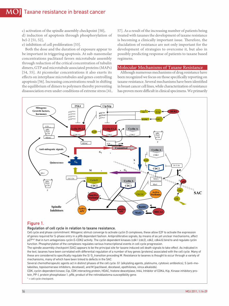
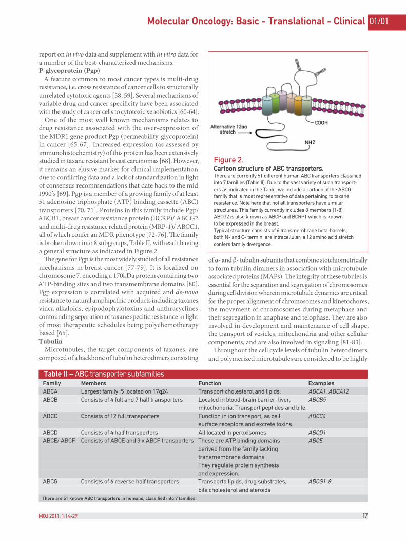
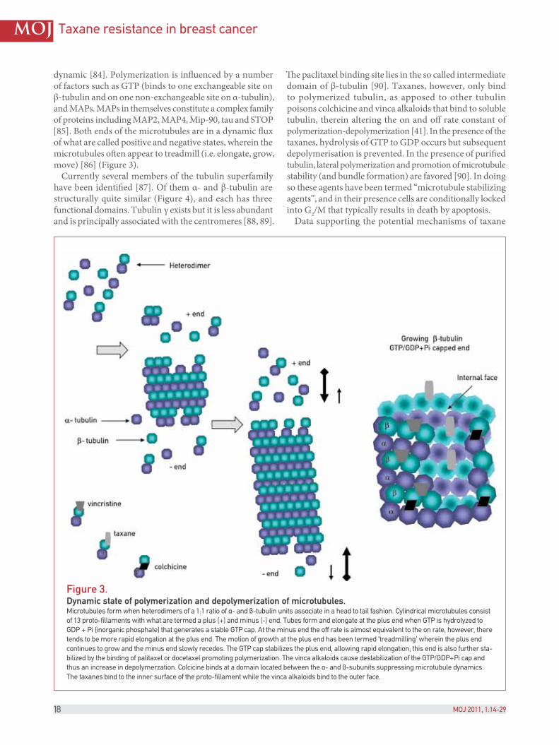
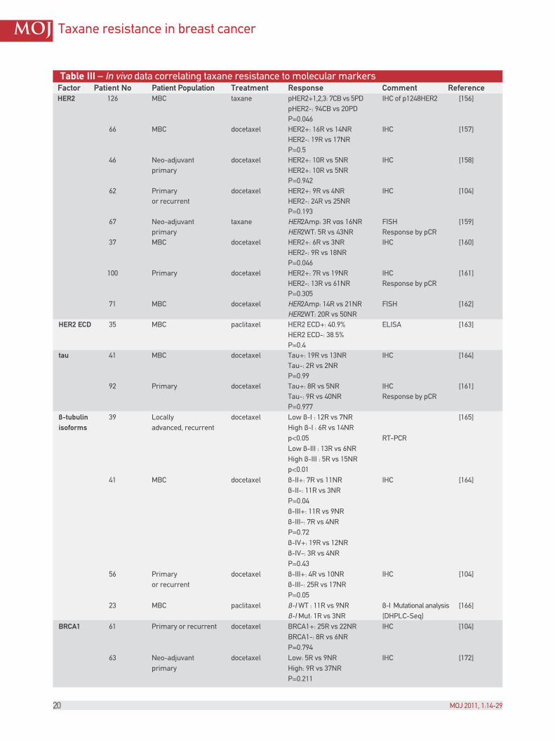
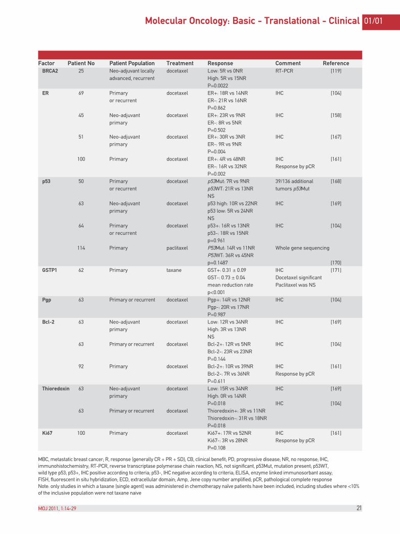
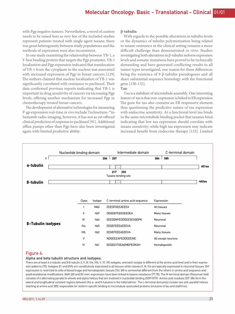


![Department of Translational Molecular Pathology, The ......Lung Cancer Biomarkers Pamela Villalobos, MD [Post-doctoral Fellow] and Department of Translational Molecular Pathology,](https://static.documents.pub/doc/80x56/5ea8a4647ff3b73b0c12a6a0/department-of-translational-molecular-pathology-the-lung-cancer-biomarkers.jpg)


![The Ten Commandments of Translational Research Informatics · translational research projects in oncology [11-15] for the past nine years, as well as the Dutch translational research](https://static.documents.pub/doc/80x56/5e85fcbf0ce01a00082536b4/the-ten-commandments-of-translational-research-informatics-translational-research.jpg)


