81
Molecular Oncology Chapter 14
| Date post: | 27-Dec-2015 |
| Category: |
Documents |
| Upload: | harvey-parrish |
| View: | 217 times |
| Download: | 0 times |

Molecular Oncology
Chapter 14
DYSPLASIA
• is an abnormality of both differentiation and maturation
• dysplasia is a condition of disordered cell growth and proliferation which may arise de novo or from tissues already showing pathological hyperplasia, metaplasia, or chronic irritation and inflammation
• early stage dysplasia- reversible- if the stimulus is removed
• advanced dysplasia can progress to neoplasia- cancer

Differences between dysplasia and cancer
• differs in two important respects:-1. invasiveness
• in dysplasia the abnormal cell proliferation does never invade the BM. Complete removal of dysplastic tissue is curative
• cancer in contrast, invades the BM and spreads through lymphatic and blood vessels, thus excision may not be curative
-2. reversibility• d. may sometimes return to normal- unlike cancer which is
irreversible

Classifications of Neoplasms• tumor- neoplasia- is an abnormality of cellular differentiation,
maturation and control of growth• tumor- the term can be applied to any swelling- inflammatory
tumor but most commonly it is used to denote suspected neoplasm• neoplasms are - benign and malignant - depends on several
features, chiefly the ability of malignant tumor to spread from the site of origin
• cancer- denotes a malignant tumor• definition of neoplasm: a neoplasm is an abnormal mass of tissue,
the growth of which exceeds and is uncoordinated with that of the surrounding normal tissues and persists in the same excessive manner after cessation of the stimuli that evoked the change.

• Cancer is usually divided into two broad categories: – Solid tumors– Hematological malignancies
Carcinomas
• EPITHELIAL TUMORS• adenocarcinoma- if derived from glandular
epithelium• squamous carcinoma, transitional carcinoma-
on the basis of origin• names may include the organ of origin or
description of microscopy- e.g. clear cell adenocarcinoma of the kidney, papillary carcinoma of thyroid, etc.
Sarcomas• MESENCHYMAL TUMORS.• malignant mesenchymal tumors are named after the cell of
origin to which is added the suffix – Leiomyosarcoma (smooth muscle), rhabdomyosarcoma(skeletal
muscle), liposarcoma, fibrosarcoma, chondrosarcoma, osteosarcoma, hemangiosarcoma, meningiosarcoma,
• Nomenclature exceptional to these rules-is used for hematological malignancies:
• leukemias- neoplasms of blood-forming organs- leukemias are subclassified on the basis of their clinical course- acute and chronic and the cell of origin- myeloid and lymphocytic, lymphoblastic
Cancer is caused by nonlethal genetic mutations affecting certain genes.
• Oncogenes, as proto-oncogenes, normally promote cell division or cell survival.– Oncogene mutations are usually a gain of function
and dominant.• Tumor suppressors: genes normally arrest cell
division.– Tumor-suppressor gene mutations are usually a
loss of function and recessive.

Cancer is caused by nonlethal genetic mutations.
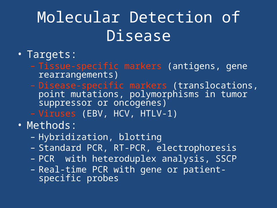
Molecular Detection of Disease
• Targets:– Tissue-specific markers (antigens, gene rearrangements)– Disease-specific markers (translocations, point mutations,
polymorphisms in tumor suppressor or oncogenes)– Viruses (EBV, HCV, HTLV-1)
• Methods:– Hybridization, blotting – Standard PCR, RT-PCR, electrophoresis – PCR with heteroduplex analysis, SSCP – Real-time PCR with gene or patient-specific probes
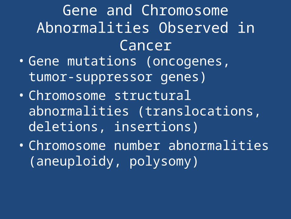
Gene and Chromosome Abnormalities Observed in Cancer
• Gene mutations (oncogenes, tumor-suppressor genes)
• Chromosome structural abnormalities (translocations, deletions, insertions)
• Chromosome number abnormalities (aneuploidy, polysomy)

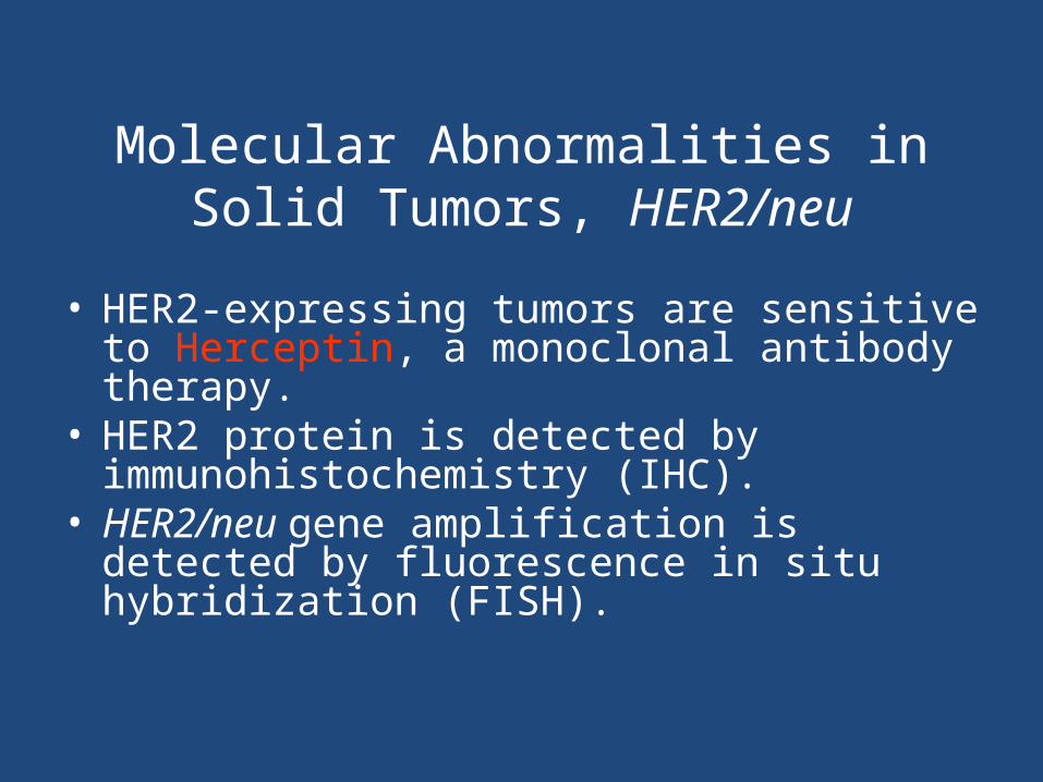
Molecular Abnormalities in Solid Tumors, HER2/neu
• HER2 (Human Epidermal Growth Factor Receptor 2) also known as Neu, ErbB-2, CD340 (cluster of differentiation 340) or p185 is a protein that in humans is encoded by the ERBB2 gene. HER2 is a member of the epidermal growth factor receptor (EGFR/ErbB) family.

Molecular Abnormalities in Solid Tumors, HER2/neu
• Amplification or over-expression of this gene has been shown to play an important role in the pathogenesis and progression of certain aggressive types of breast cancer and in recent years it has evolved to become an important biomarker and target of therapy for the disease.
Molecular Abnormalities in Solid Tumors, HER2/neu
• HER2-expressing tumors are sensitive to Herceptin, a monoclonal antibody therapy.
• HER2 protein is detected by immunohistochemistry (IHC).
• HER2/neu gene amplification is detected by fluorescence in situ hybridization (FISH).

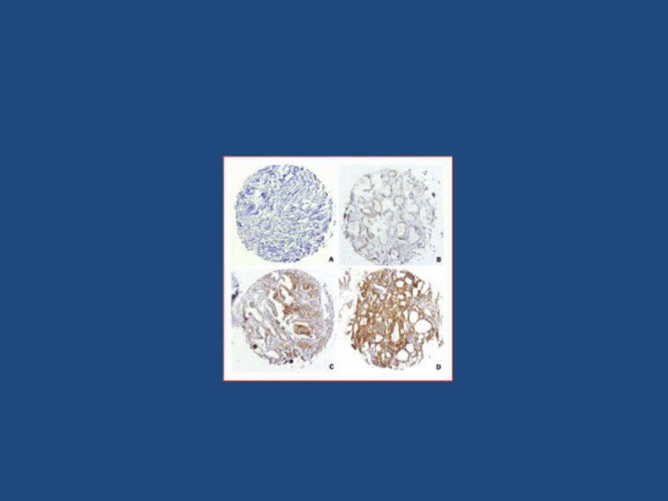
• IHC, or ImmunoHistoChemistry, is a special staining process performed on fresh or frozen breast cancer tissue removed during biopsy. IHC is used to show whether or not the cancer cells have HER2 receptors and/or hormone receptors on their surface
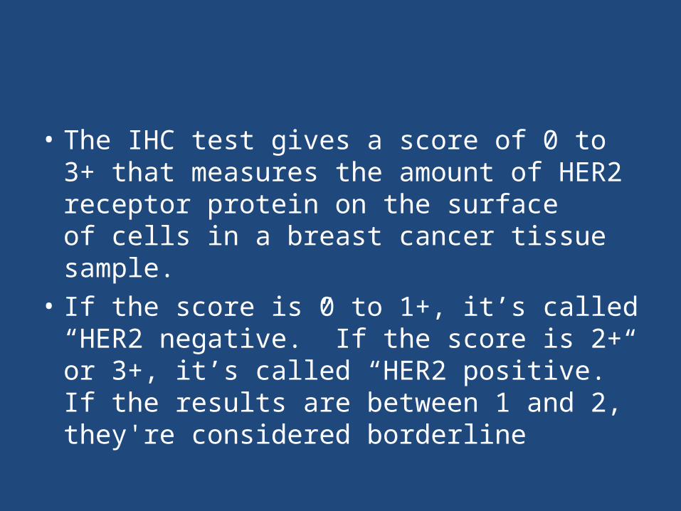
• The IHC test gives a score of 0 to 3+ that measures the amount of HER2 receptor protein on the surface of cells in a breast cancer tissue sample.
• If the score is 0 to 1+, it’s called “HER2 negative.” If the score is 2+ or 3+, it’s called “HER2 positive.” If the results are between 1 and 2, they're considered borderline

Molecular Abnormalities in Solid Tumors, EGFR
• The EGFR oncogene encodes another of the same family of epidermal growth factor receptors.
• This gene is mutated or amplified in several types of cancer cells.• Tumors with activating mutations in EGFR are sensitive to tyrosine
kinase inhibitors (TKI).• EGFR protein is detected by IHC.• EGFR gene and chromosome abnormalities are detected by FISH.• EGFR gene mutations are detected by SSCP, SSP-PCR, or direct
sequencing.

The EGFR Gene Family
Molecular Abnormalities in Solid Tumors, K-ras
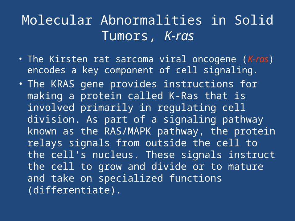
• The Kirsten rat sarcoma viral oncogene (K-ras) encodes a key component of cell signaling.
• The KRAS gene provides instructions for making a protein called K-Ras that is involved primarily in regulating cell division. As part of a signaling pathway known as the RAS/MAPK pathway, the protein relays signals from outside the cell to the cell's nucleus. These signals instruct the cell to grow and divide or to mature and take on specialized functions (differentiate).

• The K-Ras protein is a GTPase, which means it converts a molecule called GTP into another molecule called GDP. The K-Ras protein acts like a switch, and it is turned on and off by the GTP and GDP molecules. To transmit signals, the K-Ras protein must be turned on by attaching (binding) to a molecule of GTP. The K-Ras protein is turned off (inactivated) when it converts the GTP to GDP. When the protein is bound to GDP, it does not relay signals to the cell's nucleus.
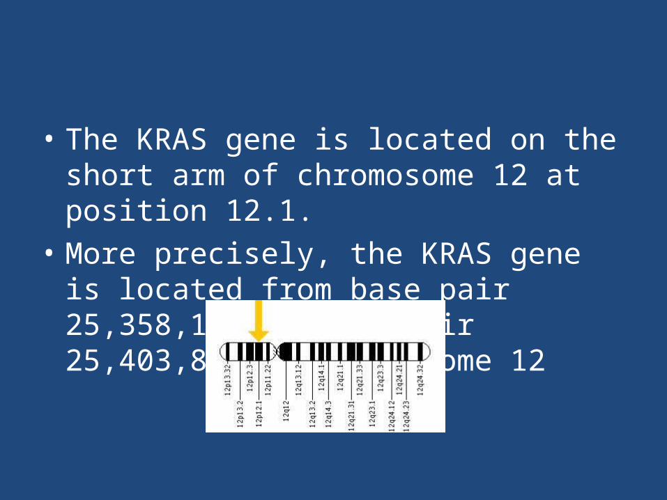
• The KRAS gene is located on the short arm of chromosome 12 at position 12.1.
• More precisely, the KRAS gene is located from base pair 25,358,179 to base pair 25,403,853 on chromosome 12
• K-ras mutations are associated with tumor malignancy and may affect response to some therapies.
• K-ras gene mutations are detected by SSCP or direct sequencing.
• Mutations in K-ras are the most common oncogene mutations in cancer.
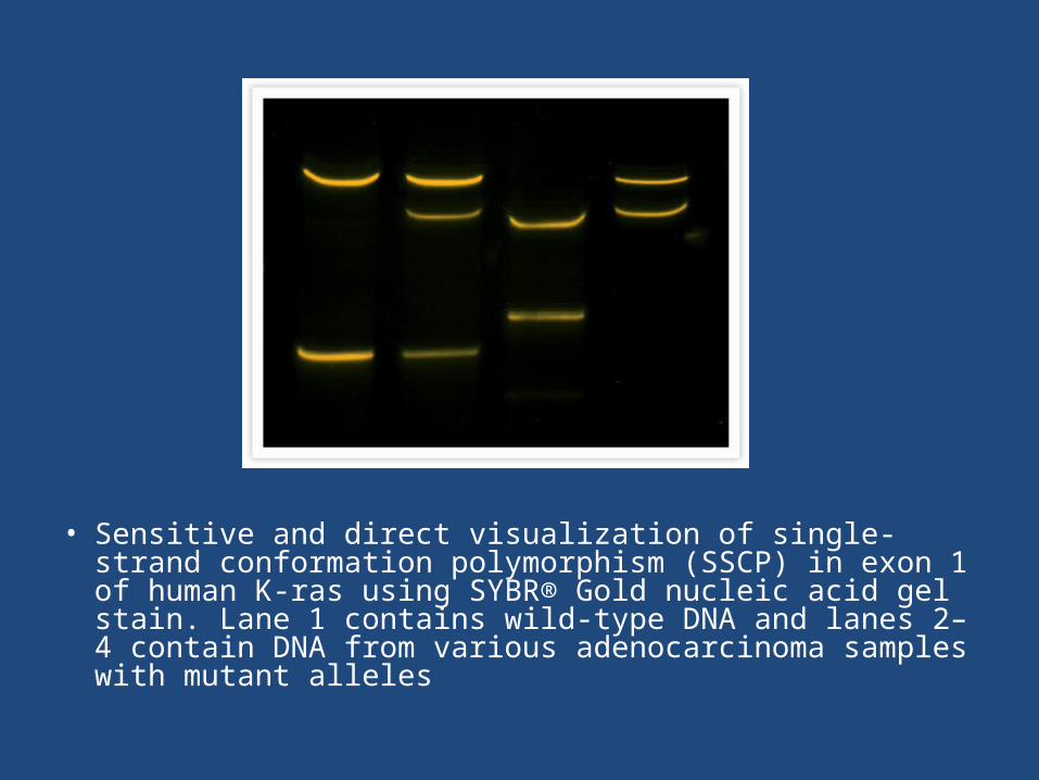
• Sensitive and direct visualization of single-strand conformation polymorphism (SSCP) in exon 1 of human K-ras using SYBR® Gold nucleic acid gel stain. Lane 1 contains wild-type DNA and lanes 2–4 contain DNA from various adenocarcinoma samples with mutant alleles
Molecular Abnormalities in Solid Tumors, TP53
• The 53-kilodalton tumor-suppressor gene (TP53) encodes a transcription factor.
• TP53 is mutated in half of all types of cancer.• Loss of TP53 function is an indicator of poor
prognosis in colon, lung, breast, and other cancers.• Mutant p53 protein is detected by IHC.• TP53 gene mutations are detected by a variety of
methods, including SSCP and direct sequencing.

Other Genes Associated with Solid Tumors
• Ewing sarcoma, EWS• Synovial sarcoma translocation, chromsome 18; synovial
sarcoma breakpoint 1 and 2, SYT-SSX1, SYT-SSX2• Paired box–Forkhead in rhabdomyosarcoma, PAX3-FKHR,
PAX7-FKHR• Ataxia telangiectasia mutated gene, ATM• Von Hippel-Lindau gene, VHL• V-myc avian myelocytomatosis viral-related oncogene,
neuroblastoma-derived, MYCN or n-myc• Rearranged during transfection (RET) protooncogene
Inherited Cancer Gene Mutations
• Inherited tumor suppressor gene mutations are recessive for the malignant phenotype.
• Tumor suppressor gene mutations are dominant with respect to increased risk of malignancy.
• Loss of heterozygosity exposes the recessive mutant allele in a hemizygous state.
• This is explained by the two-hit hypothesis.
• Tumor suppressor genes, which code for proteins that inhibit uncontrolled cell proliferation, are frequently mutated in human cancer.
• Heterozygous cells, cells with a single copy of a particular tumor suppressor gene, will not undergo aberrant cell proliferation leading to tumor formation.
• However, if the normal copy of the gene is lost, the cell becomes predisposed to abnormal growth and if enough mutations accumulate, a tumor may form. Losing the gene is known as loss of heterozygosity (LOH) the mechanisms of which are not fully understood.

Knudson two-hit hypothesis of tumorigenesis
• First Hit: The first hit is classically thought of as a point mutation that inactivates one copy of a tumor suppressor gene (TSG), such as Rb1. In hereditary cancer syndromes, individuals are born with the first hit. The individual does not develop cancer at this point because the remaining TSG on the other allele is still functioning normally.
• Second Hit: The second hit is classically thought of as a large deletion that results in loss of the remaining functioning TSG allele. This leaves only a non-functioning copy of the TSG, and the individual goes on to develop cancer.

Two-Hit Hypothesis
Normal At risk Affected
Affected
Loss of heterozygosity
At risk (inherited mutation)
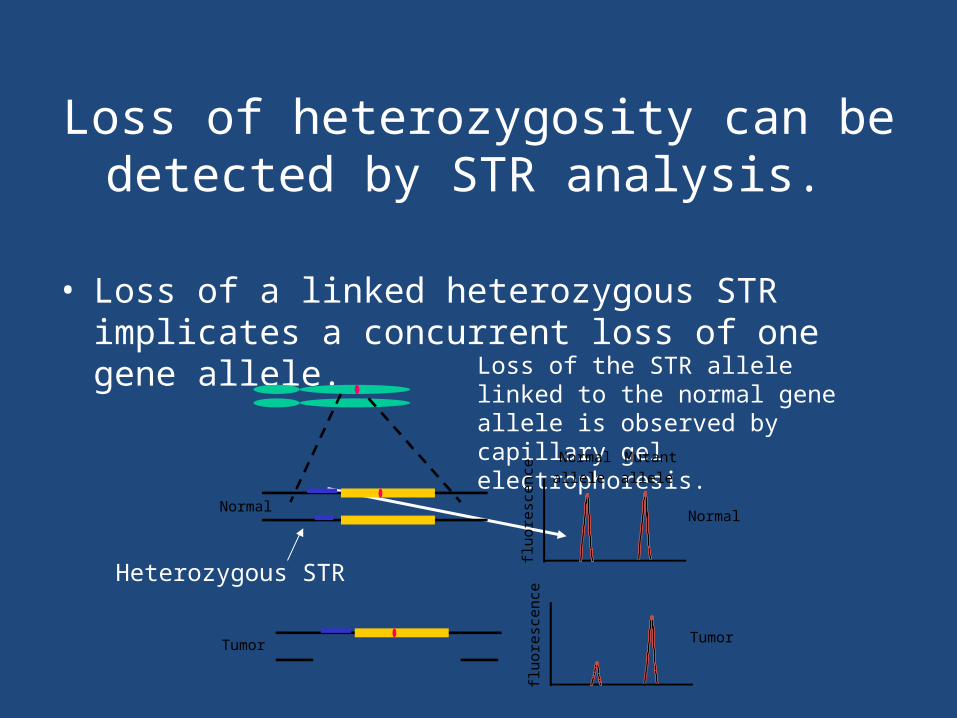
Loss of heterozygosity can be detected by STR analysis.
• Loss of a linked heterozygous STR implicates a concurrent loss of one gene allele.
Loss of the STR allele linked to the normal gene allele is observed by capillary gel electrophoresis.
Heterozygous STR
Normal allele
Mutantallele
fluor
esce
nce
fluor
esce
nce
Normal
Tumor
Normal
Tumor
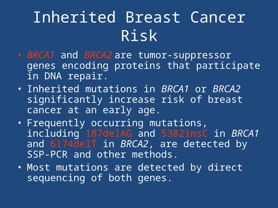
Inherited Breast Cancer Risk
• BRCA1 and BRCA2 are tumor-suppressor genes encoding proteins that participate in DNA repair.
• Inherited mutations in BRCA1 or BRCA2 significantly increase risk of breast cancer at an early age.
• Frequently occurring mutations, including 187delAG and 5382insC in BRCA1 and 6174delT in BRCA2, are detected by SSP-PCR and other methods.
• Most mutations are detected by direct sequencing of both genes.

Detection of BRCA1 185delAG by SSP-PCR
230 bp180 bp120 bp
The 180 bpproduct indicatesthe presence ofmutation.
Mutation-specificprimer
X
180 bp MW + m m + B
Agarose gel
MW = MW standard+ = normalm = mutantB = reagent blank
• http://learningobjects.wesleyan.edu/cancer/molecular_basis/
Replication Error (RER)
• Microsatellites (short tandem repeats) are sensitive to errors during DNA replication.
• These errors are normally corrected by the mismatch repair system (MMR).
• Components of the MMR system are encoded by MLH1, MSH2, and several other genes.
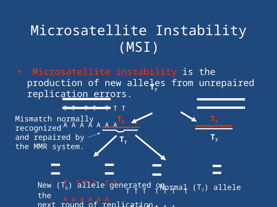
Microsatellite Instability (MSI)
• Microsatellite instability is the production of new alleles from unrepaired replication errors.
T T T T T T T A A A A A A A
T T T T T T T T T T T T TA A A A A A A A A A A A A
New (T6) allele generated on thenext round of replication.
Mismatch normally recognizedand repaired bythe MMR system.
T7
T7
T7
T6
T7
Normal (T7) allele

Hereditary Nonpolyposis Colorectal Carcinoma
• Hereditary nonpolyposis colorectal carcinoma (HNPCC) accounts for about 5% of colon cancer.
• HNPCC is the most common form of hereditary colon cancer.
• HNPCC is associated with mutations in genes encoding components of the MMR system, most frequently MLH1 and MSH2.
HNPCC and MSI
• 85%–90% of HNPCC tumors have MSI.• Mutations in genes of the MMR system (loss
of function) are inferred by testing for MSI.• MSI analysis determines gene function. Direct
sequencing is used to detect the actual gene mutation.
HNPCC and MSI
• MSI is analyzed by assessing stability of at least five microsatellite loci as recommended by the National Cancer Institute.
Marker Repeating UnitBAT25mononucleotideBAT26mononucleotideD5S346 dinucleotideD2S123 dinucleotideD17S250 dinucleotide
HNPCC and MSI
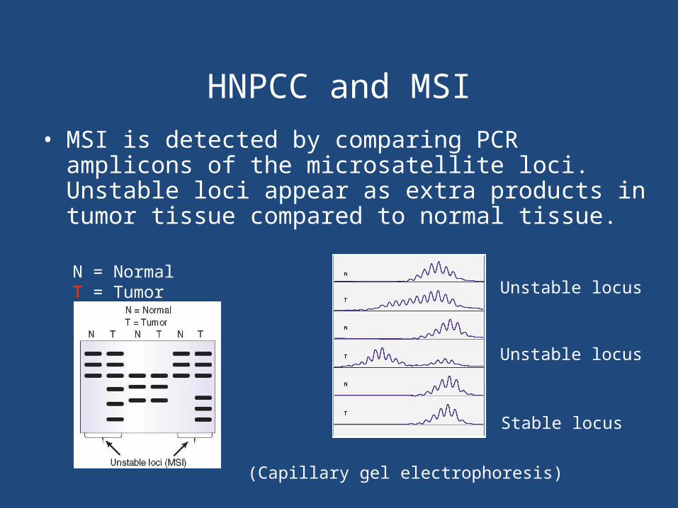
(Capillary gel electrophoresis)
Unstable locus
Stable locus
Unstable locusN = NormalT = Tumor
• MSI is detected by comparing PCR amplicons of the microsatellite loci. Unstable loci appear as extra products in tumor tissue compared to normal tissue.

Hematological Malignancies
• Hematologic cancers, (cancers of the blood, bone marrow, and lymph nodes) include leukemia, lymphoma, and myeloma.
• Every year, more than 100,000 cases of blood, bone marrow, and lymph node cancers are diagnosed in the United States, and more than 50,000 people die from these cancers.
• Among children and teens less than 20 years old, leukemia is the most common cancer and the leading cause of cancer death

Leukemia
• is a cancer of the bone marrow and blood. The two main types of leukemia are lymphocytic leukemia, which involves an increase of white blood cells called lymphocytes; and myelogenous leukemia (also known as myeloid or myelocytic leukemia), which involves an increase in white blood cells called granulocytes.
• Leukemia can be acute or chronic. Acute forms of leukemia progress rapidly, while chronic forms of leukemia progress slowly

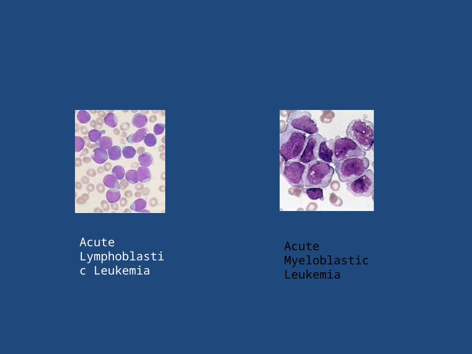
Acute Lymphoblastic Leukemia
Acute Myeloblastic Leukemia

Lymphoma
• Lymphoma is a general term for cancers that start in the lymph system; mainly the lymph nodes.
• The two main types of lymphoma are Hodgkin lymphoma and non-Hodgkin lymphoma.
• Hodgkin lymphoma spreads in an orderly manner from one group of lymph nodes to another.
• Non-Hodgkin lymphoma spreads through the lymphatic system in a non-orderly manner.
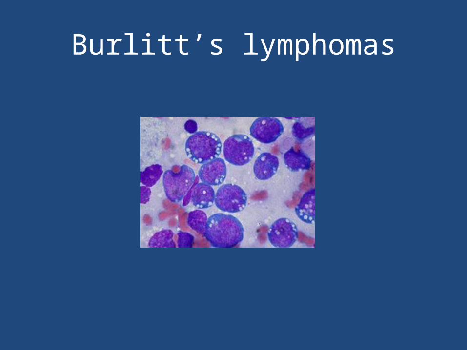
Burlitt’s lymphomas
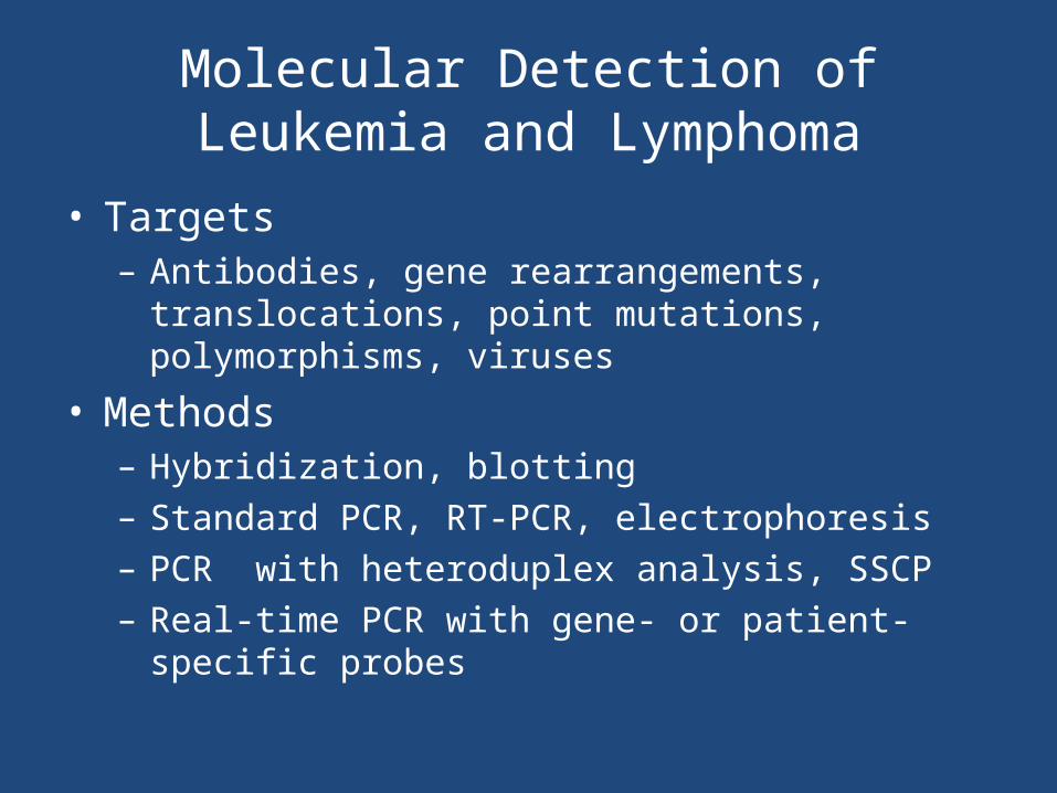
Molecular Detection of Leukemia and Lymphoma
• Targets– Antibodies, gene rearrangements, translocations, point
mutations, polymorphisms, viruses
• Methods– Hybridization, blotting – Standard PCR, RT-PCR, electrophoresis– PCR with heteroduplex analysis, SSCP– Real-time PCR with gene- or patient-specific probes
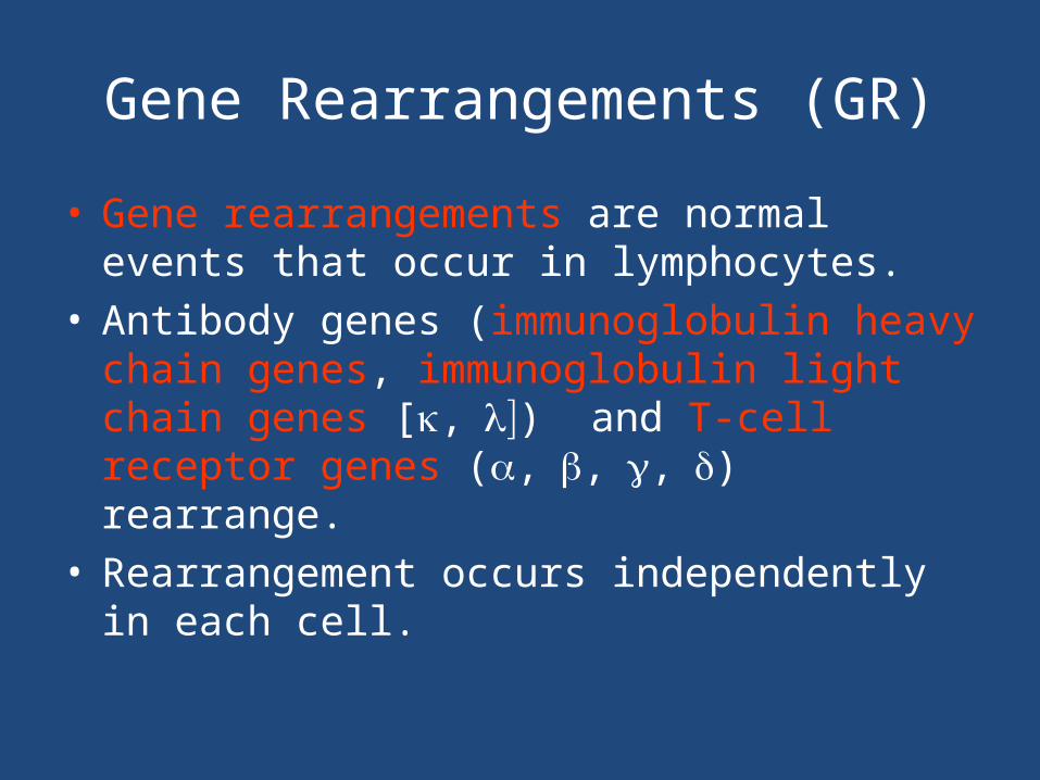
Gene Rearrangements (GR)
• Gene rearrangements are normal events that occur in lymphocytes.
• Antibody genes (immunoglobulin heavy chain genes, immunoglobulin light chain genes [,) and T-cell receptor genes (,,,) rearrange.
• Rearrangement occurs independently in each cell.

Immunoglobulin and T-Cell Receptor Gene Rearrangements
IgH GR IgH GR + IgL GR IgH + IgL GR
Early B cell precursor Pre-B B cell Mature PC
TCR and GR TCR and GR
Early thymocytes Common thymocytes
Cytotoxic T
Helper T
Lymphoidstem cell
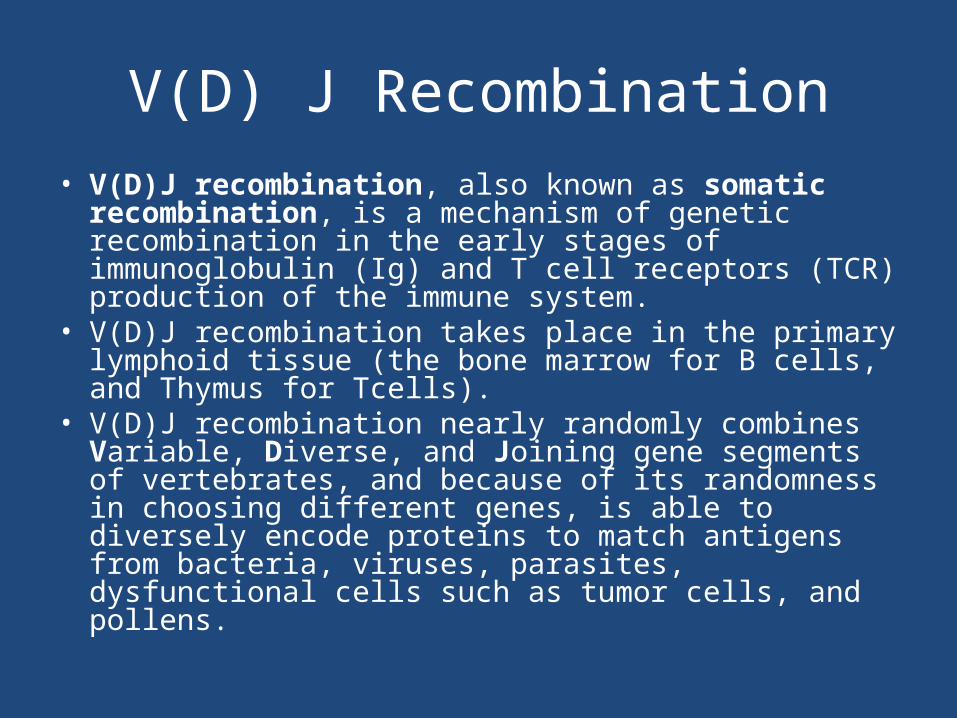
V(D) J Recombination• V(D)J recombination, also known as somatic
recombination, is a mechanism of genetic recombination in the early stages of immunoglobulin (Ig) and T cell receptors (TCR) production of the immune system.
• V(D)J recombination takes place in the primary lymphoid tissue (the bone marrow for B cells, and Thymus for Tcells).
• V(D)J recombination nearly randomly combines Variable, Diverse, and Joining gene segments of vertebrates, and because of its randomness in choosing different genes, is able to diversely encode proteins to match antigens from bacteria, viruses, parasites, dysfunctional cells such as tumor cells, and pollens.
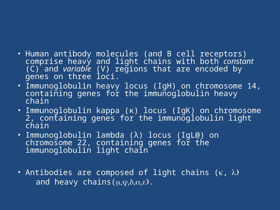
• Human antibody molecules (and B cell receptors) comprise heavy and light chains with both constant (C) and variable (V) regions that are encoded by genes on three loci.
• Immunoglobulin heavy locus (IgH) on chromosome 14, containing genes for the immunoglobulin heavy chain
• Immunoglobulin kappa (κ) locus (IgK) on chromosome 2, containing genes for the immunoglobulin light chain
• Immunoglobulin lambda (λ) locus (IgL@) on chromosome 22, containing genes for the immunoglobulin light chain
• Antibodies are composed of light chains (, and heavy chains(.

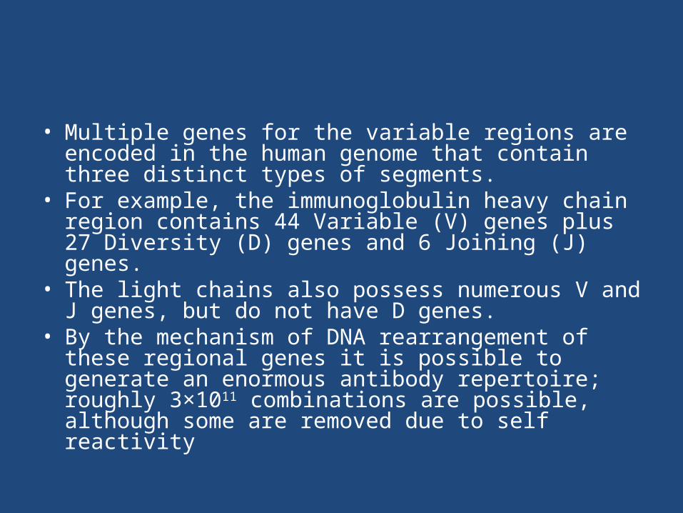
• Multiple genes for the variable regions are encoded in the human genome that contain three distinct types of segments.
• For example, the immunoglobulin heavy chain region contains 44 Variable (V) genes plus 27 Diversity (D) genes and 6 Joining (J) genes.
• The light chains also possess numerous V and J genes, but do not have D genes.
• By the mechanism of DNA rearrangement of these regional genes it is possible to generate an enormous antibody repertoire; roughly 3×10¹¹ combinations are possible, although some are removed due to self reactivity
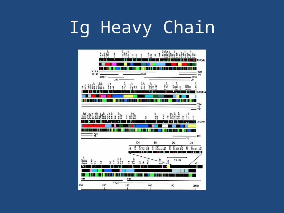
Ig Heavy Chain

• In the developing B cell, the first recombination event to occur is between one D and one J gene segment of the heavy chain locus. Any DNA between these two genes is deleted.
• This D-J recombination is followed by the joining of one V gene, from a region upstream of the newly formed DJ complex, forming a rearranged VDJ gene.
• All other genes between V and D segments of the new VDJ gene are now deleted from the cell’s genome.
• Primary transcript (unspliced RNA) is generated containing the VDJ region of the heavy chain and both the constant mu and delta chains (Cμ and Cδ). (i.e. the primary transcript contains the segments: V-D-J-Cμ-Cδ).
• The primary RNA is processed to add a polyadenylated (poly-A) tail after the Cμ chain and to remove sequence between the VDJ segment and this constant gene segment.

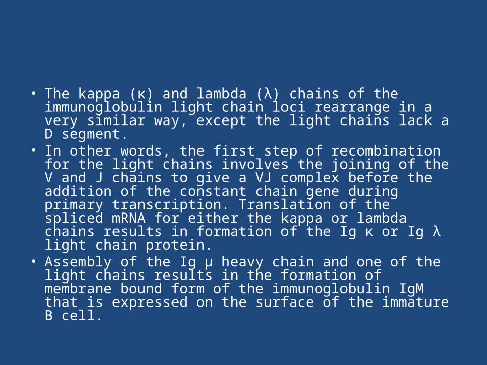
• The kappa (κ) and lambda (λ) chains of the immunoglobulin light chain loci rearrange in a very similar way, except the light chains lack a D segment.
• In other words, the first step of recombination for the light chains involves the joining of the V and J chains to give a VJ complex before the addition of the constant chain gene during primary transcription. Translation of the spliced mRNA for either the kappa or lambda chains results in formation of the Ig κ or Ig λ light chain protein.
• Assembly of the Ig μ heavy chain and one of the light chains results in the formation of membrane bound form of the immunoglobulin IgM that is expressed on the surface of the immature B cell.

Gene Rearrangements
• GR may be used to detect leukemias and lymphomas arising from cells that have rearranged their immunoglobulin (Ig) or T-cell receptor (TCR) genes.
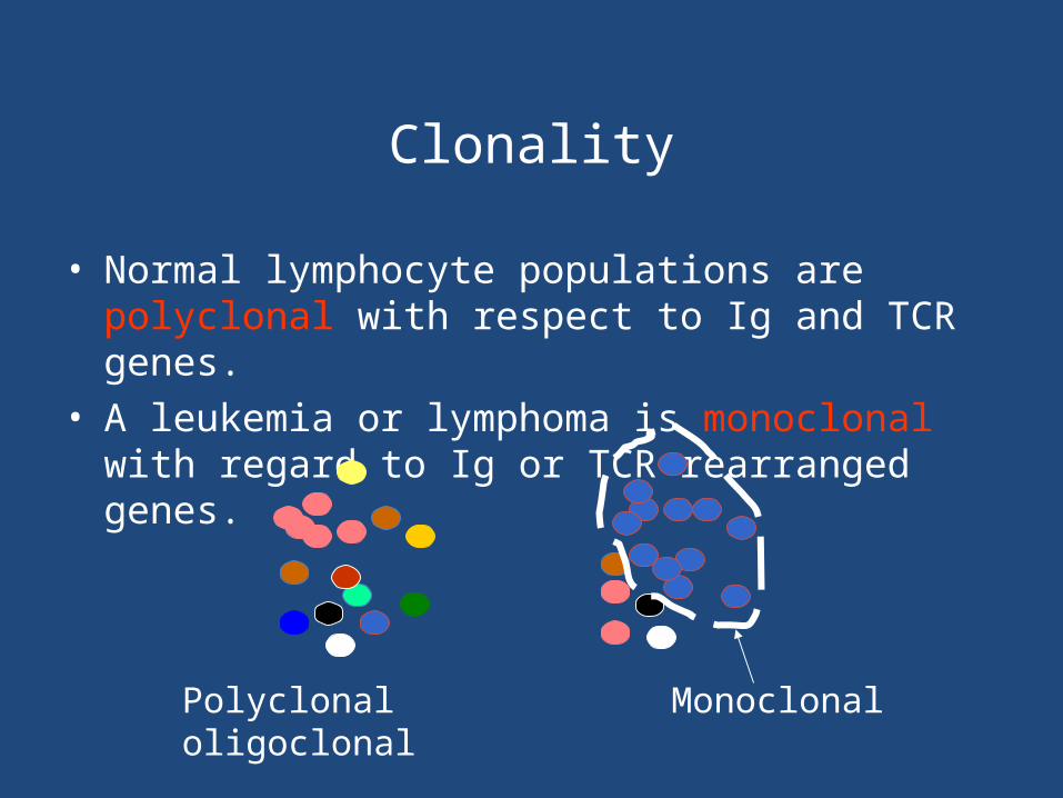
Clonality
• Normal lymphocyte populations are polyclonal with respect to Ig and TCR genes.
• A leukemia or lymphoma is monoclonal with regard to Ig or TCR rearranged genes.
Polyclonal Monoclonaloligoclonal
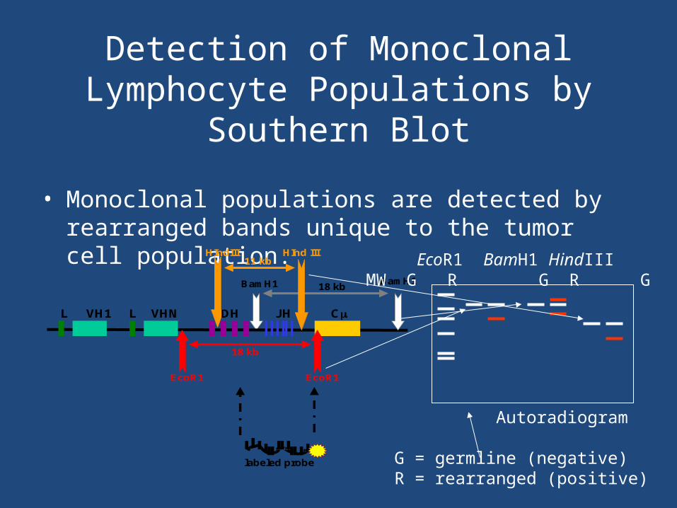
Detection of Monoclonal Lymphocyte Populations by Southern Blot
• Monoclonal populations are detected by rearranged bands unique to the tumor cell population.
L VH1 L VHN DH JH C
EcoR1 EcoR1
18 kb
BamH1 BamH118 kb
HIndIII HInd III11 kb
labeled probe
EcoR1 BamH1 HindIII MW G R G R G R
Autoradiogram
G = germline (negative)R = rearranged (positive)

Detection of Monoclonal Lymphocyte Populations by PCR
• Monoclonal populations are detected by sharp bands unique to the tumor cell population.
Monoclonal populations will yield a single PCR product.
Normal (polyclonal) populations will yield a polyclonal PCR product.
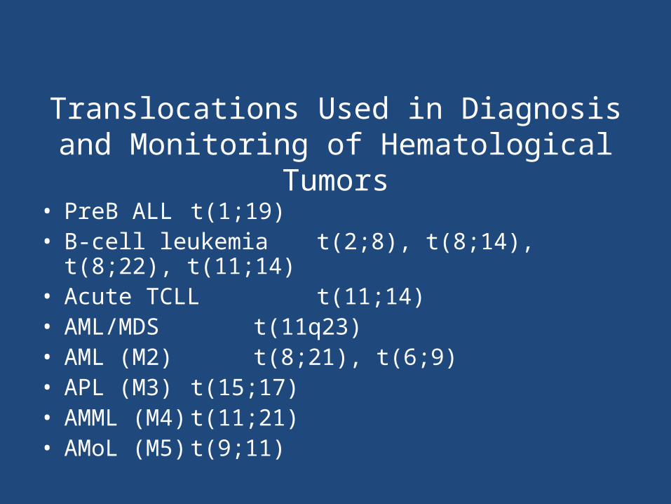
Translocations Used in Diagnosis and Monitoring of Hematological Tumors
• PreB ALL t(1;19)• B-cell leukemia t(2;8), t(8;14), t(8;22), t(11;14)• Acute TCLL t(11;14)• AML/MDS t(11q23)• AML (M2) t(8;21), t(6;9)• APL (M3) t(15;17)• AMML (M4) t(11;21)• AMoL (M5) t(9;11)
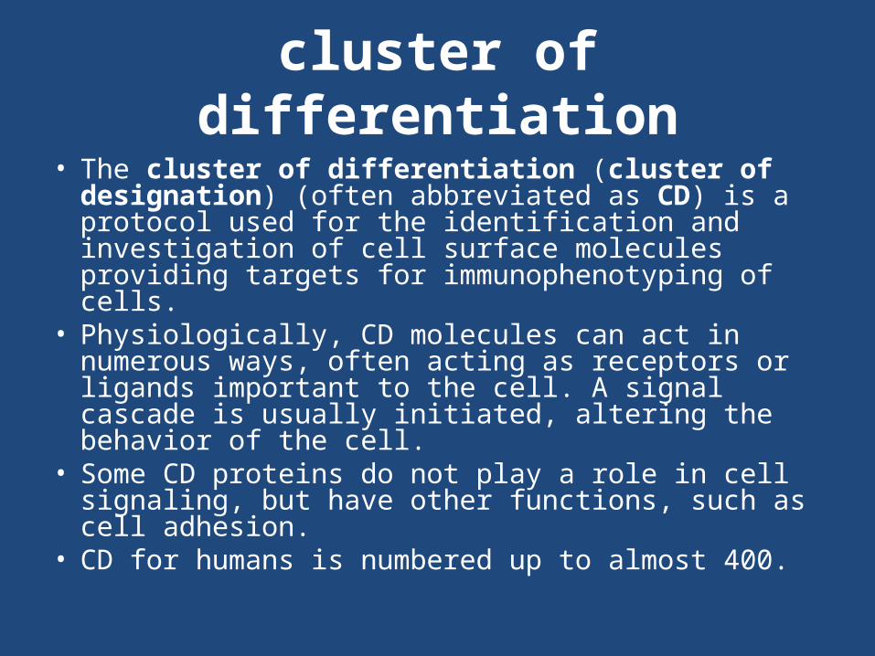
cluster of differentiation• The cluster of differentiation (cluster of designation)
(often abbreviated as CD) is a protocol used for the identification and investigation of cell surface molecules providing targets for immunophenotyping of cells.
• Physiologically, CD molecules can act in numerous ways, often acting as receptors or ligands important to the cell. A signal cascade is usually initiated, altering the behavior of the cell.
• Some CD proteins do not play a role in cell signaling, but have other functions, such as cell adhesion.
• CD for humans is numbered up to almost 400.


CD MarkersType of cell CD markers
stem cells CD34+, CD31-
all leukocyte groups CD45+
Granulocyte CD45+, CD15+, CD24+, CD114+, CD182+
Monocyte CD45+, CD14+, CD114+, CD11a, CD91+
T lymphocyte CD45+, CD3+
T helper cell CD45+, CD3+, CD4+
T regulatory cell CD4, CD25, and Foxp3
Cytotoxic T cell CD45+, CD3+, CD8+
B lymphocyte CD45+, CD19+ or CD45+, CD20+, CD24+, CD 22+
Thrombocyte CD45+, CD61+
Natural killer cell CD16+, CD56+, CD3-, CD31, CD30

Burkitt lymphoma
• The tumor cells in Burkitt’s lymphoma generally strongly express markers of B cell differentiation (CD20, CD22, CD19) as well as CD10, and BCL6. The tumor cells are generally negative for BCL2 and TdT. The high mitotic activity of Burkitt’s lymphoma is confirmed by nearly 100% of the cells staining positive for Ki67
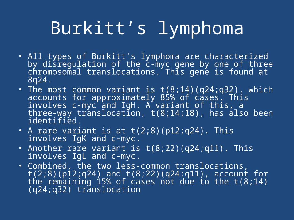
Burkitt’s lymphoma• All types of Burkitt's lymphoma are characterized by disregulation
of the c-myc gene by one of three chromosomal translocations. This gene is found at 8q24.
• The most common variant is t(8;14)(q24;q32), which accounts for approximately 85% of cases. This involves c-myc and IgH. A variant of this, a three-way translocation, t(8;14;18), has also been identified.
• A rare variant is at t(2;8)(p12;q24). This involves IgK and c-myc.• Another rare variant is t(8;22)(q24;q11). This involves IgL and c-
myc.• Combined, the two less-common translocations, t(2;8)(p12;q24)
and t(8;22)(q24;q11), account for the remaining 15% of cases not due to the t(8;14)(q24;q32) translocation
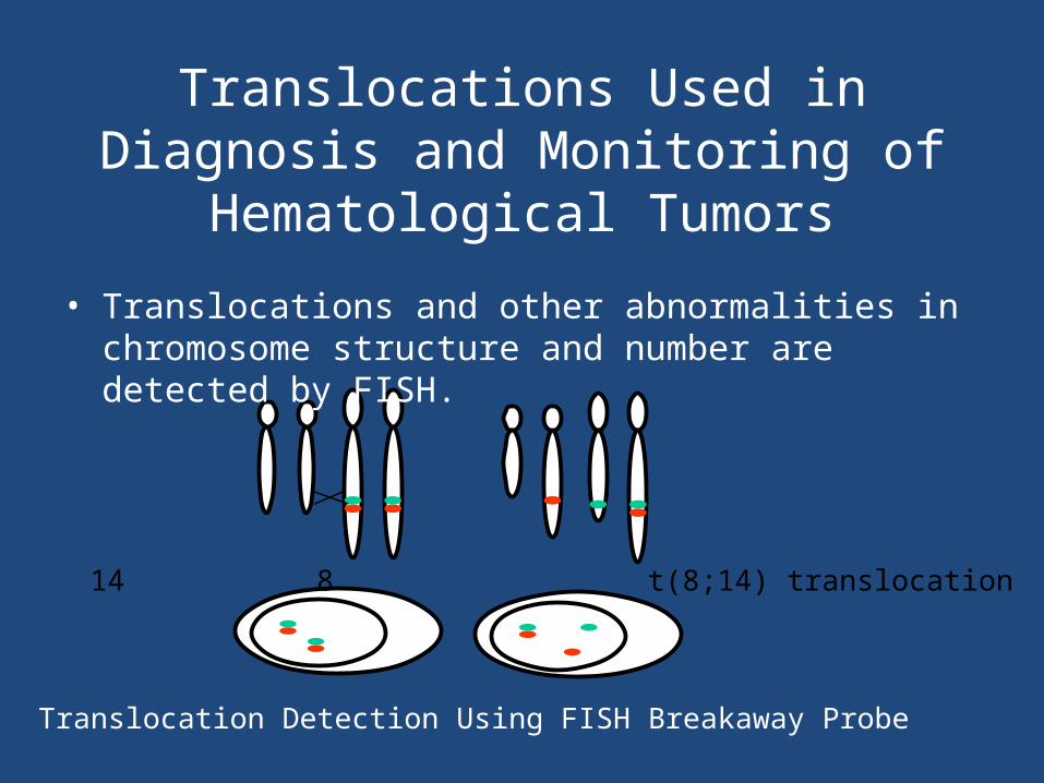
14 8 t(8;14) translocation
Translocation Detection Using FISH Breakaway Probe
Translocations Used in Diagnosis and Monitoring of Hematological Tumors
• Translocations and other abnormalities in chromosome structure and number are detected by FISH.

Translocations Used in Diagnosis and Monitoring of Hematological Tumors
• Translocations are detected with higher sensitivity using PCR.
• qPCR may be used to quantify tumor load during patient monitoring.
• FISH is recommended for initial diagnosis. PCR is better for monitoring.

Translocations Used in Diagnosis and Monitoring of Hematological Tumors
• CML t(9;22), t(11;22)• ALL t(9;22), t(12;21), t(8;14), t(2;8), t(8;22),
t(11q)• Burkitt t(8;14), t(2;8), t(8;22)• DLBCL t(3q27), t(14;18); t(8;14)• TCL t(8;14)• Follicular t(14;18), t(8;14)• MCL t(11;14)• MM t(14q32)
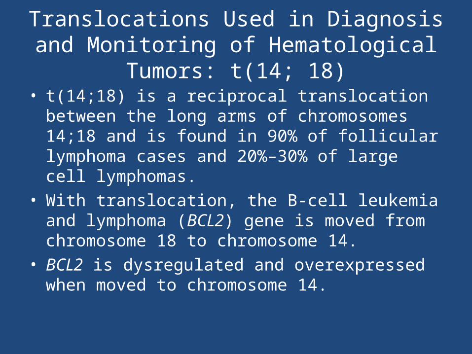
Translocations Used in Diagnosis and Monitoring of Hematological Tumors: t(14; 18)
• t(14;18) is a reciprocal translocation between the long arms of chromosomes 14;18 and is found in 90% of follicular lymphoma cases and 20%–30% of large cell lymphomas.
• With translocation, the B-cell leukemia and lymphoma (BCL2) gene is moved from chromosome 18 to chromosome 14.
• BCL2 is dysregulated and overexpressed when moved to chromosome 14.
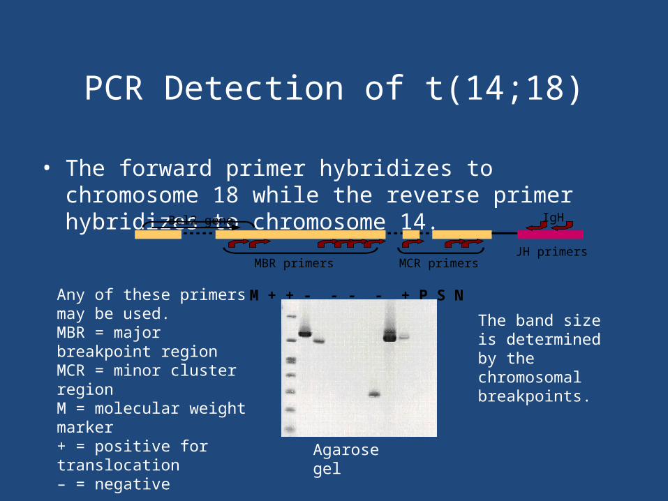
PCR Detection of t(14;18)
• The forward primer hybridizes to chromosome 18 while the reverse primer hybridizes to chromosome 14.
M + + - - - - + P S NAny of these primers may be used.MBR = major breakpoint regionMCR = minor cluster regionM = molecular weight marker+ = positive for translocation– = negative
MCR primersJH primers
IgH
MBR primers
Bcl2 gene
The band size is determined by the chromosomal breakpoints.
Agarose gel

Translocations Used in Diagnosis and Monitoring of Hematological Tumors: t(9;22)
• t(9;22) is a reciprocal translocation between the long arms of chromosomes 9;22 and is found in chronic myelogenous leukemia and acute lymphoblastic leukemia.
• This translocation forms a chimeric gene between the breakpoint cluster region (BCR) gene on chromosome 22 and the Abelson leukemia virus (ABL) gene on chromosome 9.
• The translocated chromosome is the Philadelphia chromosome.

b3a2AAAAA
FusionmRNA(8.5 kb)
Fusionprotein p210 BCRABL
e1 b1 2 3 a2 3 4....
Translocations Used in Diagnosis and Monitoring of Hematological Tumors: t(9; 22)
• The chimeric gene, BCRABL, produces an abnormal protein that drives the tumor cell phenotype.

BCR ABLPhiladelphia chromosome
Splicing
Reverse transcription
cDNA
BCRABL
cDNA made from patient mRNA is amplified if the translocation is present.
Detection of t(9;22) by RT-PCR

1 = molecular weight standard2–5 = positive for translocation6 = negative7–11 = amplification controls12 = blank
The band size is determined by different chromosome 22 breakpoints.
Agarose gel
Translocationproducts(BCRABL)
1 2 3 4 5 6 7 8 9 10 11 12
Translocationproducts(ABL)
Detection of t(9;22) by RT-PCR
1 2 3 4 5 6 7 8 9 10 11 12

1 2 3 4 5 6 7 8 9 10 11 12
Quantification by qPCR (TaqMan)
• For qPCR, use a standard curve of tumor cells diluted into normal cells.
• For RT-qPCR, use a standard curve of transcripts of known copy numbers diluted into normal RNA.

VIRAL CARCINOGENESIS
• -a variety of DNA and RNA viruses are known to cause cancers in animals, but only few of them have been linked with human tumors
• Human papilloma virus (HPV)-some types of HPV cause benign squamous papillomas (warts)- types 1, 2, 4, 7-HPV 16 and 18 are present in over than 90% of cases of squamous cell ca of the uterine cervix
• -genital warts are associated with HPV-6 and HPV-11

Epstein-Barr virus (EBV)
• -is associated with at least two human tumors-Burkitt lymphoma- is a high grade tumor of B-lymphocytes- endemic in central Africa- all patients carry EBV genome in tumor cells-undifferentiated nasopharyngeal cancer- is endemic in southern China and in Eskimos- EBV genome is found in all such tumors
• hepatitis B virus (HBV)• -there is close association of HBV infection and liver
cancer, though mechanism by which HBV causes cancer is uncertain and probably multifactorial
• Human T-cell leukemia virus type 1 (HTLV-1)• -this virus has strong afinity to CD4+ T-lymphocytes• -HTLV-1-associated leukemia/lymphoma is endemic in
Japan and Caribbean• -HTLV-1 proviral DNA is detected in DNA of leukemic
cells

Summary
• Molecular testing analyzes tissue-specific and tumor-specific (mutation) targets.
• Genome, chromosome, and gene mutations are useful targets for diagnosis and detection of solid tumors.
• Microsatellite instability is a test for function of the DNA mismatch repair system, which may be mutated in hereditary colon cancer.
• Ig and TCR gene rearrangements are tissue-specific markers for certain lymphomas and leukemias.
• Translocations are tumor-specific markers for some hematological disorders.


