Molecular pathogenesis and extraovarian origin ofepithelial ovarian cancer—Shifting the paradigmRobert J. Kurman MD⁎, Ie-Ming Shih MD, PhD
Division of Gynecologic Pathology, Departments of Pathology, Gynecology and Obstetrics and Oncology, The Johns HopkinsUniversity School of Medicine, Baltimore, MD 21231, USA
Received 25 January 2011; revised 23 March 2011; accepted 23 March 2011
919Molecular pathogenesis and extraovarian origin of EOC
Paradigms, as defined by Kuhn [1] in his seminal work 1. Molecular pathogenesis of epithelial
“The Structure of Scientific Revolutions,” are the best waysof explaining progress in science. Kuhn believed thattextbooks, which describe progress, as a cumulative,incremental process leading to a growing corpus of scientificknowledge, present an unrealistic and biased view. Instead,he felt that a more accurate depiction could be gleaned bylooking at what scientists do most of the time, which hetermed normal science, and normal science is governed byparadigms. A paradigm generates a consensus amongscientists working in a particular field about how work inthat field should be done. It also identifies puzzles, assuresscientists that each puzzle has a solution, and providesstandards for evaluating solutions. Normal science involvesshowing how nature can be fitted into the categories providedby the paradigm. When puzzles arise that repeatedly resistsolutions a crisis of confidence occurs. During a crisis, theparadigm is subjected to testing and might be rejected. If thatoccurs, a new paradigm replaces the previous one and ascientific revolution has occurred.
The operative paradigm of ovarian carcinogenesis is thatepithelial ovarian cancer (EOC) is composed of severaldifferent types, but because the vast majority is high-gradeserous carcinoma (HGSC), differences between the types areobscured, and therefore, EOC is regarded as a single disease.Moreover, because carcinomas in the female pelvis tend toinvolve the ovary, often as the dominant mass, they have allbeen regarded as ovarian in origin. This paradigm is based ontaxonomy, specifically morphologic classifications, whichtook shape in the 1930s and 1940s [2-4], matured in the1950s and 1960s [5], and culminated with the World HealthOrganization classification in 1973 [6]. The histologicclassifications created a structure that provided the basisfor performing clinicopathologic studies, but apart fromthese studies, the tools necessary to study pathogenesis werenot available, and therefore, our understanding of ovariancarcinogenesis was limited.
Arguably, research within this paradigm has failed toidentify the precursor of EOC [7-19], and as a result, currentmanagement is empirical. Despite advances in radical surgeryand cytotoxic chemotherapy, overall survival has notchanged in more than 50 years. In the last 2 decades,attention has focused on early detection, but unfortunately,this strategy has also failed to provide a survival benefit.Accordingly, there are “persistent puzzles” that have resistedsolutions, and hence, a “crisis of confidence” exists.
The introduction of molecular biology and the develop-ment of new methods of tissue sampling are now ushering ina paradigm shift, which can be considered “revolutionary.”The concepts that are emerging and shaping this newparadigm are novel and highly provocative. Some of themwill be confirmed, and others will be modified or discarded,as scientists in the process of performing “normal science”within the framework of the new paradigm attempt to clarifyand resolve issues that have frustrated progress in reducingthe burden of this disease.
ovarian carcinoma
The introduction of the “borderline (low malignantpotential)” category was an important step in refining themorphologic classification of EOC by identifying a group oftumors, defined as lacking destructive invasive growth thathad a significantly better outcome than the invasivecarcinomas. Because it was rare to find a borderline tumorcoexisting with an invasive carcinoma, it was generallybelieved that they were unrelated. In 1996, a relationshipbetween serous borderline tumor (SBT) and invasive serouscarcinoma was described based on the subdivision of SBTinto 2 groups. One group designated “atypical proliferativeserous tumor (APST)” behaved in a benign fashion, and asecond, smaller group designated “micropapillary serouscarcinoma (MPSC)” also termed “noninvasive low-gradeserous carcinoma” behaved like a low-grade malignanttumor [20]. Moreover, this latter subset was closelyassociated with invasive low-grade serous carcinoma(LGSC), and the investigators proposed that MPSC wasthe immediate precursor of LGSC. The key element leadingto this conclusion was the recognition that LGSC was adistinct entity that differed from HGSC in several ways (seebelow). Before this, serous carcinoma was graded well,moderately, and poorly differentiated, with the implicationthat serous carcinoma was a spectrum of disease in whichwell-differentiated carcinoma (LGSC) progressed to poorlydifferentiated carcinoma (HGSC). After this morphologicstudy linking APST to MPSC and LGSC, a series ofmolecular genetic studies was performed, which culminatedin the proposal of a dualistic model to explain thepathogenesis of EOC [21].
1.1. Dualistic model of carcinogenesis
Briefly, the dualistic model accommodates and confirmsthe heterogeneous nature of EOC and places the majorhistologic types into 2 groups (types I and II) based on theirdistinctive clinicopathologic and molecular genetic features.It also links specific histologic types with their putativeprecursor lesions. Thus, type I tumors comprise LGSCs, low-grade endometrioid, clear cell, and mucinous carcinomas,which develop in a stepwise fashion from well-establishedprecursor lesions, such as borderline tumors and endometri-osis. They typically present as large masses that are confinedto one ovary (stage Ia), are indolent, and have a goodprognosis. The type I tumors are relatively genetically stableand typically display a variety of somatic sequence mutationsthat include KRAS, BRAF, PTEN, PIK3CA, CTNNB1 (thegene-encoding β-catenin), ARID1A, and PPP2R1A but veryrarely TP53 [21-23]. In contrast, type II tumors compriseHGSC (usual type of serous carcinoma), high-gradeendometrioid carcinoma, malignant mixed mesodermaltumors (carcinosarcomas), and undifferentiated carcinomas,
920 R. J. Kurman, I. -M. Shih
which present in advanced stage (stages II-IV) in more than75% of cases; they grow rapidly and are highly aggressive.Type II tumors, of which HGSC is the prototypic type, arechromosomally highly unstable and harbor TP53 mutationsin more than 95% of cases [24]; they rarely display themutations found in the type I tumors. BRCA inactivation,either by mutation or inactivation of expression of BRCAand its downstream genes via promoter methylation, occursin up to 40% to 50% of HGSC [25]. BRCA inactivation hasnot been reported in the type I tumors.
1.2. Serous tumors
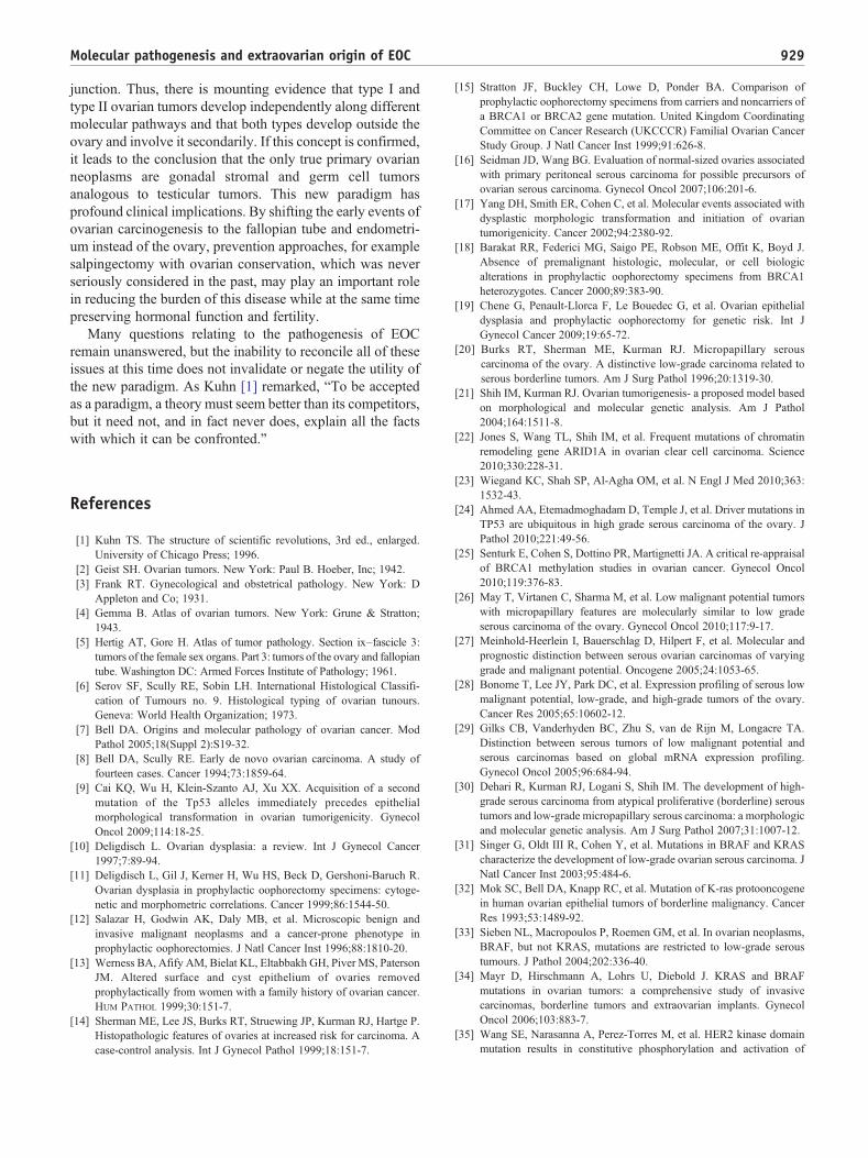
The relationship of APST and MPSC to LGSC based onmorphologic studies was supported by mutational analysis,gene expression studies, and methylation profiling demon-strating that these 3 tumor types shared molecularalterations that differed dramatically from HGSC [25-30].Initial molecular genetic studies focused on individualgenes (Fig. 1), but more recent studies have highlighted theimportance of molecular signaling pathways (Fig. 2). Forexample, the MAPK signaling pathway is important for thecellular response to a variety of growth and differentiationfactors, and activating mutations in KRAS or one of itsdownstream effectors, BRAF, (mutations of KRAS andBRAF are mutually exclusive) results in constitutiveactivation of mitogen-activated protein kinase (MAPK)-mediated signaling in more than half of APSTs, MPSCs,and LGSCs [31-34]. In addition, a 12-base-pair insertionmutation of ERBB2 (encoding HER2/neu), which activatesan upstream regulator of K-Ras, has been detected in 9% ofthese tumors. Interestingly, tumors with ERBB2 mutationslack KRAS and BRAF mutations [35,36]. Accordingly,60% to 70% of APSTs, MPSCs, and LGSCs express active
High-g
ARID1ACTNNB1PTENPIK3CAPPP2R1A
Mutation
KRASBRAFERBB2PIK3CAMutation
ARID1APIK3CAZNF217PPP2R1A
Mutation
KRASMutation
Low-grade serous
Endometrioid
Clear cell
Mucinous
TPC
Type I
Typ
Fig. 1 Prevalence of histologic types of EOC an
MAPK [37]; they rarely harbor TP53 mutations. Recentstudies have further clarified the molecular pathogenesis ofAPST, MPSC, and LGSC. First, KRAS and BRAFmutations have not been detected in serous cystadenomas,the putative precursor of SBTs, but laser capture microdis-section studies have detected these mutations in theadenoma epithelium and APST epithelium in serouscystadenomas containing small APSTs, suggesting thatthese mutations occur early in the development of APST[38]. In an attempt to elucidate the relationship of APST toLGSC, a recent study compared the gene expressionprofiles of APST, MPSC, and LGSC and found thatMPSC is closer molecularly to invasive LGSC than toAPST [26] and that the genes involved in MAPK signalingshowed higher expression in MPSC than in APST. Inaddition, a previous study reporting that MPSC harbors apattern of chromosomal imbalance distinct from that ofAPST [39] confirms the proposal that LGSC develops in astepwise fashion from cystadeno(fibro)ma to APST andMPSC, supporting the biologic role of the KRAS-BRAF-MEK-MAPK pathway in the development of LGSC. Byglobally profiling the epigenetic landscape, we haverecently reported that the methylation profiles in LGSCare closer to APST and serous cystadenoma than HGSC[30]. This finding lends further support to the dualisticmodel of ovarian serous carcinogenesis.
In contrast to LGSC, HGSC harbors TP53 mutations inmore than 95% of cases [25] but rarely contains KRAS orBRAF mutations. Aside from TP53 mutations, no othermutations are consistently found in sporadic (nonfamilial)HGSCs including mutations of BRCA1 and BRCA2, whichcharacterize familial HGSC (The Cancer Genome Atlas,unpublished). On the other hand, inactivation of the BRCA1/2genes by other mechanisms, such as hypermethylation of
Fig. 2 Schematic illustration of pathway alterations involved in the development of LGSC. The cardinal molecular genetic changes includesomatic mutations in KRAS, BRAF, and occasionally ERRB2 (encoding Her2/Neu) and PIK3CA. The mutated gene products constitutivelyactivate the signaling pathways that regulate cellular proliferation and survival and promote tumor initiation and progression through severalmechanisms including up-regulation of glucose transporter-1. The size of the boxes containing specific genes reflects the relative frequency ofthe mutation, and the thickness of the arrows indicates the relative contribution of the pathway alterations to tumor development.
921Molecular pathogenesis and extraovarian origin of EOC
the BRCA1 promoter, occurs relatively frequently, and as aresult, inactivation of BRCA1/2 by mutation or othermechanisms occurs in 40% to 50% of sporadic HGSCs[26]. The most striking molecular feature of HGSC is diffuseand high levels of DNA copy number gains or losses, whichinclude CCNE1 (cyclin E1), NOTCH3, AKT2, RSF1, andPIK3CA loci [40]. Despite their distinct molecular signatures,LGSC, and even an APST, is sometimes clonally associatedwith a synchronous HGSC , suggesting that such progressionrarely does occur [41].
1.3. Clear cell and endometrioid tumors
After serous carcinoma, endometrioid and clear cellcarcinomas are the most frequent types of EOC accountingfor approximately 15% to 20% of EOC in Westerncountries. The molecular genetic alterations that underliethe development of these tumors are now beginning toemerge. Based on genome-wide mutational analysis, the
most common molecular genetic changes in clear cellcarcinoma are a somatic inactivating mutation of ARID1A[22,23] (a tumor suppressor gene detected in about 50% ofcases), an activating mutation of PIK3CA in about 50% oftumors [42], and deletion of PTEN (a tumor suppressorgene involved in the PI3K/PTEN signaling pathway) inabout 20% [43], supporting the role of an aberrant PI3K/PTEN pathway in the development of clear cell carcinoma.In addition, single nucleotide polymorphism (SNP) arrayanalysis has identified frequent amplification of theZNF217 (zinc finger protein 217) locus and deletion ofthe CDKN2A/2B locus in clear cell carcinomas, suggestingthat the pathways involving these 2 genes are alsoimportant in their development.
Like clear cell carcinoma, mutations that deregulate PI3K/PTEN signaling are common in low-grade endometrioidcarcinoma, and in fact, mutation of the tumor suppressorgene PTEN, which occurs rarely in other types of EOC, hasbeen reported in approximately 20% of ovarian low-grade
922 R. J. Kurman, I. -M. Shih
endometrioid carcinomas [44,45]. Another mechanism bywhich activation of PI3K signaling occurs is throughactivating mutations of PIK3CA, which has been detectedin 20% of low-grade endometrioid carcinomas [42]. TheWnt/β-catenin signaling pathway, which is involved in theregulation of several important cellular processes includingproliferation, motility, and survival, is deregulated in up to40% of ovarian endometrioid carcinomas, usually on thebasis of activating mutations of CTNNB1, the gene thatencodes β-catenin [46]. Notably, mutation of CTNNB1 hasbeen associated with squamous differentiation, low tumorgrade, and a favorable outcome, features that characterizelow-grade endometrioid carcinoma [47-50].
Although low-grade endometrioid carcinomas are easilyrecognized, the distinction of high-grade endometrioidcarcinoma from HGSC can be very difficult. Somepathologists even question the existence of high-gradeendometrioid carcinoma, regarding it instead as a variant ofHGSC, whereas others classify high-grade endometrioidcarcinoma as “mixed high-grade endometrioid carcinomaand HGSC” or as “HGSC with features of endometrioidcarcinoma.” It is therefore of interest that in a study ofovarian endometrioid carcinomas of all grades, low-gradeendometrioid carcinomas were characterized by mutationsthat deregulate the canonical Wnt/β-catenin and PI3K/PTENsignaling pathways and lacked TP53 mutations, whereashigh-grade endometrioid carcinomas lacked Wnt/β-cateninor PI3K/PTEN signaling pathway defects and frequentlyharbored TP53 mutations [47]. A few high-grade endome-trioid carcinomas did, in addition to TP53 mutation, displaymolecular changes found in the low-grade endometrioidcarcinomas, suggesting that some low-grade endometrioidcarcinoma may progress to high-grade carcinoma. Thefindings parallel those seen in the serous tumors, namely,that generally, low- and high-grade tumors developindependently but that rarely progression of a low-grade toa high-grade tumor occurs. The similar high frequency ofTP53 mutations in high-grade endometrioid carcinoma asin HGSC suggests that both develop in a similar fashion, viaTP53 mutation, and that most high-grade endometrioidcarcinoma is closely related to or is a variant of HGSC.
Morphologic studies over the past 2 to 3 decades haverepeatedly shown an association of endometrioid and clearcell carcinoma with endometriosis, and early moleculargenetic studies demonstrated loss of hybridization (LOH) inthe same chromosomal regions in endometrioid carcinomaand adjacent endometriosis [51], confirming a clonalrelationship between endometriosis and endometrioid carci-noma. In addition, a recent study reported mutation of AR-ID1A in atypical endometriosis adjacent to clear cellcarcinoma but not in distant sites of endometriosis furtherlinking endometriosis to clear cell carcinoma and therebyproviding further evidence that endometriosis is the likelyprecursor of endometrioid and clear cell carcinoma [23].Although both clear cell and endometrioid carcinomas arederived from endometriosis and share some molecular
genetic features, such as mutation of ARID1A and deletionof PTEN, they clearly adopt different molecular programsfor their development, as is evident by their distinctlydifferent morphologic phenotype and clinical behavior. Forexample, canonical Wnt signaling pathway defects andmicrosatellite instability, which occur frequently in low-grade endometrioid carcinoma, have only rarely beendetected in clear cell carcinoma [46]. Also, it has beenrecently demonstrated that compared with the other types ofEOC, clear cell carcinoma has significantly longer telomeres,and this finding correlates with poor outcome [52].
Finally, morphologic studies have linked the endocer-vical-type mucinous borderline tumor, also referred to as“atypical proliferative seromucinous tumor,” to endome-triosis in about a third of cases [53]. We are unaware ofany molecular genetic studies of these neoplasms;however, we have recently detected ARID1A mutationsin 2 of these neoplasms, suggesting that they are moreclosely related to endometrioid than to serous ormucinous tumors (unpublished data) further confirmingthe role of endometriosis as a precursor of a variety of“endometrioid-related” ovarian neoplasms.
1.4. Mucinous tumors
These tumors have been the least studied histologic typesprobably because of their relative rarity (approximately 3%of EOC). KRAS mutations occur in up to 75% of primarymucinous carcinomas, and using KRAS as a molecularmarker, laser capture microdissection studies have shown theidentical KRAS mutation in mucinous carcinomas andadjacent mucinous cystadenomas and borderline tumors[32,54,55], supporting the morphologic continuum andtumor progression in ovarian mucinous neoplasms.
In summary, each of the major histologic types of EOCis associated with a different set of cell signaling pathwaysabnormalities, which for the type I tumors are shared withtheir respective precursor lesions (borderline tumors andendometriosis) supporting their stepwise progression. Incontrast, the type II tumors, aside from a very highfrequency of TP53 mutations and molecular alterations ofBRCA1/2, are characterized by marked genetic instabilityand lack other mutations. The identification and character-ization of their precursor lesions have only recently beenrecognized (see below).
2. Origin of epithelial ovarian carcinoma
2.1. Serous tumors
The conventional view of the origin of serous tumor hasbeen that they were derived from the ovarian surfaceepithelium or cortical inclusion cysts. Therefore, there wassurprise and skepticism when a group of Dutch investigators
Fig. 3 Spread of STIC from the fimbria to the ovarian surface.(Reprinted with permission from: Kurman RJ, Shih IM. The originand pathogenesis of epithelial ovarian cancer: a proposed unifyingtheory. Am J Surg Pathol 2010;34:433-43 [70].)
Fig. 5 Fimbria with 2 STICs and an associated papillary invasiveHGSC highlighted by p53 immunostain.
923Molecular pathogenesis and extraovarian origin of EOC
in 2001 first described tubal intraepithelial carcinomas, laterdesignated “serous tubal intraepithelial carcinomas (STICs)”and occult invasive HGSCs in the fallopian tube that closelyresembled ovarian HGSC, in women with a geneticpredisposition to ovarian cancer [56]. Similar lesions werenot found in the ovaries of the same women. In hindsight, thefailure to identify the tubal carcinomas in the past wasbecause it was assumed that precursors of ovarian carcinomawould logically be in the ovaries, and therefore, the fallopiantubes were not carefully examined [7-19]. It was subse-quently proposed that implantation of malignant cells fromthe tubal carcinoma to the ovary develops into a tumor mass
Fig. 4 Tubal origin of ovaria
that gives the impression that the tumor originated in theovary [57,58] (Fig. 3). Additional studies in which fallopiantubes were carefully examined confirmed that STICs andsmall, early invasive tubal carcinomas occurred not only inwomen with a genetic predisposition for the development ofovarian cancer but also in 50% to 60% of women withoutknown BRCA mutations (sporadic ovarian cancer; Fig. 4)[59-67]. Moreover, these carcinomas were almost alwaysdetected in the fimbria (Fig. 5), and it has been proposed thatearliest neoplastic change begins in secretory-type cells[63,66]. Further evidence supporting the proposal that STICsare precursors was the identification of STICs in womenwithout ovarian cancer as well as the presence of identicalTP53 mutations in STICs and concomitant ovarian
ig. 7 Development of a cortical inclusion cyst from tubalpithelium. (Reprinted with permission from: Kurman RJ, Shih IM.he origin and pathogenesis of epithelial ovarian cancer: a proposednifying theory. Am J Surg Pathol 2010;34:433-43 [70].)
924 R. J. Kurman, I. -M. Shih
HGSCs, indicating a clonal relationship between them[66,68]. A gene profiling study showing that the geneexpression profile of HGSC is more closely related tofallopian tube epithelium than to ovarian surface epithelium[69], and immunohistochemical studies showing that HGSCexpresses PAX8, a Müllerian marker, but not calretinin, amesothelial marker (ovarian surface epithelium has amesothelial not a Müllerian morphologic phenotype), lendsfurther support to the proposal that the tubal lesions areprecursors of HGSC and not the ovarian surface epithelium[70]. Further confirmation of the link between STICs andHGSC is the demonstration that both STICs and concomitantovarian HGSCs, besides coexpressing p53, also coexpressp16, FAS, Rsf-1, and cyclin E1 [71] (Fig. 6). In addition, arecent study showed that STICs, like other precancerouslesions, have relatively short telomeres [72].
As previously noted, in studies of ovarian and primaryperitoneal HGSC in which the fallopian tubes werecompletely sectioned using the Sectioning and ExtensivelyExamining the Fimbria (SEE-FIM) protocol [63], STICswere identified in 50% to 60% of cases [66,67]. This raisesthe question as to the source of the remaining ovariancarcinomas that lack evidence of tubal involvement. Onepossibility is that small STICs were missed despite completesampling of the tubes, and in fact, it has been shown thatleveling the fallopian tube blocks can detect additionalSTICs not found in the original sections [67]. A second
925Molecular pathogenesis and extraovarian origin of EOC
possibility is that the invasive carcinoma overgrew andobliterated the STIC. Another possibility is that thecarcinoma developed from ovarian cortical inclusion cysts.Although it is generally stated that these cysts develop byinvagination of ovarian surface epithelium, there is reason tobelieve that during ovulation, as the fimbria come into closecontact with the ovary, tubal epithelial cells implant on thedisrupted ovarian surface to form a cortical inclusion cyst[70] (Fig. 7). Parenthetically, tubal epithelial cells are easilydislodged for culture in the laboratory by flushing thefallopian tube [56] (I.-M. Shih, unpublished data). Inaddition, ovulation itself with the release of follicular fluid,which has been shown to contain reactive oxygen species(free radicals), and possibly associated changes in themicroenvironment, such as inflammation, may play a rolein early ovarian carcinogenesis. This is consistent withepidemiologic evidence linking decreased ovulation (as aresult of either oral contraceptive usage or multiplepregnancies) with a decreased risk of ovarian cancer[73,74]. Therefore, some HGSCs may develop from ovariancortical inclusion cysts [75], but these cysts could be derivednot from the ovarian surface epithelium but from implantedfimbrial tubal epithelium [70] (Fig. 8). Also, because thefallopian tubes are now being more carefully studied,additional abnormalities in the fallopian tube epithelium
Fig. 8 Development of low-grade (type I pathway with KRAS orBRAF mutation) and HGSC (type II pathway with TP53 mutation)from tubal epithelium by way of a cortical inclusion cyst andcystadenoma or an intraepithelial carcinoma (STIC) implantingdirectly on the ovary developing into a HGSC. (Reprinted withpermission from: Kurman RJ, Shih IM. The origin and pathogenesisof epithelial ovarian cancer: a proposed unifying theory. Am J SurgPathol 2010;34:433-43 [70].)
have been discovered. These include cytologic abnormalitiesthat fall short of STICs, which we have tentatively designated“serous tubal intra-epithelial lesions (STILs)” (Fig. 6) andtermed by others “tubal intraepithelial lesions in transition(TILT)” [76]. In addition, short stretches of normal appearingfallopian tube epithelium that strongly expresses p53, and inwhich TP53 mutations have been identified in some cases,have been termed “p53 signatures” [68]. Although theselesions may represent the very early events in serouscarcinogenesis, it is not clear, at this time, whether STILsand p53 signatures are precursor lesions or that they arebenign “reactive” changes that overexpress p53 and have nobiologic relevance to neoplasia. It is conceivable that someare precursors and others are not; clearly, this is an area thatrequires further investigation.
It appears that LGSC may also be derived from fallopiantube epithelium. Careful examination of fallopian tubes inwomen with APSTs discloses what we have recentlydescribed as “papillary tubal hyperplasia” (unpublisheddata). This lesion is characterized by small, papillary clustersof bland-appearing tubal epithelium (both secretory andciliated cells) that are often associated with psammomabodies. Varying numbers of these clusters can be detected inthe tubal lumen and appear to bud from the tubal epitheliumin a high proportion of women with APSTs (unpublisheddata). We speculate that these detached clusters of tubalepithelium pass through the tube and implant on the ovarywhere they can develop into APSTs or implant on the pelvicor abdominal peritoneum to produce noninvasive implants.
2.2. Clear cell and endometrioid tumors
As previously noted, it is well established by morphologicand, more recently, by molecular genetic studies that low-grade endometrioid and clear cell carcinomas develop fromendometriotic cysts (endometriomas) and are frequentlyassociated with implants of endometriosis elsewhere in thepelvis [77]. Although the precise origin of endometriosis hasnot been completely established, specifically, whether itdevelops in situ in the peritoneum through a process ofmetaplasia or from retrograde menstrual flow, the prepon-derance of data favors the latter mechanism [78]. Admittedly,the former theory is more difficult to prove experimentally.Thus, if retrograde menstruation accounts for most cases ofendometriosis, it is logical to assume that endometrioid andclear cell tumors develop from endometrial tissue thatimplanted on the ovary, and therefore, the ovary is involvedsecondarily [79] (Fig. 9).
Of further interest has been the observation that theeutopic endometrium in women with endometriosis exhibitsintrinsic molecular abnormalities, including activation ofoncogenic pathways [78]. Presumably, these changes permitthe endometrial tissue to implant, survive, and invadeovarian and peritoneal tissue. This hypothesis, by whichendometrioid and clear cell carcinoma develop from
Fig. 9 Development of low-grade endometrioid (EM) and clearcell (CC) carcinoma from endometriosis by retrograde menstrua-tion. (Reprinted with permission from: Kurman RJ, Shih IM. Theorigin and pathogenesis of epithelial ovarian cancer: a proposedunifying theory. Am J Surg Pathol 2010;34:433-43 [70].)
926 R. J. Kurman, I. -M. Shih
endometrial tissue implanted on the ovary, is supported byepidemiologic evidence showing that the protective effect fortubal ligation is seen only for endometrioid and clear cellcarcinoma because tubal ligation would interrupt passage ofendometrial tissue from entering the peritoneal cavity butwould not interfere with tubal cells from the fimbriaimplanting on the ovary and developing into HGSC [80].
2.3. Mucinous tumors
Studies over the last decade have shown that mostgastrointestinal-type tumors involving the ovary are second-ary [81,82] and that, in fact, primary mucinous carcinomas ofthe ovary are one of the least common types of EOCcomprising about 3% of EOC. Malignant Brenner tumors arethe least common type of EOC. The origin of these mucinoustumors and Brenner tumors is puzzling, because unlikeserous, endometrioid, and clear cell tumors, they do notdisplay a Müllerian phenotype. Although it has been arguedthat mucinous tumors bear some relationship to theendocervix, the mucinous epithelium that characterizesthem more closely resembles gastrointestinal mucosa. Itseems unlikely that they develop from cortical inclusioncysts, because mucinous metaplasia involving corticalinclusion cysts is a very rare finding. On the other hand,the association of Brenner tumors and mucinous tumors hasbeen recognized for many years. In a provocative study ofmucinous cystadenomas and Brenner tumors, it was reportedthat after extensive sectioning, mucinous cystadenomas
contained foci of Brenner tumor in 18% of cases [83].Interestingly, mucinous tumors were frequently associatedwith Walthard cell nests that are composed of benigntransitional-type epithelium, frequently found in paraovarianand paratubal locations. This raises the possibility thatmucinous tumors and Brenner tumors have the samehistogenesis arising from these microscopic transitional cellnests at the tubal-peritoneal junction, which would beconsistent with their nonMüllerian appearance [84]. Thestudy reported that Brenner tumors are small (median size,0.5 cm), whereas mucinous cystadenomas are large (mediansize, 9 cm). The investigators then went on to speculate thatas a small Brenner tumor grows, the mucinous componentbecomes dominant, resulting in the development of amucinous cystadenoma that, as it enlarges, compresses andeventually obliterates the adjacent ovary and Brenner tumorgiving the appearance that it arose in the ovary. The findingsin this study are intriguing but must be regarded aspreliminary. Additional morphologic and molecular geneticstudies are necessary to determine whether this concept isvalid. Another subset of gastrointestinal-type mucinoustumors can arise from ovarian mature cystic teratomas [85].
In summary, the data support the view that serous tumorsdevelop from the fallopian tube, that endometrioid and clearcell tumors arise from endometrial tissue passing through thefallopian tube resulting in endometriosis, and that Brennerand mucinous tumors develop from transitional-type epithe-lium located at the tubal-peritoneal junction [84]. The conceptthat EOC originates outside the ovary and involves itsecondarily has emerged only recently, because in the past,the default diagnosis of carcinomas involving the pelvis andabdomen was that these were ovarian. A carcinoma isclassified as tubal in origin only when the bulk of the tumorinvolves the fallopian tube rather than the ovary, and there isevidence of an intraepithelial (in situ) tubal carcinoma [86].Similarly, HGSC that extensively involves the peritoneum,omentum, and other abdominal organs is classified as primaryovarian, if there is as little as 5 mm of tumor involving theovaries. Although the recent data suggesting that EOC arisesin extraovarian sites and involves the ovaries secondarily arecompelling; serous neoplasms (low and high grade) involvethe ovaries and other pelvic and abdominal organs, such asthe omentum and mesentery, much more extensively than thefallopian tubes. Similarly, although endometrioid and clearcell carcinomas develop from endometriosis that frequentlyinvolve multiple sites in the pelvis, these neoplasms arealmost always confined to the ovaries. It is likely that thepropensity for growth in the ovary is multifactorial, but theprecise reasons for this are unknown.
3. The new paradigm and itsclinical implications
The aforementioned molecular genetic and morphologicstudies have provided new insight into the pathogenesis and
927Molecular pathogenesis and extraovarian origin of EOC
origin of EOC and, in so doing, have ushered in a newparadigm. These studies provide compelling evidence thatcontrary to what was previously believed, EOC is notprimarily ovarian in origin but rather secondary leading tothe conclusion that the only true primary ovarian neoplasmsare gonadal stromal and germ cell tumors analogous totesticular tumors. This is not merely of academic interestbecause it also has profound clinical implications. Given thedistinctly different morphologic, molecular genetic, andclinical features of the diverse group of tumors classified asEOC, it is important to evaluate the triad of early detection,treatment, and prevention according to whether tumors aretype I or II. Moreover, the various histologic types thatconstitute the type I group must be considered individuallybecause there is significant diversity in their pathogenesisthat will have a direct impact on how they are managed.
3.1. Early detection
The dualistic model highlights the heterogeneity ofovarian carcinoma and points out that one screening testwill not be effective in detecting all the different types ofovarian carcinomas. Type I tumors (low-grade serous, low-grade endometrioid, clear cell, and mucinous) are slowgrowing and attain a large size while still confined to theovary. They are easily detected by pelvic examination and/or transvaginal ultrasound. Moreover, they constitute only25% of ovarian cancers and account for approximately10% of ovarian cancer deaths [87]. Therefore, it can beargued that the development of a biomarker screening testis not urgently needed for type I tumors. More importantly,the recognition that type II tumors [high-grade serous andundifferentiated carcinomas, and malignant mixed meso-dermal tumors (carcinosarcomas)] represent 75% of allovarian carcinomas, are responsible for 90% of ovariancancer deaths [87], and originate outside the ovaryunderscores the importance of diagnosing these tumorsearly in their evolution. Unfortunately, screening ap-proaches designed to detect them while confined to theovary have been unsuccessful [87] and are not likely tosucceed, because these tumors are almost never confined tothe ovary at diagnosis. This has been clearly demonstratedby a study of nearly 400 carefully staged patients from theWashington Center Hospital in Washington, DC, which isa primary care hospital that found that less than 1.1% ofHGSCs were confined to the ovary at diagnosis [82], and areport from the British Columbia Tumor Registry, whichfound that only 0.5% of HGSCs were limited to the ovaryat diagnosis [88]. The futility of detecting early-stageovarian cancer was recently underscored in a large multi-institutional prospective study (Prostate, Lung, Colorectal,and Ovarian Cancer Screening Trial) in which, despiteintensive annual screening of nearly 35 000 women withcancer antigen 125 test and transvaginal ultrasound, 70% ofthe women presented with advanced-stage disease, whichwas no different from unscreened populations [89]. For
type II tumors, the goal in screening should be thedetection of low-volume disease even if outside the ovaryrather than stage I disease (tumor confined to the ovary).This can only be accomplished by developing a panel ofsensitive and specific biomarkers that are expressed early inovarian carcinogenesis.
3.2. Treatment
Treatment of type I and type II tumors, like earlydetection, must be individualized. Type I tumors aregenerally low grade, slow growing, and localized to theovary at diagnosis spreading late in their evolution.Accordingly, when confined to the ovary, salpingo-oopho-rectomy probably suffices. On the other hand, when thesetumors have spread beyond the ovary, chemotherapeuticagents that are effective against the more rapidly proliferat-ing type II tumors are not as effective for the slow-growingtype I tumors. Therefore, new therapeutic approaches foradvanced-stage type I tumors are needed. Because deregu-lation of signaling pathways as a result of somatic mutationof genes is responsible for driving progression in type Itumors, these genes could provide potential targets fortherapeutic intervention. For example, in many type Icarcinomas, there is constitutive activation of the MAPKsignaling pathway because of mutations in ERBB2, KRAS,or BRAF, the upstream regulators of MAPK. It is thereforeconceivable that MAPK kinase inhibitors could prolongdisease-free interval and improve overall survival in patientswith such advanced-stage type I tumors when combined withconventional therapeutic modalities.
In contrast to the type I tumors, treatment for type IItumors should be initiated on the basis of detection ofsensitive and specific biomarkers before the appearance ofmorphologically recognizable disease, when therapy willlikely be more effective. A related and unresolved question iswhat should be the management of a patient in whom anSTIC is diagnosed but who has no other evidence of disease.The finding of positive pelvic washings in patients with onlyan STIC indicates that these microscopic lesions can shedmalignant cells [59]. This clinical dilemma highlights theimportance of an accurate diagnosis of an STIC. Because thisis a recently described entity and pathologists have limitedexperience with it, this can be extremely challenging. Arecent study showed that even among expert gynecologicpathologists, the reproducibility of a diagnosis of STIC wasonly moderate [90]. We have developed an algorithm thatuses p53 and Ki-67 immunostaining in conjunction withmorphology to make a diagnosis. With this algorithm, wewere able to achieve high reproducibility (κ = 0.73) (K.Visvanathan et al, submitted for publication; see http://www.ovariancancerprevention.org).
The frequent inactivation of the DNA repair pathwaysinvolving BRCA1/2 offers a new approach to treatment bytaking advantage of BRCA pathway inactivation to inducecell death using small molecule inhibitors that suppress other
DNA repair pathways. In fact, the feasibility and efficacy ofthis approach have recently been demonstrated in preclinicaland clinical studies of ovarian cancer patients with poly(ADP-ribose) polymerase inhibitors such as olaparib andAG014699. It is therefore likely that poly(ADP-ribose)polymerase inhibitors will be effective in treating sporadicand hereditary ovarian type II carcinomas as a monotherapyor in combination with other cytotoxic reagents [91-94].
3.3. Prevention
The mounting evidence that ovarian cancer does notdevelop in the ovary and the lack of success of ovariancancer screening provide a strong argument for directingefforts at prevention. It has been well established inepidemiologic studies that reducing the number of ovula-tions in woman's life has a significant protective effect.Thus, the risk of EOC is reduced by as much as 50% forwomen using oral contraceptives for 5 or more years [74],and parity compared with nulliparity confers approximatelya 50% decrease in risk [95]. These data provide strongevidence that ovulation plays an important role in ovariancarcinogenesis, and as previously described, implantation oftubal epithelium from the fimbria on denuded ovariansurface epithelium at the site of ovulation may be the culprit.Accordingly, the entire approach to prophylaxis, not only forwomen at high risk of developing ovarian cancer but also forthe general female population, needs to be reevaluated in thelight of the evolving new paradigm of ovarian carcinogen-esis. The traditional approach for reducing risk for womenwith a family history of ovarian carcinoma or who are foundto have BRCA1/2 mutations has been hysterectomy andbilateral salpingo-oophorectomy. The ovarian tumors thatdevelop are almost always HGSC, and there has been noconvincing evidence that these women are at a higher risk ofdeveloping uterine serous carcinoma. Therefore, if it can beunequivocally shown that the HGSC in these womendevelop almost exclusively in the fimbria, then salpingec-tomy or fimbriectomy alone would be sufficient to reducethe risk of ovarian cancer while providing the additionalbenefit of conserving ovarian function and preservingfertility. This approach would have to be evaluated in arandomized clinical trial comparing it to the standardtreatment of bilateral salpingo-oophorectomy.
For women who are not considered to be at high risk butwho undergo a hysterectomy for benign uterine disease,many gynecologists have argued that bilateral oophorecto-my should be carried out to reduce the risk of developingovarian cancer. However, in a recent prospective study ofnearly 30 000 women in the Nurses' Health Study, it wasshown that compared with ovarian conservation, bilateraloophorectomy at the time of hysterectomy was associatedwith an increased risk of death from all causes as well asbeing associated with at increased risk of nonfatal coronaryheart disease [96]. Accordingly, for women undergoing ahysterectomy for benign uterine disease, removal of only
the fallopian tubes with sparing of the ovaries wouldimprove quality of life and overall survival while stillreducing the risk of ovarian carcinoma. Such an approachhas important public health implications, as approximately300 000 women in the United States undergo electiveoophorectomy each year [96]. Finally, for young womenwho are not at high risk but who are seeking a morepermanent form of contraception, fimbriectomy or salpin-gectomy instead of tubal ligation (tubal ligation leaves thefimbria intact, and STICs are almost always confined to thefimbria) could be performed, thereby reducing their risk ofdeveloping EOC.
4. Conclusions
Recent morphologic, immunohistochemical, and molec-ular genetic studies have led to the development of a newparadigm for the pathogenesis and origin of EOC. Theparadigm is based on a dualistic model of carcinogenesis thatdivides EOC into 2 broad categories designated types I andII. Type I tumors are generally indolent, present in stage I(tumor confined to the ovary), and develop from borderlinetumors and endometriosis. They are characterized by specificmutations, including KRAS, BRAF, ERBB2, CTNNB1,PTEN, PIK3CA, ARID1A, and PPP2R1A but rarely TP53and are relatively stable genetically.
Type II tumors are aggressive, present in advanced stage,and develop from intraepithelial carcinomas in the fallopiantube. They have a very high frequency of TP53 mutationsbut rarely harbor the mutations detected in type I tumors. Inaddition, type II tumors have molecular alterations thatperturb expression of BRCA either by mutation of the geneor by promotor methylation. They are also genetically highlyunstable. Recent studies indicate that both type I and type IItumors develop from extraovarian tissue that implants on theovary. In addition, the precursor lesions of type I and type IItumors are being characterized and have been linked to theirrespective carcinomas. Thus, the fallopian tube appears to bethe source of LGSC and HGSC. We believe that the low-grade serous tumors develop from a recently described lesiondesignated “papillary tubal hyperplasia” and the high-gradecarcinomas from an intraepithelial carcinoma designatedSTIC. Another possible mechanism for the development ofHGSC is dislodgement of normal tubal epithelium from thefimbria, which implants on the site of rupture whereovulation occurred, resulting in the formation of an inclusioncyst that may then undergo malignant transformation.Endometrioid and clear cell carcinomas may also originatefrom nonovarian, Müllerian-type tissue, because it is widelyaccepted that these tumors develop from endometriosis thatis thought to develop as a result of retrograde menstruation.The origin of mucinous and transitional cell (Brenner)tumors is still not well established, although recent datasuggest a possible origin from transitional epithelial nestslocated in paraovarian locations at the tuboperitoneal
929Molecular pathogenesis and extraovarian origin of EOC
junction. Thus, there is mounting evidence that type I andtype II ovarian tumors develop independently along differentmolecular pathways and that both types develop outside theovary and involve it secondarily. If this concept is confirmed,it leads to the conclusion that the only true primary ovarianneoplasms are gonadal stromal and germ cell tumorsanalogous to testicular tumors. This new paradigm hasprofound clinical implications. By shifting the early events ofovarian carcinogenesis to the fallopian tube and endometri-um instead of the ovary, prevention approaches, for examplesalpingectomy with ovarian conservation, which was neverseriously considered in the past, may play an important rolein reducing the burden of this disease while at the same timepreserving hormonal function and fertility.
Many questions relating to the pathogenesis of EOCremain unanswered, but the inability to reconcile all of theseissues at this time does not invalidate or negate the utility ofthe new paradigm. As Kuhn [1] remarked, “To be acceptedas a paradigm, a theory must seem better than its competitors,but it need not, and in fact never does, explain all the factswith which it can be confronted.”
References
[1] Kuhn TS. The structure of scientific revolutions, 3rd ed., enlarged.University of Chicago Press; 1996.
[2] Geist SH. Ovarian tumors. New York: Paul B. Hoeber, Inc; 1942.[3] Frank RT. Gynecological and obstetrical pathology. New York: D
Appleton and Co; 1931.[4] Gemma B. Atlas of ovarian tumors. New York: Grune & Stratton;
1943.[5] Hertig AT, Gore H. Atlas of tumor pathology. Section ix–fascicle 3:
tumors of the female sex organs. Part 3: tumors of the ovary and fallopiantube. Washington DC: Armed Forces Institute of Pathology; 1961.
[6] Serov SF, Scully RE, Sobin LH. International Histological Classifi-cation of Tumours no. 9. Histological typing of ovarian tunours.Geneva: World Health Organization; 1973.
[7] Bell DA. Origins and molecular pathology of ovarian cancer. ModPathol 2005;18(Suppl 2):S19-32.
[8] Bell DA, Scully RE. Early de novo ovarian carcinoma. A study offourteen cases. Cancer 1994;73:1859-64.
[9] Cai KQ, Wu H, Klein-Szanto AJ, Xu XX. Acquisition of a secondmutation of the Tp53 alleles immediately precedes epithelialmorphological transformation in ovarian tumorigenicity. GynecolOncol 2009;114:18-25.
[10] Deligdisch L. Ovarian dysplasia: a review. Int J Gynecol Cancer1997;7:89-94.
[11] Deligdisch L, Gil J, Kerner H, Wu HS, Beck D, Gershoni-Baruch R.Ovarian dysplasia in prophylactic oophorectomy specimens: cytoge-netic and morphometric correlations. Cancer 1999;86:1544-50.
[12] Salazar H, Godwin AK, Daly MB, et al. Microscopic benign andinvasive malignant neoplasms and a cancer-prone phenotype inprophylactic oophorectomies. J Natl Cancer Inst 1996;88:1810-20.
[13] Werness BA, Afify AM, Bielat KL, Eltabbakh GH, Piver MS, PatersonJM. Altered surface and cyst epithelium of ovaries removedprophylactically from women with a family history of ovarian cancer.HUM PATHOL 1999;30:151-7.
[14] Sherman ME, Lee JS, Burks RT, Struewing JP, Kurman RJ, Hartge P.Histopathologic features of ovaries at increased risk for carcinoma. Acase-control analysis. Int J Gynecol Pathol 1999;18:151-7.
[15] Stratton JF, Buckley CH, Lowe D, Ponder BA. Comparison ofprophylactic oophorectomy specimens from carriers and noncarriers ofa BRCA1 or BRCA2 gene mutation. United Kingdom CoordinatingCommittee on Cancer Research (UKCCCR) Familial Ovarian CancerStudy Group. J Natl Cancer Inst 1999;91:626-8.
[16] Seidman JD, Wang BG. Evaluation of normal-sized ovaries associatedwith primary peritoneal serous carcinoma for possible precursors ofovarian serous carcinoma. Gynecol Oncol 2007;106:201-6.
[17] Yang DH, Smith ER, Cohen C, et al. Molecular events associated withdysplastic morphologic transformation and initiation of ovariantumorigenicity. Cancer 2002;94:2380-92.
[18] Barakat RR, Federici MG, Saigo PE, Robson ME, Offit K, Boyd J.Absence of premalignant histologic, molecular, or cell biologicalterations in prophylactic oophorectomy specimens from BRCA1heterozygotes. Cancer 2000;89:383-90.
[19] Chene G, Penault-Llorca F, Le Bouedec G, et al. Ovarian epithelialdysplasia and prophylactic oophorectomy for genetic risk. Int JGynecol Cancer 2009;19:65-72.
[20] Burks RT, Sherman ME, Kurman RJ. Micropapillary serouscarcinoma of the ovary. A distinctive low-grade carcinoma related toserous borderline tumors. Am J Surg Pathol 1996;20:1319-30.
[21] Shih IM, Kurman RJ. Ovarian tumorigenesis- a proposed model basedon morphological and molecular genetic analysis. Am J Pathol2004;164:1511-8.
[22] Jones S, Wang TL, Shih IM, et al. Frequent mutations of chromatinremodeling gene ARID1A in ovarian clear cell carcinoma. Science2010;330:228-31.
[23] Wiegand KC, Shah SP, Al-Agha OM, et al. N Engl J Med 2010;363:1532-43.
[24] Ahmed AA, Etemadmoghadam D, Temple J, et al. Driver mutations inTP53 are ubiquitous in high grade serous carcinoma of the ovary. JPathol 2010;221:49-56.
[25] Senturk E, Cohen S, Dottino PR, Martignetti JA. A critical re-appraisalof BRCA1 methylation studies in ovarian cancer. Gynecol Oncol2010;119:376-83.
[26] May T, Virtanen C, Sharma M, et al. Low malignant potential tumorswith micropapillary features are molecularly similar to low gradeserous carcinoma of the ovary. Gynecol Oncol 2010;117:9-17.
[27] Meinhold-Heerlein I, Bauerschlag D, Hilpert F, et al. Molecular andprognostic distinction between serous ovarian carcinomas of varyinggrade and malignant potential. Oncogene 2005;24:1053-65.
[28] Bonome T, Lee JY, Park DC, et al. Expression profiling of serous lowmalignant potential, low-grade, and high-grade tumors of the ovary.Cancer Res 2005;65:10602-12.
[29] Gilks CB, Vanderhyden BC, Zhu S, van de Rijn M, Longacre TA.Distinction between serous tumors of low malignant potential andserous carcinomas based on global mRNA expression profiling.Gynecol Oncol 2005;96:684-94.
[30] Dehari R, Kurman RJ, Logani S, Shih IM. The development of high-grade serous carcinoma from atypical proliferative (borderline) seroustumors and low-grade micropapillary serous carcinoma: a morphologicand molecular genetic analysis. Am J Surg Pathol 2007;31:1007-12.
[31] Singer G, Oldt III R, Cohen Y, et al. Mutations in BRAF and KRAScharacterize the development of low-grade ovarian serous carcinoma. JNatl Cancer Inst 2003;95:484-6.
[32] Mok SC, Bell DA, Knapp RC, et al. Mutation of K-ras protooncogenein human ovarian epithelial tumors of borderline malignancy. CancerRes 1993;53:1489-92.
[33] Sieben NL, Macropoulos P, Roemen GM, et al. In ovarian neoplasms,BRAF, but not KRAS, mutations are restricted to low-grade seroustumours. J Pathol 2004;202:336-40.
[34] Mayr D, Hirschmann A, Lohrs U, Diebold J. KRAS and BRAFmutations in ovarian tumors: a comprehensive study of invasivecarcinomas, borderline tumors and extraovarian implants. GynecolOncol 2006;103:883-7.
[35] Wang SE, Narasanna A, Perez-Torres M, et al. HER2 kinase domainmutation results in constitutive phosphorylation and activation of
930 R. J. Kurman, I. -M. Shih
HER2 and EGFR and resistance to EGFR tyrosine kinase inhibitors.Cancer Cell 2006;10:25-38.
[36] Nakayama K, Nakayama N, Kurman RJ, et al. Sequence mutations andamplification of PIK3CA and AKT2 genes in purified ovarian serousneoplasms. Cancer Biol Ther 2006;5:779-85.
[37] Hsu CY, Bristow R, Cha MS, et al. Characterization of active mitogen-activated protein kinase in ovarian serous carcinomas. Clin Cancer Res2004;10:6432-6.
[38] Ho CL, Kurman RJ, Dehari R, Wang TL, Shih IM. Mutations ofBRAF and KRAS precede the development of ovarian serousborderline tumors. Cancer Res 2004;64:6915-8.
[39] Staebler A, Heselmeyer-Haddad K, Bell K, et al. Micropapillary serouscarcinoma of the ovary has distinct patterns of chromosomalimbalances by comparative genomic hybridization compared withatypical proliferative serous tumors and serous carcinomas. HUM
PATHOL 2002;33:47-59.[40] Nakayama K, Nakayama N, Jinawath N, et al. Amplicon profiles in
ovarian serous carcinomas. Int J Cancer 2007;120:2613-7.[41] Shih Ie M, Chen L, Wang CC, et al. Distinct DNAmethylation profiles
in ovarian serous neoplasms and their implications in ovariancarcinogenesis. Am J Obstet Gynecol 2010;584:e1-e22.
[42] Campbell IG, Russell SE, Choong DY, et al. Mutation of the PIK3CAgene in ovarian and breast cancer. Cancer Res 2004;64:7678-81.
[43] Sato N, Tsunoda H, Nishida M, et al. Loss of heterozygosity on10q23.3 and mutation of the tumor suppressor gene PTEN in benignendometrial cyst of the ovary: possible sequence progression frombenign endometrial cyst to endometrioid carcinoma and clear cellcarcinoma of the ovary. Cancer Res 2000;60:7052-6.
[44] Obata K, Morland SJ, Watson RH, et al. Frequent PTEN/MMACmutations in endometrioid but not serous or mucinous epithelialovarian tumors. Cancer Res 1998;58:2095-7.
[45] Catasus L, Bussaglia E, Rodrguez I, et al. Molecular genetic alterationsin endometrioid carcinomas of the ovary: similar frequency ofbeta-catenin abnormalities but lower rate of microsatellite instabil-ity and PTEN alterations than in uterine endometrioid carcinomas.HUM PATHOL 2004;35:1360-8.
[46] Cho KR, Shih I-E. Ovarian cancer. Ann Rev Pathol Mech Dis 2009;4:287-313.
[47] Wu R, Hendrix-Lucas N, Kuick R, et al. Mouse model of humanovarian endometrioid adenocarcinoma based on somatic defects in theWnt/B-catenin and PI3K/Pten signaling pathways. Cancer Cell2007;11:321-33.
[48] Willner J, Wurz K, Allison KH, et al. Alternate molecular geneticpathways in ovarian carcinomas of common histological types. HUM
PATHOL 2007;38:607-13.[49] Gamallo C, Palacios J, Moreno G, Calvo de Mora J, Suarez A, Armas
A. Beta-catenin expression pattern in stage I and II ovariancarcinomas: relationship with beta-catenin gene: mutations, clinico-pathological features, and clinical outcome. Am J Pathol 1999;155:527-36.
[50] Saegusa M, Okayasu I. Frequent nuclear beta-catenin accumulationand associated mutations in endometrioid-type endometrial andovarian carcinomas with squamous differentiation. J Pathol 2001;194:59-67.
[51] Jiang X, Morland SJ, Hitchcock A, Thomas EJ, Campbell IG. Allelotyping of endometriosis with adjacent ovarian carcinoma revealsevidence of a common lineage. Cancer Res 1998;58:1707-12.
[52] Kuhn E, Meeker A, Visvanathan K, Gross AL, Wang T-L, Kurman RJ,Shih I-M. Telomere length in different histologic types of epithelialovarian carcinoma with emphasis on clear cell carcinoma. Am J SurgPathol 2010;34:829-36.
[53] Rutgers JL, Scully RE. Ovarian Müllerian mucinous papillarycystadenomas of borderline malignancy. A clinicopathologic analysis.Cancer 1988;61:340-8.
[54] Ichikawa Y, Nishida M, Suzuki H, et al. Mutation of K-rasprotooncogene is associated with histological subtypes in humanmucinous ovarian tumors. Cancer Res 1994;54:33-5.
[55] Gemignani ML, Schlaerth AC, Bogomolniy F, et al. Role of KRASand BRAF gene mutations in mucinous ovarian carcinoma. GynecolOncol 2003;90:378-81.
[56] Piek JM, van Diest PJ, Zweemer RP, et al. Dysplastic changes inprophylactically removed Fallopian tubes of women predisposed todeveloping ovarian cancer. J Pathol 2001;195:451-6.
[57] Piek JM, van Diest PJ, Zweemer RP, Kenemans P, Verheijen RH.Tubal ligation and risk of ovarian cancer. Lancet 2001;358:844.
[59] Callahan MJ, Crum CP, Medeiros F, et al. Primary fallopian tubemalignancies in BRCA-positive women undergoing surgery forovarian cancer risk reduction. J Clin Oncol 2007;25:3985-90.
[60] Carcangiu ML, Radice P, Manoukian S, et al. Atypical epithelialproliferation in fallopian tubes in prophylactic salpingo-oophorectomyspecimens from BRCA1 and BRCA2 germline mutation carriers. Int JGynecol Pathol 2004;23:35-40.
[61] Paley PJ, Swisher EM, Garcia RL, et al. Occult cancer of the fallopiantube in BRCA-1 germline mutation carriers at prophylactic oophorec-tomy: a case for recommending hysterectomy at surgical prophylaxis.Gynecol Oncol 2001;80:176-80.
[62] Finch A, Shaw P, Rosen B, Murphy J, Narod SA, Colgan TJ. Clinicaland pathologic findings of prophylactic salpingo-oophorectomies in159 BRCA1 and BRCA2 carriers. Gynecol Oncol 2006;100:58-64.
[63] Medeiros F, Muto MG, Lee Y, et al. The tubal fimbria is a preferredsite for early adenocarcinoma in women with familial ovarian cancersyndrome. Am J Surg Pathol 2006;30:230-6.
[64] Colgan TJ, Murphy J, Cole DE, Narod S, Rosen B. Occult carcinomain prophylactic oophorectomy specimens: prevalence and associationwith BRCA germline mutation status. Am J Surg Pathol 2001;25:1283-9.
[65] Shaw PA, Rouzbahman M, Pizer ES, Pintilie M, Begley H. Candidateserous cancer precursors in fallopian tube epithelium of BRCA1/2mutation carriers. Mod Pathol 2009;22:1133-8.
[66] Kindelberger DW, Lee Y, Miron A, et al. Intraepithelial carcinoma ofthe fimbria and pelvic serous carcinoma: evidence for a causalrelationship. Am J Surg Pathol 2007;31:161-9.
[67] Przybycin CG, Kurman RJ, Ronnett BM, Shih IM, Vang R. Are allpelvic (nonuterine) serous carcinomas of tubal origin? Am J SurgPathol 2010;34:1407-16.
[68] Lee Y, Miron A, Drapkin R, et al. A candidate precursor to serouscarcinoma that originates in the distal fallopian tube. J Pathol 2007;211:26-3569.
[69] Marquez RT, Baggerly KA, Patterson AP, et al. Patterns of geneexpression in different histotypes of epithelial ovarian cancer correlatewith those in normal fallopian tube, endometrium, and colon. ClinCancer Res 2005;11:6116-612670.
[70] Kurman RJ, Shih IM. The origin and pathogenesis of epithelial ovariancancer: a proposed unifying theory. Am J Surg Pathol 2010;34:433-43.
[71] Smith Sedhev A, Kurman RJ, Kuhn E, Shih I-M. Serous tubalintraepithelial carcinoma upregulates markers associated with high-grade serous carcinomas including Rsf-1 (HBXAP), cyclin E and fattyacid synthase. Mod Pathol 2010;23:844-85572.
[72] Kuhn E, Meeker A, Wang TL, Sehdev AS, Kurman RJ, Shih I-M.Shortened telomeres in serous tubal intraepithelial carcinoma: an earlyevent in ovarian high-grade serous carcinogenesis. Am J Surg Pathol2010;34:829-36.
[73] Beral V, Bull D, Green J, Reeves G. Ovarian cancer and hormonereplacement therapy in the Million Women Study. Lancet 2007;369:1703-10.
[74] Risch HA, Weiss NS, Lyon JL, Daling JR, Liff JM. Events ofreproductive life and the incidence of epithelial ovarian cancer. Am JEpidemiol 1983;117:128-39.
[75] Pothuri B, Leitao MM, Levine DA, et al. Genetic analysis of the earlynatural history of epithelial ovarian carcinoma. PLoS One 2010;5:e10358.
931Molecular pathogenesis and extraovarian origin of EOC
[76] Jarboe E, Folkins A, Nucci MR, et al. Serous carcinogenesis in thefallopian tube: a descriptive classification. Int J Gynecol Pathol 2008;27:1-9.
[77] Veras E, Mao TL, Ayhan A, et al. Cystic and adenofibromatous clearcell carcinomas of the ovary: distinctive tumors that differ in theirpathogenesis and behavior: a clinicopathologic analysis of 122 cases.Am J Surg Pathol 2009;33:844-53.
[78] Bulun SE. Endometriosis. N Engl J Med 2009;360:268-79.[79] Martin DC. Cancer and endometriosis: do we need to be concerned?
Semin Reprod Endocrinol 1997;15:319-24.[80] Rosenblatt KA, Thomas DB. Reduced risk of ovarian cancer in women
with a tubal ligation or hysterectomy. The World Health OrganizationCollaborative Study of Neoplasia and Steroid Contraceptives. CancerEpidemiol Biomarkers Prev 1996;5:933-5.
[81] Riopel MA, Ronnett BM, Kurman RJ. Evaluation of diagnostic criteriaand behavior of ovarian intestinal-type mucinous tumors: atypicalproliferative (borderline) tumors and intraepithelial, microinvasive,and metastatic carcinomas. Am J Surg Pathol 1999;23:617-35.
[82] Seidman JD, Cho KR, Ronnett BM, Kurman RJ. Surface epithelialtumors of the ovary. In: Kurman RJ, Ellenson LH, Ronnett BM,editors. Blaustein's pathology of the female genital tract. 6th ed. NewYork: Springer-Verlag; 2011. p. 679-784.
[83] Seidman JD, Khedmati F. Exploring the histogenesis of ovarianmucinous and transitional cell (Brenner) neoplasms and theirrelationship with Walthard cell nests: a study of 120 tumors. ArchPathol Lab Med 2008;132:1753-60.
[84] Seidman JD, Yemelyanova A, Zaino RJ, Kurman RJ. The fallopiantube–peritoneal junction: a potential site of carcinogenesis. Int JGynecol Pathol 2010;30:4-11.
[85] Vang R, Gown AM, Zhao C, et al. Ovarian mucinous tumorsassociated with mature cystic teratomas. Morphologic and immuno-histochemical analysis identifies a subset of potential teratomatousorigin that shares features of lower gastrointestinal tract mucinoustumors more commonly encountered in the ovary. Am J Surg Pathol2007;31:854-69.
[86] Sedlis A. Primary carcinoma of the fallopian tube. Obstet GynecolSurv 1961;16:209-26.
[87] Guth U, Huang DJ, Bauer G, et al. Metastatic patterns at autopsy inpatients with ovarian carcinoma. Cancer 2007;110:1272-80.
[88] Salvador S, Gilks B, Kobel M, et al. The fallopian tube: primary site ofmost pelvic high-grade serous carcinomas. Int J Gynecol Cancer2009;19:58.
[89] Partridge E, Kreimer AR, Greenlee RT, et al. Results from four roundsof ovarian cancer screening in a randomized trial. Obstet Gynecol2009;113:775-82.
[90] Carlson JW, Jarboe EA, Kindelberger D, Nucci MR, Hirsch MS,Crum CP. Serous tubal intraepithelial carcinoma: diagnosticreproducibility and its implications. Int J Gynecol Pathol 2010;29:310-4.
[91] Drew Y, Mulligan EA, Vong WT, et al. Therapeutic Potential of Poly(ADP-ribose) Polymerase Inhibitor AG014699 in Human Cancers WithMutated orMethylated BRCA1 or BRCA2. J Natl Cancer Inst 2011;103:334-46.
[92] Audeh MW, Carmichael J, Penson RT, et al. Oral poly(ADP-ribose)polymerase inhibitor olaparib in patients with BRCA1 or BRCA2mutations and recurrent ovarian cancer: a proof-of-concept trial.Lancet 2010;376:245-51.
[93] Tutt A, Robson M, Garber JE, et al. Oral poly(ADP-ribose)polymerase inhibitor olaparib in patients with BRCA1 or BRCA2mutations and advanced breast cancer: a proof-of-concept trial. Lancet2010;376:235-44.
[94] Konstantinopoulos PA, Spentzos D, Karlan BY, et al. Gene expressionprofile of BRCAness that correlates with responsiveness to chemo-therapy and with outcome in patients with epithelial ovarian cancer. JClin Oncol 2010;28:3555-61.
[96] Parker WH, Broder MS, Chang E, et al. Ovarian conservation at thetime of hysterectomy and long-term health outcomes in the nurses'health study. Obstet Gynecol 2009;113:1027-37.