“MOL 17319” 1 Title: Neurochemical characterization of a neuroprotective compound from Parawixia bistriata spider venom that inhibits synaptosomal uptake of GABA and glycine. Authors: Renê Oliveira Beleboni; Renato Guizzo; Andréia Cristina Karklin Fontana; Andrea Baldocchi Pizzo; Ruither Oliveira Gomes Carolino; Leonardo Gobbo-Neto; Norberto Peporine Lopes; Joaquim Coutinho-Netto; Wagner Ferreira dos Santos 1 . Department of Biochemistry and Immunology, Ribeirão Preto School of Medicine, University of São Paulo (R.O.B.; R.O.G.C.; J.C.N.); Department of Biology, Ribeirão Preto Faculty of Philosophy, Sciences and Literature, University of São Paulo (R.G.; A.B.P.; W.F.S); Department of Physics and Chemistry, Faculty of Pharmaceutical Sciences of Ribeirão Preto, University of São Paulo (L.G.; N.P.L.); Department of Neurobiology, School of Medicine, University of Pittsburgh, Pittsburgh, Pennsylvania, USA (A.C.K.F); Department of Biotecnology, University of Ribeirão Preto (R.O.B). Molecular Pharmacology Fast Forward. Published on March 21, 2006 as doi:10.1124/mol.105.017319 Copyright 2006 by the American Society for Pharmacology and Experimental Therapeutics. This article has not been copyedited and formatted. The final version may differ from this version. Molecular Pharmacology Fast Forward. Published on March 21, 2006 as DOI: 10.1124/mol.105.017319 at ASPET Journals on June 29, 2020 molpharm.aspetjournals.org Downloaded from
Transcript
“MOL 17319”
1
Title: Neurochemical characterization of a neuroprotective compound from Parawixia
bistriata spider venom that inhibits synaptosomal uptake of GABA and glycine.
Andrea Baldocchi Pizzo; Ruither Oliveira Gomes Carolino; Leonardo Gobbo-Neto;
Norberto Peporine Lopes; Joaquim Coutinho-Netto; Wagner Ferreira dos Santos1.
Department of Biochemistry and Immunology, Ribeirão Preto School of Medicine,
University of São Paulo (R.O.B.; R.O.G.C.; J.C.N.); Department of Biology, Ribeirão
Preto Faculty of Philosophy, Sciences and Literature, University of São Paulo (R.G.;
A.B.P.; W.F.S); Department of Physics and Chemistry, Faculty of Pharmaceutical
Sciences of Ribeirão Preto, University of São Paulo (L.G.; N.P.L.); Department of
Neurobiology, School of Medicine, University of Pittsburgh, Pittsburgh, Pennsylvania,
USA (A.C.K.F); Department of Biotecnology, University of Ribeirão Preto (R.O.B).
Molecular Pharmacology Fast Forward. Published on March 21, 2006 as doi:10.1124/mol.105.017319
Copyright 2006 by the American Society for Pharmacology and Experimental Therapeutics.
This article has not been copyedited and formatted. The final version may differ from this version.Molecular Pharmacology Fast Forward. Published on March 21, 2006 as DOI: 10.1124/mol.105.017319
This article has not been copyedited and formatted. The final version may differ from this version.Molecular Pharmacology Fast Forward. Published on March 21, 2006 as DOI: 10.1124/mol.105.017319
The major contribution of this work is the isolation of a neuroprotective compound
referred to as FrPbAII (2-amino-5-ureidopentanamide, Mr = 174), from Parawixia
bistriata spider venom, and an investigation of its mode of action. FrPbAII inhibits
synaptosomal GABA uptake in a dose-dependent manner and probably does not act on
Na+, K+, Ca2+ channels, GABAB receptors or γ-aminobutyrate: α-ketoglutarate
aminotransferase enzyme (GABA-T), therefore, it is not directly dependent on these
structures for its action. Direct increase of GABA release and reverse transport are also
ruled out as mechanisms of FrPbAII activities, as well as unspecific actions on pore
membrane formation. Moreover, FrPbAII is selective for GABA and glycine transporters,
having slight or no effects on monoamines or glutamate transporters. According to our
experimental glaucoma data in rat retina, FrPbAII is able to cross the blood-retina barrier
and promote effective protection of retinal layers submitted to ischemic conditions.
These studies are of relevance by providing a better understanding of neurochemical
mechanisms involved in brain function, and for possible development of new
neuropharmacological and therapeutic tools.
This article has not been copyedited and formatted. The final version may differ from this version.Molecular Pharmacology Fast Forward. Published on March 21, 2006 as DOI: 10.1124/mol.105.017319
γ-aminobutyric acid (GABA) is the most predominant inhibitory neurotransmitter in
the mammalian central nervous system (Hendry et al., 1987). Also, it has been well
established that a detailed understanding of the GABA pathways and new approaches
to identify targets and drugs for the treatment of neural diseases are of relevance (Wong
et al., 2003; Beleboni et al., 2004a).
Following endogenous synthesis, the build-up of GABA in synaptic vesicles is
directed by a Na+-independent proton-electrochemical gradient generated at the cost of
a H+-ATPase transporter (Christensen et al., 1991). Release of GABA occurs either by
classical Ca2+-dependent exocytosis or through a Ca2+-independent mechanism
probably involving reverse transport of the neurotransmitter (Agostinho et al., 1994).
Once released, GABA acts through specific receptors localized in pre- and post-synaptic
membranes, which may be classified as ionotropic (GABAA and GABAC) or metabotropic
(GABAB) receptors (Bormann 2000; Bowery et al., 2002).
The physiological GABA concentrations in synaptic cleft are maintained by
neuronal and glial membrane transporters. GABA transporters are proteins of the Na+-
and Cl--dependent transporter family, composed of several sub-families like the choline,
monoamine, taurine, glycine and betaine amino acid transporters (Worral and Willians,
1994). Up to now, the existence of four subtypes of GABA transporters has been
postulated. The nomenclature of these proteins is not clear enough, varying according to
the species from which they were cloned, that includes mice, rats and humans (Sarup et
al., 2003).
This article has not been copyedited and formatted. The final version may differ from this version.Molecular Pharmacology Fast Forward. Published on March 21, 2006 as DOI: 10.1124/mol.105.017319
Spider venoms are useful sources of bioactive molecules and show a wide range
of pharmacological effects on synaptic transmission. Neurotoxins from these venoms are
evolutionary products that could function to immobilize prey or also in self-defense.
Several of these molecules show high affinity for ion channels, receptors and
transporters in invertebrates and vertebrates (for review, see Beleboni et al., 2004b).
These molecules represent a rich source of useful probes for understanding of synaptic
transmission events, for identifying insecticide targets and as aids for the designing of
novel drugs for the treatment of neurological disorders (Harvey et al., 1998; Escoubas et
al., 2000).
Parawixia bistriata is a social spider found in the South American “cerrados”. The
injection of its venom produces irreversible and dose-dependent paralysis in termites
(Fontana et al., 2000). Intracerebroventricular injection of the venom, as well as of its
more purified fractions (including P. bistriata fraction AII; FrPbAII), abolishes convulsive
tonic-clonic seizures induced by picrotoxin, bicuculline and pentylenetetrazole in rats
(Cairrão et al., 2002). Also, a previous report has shown that a highly purified
component of the venom (P. bistriata toxin 1.2.3; PbTx1.2.3) (Mr = 437) enhances
glutamate uptake by a mechanism that seems to be independent from glutamate
receptor activation. In addition, PbTx1.2.3 prevents neuronal death during retinal
ischemia by enhancing glutamate clearance (Fontana et al., 2003).
Considering the recognized relevance of GABA neurotransmission, as well as of
spider venoms as a rich source of bioactive substances, the major aims of this work
were to identify a novel neuroprotective compound from P. bistriata spider venom,
FrPbAII, and to investigate its mode of action in synaptosomes. According to our results,
This article has not been copyedited and formatted. The final version may differ from this version.Molecular Pharmacology Fast Forward. Published on March 21, 2006 as DOI: 10.1124/mol.105.017319
FrPbAII could serve as a basis for designing of therapeutic drugs that decrease GABA
clearance hence decreasing the neuronal damage.
This article has not been copyedited and formatted. The final version may differ from this version.Molecular Pharmacology Fast Forward. Published on March 21, 2006 as DOI: 10.1124/mol.105.017319
(15 Ci/mmol) and L-[G-3H]glutamate (49 Ci/mmol) were obtained from Amersham
Biosciences (UK). Reagents for Krebs-phosphate buffer, tetrodotoxin (TTX), α-
ketoglutaric acid, and unlabelled neurotransmitters were from Sigma (St. Louis, MO,
USA). Nipecotic acid, tetraethylammonium (TEA) and baclofen were from RBI
(Massachusetts, USA).
3,5-Diaminobenzoic acid was from ACROS Organics (New Jersey, USA), and
thiopental from Cristalia (Brazil). Scintillation cocktail ScintiVerse was obtained from
Fisher Scientific (UK). Solutions for histological analyses and all other reagents were
from Reagen, Vetec (Brazil) or Merck (Germany).
Spider collection and preparation of venom extract
P. bistriata specimens were collected in the region of Ribeirão Preto, São Paulo
State, Brazil. Upon arrival in the laboratory, spiders were frozen and stored at -20°C.
The venom sacs were removed, crushed in Milli-Q water at 0-4°C, and the extract boiled
for 10 min. After, this venom extract was cleared by centrifugation at 3,000 x g for 10
min, and the supernatant was lyophilized and weighted.
This article has not been copyedited and formatted. The final version may differ from this version.Molecular Pharmacology Fast Forward. Published on March 21, 2006 as DOI: 10.1124/mol.105.017319
Fractionation of the venom extract by reverse phase HPLC
HPLC procedures was performed on a Shimadzu LC-6A apparatus with a
ultraviolet detector SPD-6AV coupled with an auto injector (SIL-10ADvp, Shimadzu) or
on a Shimadzu LC-6AD apparatus with a Diode Array Detector (SPD-M10Avp,
Shimadzu), associated with an auto injector (SIL-10AF, Shimadzu), both using the
software CLASS-VP 6.14.
FrPbAII was obtained to homogeneity by two chromatographic steps carried out
at room temperature. In the first, Milli Q water (solvent A) and acetonitrile (solvent B)
were degassed prior to use. The lyophilized venom extract was dissolved in Milli-Q
water (40 mg/ml), filtered on Millipore filters, and applied onto a reverse phase HPLC
column (PREP-ODS 20 x 250 mm, 5 µm) previously equilibrated with 1% (v/v) of
Solvent B. This sample was eluted by a linear gradient from 1-100% of Solvent B (v/v)
with a hold of 5 min at 20% (v/v) of Solvent B. The flow rate was 8.0 ml/min, and elutes
were continuously monitored at 215 nm. Seven fractions were collected in ice bath,
lyophilized, dissolved in Krebs-phosphate buffer, and assayed for effects on
synaptosomal GABA uptake, as described below.
In the second step, the active fraction referred to as to FrPbAI (Parawixia bistriata
fraction AI; retention time of 7.25 min) (1.37 mg/ml) was chromatographed on a Shim-
pack CLC-C8 (M) (4.6 x 250 mm, 5 µm) analytical column, coupled to a pre-column
Shim-pack, CLC G-C8 (4 x 10 mm), following an isocratic profile of Milli-Q
water/Metanol/TFA (99:1:0.1, v/v), for 8 min, at a flow rate of 1.0 ml/min. Again, eluates
were continuously monitored at 215 nm. Five eluted fractions were collected in ice bath,
This article has not been copyedited and formatted. The final version may differ from this version.Molecular Pharmacology Fast Forward. Published on March 21, 2006 as DOI: 10.1124/mol.105.017319
lyophilized, dissolved in Krebs-phosphate buffer, and that able to inhibit synaptosomal
GABA uptake referred to as FrPbAII (Retention time of 4.1 min).
Electrospray Mass Spectrometry
Electrospray Ionization Tandem Mass Spectrometry (ESI-MS/MS) was performed
on a Quattro-LC instrument (Micromass, UK). High resolution q-TOF ESI-MS spectrum
was acquired on an UltrOTOF apparatus (Bruker Daltonics, Billerica, USA). Solutions
were infused into the ESI source using a Harvard Apparatus model 1746 (Holliston, MA)
syringe pump, at a flow rate of 10 µL/min. Collision-induced dissociation was performed
on the isolated protonated molecule using argon as collision gas.
Nuclear Magnetic Ressonance
1H and 2D Heteronuclear Multiple Quantum Coherence (HMQC), Heteronuclear
Multiple Bond Correlation (HMBC), Correlation Spectroscopy (COSY 1H-1H) NMR
spectra were recorded at 500MHz on a Bruker Avance DRX-500. Chemical shifts (δ)
were referenced to TMS signal.
Preparation of synaptosomes
Male Wistar rats (200-250g) were bred at University of São Paulo (Ribeirão
Preto/SP, Brazil). Animals were kept on a 12:12 h light:dark cycle, at room temperature,
supplied with food and water ad libitum. Animals were decapitated without the use of
anesthetics. All procedures followed the guidelines established by the Guide for the
Care and Use of Laboratory Animals as adopted and promulgated by the U.S. National
Institutes of Health. Every effort was made to avoid unnecessary stress and pain to the
experimental animals.
This article has not been copyedited and formatted. The final version may differ from this version.Molecular Pharmacology Fast Forward. Published on March 21, 2006 as DOI: 10.1124/mol.105.017319
[3H]Serotonine, 5 nM; [3H]Noradrenaline, 50 nM or [3H]L-glutamate, 100 nM; all at final
concentration), to synaptosomal suspensions (100 µg of protein/ml, final concentration),
and incubated for 3 min (for GABA and glutamate assays), 5 min (for noradrenaline
assays), 10 min (for dopamine and serotonine assays), or 15 min (for glycine assays) at
the above specified temperatures. The time and temperature of incubation were
This article has not been copyedited and formatted. The final version may differ from this version.Molecular Pharmacology Fast Forward. Published on March 21, 2006 as DOI: 10.1124/mol.105.017319
adjusted for 5 min at 25°C, for GABA or glycine uptake assays, when rat retina
synaptosomes were used. The uptake rate measured under these conditions was found
to be linear over time and amount of tissue used. For [3H]Dopamine, [3H]Serotonine and
[3H]Noradrenaline, ascorbic acid and pargyline (1.7 mM and 80 µM, final concentrations,
respectively) were added to Krebs-phosphate buffer to avoid monoamines oxidation or
degradation.
In order to evaluate FrPbAII effects on synaptosomal GABA uptake in the
presence of agents that alter GABAB receptor and ion channels activities, synaptosomes
from rat cerebral cortex (100 µg of protein/ml) were incubated for 3 min at 25°C, with 10
nM of [3H]GABA in the presence or absence of FrPbAII (24 µg/ml); and at final
concentrations of TTX (5 µM); CdCl2 (1.0 mM); TEA (5.0 mM) or Baclofen (0.1mM).
Incubations were carried out in triplicate, and reactions were interrupted by
centrifugation (3,000 x g, for 3 min at 4°C). Supernatants were discarded; pellets were
washed twice with ice-cold distilled water, homogenized in 10% tricloroacetic acid (TCA)
and centrifuged at 3.000 x g, for 3 min at 4°C. Aliquots of supernatants were transferred
to scintillation vials containing 5 ml of the biodegradable scintillation cocktail ScintiVerse,
and their radioactivity were quantified in a scintillation counter (Beckman, LS-6800) with
a counting efficiency of 35-40% for [3H]. Non-specific uptake was estimated in parallel
probes with nipecotic acid (6 mM, final concentration) (for GABA assays), and using
non-labeled neurotransmitters (1mM, final concentrations, for glutamate and glycine) or
low temperature (0-4°C, for all other assays); values obtained were subtracted from
those of the total uptake.
This article has not been copyedited and formatted. The final version may differ from this version.Molecular Pharmacology Fast Forward. Published on March 21, 2006 as DOI: 10.1124/mol.105.017319
Dose-response curves were fitted to the Hill equation in non-linear regression
analyses using the GraphPad Prism version 3.02 for Windows (GraphPad Software,
San Diego, California, USA). Results were expressed as averaged percent control
uptake values with their standard error of the mean (S.E.M.). Statistical significance was
assessed using Student’s t-test; *p<0.05 values were considered significant.
Saturation curves were performed as described above, in the presence of
increasing concentrations of unlabeled GABA (4.5 nM to 10 µM, final concentration).
The kinetic values KM and Vmax for synaptosomal GABA uptake were obtained by means
of Michaelis-Menten curves. All data are presented as means with their S.E.M. Briefly,
high affinity GABA uptake assays were initiated by the [3H]GABA (10 nM, final
concentration) to synaptosomes (100 µg of protein/ml, final concentration) in triplicate.
Statistical analyses of Vmax. and KM values, obtained in each experiment in the presence
or absence of FrPbAII (24 µg/ml, final concentration), were performed using Student’s t-
test for paired data (*p<0.05).
GABA release assays
Synaptosomes (3 mg/ml) were pre-loaded with 0.5 µM [3H]GABA in Krebs-
phosphate buffer, for 20 min at 25°C. Samples were centrifuged for 3 min at 7.200 x g at
4°C, and pellets washed three times with cold buffer. To assess neurotransmitter
release, the final pellet was resuspended in Krebs-phosphate buffer and incubated for 3
min at 25°C in the absence or presence of increasing concentrations of FrPbAII. These
concentrations are representative of those used to access the FrPbAII activity in GABA
uptake assays. Neurotransmitter release was also measured in the presence of 50 mM
KCl and 5 µM TTX, to verify the functional properties of synaptosomal preparations.
This article has not been copyedited and formatted. The final version may differ from this version.Molecular Pharmacology Fast Forward. Published on March 21, 2006 as DOI: 10.1124/mol.105.017319
To obtain whole brain homogenate preparations, rat brains were removed and
homogenized in 5 ml of ice-cold water using a Potter-Elhvejen, Labo-Stirrer LS-50-
Yamato homogenizer. Protein content was determined by the Lowry method (Lowry et
al., 1951), as modified by Hartree (1972). GABA-T activity was measured based on the
rate of succinic semialdehyde formation, according to Salvador and Albers (1959). The
incubation medium contained α-ketoglutaric acid (10 mM), GABA (50 mM) and 0.2 ml of
brain homogenate (150-250 µg of protein/ml), in the presence or absence of FrPbAII (24
µg/ml) in a final volume of 1 ml. Samples were incubated for 60 min at 38°C, cooled in
an ice bath and centrifuged for 15 min at 3.000 x g at 4°C. Aliquots of 0.3 ml of
supernatants were collected and added to 0.3 ml of a 3,5-diaminobenzoic acid solution
(0.2 M; pH 6.0), and samples were heated for 60 min at 60°C. After these procedures,
the samples were measured in Spectrofluorometer RS-540 (Shimadzu). The excitation
and emission wavelength were set to 405 and 505 nm, respectively. For tissue blanks
the incubation at 38°C was omitted.
Effects of FrPbAII in experimental glaucoma model
This article has not been copyedited and formatted. The final version may differ from this version.Molecular Pharmacology Fast Forward. Published on March 21, 2006 as DOI: 10.1124/mol.105.017319
Male Wistar rats (230-250g) were intraperitoneally anesthetized with thiopental
(50 mg/kg), and their retinas submitted to ischemia according to Louzada-Jr. et al.
(1992) and Fontana et al. (2003). Intraocular pressure was increased to 155 mmHg, by
cannulating in the anterior chamber of the eye, with a sterile 27-gauge needle attached
to a manometer/pump and an air reservoir. Ischemia was induced for 60 min, after
which intraocular pressure was reduced to normal levels for 45 min (reperfusion period).
The left retina of each animal was subjected to the experimental conditions, ischemia
(n=5) and ischemia/reperfusion (n=5), while the right retinas served as a non-ischemic
control (n=10). 25 µl of FrPbAII (6 mg/ml) were intravenously (i.v.) injected 15 min prior
to ischemia (n=5) and ischemia/reperfusion (n=5) and the right retinas (contralateral
retinas) served as a non-ischemic control (n=10). The animals were then sacrificed, the
left and right eyes rapidly enucleated and fixed in Bouin’s solution (75% picric acid, 25%
formalin and 5% acetic acid) for 24 h. After fixation, cornea, aqueous humor, lens,
vitreous humor were removed and the eyecups dehydrated in 70-100% ethanol and
embedded in paraffin. Retinas were sectioned at 5 µm, approximately 1 mm from the
emergency of the optic nerve, stained with hematoxilin-eosin and examined using a
Zeiss Axiophot microscope. For each experimental group, five microscopic fields (160X
or 636x474 pixels) of one sagital section at the superior retina were captured by light
microscopy and digitalized with an analogic camera (JVC TK1270) connected to the
microscope and a computer. A computer program KS 400 (Carl Zeiss Vision, Germany)
was used to quantify the cell counts manually (means of cells ± SEM) in established
areas (in mm2), the outer nuclear layer (ONL, 47.16), inner nuclear layer (INL, 40.40)
and ganglionar cell layer (GCL, 29.52). Significance of recorded differences between
This article has not been copyedited and formatted. The final version may differ from this version.Molecular Pharmacology Fast Forward. Published on March 21, 2006 as DOI: 10.1124/mol.105.017319
groups were determined using ANOVA (*p<0.05). Qualitative analysis was performed to
characterize histological damages as decreases of cell numbers, cytoplasm
vacuolization, edema, disorganization and pyknotic nuclei.
This article has not been copyedited and formatted. The final version may differ from this version.Molecular Pharmacology Fast Forward. Published on March 21, 2006 as DOI: 10.1124/mol.105.017319
Effects of the venom extract and purified venom fractions (FrPbAI and AII) on
synaptosomal GABA uptake are dose-dependent
Preliminary experiments assessing the effects of P. bistriata boiled crude venom
on GABA uptake into cortical synaptosomes, indicated that this venom extract inhibits
this process (Fontana et al., 2003). To further explore these observations, we examined
the dose-dependence of the venom extract on this system. Fig. 1a shows the dose-
response curve of spider venom extract on GABA uptake. The maximum inhibition (~
98.20%) was obtained in the presence of 13440 µg/ml; the IC50 was 1700 + 130 µg /ml
(final concentration).
Following these early results, the aqueous extract of venom (40 mg/ml) was
applied to a C18 column, resulting in the purification of the first active fraction, referred
to as FrPbAI (retention time of 7.25 min) (data not shown).
Fig. 1b shows the dose-response curve of FrPbAI on GABA uptake. Maximum
inhibition (~ 98.30%) was obtained in the presence of 1344 µg/ml; the IC50 was 100 + 12
µg/ml (final concentration). FrPbAI (1.37 mg/ml) was chromatographed on an analytical
C8 column, producing the second active fraction referred to as FrPbAII (retention time,
4.1 min).
Fig. 1c shows the dose-response curve of FrPbAII on GABA uptake. Maximum
inhibition (~ 96.20%) was obtained in the presence of 320 µg/ml; the IC50 was 24 +
0.019 µg/ml (final concentration). Therefore, IC50 values from the venom extract to the
FrPbAII followed a profile of increasing affinity.
This article has not been copyedited and formatted. The final version may differ from this version.Molecular Pharmacology Fast Forward. Published on March 21, 2006 as DOI: 10.1124/mol.105.017319
All other fractions obtained at each chromatographic step, which include both
highly hydrophilic and hydrophobic components, were assayed for their ability to
produce inhibition of GABA uptake, having slight or no effects on this system. Uptake
assays were regularly accompanied in parallel, by lactic acid dehydrogenase (LDH)
activity measurement or morphological examination by electron microscopy. No
morphological damage or increase in LDH level in the supernatants were detected,
indicating that the synaptosomes maintained their integrity in the presence of the venom
extract or its active fractions (data not shown).
Structural elucidation of FrPbAII
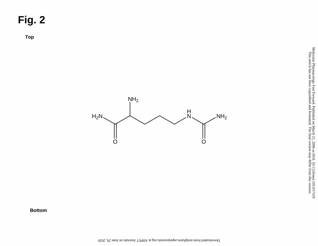
FrPbAII was chemically identified as 2-amino-5-ureidopentanamide (Fig. 2) by
NMR and ESI-MS analyses. Two multiplets (2 H each) at δ = 1.57 and 1.78ppm, two
triplets at δ = 3.09 (2 H, J = 6.8Hz) and 3.70ppm (1 H, J = 6.0Hz) were observed in 1H
NMR. Taken together with COSY 1H-1H data, these results indicate the saturated carbon
chain. HMBC and HMQC confirmed this part of the molecule and provided 13C chemical
shift data suggesting the presence of an amide carbonil (δ = 175.5 ppm) in one edge of
the molecule. The other functional groups linked to the carbon chain were proposed
based in chemical shifts of 1H and 13C provided by NMR spectra.
Positive ESI-MS scan of FrPbAII showed the most intense signal at m/z 175. ESI-
MS/MS experiments undertaken with this peak confirmed that all other peaks present in
ESI-MS spectrum were its daughters, formed by in source dissociation. Therefore, [M +
H]+ 175 give us the Mr 174 to FrPbAII. In ESI-MS/MS experiments it could be observed
the following three competitive losses, which confirmed all the functional groups linked
to the carbon chain: loss of NH3 (17 m.u.), affording m/z 158; or a loss of CONH3 (45
This article has not been copyedited and formatted. The final version may differ from this version.Molecular Pharmacology Fast Forward. Published on March 21, 2006 as DOI: 10.1124/mol.105.017319
175.1182 for the protonated parent ion [M + H]+. Taken together with NMR and MS/MS
fragmentation data, this information confirmed the molecular formula C6H15N4O2+ (4.6
ppm error) for [PbFrAII + H]+, since no other reasonable molecular formula is possible
within a 50 ppm error.
FrPbAII does not affect maximum transport velocity but does alter apparent
transport affinities
By measuring the synaptosomal GABA uptake over a range of substrate
concentrations, the transport activity was shown to possess both saturability and high
affinity. Fig. 3 shows the alteration of apparent transport affinities evoked by 24 µg/ml of
FrPbAII (IC50) (final concentration); it also shows that the Vmax of GABA transport was
not modified. The control values obtained for KM and Vmax were 1.17 + 0.2 µM and 8.19
+ 0.3 pmol/min/mg, respectively. In the presence of FrPbAII, the KM value was increased
to 2.22 + 0.2 µM and Vmax was 8.15 + 0.2 pmol/min/mg, a value quite similar to that
obtained in control. Therefore, KM was increased whereas Vmax was not significantly
changed, suggesting a competitive inhibition.
Uptake inhibition is not caused by alterations in GABA release or GABA-T activity
Synaptosomes can also mediate GABA release in a Ca+2-dependent manner, or
by means of reverse transport, especially when GABA-T is inhibited (Bernath and
Zigmond, 1988). In order to confirm that GABA release was not directly affected in our
experiments, we measured the neurotransmitter release in the presence or absence of
FrPbAII, at a representative range of concentrations used in the GABA uptake assay.
This article has not been copyedited and formatted. The final version may differ from this version.Molecular Pharmacology Fast Forward. Published on March 21, 2006 as DOI: 10.1124/mol.105.017319
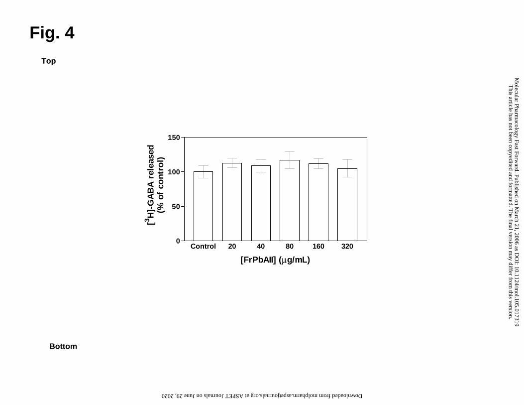
Fig. 4 shows the effect of FrPbAII (20-320 µg/ml, final concentration) on GABA basal
release. Spontaneous release of GABA was not altered by FrPbAII. Control samples
were incubated in the presence of KCl (50 mM) or TTX (5 µM), in order to verify the
functional integrity of preparation. As expected, basal GABA release as increased by
KCl, and TTX did not alter the process (data not shown). In addition, our experiments
measuring GABA-T activity in the presence of FrPbAII (24 µg/ml, final concentration) did
not showed significant alterations (data not shown).
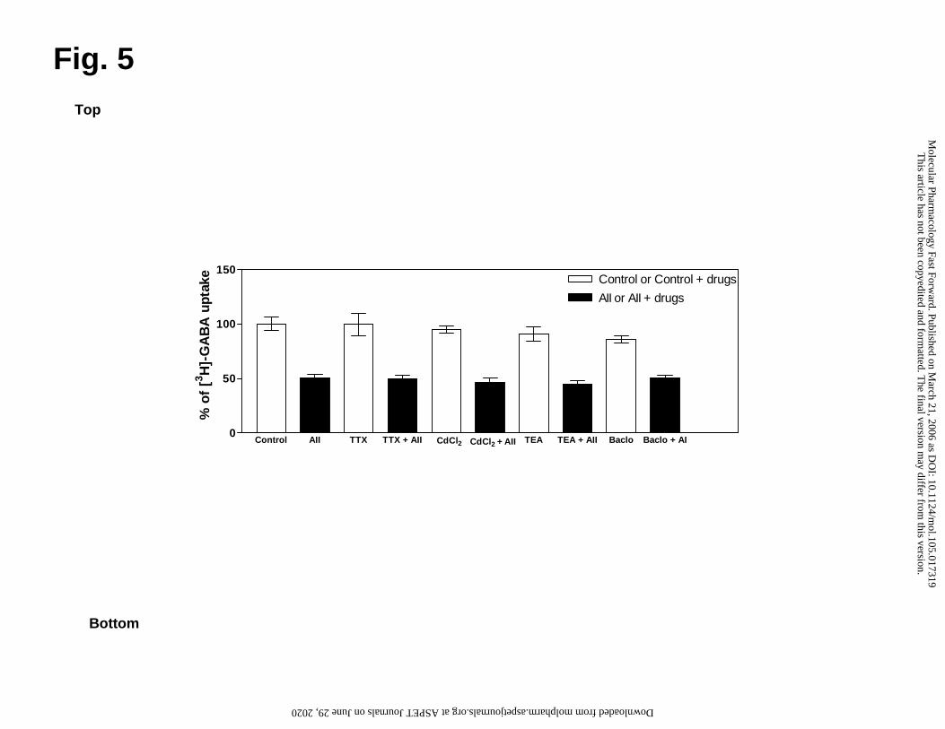
FrPbAII effects are maintained even in the presence of ion channel inhibitors or a
GABAB receptor agonist
Fig. 5 shows that the inhibition of synaptosomal GABA uptake caused by 24
µg/ml of FrPbAII (approximately 50%) is maintained even when the experiment was
performed using ion channels inhibitors (TTX, 5 µM; CdCl2, 1.0 mM; and TEA, 5.0 mM,
final concentrations) or a GABAB receptor agonist (Baclofen, 0,1 mM, final
concentration).
FrPbAII is selective for GABA and glycine systems
Monoamines and glycine transporters are homologous with GABA transporters;
glutamate transporters are only similar, but also involved in ischemic damage. To verify
the selectivity of the action of FrPbAII, synaptosomal uptake of [3H]Glycine,
[3H]Serotonine, [3H]Dopamine, [3H]Noradrenaline and [3H]L-glutamate was studied in the
presence or absence of increasing concentrations of FrPbAII (20–320 µg/ml), at same
range used in the [3H]GABA uptake in this assay. Fig. 6 shows that FrPbAII is a
selective inhibitor of [3H]GABA and [3H]Glycine uptake, having slight or no effect on
other homologous or glutamate transporters.
This article has not been copyedited and formatted. The final version may differ from this version.Molecular Pharmacology Fast Forward. Published on March 21, 2006 as DOI: 10.1124/mol.105.017319
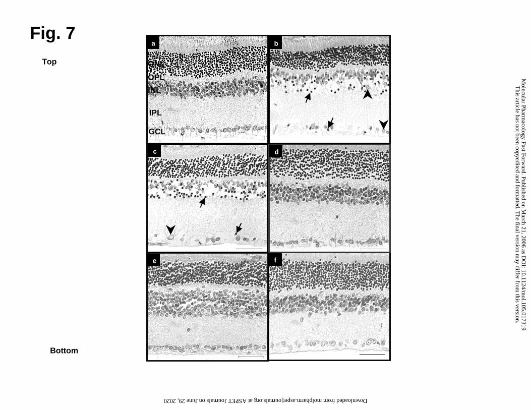
Neuroprotective effects of FrPbAII in experimental glaucoma
To investigate the effect of FrPbAII in vivo, rat retinas were submitted to
experimental glaucoma. In these experiments, rats received i.v. injections of 25 µL
FrPbAII (6mg/ml) or saline 15 min prior to ischemic treatment. Fig. 7 shows micrographs
of non-ischemic control retinas, subjected to ischemia or to ischemia followed by
reperfusion. Marked histological alterations could be observed when comparing non-
ischemic control (Fig. 7a), ischemic (Fig. 7b) and ischemic/reperfused retinas (Fig. 7c).
Compared to non-ischemic control retinas, ischemic retinas (Fig. 7b), showed
cytoplasm vacuolization, pyknotic nuclei and a decrease in cell number in the GCL. The
INL displayed greater edema, pyknotic nuclei and cellular disorganization. The ONL
exhibited decreased cell number, compared to control retinas. Edema was also
observed in the inner plexiform layer (IPL).
In the ischemia/reperfusion retinas (Fig. 7c) the GCL showed lower cell density,
and increased vacuolization and number of pyknotic nuclei as well as cellular
disorganization. The INL also had fewer cells and those remaining had a greater amount
of pyknotic nuclei and cytoplasm vacuolization, as well as, enhanced edema and cellular
disorganization. There were also fewer cells in the ONL.
Compared to ischemic and ischemic/reperfused retinas (Fig. 7b and 7c), retinas
of animals treated with FrPbAII (Fig. 7d, 7e and 7f) displayed normal cellular
morphology and a remarkable reduction in cell death in all layers. Fig. 7d represents
another control, a non-ischemic retina of animal treated with FrPbAII that showed no
signs of cell damage. Fig. 7e shows ischemia-induced retinas treated with FrPbAII. No
degeneration, pyknotic nuclei or disorganization of the cells, but some cell loss were
This article has not been copyedited and formatted. The final version may differ from this version.Molecular Pharmacology Fast Forward. Published on March 21, 2006 as DOI: 10.1124/mol.105.017319
observed. Fig. 7f shows ischemic/reperfused retinas treated with FrPbAII, where
protection was observed in all layers and there was a decreased in cell loss.
The number of cells in control, ischemic and ischemic/reperfused retinas with or
without treatment with FrPbAII are presented in Table 1. There was a decrease in
number of cells in all layers following ischemia and in those that were reperfused
following ischemia. The cell numbers were significantly decreased by 38% in the ONL,
54% in the INL and 51% in the GCL after ischemia and were decreased by 40% in the
ONL, by 63% in the INL, and 58% in the GCL in the retinas subjected to
ischemia/reperfusion compared to the non-ischemic control retinas (*p<0.05). In
contrast, retinas of animals treated with FrPbAII, showed significant protection of 27% in
ONL; 57% in INL and 50% in GCL after ischemia and of 20% in ONL; 81% in INL and
44% in GCL in ischemia/reperfusion compared to ischemic and ischemic/reperfusion
conditions (*p<0.05), respectively (Table 1).
FrPbAII is also able to inhibit GABA and glycine uptake in synaptosomes from rat
retinas
Synaptosomes from rat retina were used in order to verify the direct correlation
between neuroprotection and inhibition of GABA and glycine uptake caused by FrPbAII.
In these experiments, FrPbAII also caused a dose-dependent inhibition of
neurotransmitter uptake (Fig. 8), as it was demonstrated by our assays using
synaptosomes from rat cerebral cortex (Fig. 1c). IC50 values for FrPbAII in GABA and
glycine uptake were 13.80 and 13.10 µg/ml, respectively.
This article has not been copyedited and formatted. The final version may differ from this version.Molecular Pharmacology Fast Forward. Published on March 21, 2006 as DOI: 10.1124/mol.105.017319
In this work, we show the novel neuroprotective compound from P. bistriata
venom extract (FrPbAII), which inhibits synaptosomal GABA uptake in a dose-
dependent manner (Fig. 1c) and probably by a competitive antagonism (Fig. 3). In
addition to this neuroprotective property, FrPbAII has previously been reported as a
significant anticonvulsant against seizures induced in rats (Cairrão et al., 2002).
Synaptosomes are recognized as a useful model to neurochemical studies since
they retain all machinery for the uptake, storage, release of neurotransmitters and ionic
conductance (Gray and Whittaker, 1962, Bicalho et al., 2002, Wang and Sihra, 2003). In
our studies, morphological examination of the synaptosomal preparations by electron
microscopy demonstrated that membrane integrity was maintained, and levels of LDH
were not changed when synaptosomes were incubated with either the venom extract,
FrPbAI or FrPbAII (results not shown). These results indicate that the observed uptake
inhibition was not due to plasma membrane disruption or pore formation.
Synaptosomes mediate residual GABA release during the uptake process. If
FrPbAII was elevating basal release, it could enhance the unlabeled/labeled GABA ratio
in the assay buffer producing an apparent and unreal decrease of neurotransmitter
transport. Therefore, we also examined the effect of FrPbAII on GABA efflux. The
synaptosomal release of neurotransmitter was stimulated by K+ and was not altered by
TTX, confirming the functional integrity of the preparation (data not shown). FrPbAII did
not affect the levels of basal GABA release (Fig. 4), an indication that it does not act
indirectly by altering tonic or depolarization-dependent GABA release or even via
membrane pore formation.
This article has not been copyedited and formatted. The final version may differ from this version.Molecular Pharmacology Fast Forward. Published on March 21, 2006 as DOI: 10.1124/mol.105.017319
GABAB receptors are present on neural terminals throughout the CNS, acting as
autoreceptors when localized on the pre-synaptic membrane. In this case, their
activation inhibits release of further synaptic vesicles through the suppression of high-
threshold Ca2+ channels (Harayama et al., 1998).
Upon the uptake process, GABA can be catabolized by the action of GABA-T.
The enzyme inhibition increases GABA concentration in the brain, especially by reverse
transport, decreasing susceptibility to convulsions and epileptic conditions (Sherif and
Ahmed 1995). This process is distinguished from exocytotic release by being non-
vesicular and independent of Ca2+ influx via selective voltage-dependent channels
(Agostinho et al., 1994; Beleboni et al., 2004a).
If FrPbAII is able to inhibit GABA-T, K+ channel activity and GABAB receptors
function, or to enhance Na+ or Ca2+ channels activity, these effects also could be
responsible for an apparent and unreal decrease of neurotransmitter uptake, ruling out
the possibility of a direct effect of FrPbAII on the GABA transporter.
In order to evaluate whether inhibition of GABA uptake by FrPbAII is mediated by
an indirect mechanism through GABAB receptors or ion channels, we investigated
whether channel and receptor agonists or antagonists could modify its effects on GABA
uptake. These agents included TTX a Na+ channel blocker, cadmium chloride a Ca2+
channel blocker, TEA a K+ channel blocker, and baclofen a GABAB receptor agonist.
FrPbAII-inhibited GABA uptake was unchanged by the blockade of voltage-
dependent Na+ channels, Ca2+ or K+ channels, when these blockers were tested
concentrations known to produce effective inhibition of channel activity (Fig. 5). Despite
of its potential effects on neuronal GABA concentration, baclofen did not alter the ability
of FrPbAII to inhibit GABA uptake into synaptosomes, suggesting that those effects are
This article has not been copyedited and formatted. The final version may differ from this version.Molecular Pharmacology Fast Forward. Published on March 21, 2006 as DOI: 10.1124/mol.105.017319
not dependent on GABAB receptor inhibition (Fig. 5). Moreover, GABA-T activity is
maintained in the presence of FrPbAII, indicating that reverse transport is not involved in
FrPbAII effects (data not shown). Therefore, FrPbAII effect can not be directly
dependent on Na+, K+, Ca2+ channels, GABAB receptors or GABA-T for its action. These
results associated to those demonstrated on Fig. 4 rule out the possibility that FrPbAII
could act by inhibition or activation of these structures, indicating its selectivity of action.
As demonstrated on Fig. 6, FrPbAII is highly selective for GABA and glycine
transporters, having slight or no effects on homologous (serotonine, dopamine,
noradrenaline) or glutamate transporters. These results reinforce the concept of a
remarkable selectivity of FrPbAII, a desirable attribute for future development of new
therapeutic drug or pharmacological tools.
GABA and glycine are relevant inhibitory transmitters in retinal synapses, as it
was well established in the CNS, and many reports suggest a heterogeneous
distribution of GABA/glycine receptors and transporters in the vertebrate retina (Honda
et al., 1995; Wässle et al., 1998; Gadea et al., 1999). The organization and accessibility
of the retina has made it the best-characterized system for examining the physiology
and function of amino acid transporters and an excellent model to study the effects of
drugs on ischemia/reperfusion in the CNS (Louzada Jr et al., 1992; Eliasof et al., 1998).
It is well-established that drugs acting on GABA and glycine uptake may provide
an effective means for protecting the brain against neuronal injury and for treating
epilepsy, as illustrated by the clinical use of tiagabine (Meldrum 1997; Fisher and
Bogousslavsky 1998). Ichinose and Lukasiewicz (2002) using the selective GAT-1
blocker NO-711 showed that blockade of GABA uptake resulted in increased activation
of GABAC receptors. This compound has proved to be effective as an anticonvulsant in
This article has not been copyedited and formatted. The final version may differ from this version.Molecular Pharmacology Fast Forward. Published on March 21, 2006 as DOI: 10.1124/mol.105.017319
animal models and as a neuroprotector against ischemia of CA1 pyramidal neurons in
gerbil (O'Connell et al., 2001). Thus, to obtain insight into the possible neuroprotective
actions, consequent to inhibition of GABA and glycine uptake, we examined FrPbAII
effects during retinal ischemia and ischemia followed by reperfusion, using the
experimental glaucoma model. In our experiments, FrPbAII was found to protect
neurons from injury in all retinal layers, particularly in the INL (Fig. 7 and Table 1).
Thus, inhibition of GABA and glycine uptake by FrPbAII can increase GABA and
glycine levels on the synaptic cleft promoting an activation of their receptors. These
processes hyperpolarize neurons leading to a reduced transmitter release and/or action
potential firing, effects that can act in the ischemic cascade promoting a neuroprotective
effect of the retina in the experimental glaucoma model. Similar conclusions could be
taken to explain our previous results, indicating a marked anticonvulsant activity
presented by FrPbAII (Cairrão et al., 2002).
The neuroprotective effects of FrPbAII are observed in both ischemic and
ischemic/reperfused retinas. Louzada Jr. et al. (1992) have shown that there is a higher
percentage of glutamate release, and subsequent neuronal death during the reperfusion
period than during ischemia; nevertheless, we still observed protection when FrPbAII
was administered in this condition.
FrPbAII is also able to inhibit GABA and glycine uptake in synaptosomes
prepared from rat retina (Fig. 8). This result confirms the view that the neuroprotective
action of FrPbAII is due to inhibition of GABA and glycine uptake. Moreover, an
enhancement of glutamate uptake could be discarded as a mechanism by which
FrPbAII could act as a neuroprotective compound, since no effect on glutamate
transport is observed when synaptosomes were incubated with FrPbAII (Fig. 6). A direct
This article has not been copyedited and formatted. The final version may differ from this version.Molecular Pharmacology Fast Forward. Published on March 21, 2006 as DOI: 10.1124/mol.105.017319
FrPbAII interaction with GABAA/GABAC receptors on GABA binding site is also ruled out
as a neuroprotective mechanism, since only a very high concentration of FrPbAII is able
to induce [3H]-GABA displacement on binding assays (Zukin et a., 1974; data not
shown).
Although a number of classical GABA analogues are useful as pharmacological
tools in research epilepsy and ischemic cerebral damage, they were shown to be
inefficient in therapy due to their low permeability of the blood-brain barrier (Krogsgaard-
Larsen et al., 1998). FrPbAII protects retinas against neuronal damage when
administered intravenously, suggesting that this compound is able to cross the blood-
retina barrier, which is structurally and functionally similar to the blood-brain barrier
(Steuer et al., 2005).
To summarize, our results taken together provide an insight into the effects of a
new neuroprotective compound from P. bistriata spider venom that acts primarily and
directly on GABA and glycine transporters. In fact, the significant inhibition of GABA and
glycine uptake caused by FrPbAII associated with its selectivity and blood-retina barrier
permeability strongly suggest its high potential value in studies of transport mechanisms
and the role of uptake during inhibitory neurotransmission. Moreover, an understanding
of the structure and activity of active compounds from arthropod venoms may provide
"proof of principle" for a new class of neuroprotective and anticonvulsant drugs that act
by inhibiting GABA and glycine clearance.
This article has not been copyedited and formatted. The final version may differ from this version.Molecular Pharmacology Fast Forward. Published on March 21, 2006 as DOI: 10.1124/mol.105.017319
The authors thank Vera L.A. Epifânio, Silvia H. Epifânio (Biochemistry
Department, FMRP-USP) and José Carlos Tomaz (Physics and Chemistry Department,
FCFRP-USP) for technical assistance.
This article has not been copyedited and formatted. The final version may differ from this version.Molecular Pharmacology Fast Forward. Published on March 21, 2006 as DOI: 10.1124/mol.105.017319
Miranda A, and Santos WF (2002) Anticonvulsant and GABA uptake inhibition
properties of venom fractions from the spiders Parawixia bistriata and Scaptocosa
raptoria. Pharm Biol 40:472-477.
This article has not been copyedited and formatted. The final version may differ from this version.Molecular Pharmacology Fast Forward. Published on March 21, 2006 as DOI: 10.1124/mol.105.017319
Santos WF, and Coutinho-Netto J (2003) Purification of a neuroprotective compound
of Parawixia bistriata spider venom that enhances glutamate uptake. Br J Pharmacol
139:1297-1309.
Gadea A, Lopez E, and Lopez-Colome AM (1999) Characterization of glycine transport
in cultured Müller glial cells from the retina. Glia 26:273-279.
Gray EG, and Whittaker, VP (1962) The isolation of nerve ending from brain: an
electron-microscopic study of cell fragments derived by homogenization and
centrifugation. J Anat 96:79-87.
This article has not been copyedited and formatted. The final version may differ from this version.Molecular Pharmacology Fast Forward. Published on March 21, 2006 as DOI: 10.1124/mol.105.017319
Harvey AL, Bradley KN, Cochran SA, Rowan EG, Pratt JA, Quillfeldt J.A., and
Jerusalinsky DA (1998) What can toxins tell us for drug discovery? Toxicon 36:1635-
1640.
Harayama N, Shibuya I, Tanaka K, Kabashima N, Ueta Y, and Yamashita H (1998)
Inhibition of N- and P/Q-type calcium channels by postsynaptic GABAB receptor
activation in rat supraoptic neurones. J Physiol (Lond.) 509:371–383.
Hartree EF (1972) Determination of protein: a modification of the Lowry method that
gives a linear photometric response. Anal Biochem 48:422-427.
Hendry SH, Schwark HD, Jones EG, and Yan J (1987) Numbers and proportions of
GABA-immunoreactive neurons in different areas of monkey cerebral cortex. J
Neurosci 7:1503-1519.
Honda S, Yamamoto M, and Saito N (1995) Immunocytochemical localization of three
subtypes of GABA transporter in rat retina. Brain Res Mol Brain Res 33:319-325.
Ichinose T, and Lukasiewicz PD (2002) GABA transporters regulate inhibition in the
retina by limiting GABA(C) receptor activation. J Neurosci 22:3285-3292.
Krogsgaard-Larsen P, Frolund BF, and Falch E (1998) Inhibitors of gamma-
aminobutyric acid transport as experimental tools and therapeutic agents. Methods
Enzymol 296:165-175.
Louzada-Jr P, Dias JJ, Santos WF, Lachat JJ, Bradford, HF, and Coutinho-Netto, J
(1992) Glutamate release in experimental ischaemia of the retina: an approach using
microdialysis. J Neurochem 59:358-363.
Lowry OH, Rosenbrouch NJ, Farr AL, and Randall RJ (1951) Protein measurement with
the folin phenol reagent. J Biol Chem 193:265-275.
This article has not been copyedited and formatted. The final version may differ from this version.Molecular Pharmacology Fast Forward. Published on March 21, 2006 as DOI: 10.1124/mol.105.017319
Steuer H, Jaworski A, Elger B, Kaussmann M, Keldenich J, Schneider H, Stoll D, and
Schlosshauer B (2005) Functional characterization and comparison of the outer
blood-retina barrier and the blood-brain barrier. Invest Ophthalmol Vis Sci 46:1047-
1053.
Wang SJ and Sihra TS (2003) Opposing facilitatory and inhibitory modulation of
glutamate release elicited by cAMP production in cerebrocortical nerve terminals
(synaptosomes). Neuropharmacology 44:686–697.
Wässle H, Koulen P, Brandstatter JH, Fletcher EL, and Becker CM (1998) Glycine and
GABA receptors in the mammalian retina. Vision Res 38:1411-1430.
Wong CG, Bottiglieri T, and Snead OC 3rd. (2003) GABA, gamma-hydroxybutyric acid,
and neurological disease. Ann Neurol 54:3-12.
This article has not been copyedited and formatted. The final version may differ from this version.Molecular Pharmacology Fast Forward. Published on March 21, 2006 as DOI: 10.1124/mol.105.017319
Worrall DM, and Williams DC (1994) Sodium ion-dependent transporters for
neurotransmitters: a review of recent developments. Biochem J 297:425-436.
Zukin SR, Young AB and Snyder SH. (1974) Gamma-aminobutyric acid binding to
receptor sites in the rat central nervous system. Proc Natl Acad Sci U S A.
71(12):4802-4807.
This article has not been copyedited and formatted. The final version may differ from this version.Molecular Pharmacology Fast Forward. Published on March 21, 2006 as DOI: 10.1124/mol.105.017319
This work was supported by FAPESP (00/08101-0; 00/08010-5 and 03/06328-6),
CNPq and CAPES (R.O.G scholarship).
This article has not been copyedited and formatted. The final version may differ from this version.Molecular Pharmacology Fast Forward. Published on March 21, 2006 as DOI: 10.1124/mol.105.017319
Fig. 1 Dose response curves for the inhibitory effects of increasing concentrations of P.
bistriata venom extract (a), FrPbAI (b) or FrPbAII (c), on [3H]GABA uptake in
synaptosomes from rat cerebral cortex. Uptake assays were initiated by adding
[3H]GABA (10 nM, final concentration). Synaptosomes were pre-incubated in the
presence or absence of venom extract, FrPbAI and FrPbAII, at final concentrations of
210-13440 µg/ml, 21-1344 µg/ml, 2.5–320 µg/ml, respectively, for 3 min at 25°C.
Concentrations of venom extract and fractions are plotted on a logarithmic scale. Higher
concentrations than those shown also inhibit GABA uptake, but were not included in the
Figure. Data from four-six independent experiments, performed in triplicate, generated
an IC50 of 1700 + 130 µg/ml, 100 + 12 µg/ml and 24 + 0.019 µg/ml, for respectively,
venom extract, FrPbAI and FrPbAII in the GABA uptake assays.
Fig. 2 Chemical structure of FrPbAII.
Fig. 3 Kinetic analysis of high affinity GABA uptake by synaptosomes pre-incubated in
absence (■) or presence (�) of IC50 of FrPbAII (24 µg/ml) (final concentration). Uptake
was measured in the presence of increasing concentrations of unlabeled
neurotransmitter (from 4.5 nM to 30 µM, final concentration) and [3H]GABA (10 nM, also
at final concentration). Data are means ± S.E.M. of four independent experiments,
performed in triplicate. Inset, Eadie-Hofstee plot.
Fig. 4 Effects of final increasing concentrations of FrPbAII on [3H]GABA release.
Synaptosomes were preloaded with [3H]GABA (0.5 µM) for 20 min at 25°C. Release,
expressed as % of neurotransmitter released over the control, was initiated by the
addition of FrPbAII from 20 to 320 µg/ml. No significant difference was observed
This article has not been copyedited and formatted. The final version may differ from this version.Molecular Pharmacology Fast Forward. Published on March 21, 2006 as DOI: 10.1124/mol.105.017319
Fig. 8 Dose response curves for the inhibitory effects of increasing concentrations of
FrPbAII on [3H]GABA (a) or [3H]glycine (b) uptake in synaptosomes from rat retinas.
Uptake assays were initiated by adding [3H]GABA or [3H]glycine (at respectively, 10 nM
and 250 nM final concentrations). Synaptosomes were pre-incubated for 3 min at 25°C,
in the presence or absence of FrPbAII at final concentrations of 2.5–320 µg/ml, the
This article has not been copyedited and formatted. The final version may differ from this version.Molecular Pharmacology Fast Forward. Published on March 21, 2006 as DOI: 10.1124/mol.105.017319
same range used in assays with synaptosomes from cerebral cortex. Concentrations are
plotted on a logarithmic scale. Data from four independent experiments, performed in
triplicate, generated an IC50 of 13.80 and 13.10 mg/ml respectively, for FrPbAII in the
GABA and glycine uptake assays.
This article has not been copyedited and formatted. The final version may differ from this version.Molecular Pharmacology Fast Forward. Published on March 21, 2006 as DOI: 10.1124/mol.105.017319
This article has not been copyedited and formatted. The final version may differ from this version.Molecular Pharmacology Fast Forward. Published on March 21, 2006 as DOI: 10.1124/mol.105.017319