Zoological Journal of the Linnean Society, 2019, XX, 1–23. With 15 figures.
Molecular phylogeny and species delimitation of the genus Dicerapanorpa (Mecoptera: Panorpidae)
GUI-LIN HU1, KAI GAO1, JI-SHEN WANG1, PAUL D. N. HEBERT2 and BAO-ZHEN HUA1,*,
1Key Laboratory of Plant Protection Resources and Pest Management, Ministry of Education, College of Plant Protection, Northwest A&F University, Yangling, Shaanxi, China2Centre for Biodiversity Genomics, University of Guelph, Guelph, ON, Canada
Received 16 January 2019; revised 28 April 2019; accepted for publication 9 June 2019
Given that species is the fundamental unit in systematic biology, rigorous species delimitation is crucial for taxonomic studies, yet routine species delimitation remains an ongoing challenge in the taxonomic practice of insects. The two-horned scorpionfly Dicerapanorpa is a small genus in Panorpidae (Mecoptera) endemic to the Qinling-Bashan and Hengduan mountains, a biodiversity hotspot. However, species of Dicerapanorpa are difficult to delineate owing to marked intraspecific variation and interspecific similarity. Here, we investigate the diversity and species boundaries of Dicerapanorpa using an integrative approach based on DNA barcoding, morphological, geometric morphometric and molecular phylogenetic analyses. This integrative analyses confirmed the 13 described species of Dicerapanorpa and revealed three new species: Dicerapanorpa lativalva sp. nov., Dicerapanorpa hualongshana sp. nov. and Dicerapanorpa minshana sp. nov. Most molecular operational taxonomic units are in congruence with morphological clusters. Possible reasons for several discordances in Dicerapanorpa are tentatively discussed.
Given that species is the fundamental unit of biology, rigorous species delimitation is crucial for biodiversity studies, conservation programmes and ecological and evolutionary research (Isaac et al., 2004; de Queiroz, 2005, 2007; Bickford et al., 2007; Wiens, 2007; Bortolus, 2008). More than 25 species concepts have been proposed during the past few decades, but many of these are difficult to apply in reality and some are incompatible, leading to different conclusions concerning the boundaries and number of species (de Queiroz, 2007). Although the unified species concept is receiving more and more support (de Queiroz, 2007), routine species delimitation remains an ongoing challenge in the taxonomic practice of many insect groups to date. Recently, integrative taxonomy has been adopted by numerous insect taxonomists to delimit species boundaries in many insect groups
with good results, including the fruit fly Bactrocera dorsalis complex (Schutze et al., 2015, 2017) and jumping bristletails (Dejaco et al., 2016).
The two-horned scorpionfly, Dicerapanorpa Zhong & Hua, 2013, is a small genus of the diverse family Panorpidae in Mecoptera (Zhong & Hua, 2013a; Wang & Hua, 2017). Species of Dicerapanorpa, mainly diagnosed by two anal horns on tergum VI in males, are endemic to the Qinling-Bashan and Hengduan Mountain ranges, a biodiversity hotspot. The monophyly of the genus has been confirmed by both molecular data (Hu et al., 2015; Miao et al., 2019) and morphological studies (Ma et al., 2009, 2011, 2012). The larvae of Dicerapanorpa are eruciform with annulated processes on the dorsum, which might have adaptive significance for fossorial and soil-living habits (Ma et al., 2014). The male adults control the females during mating with a notal organ on the third abdominal tergum and two finger-like anal horns on the sixth tergum, and offer salivary masses as nuptial gifts to prolong copulation (Zhong et al., 2015).
The members of Dicerapanorpa can be categorized into two groups (Zhong & Hua, 2013a; Hu &
*Corresponding author. E-mail: [email protected][Vers ion o f Record , publ i shed onl ine 21 October 2019; http: / /zoobank.org/ urn:lsid:zoobank.org:pub: D4405014-D172-481B-B822-2C9B6479CD7]
Hua, 2019; Hu et al., 2019b). The first group (the Dicerapanorpa magna Chou in Chou et al., 1981 group), characterized by yellowish wings with distinct markings and a rostrum lacking distinct longitudinal stripes, is nearly restricted to the Qinling-Bashan Mountains, where it inhabits dense forests with herbaceous groundcover. In contrast, the second group [the Dicerapanorpa diceras (MacLachlan, 1894) group], distinguished by hyaline wings without distinct markings and a rostrum with two distinct lateral stripes, is most abundant in the Hengduan Mountains, where it occurs in open areas with direct sunlight, especially near river margins.
Species of Dicerapanorpa are largely diagnosed by the male adult morphologica l features, whereas the female morphology has received little investigation. In fact, some sympatric species exhibit morphological stasis or homoplasy owing to similar selective pressure, making the species delimitation particularly difficult. For example, the sympatric Dicerapanorpa macula Hu, Wang & Hua, 2019 and Dicerapanorpa zhongdianensis Hu, Wang & Hua, 2019 occur in Shangri-La County, Yunnan Province and exhibit similar body shape and wing coloration and pattern (Hu et al., 2019b), leading to difficulty in discriminating between the conspecific and heterospecific females. Moreover, detailed studies of D. magna revealed considerable variation in its external morphology (e.g. wing shape) and internal anatomy (e.g. number of female ovarioles and male salivary gland tubes) (Hou & Hua, 2008; Ma et al., 2011; Liu et al., 2016). A phylogeographical study further suggested that incipient speciation has probably occurred in D. magna (Hu et al., 2019a). The interspecific morphological similarities and intraspecific variations blurred the distinction between geographical population-level variation and species-level divergence in Dicerapanorpa. Therefore, the genus Dicerapanorpa needs to be investigated comprehensively through an integrative approach.
DNA barcoding (Hebert et al., 2003a, b) has disentangled taxonomic disputes and accelerated the discovery of new species in numerous taxa (e.g. Smith et al., 2006; Witt et al., 2006; Hsu et al., 2013; Mendoza et al., 2016). For the bulk of undescribed biodiversity, the DNA barcoding approach was proposed to produce potential species [i.e. operational taxonomic units (OTUs)] for approximating species description (Goldstein & DeSalle, 2011; Puillandre et al., 2012b). Molecular OTUs were subsequently evaluated with various criteria, such as morphological, geometric morphometric, geographical and phylogenetic data. Conclusive concordance among different disciplines reinforces the robustness of species hypotheses, and discordance over the number and demarcation of species
is resolved by invoking evolutionary explanations (Schlick-Steiner et al., 2010; Carstens et al., 2013). The integrative approach with multiple independent lines of evidence has effectively delivered significant advances in the resolution of taxonomically intricate instances and uncovering complex evolutionary histories (Dayrat, 2005; Padial et al., 2010; Schlick-Steiner et al., 2010; Dejaco et al., 2016).
In this study, we used an integrative approach to delimit species of Dicerapanorpa using DNA barcoding, morphological comparison, geometric morphometric analysis and multilocus phylogenetic reconstruction. The main aims are as follows: (1) to clarify species diversity in Dicerapanorpa, especially in the D. magna group; (2) to resolve the boundaries between the morphologically similar species; and (3) to ascertain whether patterns of morphological clusters are congruent with molecular OTUs.
MATERIAL AND METHODS
Sampling
Specimens of Dicerapanorpa were col lected throughout the Hengduan and Qinling-Bashan Mountains (Fig. 1) from 2009 to 2018. In total, 213 individuals of Dicerapanorpa and five specimens of outgroups from Sinopanorpa Cai & Hua in Cai et al., 2008 and Megapanorpa Wang & Hua, 2019 were sequenced (Supporting Information, Table S1). In addition, using geometric morphometric analysis we examined 66 specimens of D. magna from the Qinling Mountains, 56 of Dicerapanorpa minshana from Minshan Mountain, 47 of Dicerapanorpa hualongshana from the Bashan Mountains, 20 of Dicerapanorpa baiyunshana Zhong & Hua, 2013 from Baiyunshan Mountain and 40 of Dicerapanorpa shennongensis Zhong & Hua, 2013 from Shennongjia. The specimens examined in this study are stored at −20 °C in 75 or 100% ethanol at the Entomological Museum, Northwest A&F University, China (NWAU).
laboratory procedureS
Total genomic DNA was extracted from three legs removed from one side of each specimen, using a Genomic DNA Mini Preparation Kit with Spin Column (Beyotime, China). Four gene fragments (COI, COII, cytb and 28S rRNA) were amplified and sequenced. Primer sequences (Folmer et al., 1994; Simon et al., 1994; Whiting, 2002; Song & Liang, 2013) are shown in the Supporting Information (Table S2). Polymerase chain reaction products were sequenced bidirectionally at Shanghai Sangon Biotechnology Co. Ltd (China). All sequences were subsequently uploaded to BOLD
Dow
nloaded from https://academ
ic.oup.com/zoolinnean/advance-article-abstract/doi/10.1093/zoolinnean/zlz059/5601891 by N
(Ratnasingham & Hebert, 2007) together with the trace files and specimen information, and cross-referenced with GenBank (for detailed information, see Supporting Information, Table S1).
The DNA sequences were checked, assembled and edited with SeqMan (Swindell & Plasterer, 1997) or CodonCode Aligner v.6.0.2 (CodonCode Corporation, Dedham, MA, USA). Multiple sequence alignments were performed using MAFFT v.7.037 (Katoh et al., 2009) with the accurate algorithm of G-INS-i. Ambiguous sites at both ends of the alignment were deleted manually using BioEdit v.7.0.9.0 (Hall, 1999).
dna barcoding
Barcode index number (BIN) assignments were generated in BOLD (Ratnasingham & Hebert, 2013) based on COI barcode sequences. Delimitation of OTUs was performed using the automatic barcode gap discovery (ABGD) method (Puillandre et al., 2012a), generalized mixed
Yule coalescent (GMYC) procedure (Pons et al., 2006; Fujisawa & Barraclough, 2013) and Bayesian Poisson tree processes (bPTP) (Zhang et al., 2013).
Barcode index numbersTo assign DNA barcode sequences to a BIN, BOLD uses single linkage clustering with a threshold of 2.2%, followed by OTU refinement using Markov clustering (Ratnasingham & Hebert, 2013). All sequences were automatically assigned to a BIN in BOLD, and the BIN assignments are presented in the Supporting Information (Table S1).
Automated barcode gap discoveryThe ABGD method generates clusters based on genetic distance distributions (Puillandre et al., 2012a). The COI sequences (N = 209) were collapsed to haplotypes (N = 118) using ALTER (https://sing.
ei.uvigo.es/ALTER/) (Glez-Peña et al., 2010). The haplotypes were uploaded on http://wwwabi.snv.jussieu.fr/public/abgd/abgdweb.html. The run was based on a Kimura 2-parameter distance matrix with a relative gap of one.
Generalized mixed Yule coalescentThe GMYC model-based likelihood analysis identifies the transition point between coalescent and speciation events (Pons et al., 2006; Monaghan et al., 2009) with a fully resolved ultrametric gene tree as input. The best-fitting substitution model (TrN+I+G) for COI was determined using jModelTest v.2.1.4 (Darriba et al., 2012) based on the Bayesian information criterion. The Bayesian tree of COI haplotypes was reconstructed using BEAST v.1.8.0 (Drummond et al., 2012) with an uncorrelated lognormal relaxed clock and a Yule speciation process. The analysis was run for 40 million generations, with a sample frequency of 1000. The convergence and stability were evaluated in Tracer v.1.5 (https://beast.bio.ed.ac.uk/software/tracer/), ensuring effective sample size > 200 for all parameters. A maximum clade credibility tree was assembled using TreeAnnotator v.1.8.0 (BEAST package), with the first 25% of samples removed as burn-in. The GMYC model was implemented in the R splits package using single- and multiple-threshold analyses, separately (available at http://r-forge.r-project.org/projects/splits).
Bayesian Poisson tree processesA maximum likelihood (ML) tree of COI haplotypes was reconstructed using RAxML searches. The ML analysis was implemented in raxmlGUI v.1.31 (Silvestro & Michalak, 2012) with 1000 rapid bootstrap replicates and codon-specific partition scheme under the GTRCAT model. The bPTP analysis was carried out on the bPTP server (http://species.h-its.org/) with the ML tree as input (Zhang et al., 2013). The OTUs recognized by these four methods were compared for congruence.
morphological compariSon
Female medigynia were dissected under a Nikon SMZ1500 stereoscopic zoom microscope, macerated in cold 5% NaOH for 3–5 min and rinsed in distilled water. The female medigynia were imaged digitally using a QImaging Retiga 2000R Fast 1394 Digital CCD camera attached to the microscope. Other photographs were taken using a Scientific Digital micrography system (ZEISS SteREO Discovery V20) equipped with an auto-montage imaging system (AxioCam IC).
The images were assembled using Adobe Photoshop CS4. Morphological characters, especially genital structures, were compared with the original images and descriptions (Carpenter, 1938; Cheng, 1957; Zhong & Hua, 2013a). Taxonomic assignments were attributed to OTUs based on the integrative criteria of morphological characters, geographical distribution and molecular clusters.
morphometric analySeS of the D. magna group
To evaluate species diversity in the D. magna group, we performed a geometric morphometric analysis based on female medigynia. The images were loaded into MakeFan7 (Sheets, 2009) with a comb of 20 lines to create a standardized template for landmark digitization. TPS files were generated for images using tpsUtil v.1.46 (Rohlf, 2010). To outline the exterior margin of a medigynium, 52 landmarks were digitized on each image using tpsDig2 v.2.19 (Rohlf, 2015). Generalized Procrustes analysis (GPA) was performed in CoordGen7a from the Integrated Morphometric Package (IMP) to eliminate the non-shape variation in size, location and orientation and generate Procrustes superimposition data (Sheets, 2012). Canonical variate analysis (CVA) was conducted on the shape data using CVAGen7b (Sheets, 2012). To test for differences in shape between species, a single-factor permutation multivariate analysis of variance (MANOVA) was performed using CVAGen7b with 1000 permutations. The variation in shape was visualized in MorphoJ v.1.05f (Klingenberg, 2011).
molecular phylogeny
To reconstruct the phylogeny of Dicerapanorpa, COI sequences were concatenated with those for COII, cytb and 28S rRNA. Phylogenetic reconstruction was conducted using ML analysis and Bayesian inference (BI). The most suitable substitution model and partition scheme were determined for the combined dataset using PartitionFinder v.1.1.1 (Lanfear et al., 2012). They are as follows: TrN+I+G for the first codon position of COI, COII and cytb, HKY+I+G for the second codon position, TIM+I+G for the third codon position, and K80 for 28S rRNA.
Maximum likelihood analysis was executed in raxmlGUI v.1.31 (Silvestro & Michalak, 2012) with codon-specific partition under the GTRCAT model. Bootstrap values (BVs) were calculated with 1000 rapid bootstrap replicates. Bayesian inference was performed in MrBayes v.3.2.6 (Ronquist & Huelsenbeck, 2003) through the CIPRES Science Gateway (Miller et al., 2010). Two independent runs
Dow
nloaded from https://academ
ic.oup.com/zoolinnean/advance-article-abstract/doi/10.1093/zoolinnean/zlz059/5601891 by N
with four Markov chain Monte Carlo chains were performed for 40 million generations, with sampling every 1000 generations. The average standard deviation of split frequency < 0.01 indicates that the sampling of posterior distribution was adequate. The stationarity was evaluated with Tracer v.1.5 (http://beast.bio.ed.ac.uk/software/tracer/) by plotting log-likelihood values vs. generation number. The first 25% of the samples were discarded as burn-in, and the remaining trees were used to generate a majority consensus tree and estimate the posterior probabilities (PPs) in TreeAnnotator v.1.8.4 (BEAST package). The tree was visualized in FigTree v.1.3.1 (http://beast.bio.ed.ac.uk/figtree).
RESULTS
dna barcoding
BOLD assigned 214 COI sequences of Dicerapanorpa to 12 BINs (Fig. 2). With the ABGD method, after two extreme prior divergences were excluded, the remaining prior intraspecific distances resulted in the recognition of ten, 11 and 23 partitions. The molecular OTU count (23) was much more biologically realistic at the prior of 0.00167 in the recursive analysis. GMYC analysis is susceptible to oversplitting species, and the multiple-threshold model fragmented most morphological species into several individual clusters. Therefore, we preferred the OTU count of 26 from the single-threshold model. In contrast, the bPTP analysis produced 18 clusters with the highest Bayesian solution. Taking morphological uniqueness into consideration, we determined a final OTU count of 19.
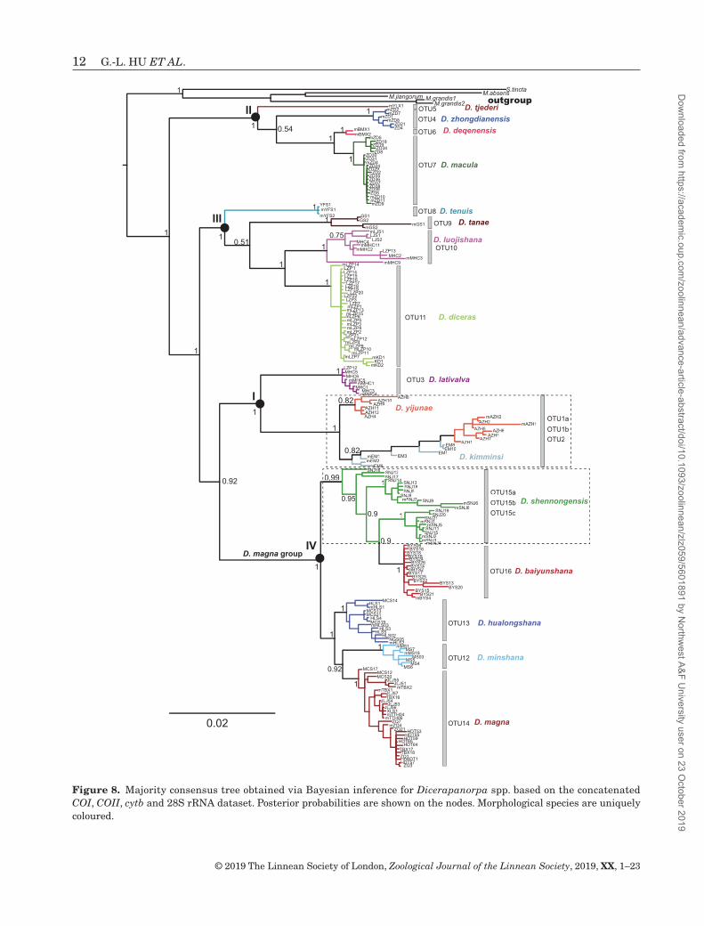
The COI haplotype tree resolved four robust clades (I–IV in Fig. 2) of Dicerapanorpa. In clade I, BIN, ABGD and GMYC clustered all samples from Anzihe Nature Reserve, Sichuan and Mount Emei, Sichuan into three OTUs (1a, 1b and 2), whereas bPTP merged these OTUs into a single cluster. In clade II, ABGD and GMYC generated four OTUs (4–7), whereas BIN grouped OTU6 and OTU7 together, and bPTP split OTU6 into two clusters. In clade III, BIN and bPTP recognized four OTUs (8–11), whereas ABGD and GMYC delineated five OTUs, splitting OTU10 into two partitions. The main discordance of these four approaches involved clade IV (i.e. the D. magna group). BIN produced only one cluster in clade IV, whereas ABGD and GMYC oversplit the samples, and the bPTP result was the most biologically realistic, with members from Shennongjia, Hubei split into three OTUs (15a–15c).
morphological compariSon
Species assignments were based primarily on overall morphological features, including the anal horn (Fig. 3), notal organ (Fig. 4), male genitalia (Fig. 5) and female medigynium (Fig. 6). Through integrative analyses of morphological characters, molecular clusters and geographical information, we recognized three new species (D. lativalva, D. minshana and D. hualongshana), which are described below. In general, morphological species corresponded well to OTUs, but several discordances existed between molecular and morphological clusters. For example, the allopatric Dicerapanorpa yijunae Hu & Hua, 2019 and Dicerapanorpa kimminsi (Carpenter, 1948) possess morphologically distinct male genitalia (Fig. 5A, B) and female medigynia (Fig. 6A, B), but lack molecular divergence (OTU1a, OTU1b and OTU2 in Fig. 2). The specimens of D. shennongensis with the same locality and conserved body, wing coloration and pattern are clustered into three OTUs (15a–15c in Fig. 2), but exhibit morphological diversity in the female medigynium (Fig. 6Q–S).
geometric morphometric analySeS
The CVA indicated that D. minshana and D. hualongshana were clearly separated from each other and from D. magna, D. baiyunshana and D. shennongensis (Fig. 7A). The sole cases of overlap involved D. baiyunshana and D. shennongensis. The permutation MANOVA confirmed significant shape differences in female medigynia of these five species in the D. magna group [F(4, 224) = 71.780, P < 0.001]. The CVA discriminated them based on female medigynia (axis 1, λ = 0.003, χ 2 = 1415.174, d.f. = 400, P < 0.001; axis 2, λ = 0.006, χ 2 = 913.085, d.f. = 297, P < 0.001; axis 3, λ = 0.068, χ 2 = 469.587, d.f. = 196, P < 0.001; axis 4, λ = 0.334, χ 2 = 192.613, d.f. = 97, P < 0.001). The first two canonical variates (CV1 and CV2) accounted for 48.69 and 34.06% of the total shape variation, respectively. The shape variables related to CV1 were a greatly elongated axis, slightly lowered posterior arms and expanded upper sides, whereas the shape variables involved in CV2 were the rapidly constricted middle sides and expanded lower sides (Fig. 7B).
molecular phylogeny
This study recovered 214 sequences of COI, 189 of COII, 198 of cytb and 166 of 28S rRNA. After alignment and the deletion of ambiguous sites, COI had a size range of 777 bp, COII of 699 bp, cytb of 567 bp and 28S rRNA
Dow
nloaded from https://academ
ic.oup.com/zoolinnean/advance-article-abstract/doi/10.1093/zoolinnean/zlz059/5601891 by N
Figure 2. Bayesian inference tree for Dicerapanorpa spp. based on mitochondrial COI haplotypes. Posterior probabilities are shown on the nodes. The right vertical bars indicate molecular partitions and final operational taxonomic units (OTUs) from delimitation analyses. Morphological species are uniquely coloured.
Dow
nloaded from https://academ
ic.oup.com/zoolinnean/advance-article-abstract/doi/10.1093/zoolinnean/zlz059/5601891 by N
of 851 bp. In total, 197 individuals with at least three loci were selected for the phylogenetic reconstruction.
The BI (Fig. 8) and ML trees (Fig. 9) reveal four identical monophyletic clades (I–IV) of Dicerapanorpa, as mentioned above (Fig. 2). The monophyly of most species was strongly supported, with a few exceptions. In clade I, D. lativalva was well supported to be monophyletic, whereas D. yijunae and D. kimminsi were paraphyletic. The samples of D. yijunae and D. kimminsi were mixed, forming a sister group to D. lativalva (PP = 1, BV = 99). In clade II, D. zhongdianensis, D. deqenensis Hu, Wang & Hua, 2019 and D. macula were strongly supported as
monophyletic clades. Dicerapanorpa deqenensis was the sister species to D. macula, but the phylogenetic positions of D. zhongdianensis and Dicerapanorpa tjederi (Carpenter, 1938) were inconsistent between the BI and ML trees. In clade III, all the species (Dicerapanorpa tanae Hu, Wang & Hua, 2019, Dicerapanorpa tenuis Hu, Wang & Hua, 2019, Dicerapanorpa luojishana Hu & Hua, 2019 and D. diceras) were well supported as monophyletic in the BI tree (Fig. 8), whereas D. luojishana was paraphyletic with D. diceras in the ML tree (Fig. 9). The phylogenetic positions of D. tanae and D. tenuis were uncertain because of the inconsistent topology in both
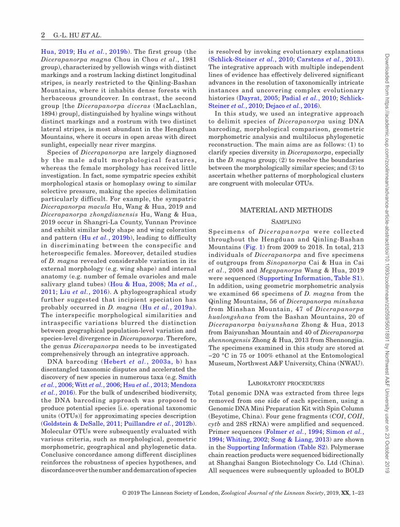
Figure 3. Anal horns for 16 species of Dicerapanorpa: A, D. yijunae; B, D. kimminsi; C, D. diceras; D, D. lativalva; E, D. zhongdianensis; F, D. luojishana; G, D. tanae; H, D. macula; I, D. deqenensis; J, D. tjederi; K, D. tenuis; L, D. minshana; M, D. hualongshana; N, D. magna; O, D. shennongensis; P, D. baiyunshana.
Dow
nloaded from https://academ
ic.oup.com/zoolinnean/advance-article-abstract/doi/10.1093/zoolinnean/zlz059/5601891 by N
trees. Clade IV was separated into two subclades with high support values (PP = 1, BV = 100). One subclade included D. shennongensis and D. baiyunshana, with D. shennongensis paraphyletic with D. baiyunshana. The other subclade included D. minshana , D. hualongshana and D. magna. Dicerapanorpa minshana was the sister species to D. magna, with both forming the sister group to D. hualongshana in the BI tree. However, the relationships among these three species were not well resolved in the ML tree.
Type materialHolotype: CHINA: ♂, Menghuocheng (29.00°N, 102.30°E), 2600 m, Shimian County, Sichuan, 26 July 2016, leg. Lu Jiang.
Figure 4. Notal organs for 16 species of Dicerapanorpa: A, D. yijunae; B, D. kimminsi; C, D. diceras; D, D. lativalva; E, D. zhongdianensis; F, D. luojishana; G, D. tanae; H, D. macula; I, D. deqenensis; J, D. tjederi; K, D. tenuis; L, D. minshana; M, D. hualongshana; N, D. magna; O, D. shennongensis; P, D. baiyunshana.
Dow
nloaded from https://academ
ic.oup.com/zoolinnean/advance-article-abstract/doi/10.1093/zoolinnean/zlz059/5601891 by N
Paratypes: Six ♂, five ♀, Menghuocheng and Liziping Nature Reserve, Shimian County, Sichuan, 22–26 July 2016, leg. Lu Jiang.
EtymologyThe specific epithet is composed of the Latin latus, wide or broad, and valvae, valves or doors, referring to the broad dorsal valves of the male aedeagus.
DiagnosisThe new species resembles D. diceras, but can be distinguished from the latter by the following characters: (1) basal branch of male paramere slender and straight (cf. broadened basally); (2) lateral branches semicircular at basal two-thirds and parallel at distal one-third (cf. curved inward); (3) dorsal valve of male aedeagus broad, reaching
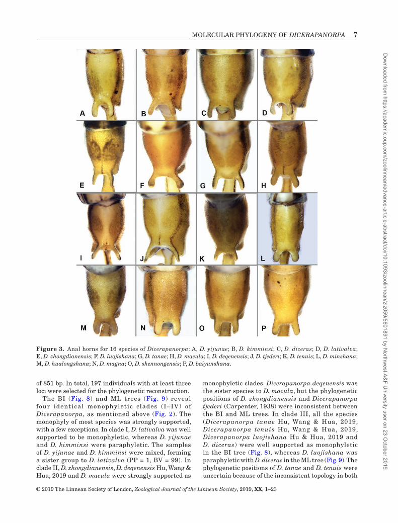
Figure 5. Male genitalia for 16 species of Dicerapanorpa: A, D. yijunae; B, D. kimminsi; C, D. diceras; D, D. lativalva; E, D. zhongdianensis; F, D. luojishana; G, D. tanae; H, D. macula; I, D. deqenensis; J, D. tjederi; K, D. tenuis; L, D. minshana; M, D. hualongshana; N, D. magna; O, D. shennongensis; P, D. baiyunshana.
Dow
nloaded from https://academ
ic.oup.com/zoolinnean/advance-article-abstract/doi/10.1093/zoolinnean/zlz059/5601891 by N
basal process of gonostylus (cf. slender, short); and (4) main plate of female medigynium pear-shaped (cf. rounded).
DescriptionHead: Head yellow. Vertex pale yellow. Ocellar triangle black. Rostrum yellow with two black
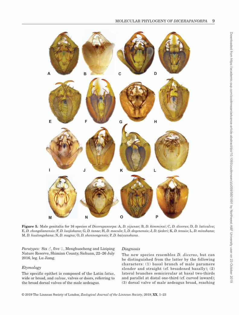
Figure 6. Female medigynium for 16 species of Dicerapanorpa: A, D. yijunae; B, D. kimminsi; C, D. lativalva; D, D. deqenensis; E, D. macula; F, D. zhongdianensis; G, D. tenuis; H, D. tanae; I, D. luojishana; J, D. tjederi; K, D. diceras; L, M, D. minshana; N, D. hualongshana; O, P, D. magna; Q–S, D. shennongensis; T, D. baiyunshana.
Dow
nloaded from https://academ
ic.oup.com/zoolinnean/advance-article-abstract/doi/10.1093/zoolinnean/zlz059/5601891 by N
Figure 7. Canonical variate analysis of the shape variables based on female medigynia of Dicerapanorpa magna, D. minshana, D. hualongshana, D. baiyunshana and D. shennongensis. A, scatter plots of canonical variates CV1 vs. CV2 are shown for different species. B, shape variations along CV1 and CV2 illustrated by deformation grid graphs.
Dow
nloaded from https://academ
ic.oup.com/zoolinnean/advance-article-abstract/doi/10.1093/zoolinnean/zlz059/5601891 by N
Figure 8. Majority consensus tree obtained via Bayesian inference for Dicerapanorpa spp. based on the concatenated COI, COII, cytb and 28S rRNA dataset. Posterior probabilities are shown on the nodes. Morphological species are uniquely coloured.
Dow
nloaded from https://academ
ic.oup.com/zoolinnean/advance-article-abstract/doi/10.1093/zoolinnean/zlz059/5601891 by N
Figure 9. Maximum likelihood tree for Dicerapanorpa spp. based on the concatenated dataset of COI, COII, cytb and 28S rRNA. Bootstrap values are shown on the nodes.
Dow
nloaded from https://academ
ic.oup.com/zoolinnean/advance-article-abstract/doi/10.1093/zoolinnean/zlz059/5601891 by N
lateral longitudinal stripes. Antenna blackish brown (Figs 10, 11A, B).
Thorax: Pronotum yellow, with long black setae along anterior margin and a black longitudinal stripe along each side. Meso- and metanotum yellow, with two black longitudinal stripes laterally. Pleura yellow. Legs cream coloured, with tarsomeres blackish (Figs 10, 11A, B).
Wings: Male holotype: forewing length 14.6 mm, width 3.7 mm; hindwing length 13.9 mm, width 3.5 mm; wing membrane hyaline, without distinct markings
(Fig. 11A). Female: forewing length 15.4–16.6 mm, width 3.8–4.1 mm; hindwing length 14.2–15.2 mm, width 3.8–4.1 mm; similar to male in general appearance (Figs 10B, 11B).
Abdomen: Terga I–V (T1–T5) yellowish, with two black longitudinal lateral stripes; sterna and pleura yellow (Figs 10, 11). Male: notal organ of T3 slightly prominent, bearing black setae posteriorly (Fig. 4D); T6 yellowish brown, with a pair of anal horns on posterior margin (Figs 3D, 11A); abdominal segments VII and III (A7–A8) brownish yellow, elongate, constricted at basal half and thickened at apical half, but A8 much
Figure 11. Dicerapanorpa lativalva: A, male holotype, dorsal view; B, female paratype, dorsal view; C, D, male genital bulb, dorsal and ventral views, respectively; E, female subgenital plate, ventral view; F, female medigynium, ventral view. Abbreviations: ae, aedeagus; ax, axis; bb, basal branch; bp, basal process; ep, epandrium; gcx, gonocoxite; gs, gonostylus; hv, hypovalve; lb, lateral branch; mb, mesal branch; mp, main plate; mt, median tooth; pa, posterior arm; sgp, subgenital plate. Scale bars: 5.0 mm (A, B); 0.5 mm (C, D); 0.1 mm (E, F).
Figure 10. Habitus of Dicerapanorpa lativalva: A, male; B, female.
Dow
nloaded from https://academ
ic.oup.com/zoolinnean/advance-article-abstract/doi/10.1093/zoolinnean/zlz059/5601891 by N
Male genitalia: Genital bulb spherical, yellowish brown. Epandrium broad basally and narrowed distally, terminating with a shallow, broad, U-shaped emargination, extending over apex of gonocoxite (Fig. 11C). Hypovalve greatly broadened toward apex and slightly curved inward distally, bearing long bristles along inner margin (Figs 5D, 11D). Gonostylus smoothly curved, bearing a developed basal process and a sharp median tooth along inner margin. Parameres trifurcate: basal branches slender and straight, nearly parallel, reaching basal process of gonostylus; mesal branches divergent at base and convergent at apex, reaching median tooth of gonostylus; lateral branches semicircular at basal two-thirds and parallel at distal one-third. Ventral valves of aedeagus membranous and slender, reaching apex of gonocoxite; dorsal valves broadened and elongate, reaching basal process of gonostylus.
Female geni ta l ia : Subgeni ta l p late broad , trapezoidal, terminating in a ligulate process (Fig. 11E). Medigynium elongate and pear-shaped, folded ventrally on each side. Main plate nearly rectangular, twice as long as posterior arms. Axis concealed in main plate, slightly protruding at apex (Figs 6C, 11F).
Type materialHolotype: CHINA: ♂, Hualongshan Mountain (32.01°N, 109.36°E), 2100 m, Pingli County, Shaanxi, 12 July 2015, leg. Bao-Zhen Hua.
Paratypes: Seven ♀, Mount Nangongshan (32.29°N, 109.06°E), Langao County, Shaanxi, 11 June 2013, leg. Jing Chen and Qin-Xiao Chen; Five ♂, three ♀, Hualongshan Mountain, Pingli County, Shaanxi, 24 June 2018, leg. Kai Gao, Yuan Hua and Yu-Ru Yang; one ♂, 21 ♀, Mount Nangongshan, Langao County, Shaanxi, 26 June 2018, leg. Yuan Hua and Kai Gao; four ♂, ten ♀, Chengkou County (31.84°N, 109.107°E), Chongqing, 20 June 2018, leg. Kai Gao.
EtymologyThe specific epithet refers to the type locality, Hualongshan Mountain.
DiagnosisThis new species resembles D. shennongensis, but can be readily differentiated from the latter by the following characters: (1) mesal branches of male parameres convergent distally (cf. parallel); and (2) main plate of female medigynium broad, posterior arms short (cf. slender and long).
DescriptionHead: Head mostly yellow. Vertex yellowish. Ocellar triangle black. Rostrum yellow, without distinct black longitudinal stripes. Antenna blackish brown (Fig. 12A, B).
Thorax: Pro-, meso- and metanotum yellowish, bearing black stout setae anteriorly and two black longitudinal stripes laterally. Pleura pale yellow. Legs yellowish brown (Fig. 12A, B).
Wings: Male holotype: forewing length 15.8 mm, width 4.2 mm, yellowish with dark brown markings; apical band enclosing a large hyaline window; pterostigmal band with a broad basal branch and a reduced separated distal branch; basal band extremely reduced; marginal and basal spots indistinct; hindwing length 13.9 mm, width 4.0 mm, with more degenerated markings (Fig. 12A). Female: forewing length 14.8–17.2 mm, width 4.0–4.9 mm, basal band reduced, extending from vein R1 to 1A; pterostigma and apical bands complete; marginal spot extending from vein R1 to R4 + 5. Hindwing length 13.8–16.1 mm, width 4.0–4.8 mm, similar to forewing in general appearance (Fig. 12B).
Abdomen: T1–T5 pale yellow, with two black longitudinal stripes laterally (Fig. 12A, B). Male: notal organ of T3 well developed, bearing thick setae on posterior margin (Fig. 4M); T6 yellowish brown, with a pair of digitate anal horns posteriorly (Fig. 3M); abdominal segment VII (A7) yellowish brown, elongate, constricted basally and abruptly dilated distally; A8 similar to A7, but much thinner apically (Fig. 12A). Female: abdominal segments gradually narrowed distally (Fig. 12B).
Male genitalia: Genital bulb yellowish brown, elliptic. Epandrium broad at base, gradually narrowing toward apex, with a deep U-shaped terminal emargination (Fig. 12C). Hypovalve not reaching apex of gonocoxite, with long bristles along inner margin (Figs 12D, 13C). Gonostylus shorter than gonocoxite, with a developed pentagon-shaped basal process and a small, sharp median tooth (Fig. 12C, D). Parameres trifurcate: basal branches short, nearly parallel; mesal branch elongate, curved inward at apex, reaching basal process of gonostylus; lateral branch incurved, reaching or
Dow
nloaded from https://academ
ic.oup.com/zoolinnean/advance-article-abstract/doi/10.1093/zoolinnean/zlz059/5601891 by N
Figure 13. Scanning electron micrographs of male (A–C) and female genitalia (D–G) of Dicerapanorpa hualongshana: A, male genital bulb, with hypandrium removed, ventral view; B, magnification of A to show the male aedeagus, ventral view; C, right hypovalve, ventral view; D, E, female subgenital plate; F, G, medigynium. Scale bars: 0.2 mm.
Figure 12. Dicerapanorpa hualongshana: A, male holotype, dorsal view; B, female paratype, dorsal view; C, D, male genital bulb, dorsal and ventral views, respectively; E, female subgenital plate, ventral view; F, female medigynium, ventral view. Abbreviations: ae, aedeagus; ax, axis; bb, basal branch; bp, basal process; ep, epandrium; gcx, gonocoxite; gs, gonostylus; hv, hypovalve; lb, lateral branch; mb, mesal branch; mp, main plate; mt, median tooth; pa, posterior arm; sgp, subgenital plate. Scale bars: 5.0 mm (A, B); 0.5 mm (C, D); 0.1 mm (E, F).
Dow
nloaded from https://academ
ic.oup.com/zoolinnean/advance-article-abstract/doi/10.1093/zoolinnean/zlz059/5601891 by N
exceeding apex of ventral valve. Ventral valves of aedeagus long, slender; dorsal valves sclerotized, not reaching apex of gonocoxite (Fig. 12D).
Female genitalia: Subgenital plate ovoid, terminating in a ligulate process, covered with long bristles caudally (Fig. 12E). Main plate of female medigynium broad, nearly rectangular (Fig. 12F). Posterior arms shorter than main plate. Axis concealed in main plate, slightly protruding beyond main plate at apex.
RemarksDifferent individuals of this species exhibit variations in male aedeagus, paramere (Figs 4M, 13A, B), female subgenital plate (Fig. 13D, E) and medigynium (Figs 6N, 13F, G).
Type materialHolotype: CHINA: ♂, Laohegou Nature Reserve (32.47°N, 104.73°E), Minshan Mountain, 1800 m, Pingwu County, Sichuan, 6 May 2013, leg. Shuang Xue.
Paratypes: Thirteen ♂, three ♀, same data as holotype; 13 ♂, 16 ♀, same locality as holotype, 28 May 2018, leg. Kai Gao.
EtymologyThe specific epithet refers to the type locality, Minshan Mountain.
DiagnosisThis new species can be differentiated readily from D. magna by the following characters: (1) male genital bulb spherical (cf. long and elliptical); (2) basal branch of male paramere short, hook-shaped (cf. relatively long); and (3) main plate of female medigynium nearly rectangular distally, constricted mesally and smoothly curved basally (cf. subtriangular or constricted rapidly at base).
DescriptionHead: Head yellow. Vertex yellowish. Ocellar triangle black. Antenna blackish brown. Rostrum yellowish, without black lateral longitudinal stripes (Fig. 14A, B).
Thorax: Pro-, meso- and metanotum yellow, bearing several setae anteriorly and two black longitudinal stripes laterally. Pleura light yellow. Legs yellowish brown, with tarsomeres gradually darkened toward apex (Fig. 14A, B).
Wings: Male holotype: forewing length 16.5 mm, width 4.5 mm, yellowish, apical band reduced; pterostigma band incomplete and dark brown, with reduced basal and distal branches; basal band extending from vein Rs to CuP, other markings indistinct; hindwing length 15.2 mm, width 4.3 mm, similar to forewing, but with more degenerated markings (Fig. 14A). Female: forewing length 15.5–17.6 mm, width 4.1–4.9 mm; hindwing length 14.1–16.4 mm, width 4.0–4.7 mm; similar to male in general appearance (Fig. 14B).
Abdomen: T1–T5 yellowish, with two black lateral longitudinal stripes (Fig. 14A, B). Male: notal organ developed, covered with numerous black setae posteriorly (Fig. 4L); T6 brownish yellow, bearing a pair of digitate anal horns posteriorly (Figs 3L, 14A); A7 and A8 yellowish brown, elongate, constricted at basal half and dilated at distal half, but A8 much thinner than A7 distally (Fig. 14A). Female: abdominal segments gradually narrowed caudally (Fig. 14B).
Male genitalia: Genital bulb brownish yellow, spherical. Epandrium gradually narrowing toward apex, with a rounded, U-shaped terminal emargination (Fig. 14C). Hypovalve slender, bearing long bristles along inner margin, nearly reaching apex of gonocoxite (Figs 5L, 14D). Gonostylus shorter than gonocoxite, with a well-developed trapezoidal basal process and a small sharp subtriangular median tooth. Parameres trifurcate: basal branch considerably short, hook-shaped; mesal and lateral branches curved inward, reaching or exceeding apex of gonocoxite. Ventral valves of aedeagus short; dorsal valves broadened and elongate, nearly reaching apex of gonocoxite (Figs 5L, 14D, 15A, B).
Female genitalia: Subgenital plate ovoid, terminating in a ligulate process, covered with long setae caudally (Figs 14E, 15C). Medigynium strongly sclerotized, with main plate rectangular distally, gradually constricted mesally and rounded basally. Posterior arms parallel and short, approximately half the length of main plate (Figs 6L, M, 14F).
DISCUSSION
Owing to highly intraspecific variations and interspecific similarities, species delimitation of Dicerapanorpa has been challenging. By integrating
Dow
nloaded from https://academ
ic.oup.com/zoolinnean/advance-article-abstract/doi/10.1093/zoolinnean/zlz059/5601891 by N
molecular, morphological, geometric morphometric and phylogenetic analyses, this study confirmed the 13 species of Dicerapanorpa and revealed the existence of three formerly overlooked new species (D. lativalva, D. hualongshana and D. minshana), considerably
deepening our understanding of species limits and boundaries in Dicerapanorpa.
Molecular approaches to species delimitation have developed rapidly in recent decades, and several methods are now available to infer species boundaries
Figure 15. Scanning electron micrographs of male and female genitalia of Dicerapanorpa minshana: A, male genital bulb, with hypandrium removed, ventral view; B, magnification of A to show the male aedeagus and parameres, ventral view; C, female genital plate, ventral view. Scale bars: 0.2 mm.
Figure 14. Dicerapanorpa minshana: A, male holotype, dorsal view; B, female paratype, dorsal view; C, D, male genital bulb, dorsal and ventral views, respectively; E, female subgenital plate, ventral view; F, female medigynium, ventral view. Abbreviations: ae, aedeagus; ax, axis; bb, basal branch; bp, basal process; ep, epandrium; gcx, gonocoxite; gs, gonostylus; hv, hypovalve; lb, lateral branch; mb, mesal branch; mp, main plate; mt, median tooth; pa, posterior arm; sgp, subgenital plate. Scale bars: 5.0 mm (A, B); 0.5 mm (C, D); 0.1 mm (E, F).
Dow
nloaded from https://academ
ic.oup.com/zoolinnean/advance-article-abstract/doi/10.1093/zoolinnean/zlz059/5601891 by N
(Dellicour & Flot, 2015). Our present study initially used the BIN system in BOLD to assign individuals to presumptive species, and subsequently applied three other delimitation methods (ABGD, GMYC bPTP). BIN unexpectedly grouped all samples of the five species in the D. magna group (clade IV in Fig. 2) together, which might result from the large geographical scale of sampling, as reported by Bergsten et al. (2012). The GMYC approach, especially the multiple-threshold model, greatly inflated the species count (30 OTUs), a result seen earlier in spiders (Satler et al., 2013; Hedin, 2015). In contrast, bPTP produced a relatively conservative species count (18 OTUs) and was considered to outperform the GMYC model in both simulation and empirical studies (Zhang et al., 2013; Hedin, 2015). The ABGD method was previously considered to perform poorly in delimiting species when sample sizes are low (fewer than five; Puillandre et al., 2012b), but performed well in our study. These four methods are complementary, and effectively contribute to the final molecular OTUs.
The phylogenetic trees (Figs 8, 9) clearly indicate that the genus Dicerapanorpa is a monophyletic group, consistent with previous conclusions (Ma et al., 2012; Hu et al., 2015; Miao et al., 2019). Clades I–III belong to the D. diceras group, and clade IV belongs to the D. magna group, showing the paraphyletic relationship of the D. diceras group with respect to the D. magna group. This might also suggest that the hyaline wings and the rostrum with two black longitudinal stripes in the D. diceras group are plesiomorphic characters, whereas the yellowish wings with distinct markings and the rostrum without stripes in the D. magna group are apomorphic characters.
Reciprocal monophyly is often viewed as the most important criterion for species delimitation (de Queiroz, 2007), but it is often not evident in recently diverged species (Knowles & Carstens, 2007; Cummings et al., 2008). The phylogenetic trees (Figs 8, 9) in the present four-gene dataset reveal that some species are not monophyletic. For example, D. shennongensis is paraphyletic with D. baiyunshana, and D. yijunae is paraphyletic with D. kimminsi. The potential reason for this phenomenon might be hybridization followed by introgression, incomplete lineage sorting and recent evolutionary divergence, as explained in previous studies (Funk & Omland, 2003; Knowles & Carstens, 2007; Degnan & Rosenberg, 2009). New species start from initial polyphyly and achieve monophyly through paraphyly in the speciation process (Avise, 2000). The progress may be more rapid for mitochondrial haplotypes and can be distinctive in the case of budding speciation through peripheral isolation (Frey, 1993). Ancestral (e.g. D. shennongensis) and budding species (e.g. D. baiyunshana) will remain paraphyletic until the ancestral species has lost the characteristics
of the budding species (Podani, 2013; Kaya & Çiplak, 2016). Mitochondrial genes play a dominant role in the phylogenetic reconstruction, probably leading to discordance between the phylogenetic topology and species tree. Even so, the mitochondrial genetic data, as a source of complementation to other evidence for species delimitation, do provide valuable information for investigating evolutionary histories and patterns of species diversity (Avise, 2009; Monaghan et al., 2009; Fujisawa & Barraclough, 2013).
Male genitalia are remarkably useful morphological traits for species discrimination (Song & Bucheli, 2010). Numerous taxa of insects have evolved species-specific male genitalia, and morphological divergence of male genitalia among closely related species is often dramatic (Eberhard, 1985). For example, body coloration and wing pattern are relatively conserved in Dicerapanorpa, whereas genital shape and complexity exhibit a greater divergence, as also seen in water striders (Eberhard, 2010; Rowe & Arnqvist, 2012). The paramere of male genitalia is the most phenotypically differentiated structure in Dicerapanorpa, with variation of its shape and length distinguishing closely related species, especially in the D. magna group (Fig. 5L–P). The rapid evolution and divergence of this trait have probably been driven by sexual selection through facilitating male domination and success in the process of copulation (Hosken & Stockley, 2004; Simmons, 2014). The male genital structures in scorpionflies, such as the gonostylus, hypovalve and epandrium, in addition to the anal horn and notal organ, have been shown to function in countering the female resistance and stabilizing the mating position (Ma et al., 2010; Zhong & Hua, 2013b; Zhong et al., 2015).
In contrast, female genital structures have been little studied by taxonomists, mainly because they are generally internal and require dissection (Simmons, 2014). The medigynium, an important component of female genitalia, was previously considered as possessing taxonomic value in scorpionflies (Byers, 1954; Cheng, 1957). Ma et al. (2012) concluded that the medigynium is a reliable character for species delimitation in the Panorpidae at generic or higher levels. The present study corroborates that most Dicerapanorpa species have a morphologically distinct medigynium, with considerable variation within some species, such as D. magna (Fig. 6O, P) and D. shennongensis (Fig. 6Q–S). The extensive morphological variations between and within populations in Dicerapanorpa are more complicated than the variation in male genitalia, a situation also noted in a scarab beetle (Polihronakis, 2009). The copulatory pore is situated at the posterior end of the axis of the medigynium in Dicerapanorpa, implying that the main plate and posterior arms are likely to be free from functional constraint. This might explain
Dow
nloaded from https://academ
ic.oup.com/zoolinnean/advance-article-abstract/doi/10.1093/zoolinnean/zlz059/5601891 by N
the extensive variation in the shape and length of the female medigynium in Dicerapanorpa.
Evidence from multiple characters suggests that the D. magna group consists of five species, i.e. D. baiyunshana, D. hualongshana, D. magna, D. minshana and D. shennongensis. A previous geometric morphometric study of wings supports the significant differences between D. hualongshana and D. magna (Liu et al., 2016). A phylogeographical study (Hu et al., 2019a) also suggests the presence of three genetically distinct lineages and incipient speciation in the D. magna group (i.e. D. hualongshana, D. magna and D. minshana). Geographical barriers, including mountains and the Hanshui River between the Qinling and Bashan Mountains (Su et al., 2015; Liu et al., 2016), are apparently responsible for limiting gene flow and promoting divergence among species in the D. magna group. Pleistocene glaciations could also have promoted recent divergence associated with colonization of individual sky islands, as found in grasshoppers (Knowles, 2001). Owing to divergent ecological selection, reproductive isolation between allopatric populations might have evolved. However, D. hualongshana, D. magna and D. minshana have probably experienced recurrent range contractions and expansions during the Pleistocene, leading to secondary contact between allopatric populations (Hu et al., 2019a). These species are probably able to hybridize in the contact zone, because they are unlikely to exhibit complete reproductive isolation.
Mountain regions, as naturally fragmented habitat islands, provide opportunities for new species to diversify and survive (Hewitt, 2000, 2004). Allopatric speciation is often fostered by geographical barriers within montane systems owing to natural selection or genetic drift (Vuilleumier & Monasterio, 1986; Moritz et al., 2000), leading to the exceptionally high species diversity and richness in the mountains of southwestern China. Besides, the climate changes during the Pleistocene have probably contributed to population differentiation and speciation processes in Dicerapanorpa (Hu et al., 2019a). The Dicerapanorpa species considered in this study are narrow endemics restricted to small or medium-sized regions on the mountaintops with a few exceptions in the D. magna group. Some species of Dicerapanorpa may have experienced convergent evolution, because they display nearly identical body and wing coloration, which might result from similar selective pressure in the mountain sky islands, as noted in cave-dwelling harvestmen (Derkarabetian et al., 2010; Derkarabetian & Hedin, 2014). Nevertheless, the integrative approach has uncovered taxonomic boundaries in these morphologically conserved species, such as D. diceras, D. lativalva and D. luojishana, laying a foundation for future evolutionary and systematic studies of this group.
ACKNOWLEDGEMENTS
We thank Lu Jiang, Wei Du, Ying Miao, Kai-Wen Gao and Yi-Jun Chai for collecting specimens, and Mei Liu, Zhuo Wang and Lu-Yao Yang for aid with photography. We thank Mari Kekkonen, Muhammad Ashfaq and Sujeevan Ratnasingham for providing advice on molecular analyses and Dirk Steinke for help with the morphometric analysis. We also thank the anonymous reviewer for the valuable comments on the revision of the manuscript. This research was supported by the National Natural Science Foundation of China (grant no. 31672341).
REFERENCES
Avise JC. 2000. Phylogeography: the history and formation of species. Cambridge: Harvard University Press.
Avise JC. 2009. Phylogeography: retrospect and prospect. Journal of Biogeography 36: 3–15.
Bergsten J, Bilton DT, Fujisawa T, Elliott M, Monaghan MT, Balke M, Hendrich L, Geijer J, Herrmann J, Foster GN, Ribera I, Nilsson AN, Barraclough TG, Vogler AP. 2012. The effect of geographical scale of sampling on DNA barcoding. Systematic Biology 61: 851–869.
Bickford D, Lohman DJ, Sodhi NS, Ng PK, Meier R, Winker K, Ingram KK, Das I. 2007. Cryptic species as a window on diversity and conservation. Trends in Ecology & Evolution 22: 148–155.
Bortolus A. 2008. Error cascades in the biological sciences: the unwanted consequences of using bad taxonomy in ecology. AMBIO: A Journal of the Human Environment 37: 114–118.
Byers GW. 1954. Notes on North American Mecoptera. Annals of the Entomological Society of America 47: 484–510.
Carpenter FM. 1938. Mecoptera from China, with descriptions of new species. Proceedings of the Entomological Society of Washington 40: 267–281.
Carstens BC, Pelletier TA, Reid NM, Satler JD. 2013. How to fail at species delimitation. Molecular Ecology 22: 4369–4383.
Cheng FY. 1957. Revision of the Chinese Mecoptera. Bulletin of the Museum of Comparative Zoology 116: 1–118.
Cummings MP, Neel MC, Shaw KL. 2008. A genealogical approach to quantifying lineage divergence. Evolution 62: 2411–2422.
Darriba D, Taboada GL, Doallo R, Posada D. 2012. jModelTest 2: more models, new heuristics and parallel computing. Nature Methods 9: 772.
Dayrat B. 2005. Towards integrative taxonomy. Biological Journal of the Linnean Society 85: 407–415.
Degnan JH, Rosenberg NA. 2009. Gene tree discordance, phylogenetic inference and the multispecies coalescent. Trends in Ecology & Evolution 24: 332–340.
Dejaco T, Gassner M, Arthofer W, Schlick-Steiner BC, Steiner FM. 2016. Taxonomist’s nightmare … evolutionist’s delight: an integrative approach resolves species limits in jumping bristletails despite widespread hybridization and parthenogenesis. Systematic Biology 65: 947–974.
Dow
nloaded from https://academ
ic.oup.com/zoolinnean/advance-article-abstract/doi/10.1093/zoolinnean/zlz059/5601891 by N
Dellicour S, Flot JF. 2015. Delimiting species-poor data sets using single molecular markers: a study of barcode gaps, Haplowebs and GMYC. Systematic Biology 64: 900–908.
Derkarabetian S, Hedin M. 2014. Integrative taxonomy and species delimitation in harvestmen: a revision of the western North American genus Sclerobunus (Opiliones: Laniatores: Travunioidea). PLoS ONE 9: e104982.
Derkarabetian S, Steinmann DB, Hedin M. 2010. Repeated and time-correlated morphological convergence in cave-dwelling harvestmen (Opiliones, Laniatores) from montane western North America. PLoS ONE 5: e10388.
Drummond AJ, Suchard MA, Xie D, Rambaut A. 2012. Bayesian phylogenetics with BEAUti and the BEAST 1.7. Molecular Biology and Evolution 29: 1969–1973.
Eberhard WG. 1985. Sexual selection and animal genitalia. Cambridge: Harvard University Press.
Eberhard WG. 2010. Evolution of genitalia: theories, evidence, and new directions. Genetica 138: 5–18.
Folmer O, Black M, Hoeh W, Lutz R, Vrijenhoek R. 1994. DNA primers for amplification of mitochondrial cytochrome c oxidase subunit I from diverse metazoan invertebrates. Molecular Marine Biology and Biotechnology 3: 294–299.
Frey JK. 1993. Modes of peripheral isolate formation and speciation. Systematic Biology 42: 373–381.
Fujisawa T, Barraclough TG. 2013. Delimiting species using single-locus data and the Generalized Mixed Yule Coalescent approach: a revised method and evaluation on simulated data sets. Systematic Biology 62: 707–724.
Funk DJ, Omland KE. 2003. Species-level paraphyly and polyphyly: frequency, causes, and consequences, with insights from animal mitochondrial DNA. Annual Review of Ecology, Evolution, and Systematics 34: 397–423.
Glez-Peña D, Gomez-Blanco D, Reboiro-Jato M, Fdez-Riverola F, Posada D. 2010. ALTER: program-oriented conversion of DNA and protein alignments. Nucleic Acids Research 38: W14–W18.
Goldstein PZ, DeSalle R. 2011. Integrating DNA barcode data and taxonomic practice: determination, discovery, and description. Bioessays 33: 135–147.
Hall TA. 1999. BioEdit: a user-friendly biological sequence alignment editor and analysis program for Windows 95/98/NT. Nucleic Acids Symposium Series 41: 95–98.
Hebert PDN, Cywinska A, Ball SL, deWaard JR. 2003a. Biological identifications through DNA barcodes. Proceedings of the Royal Society B: Biological Sciences 270: 313–321.
Hebert PDN, Ratnasingham S, deWaard JR. 2003b. Barcoding animal life: cytochrome c oxidase subunit 1 divergences among closely related species. Proceedings of the Royal Society B: Biological Sciences 270(Suppl 1): S96–S99.
Hedin M. 2015. High-stakes species delimitation in eyeless cave spiders (Cicurina, Dictynidae, Araneae) from central Texas. Molecular Ecology 24: 346–361.
Hewitt GM. 2000. The genetic legacy of the Quaternary ice ages. Nature 405: 907–913.
Hewitt GM. 2004. Genetic consequences of climatic oscillations in the Quaternary. Philosophical Transactions of the Royal Society B: Biological Sciences 359: 183–195.
Hosken DJ, Stockley P. 2004. Sexual selection and genital evolution. Trends in Ecology & Evolution 19: 87–93.
Hou XY, Hua BZ. 2008. Structures of the female reproductive systems in Panorpidae (Mecoptera) with remarks on their taxonomic significance. Acta Zootaxonomica Sinica 33: 427–434.
Hsu TH, Ning Y, Gwo JC, Zeng ZN. 2013. DNA barcoding reveals cryptic diversity in the peanut worm Sipunculus nudus. Molecular Ecology Resources 13: 596–606.
Hu GL, Hua BZ. 2019. Two new species of the genus Dicerapanorpa (Mecoptera: Panorpidae) from Sichuan, China. Entomotaxonomia 41: 73–79.
Hu GL, Hua Y, Hebert PDN, Hua BZ. 2019a. Evolutionary history of the scorpionfly Dicerapanorpa magna (Mecoptera, Panorpidae). Zoologica Scripta 48: 93–105.
Hu GL, Wang JS, Hua BZ. 2019b. Five new species of Dicerapanorpa Zhong & Hua (Mecoptera, Panorpidae) from Yunnan, China. Journal of Asia-Pacific Entomology 22: 159–166.
Hu GL, Yan G, Xu H, Hua BZ. 2015. Molecular phylogeny of Panorpidae (Insecta: Mecoptera) based on mitochondrial and nuclear genes. Molecular Phylogenetics and Evolution 85: 22–31.
Isaac NJ, Mallet J, Mace GM. 2004. Taxonomic inflation: its influence on macroecology and conservation. Trends in Ecology & Evolution 19: 464–469.
Katoh K, Asimenos G, Toh H. 2009. Multiple alignment of DNA sequences with MAFFT. Methods in Molecular Biology 537: 39–64.
Kaya S, Çiplak B. 2016. Budding speciation via peripheral isolation: the Psorodonotus venosus (Orthoptera, Tettigoniidae) species group example. Zoologica Scripta 45: 521–537.
Klingenberg CP. 2011. MorphoJ: an integrated software package for geometric morphometrics. Molecular Ecology Resources 11: 353–357.
Knowles LL. 2001. Did the Pleistocene glaciations promote divergence? Tests of explicit refugial models in montane grasshopprers. Molecular Ecology 10: 691–701.
Knowles LL, Carstens BC. 2007. Delimiting species without monophyletic gene trees. Systematic Biology 56: 887–895.
Lanfear R, Calcott B, Ho SYW, Guindon S. 2012. PartitionFinder: combined selection of partitioning schemes and substitution models for phylogenetic analyses. Molecular Biology and Evolution 29: 1695–1701.
Liu M, Ma N, Hua BZ. 2016. Intraspecific morphological variation of the scorpionfly Dicerapanorpa magna (Chou) (Mecoptera: Panorpidae) based on geometric morphometric analysis of wings. Contributions to Zoology 85: 1–11.
Ma N, Cai LJ, Hua BZ. 2009. Comparative morphology of the eggs in some Panorpidae (Mecoptera) and their systematic implication. Systematics and Biodiversity 7: 403–417.
Ma N, Chen HM, Hua BZ. 2014. Larval morphology of the scorpionfly Dicerapanorpa magna (Chou) (Mecoptera: Panorpidae) and its adaptive significance. Zoologischer Anzeiger 253: 216–224.
Ma N, Liu SY, Hua BZ. 2011. Morphological diversity of male salivary glands in Panorpidae (Mecoptera). European Journal of Entomology 108: 493–499.
Dow
nloaded from https://academ
ic.oup.com/zoolinnean/advance-article-abstract/doi/10.1093/zoolinnean/zlz059/5601891 by N
Ma N, Zhong W, Gao QH, Hua BZ. 2012. Female genital plate diversity and phylogenetic analyses of East Asian Panorpidae (Mecoptera). Systematics and Biodiversity 10: 159–178.
Ma N, Zhong W, Hua BZ. 2010. Genitalic morphology and copulatory mechanism of the scorpionfly Panorpa jilinensis (Mecoptera: Panorpidae). Micron 41: 931–938.
Mendoza AM, Torres MF, Paz A, Trujillo-Arias N, Lopez-Alvarez D, Sierra S, Forero F, Gonzalez MA. 2016. Cryptic diversity revealed by DNA barcoding in Colombian illegally traded bird species. Molecular Ecology Resources 16: 862–873.
Miao Y, Wang JS, Hua BZ. 2019. Molecular phylogeny of the scorpionflies Panorpidae (Insecta: Mecoptera) and chromosomal evolution. Cladistics 35(4). doi: 10.1111/cla.12357.
Miller MA, Pfeiffer W, Schwartz T. 2010. Creating the CIPRES Science Gateway for inference of large phylogenetic trees. In: Proceedings of the Gateway Computing Environments workshop (GCE). New Orleans: Institute of Electrical and Electronics Engineers, 1–8.
Monaghan MT, Wild R, Elliot M, Fujisawa T, Balke M, Inward DJ, Lees DC, Ranaivosolo R, Eggleton P, Barraclough TG, Vogler AP. 2009. Accelerated species inventory on Madagascar using coalescent-based models of species delineation. Systematic Biology 58: 298–311.
Moritz C , Patton J , Schneider C , Smith T. 2000. Diversification of rainforest faunas: an integrated molecular approach. Annual Review of Ecology and Systematics 31: 533–563.
Padial JM, Miralles A, De la Riva I, Vences M. 2010. The integrative future of taxonomy. Frontiers in Zoology 7: 16.
Podani J. 2013. Tree thinking, time and topology: comments on the interpretation of tree diagrams in evolutionary/phylogenetic systematics. Cladistics 29: 315–327.
Polihronakis M. 2009. Hierarchical comparative analysis of genetic and genitalic geographical structure: testing patterns of male and female genital evolution in the scarab beetle Phyllophaga hirticula (Coleoptera: Scarabaeidae). Biological Journal of the Linnean Society 96: 135–149.
Pons J, Barraclough T, Gomez-Zurita J, Cardoso A, Duran D, Hazell S, Kamoun S, Sumlin W, Vogler A. 2006. Sequence-based species delimitation for the DNA taxonomy of undescribed insects. Systematic Biology 55: 595–609.
Puillandre N, Lambert A, Brouillet S, Achaz G. 2012a. ABGD, Automatic Barcode Gap Discovery for primary species delimitation. Molecular Ecology 21: 1864–1877.
Puillandre N , Modica MV , Zhang Y , Sirovich L , Boisselier MC, Cruaud C, Holford M, Samadi S. 2012b. Large-scale species delimitation method for hyperdiverse groups. Molecular Ecology 21: 2671–2691.
de Queiroz K. 2005. Ernst Mayr and the modern concept of species. Proceedings of the National Academy of Sciences of the United States of America 102(Suppl 1): 6600–6607.
de Queiroz K. 2007. Species concepts and species delimitation. Systematic Biology 56: 879–886.
Ratnasingham S, Hebert PDN. 2007. BOLD: the barcode of life data system (http://www.barcodinglife.org). Molecular Ecology Notes 7: 355–364.
Ratnasingham S, Hebert PDN. 2013. A DNA-based registry for all animal species: the barcode index number (BIN) system. PLoS ONE 8: e66213.
Rohlf F. 2010. TpsUtil v1.46. Stony Brook: Department of Ecology and Evolution, State University of New York.
Rohlf F. 2015. TpsDig2. Stony Brook: Department of Ecology and Evolution, State University of New York.
Rowe L, Arnqvist G. 2012. Sexual selection and the evolution of genital shape and complexity in water striders. Evolution 66: 40–54.
Satler JD, Carstens BC, Hedin M. 2013. Multilocus species delimitation in a complex of morphologically conserved trapdoor spiders (Mygalomorphae, Antrodiaetidae, Aliatypus). Systematic Biology 62: 805–823.
Schlick-Steiner BC, Steiner FM, Seifert B, Stauffer C, Christian E, Crozier RH. 2010. Integrative taxonomy: a multisource approach to exploring biodiversity. Annual Review of Entomology 55: 421–438.
Schutze MK, Aketarawong N, Amornsak W, Armstrong KF, Augustinos AA, Barr N, Bo W, Bourtzis K, Boykin LM, Cáceres C, Cameron SL, Chapman TA, Chinvinijkul S, Chomič A, De Meyer M, Drosopoulou E, Englezou A, Ekesi S, Gariou-Papalexiou A, Geib SM, Hailstones D, Hasanuzzaman M, Haymer D, Hee AKW, Hendrichs J, Jessup A, Ji Q, Khamis FM, Krosch MN, Leblanc LUC, Mahmood K, Malacrida AR, Mavragani-Tsipidou P, Mwatawala M, Nishida R, Ono H, Reyes J, Rubinoff D, San Jose M , Shelly TE , Srikachar S , Tan KH , Thanaphum S, Haq I, Vijaysegaran S, Wee SL, Yesmin F, Zacharopoulou A, Clarke AR. 2015. Synonymization of key pest species within the Bactrocera dorsalis species complex (Diptera: Tephritidae): taxonomic changes based on a review of 20 years of integrative morphological, molecular, cytogenetic, behavioural and chemoecological data. Systematic Entomology 40: 456–471.
Schutze MK, Virgilio M, Norrbom A, Clarke AR. 2017. Tephritid integrative taxonomy: where we are now, with a focus on the resolution of three tropical fruit fly species complexes. Annual Review of Entomology 62: 147–164.
Sheets HD. 2009. MakeFan7. Buffalo: Department of Physics, Canisius College.
Sheets HD. 2012. IMP software series. Buffalo: Department of Physics, Canisius College.
Silvestro D, Michalak I. 2012. raxmlGUI: a graphical front-end for RAxML. Organisms Diversity & Evolution 12: 335–337.
Simmons LW. 2014. Sexual selection and genital evolution. Austral Entomology 53: 1–17.
Simon C, Frati F, Beckenbach A, Crespi B, Liu H, Flook P. 1994. Evolution, weighting, and phylogenetic utility of mitochondrial gene sequences and a compilation of
Dow
nloaded from https://academ
ic.oup.com/zoolinnean/advance-article-abstract/doi/10.1093/zoolinnean/zlz059/5601891 by N
conserved polymerase chain reaction primers. Annals of the Entomological Society of America 87: 651–701.
Smith MA, Woodley NE, Janzen DH, Hallwachs W, Hebert PDN. 2006. DNA barcodes reveal cryptic host-specificity within the presumed polyphagous members of a genus of parasitoid flies (Diptera: Tachinidae). Proceedings of the National Academy of Sciences of the United States of America 103: 3657–3662.
Song H, Bucheli SR. 2010. Comparison of phylogenetic signal between male genitalia and non‐genital characters in insect systematics. Cladistics 26: 23–35.
Song N , Liang AP. 2013. A preliminary molecular phylogeny of planthoppers (Hemiptera: Fulgoroidea) based on nuclear and mitochondrial DNA sequences. PLoS ONE 8: e58400.
Su LN, Li XC, Meng HZ, Gao XY, Yin H, Li K. 2015. Population genetic structure and historical demography of the ground beetle Chlaenius costiger in the Tsinling-Dabashan Mountains of central China. Genetics and Molecular Research 14: 3579–3589.
Vuilleumier F, Monasterio M. 1986. High altitude tropical biogeography. Oxford: Oxford University Press.
Wang JS, Hua BZ. 2017. An annotated checklist of the Chinese Mecoptera with description of male Panorpa guttata Navás, 1908. Entomotaxonomia 39: 24–42.
Whiting MF. 2002. Mecoptera is paraphyletic: multiple genes and phylogeny of Mecoptera and Siphonaptera. Zoologica Scripta 31: 93–104.
Wiens JJ. 2007. Species delimitation: new approaches for discovering diversity. Systematic Biology 56: 875–878.
Witt JD, Threloff DL, Hebert PDN. 2006. DNA barcoding reveals extraordinary cryptic diversity in an amphipod genus: implications for desert spring conservation. Molecular Ecology 15: 3073–3082.
Zhang J, Kapli P, Pavlidis P, Stamatakis A. 2013. A general species delimitation method with applications to phylogenetic placements. Bioinformatics 29: 2869–2876.
Zhong W, Ding G, Hua BZ. 2015. The role of male’s anal horns in copulation of a scorpionfly. Journal of Zoology 295: 170–177.
Zhong W, Hua BZ. 2013a. Dicerapanorpa, a new genus of East Asian Panorpidae (Insecta: Mecoptera: Panorpidae) with descriptions of two new species. Journal of Natural History 47: 1019–1046.
Zhong W, Hua BZ. 2013b. Mating behaviour and copulatory mechanism in the scorpionfly Neopanorpa longiprocessa (Mecoptera: Panorpidae). PLoS ONE 8: e74781.
SUPPORTING INFORMATION
Additional Supporting Information is available in the online version of this article at the publisher's web-site:
Table S1. Detailed information for all specimens used for molecular analyses. *Samples are from previous phylogeographical study (Hu et al., 2019a).Table S2. Sequences for the forward (F) and reverse (R) primers used in this study.
Dow
nloaded from https://academ
ic.oup.com/zoolinnean/advance-article-abstract/doi/10.1093/zoolinnean/zlz059/5601891 by N