Molecular phylogeny and toxin profiles of Alexandrium tamarense (Lebour) Balech (Dinophyceae) from the west coast of Greenland Claus Baggesen a , Øjvind Moestrup a , Niels Daugbjerg a, *, Bernd Krock b , Allan D. Cembella b , Sine Madsen c a Department of Biology, University of Copenhagen, Universitetsparken 4, DK-2100 Copenhagen Ø, Denmark b Alfred-Wegener Institut fu ¨r Polar- und Meeresforschung, Am Handelshafen 12, 27570 Bremerhaven, Germany c ATI Skolen, Postbox 95, 3912 Maniitsoq, Greenland 1. Introduction The marine dinoflagellate Alexandrium tamarense (Lebour) Balech occurs worldwide, but with a tendency for biogeographical bias toward temperate coastal waters (Steidinger and Tangen, 1997). This dinoflagellate is notorious as one of the most well known species to produce the tetrahydropurine neurotoxins that cause paralytic shellfish poisoning (PSP). Saxitoxin (STX) and more than two dozen naturally occurring derivatives (collectively PSP toxins) are potent neurotoxins that block the sodium-channels in cell membranes. The PSP toxin syndrome in humans is character- ized by primarily neurological symptoms – tingling and numbness in the extremities, with paralysis leading to death by respiratory arrest in severe cases (Kao and Walker, 1982; Clark et al., 1999). Most PSP toxicity events are caused by ingestion of contaminated shellfish, primarily suspension-feeding bivalve molluscs, which accumulate the dinoflagellate toxins in their flesh (Bricelj and Shumway, 1998). A. tamarense is also capable of forming Harmful Algal Blooms (HABs), in some cases responsible for marine faunal mortalities, including fish kills (Cembella et al., 2002). In recent years A. tamarense has received heightened interest due to the fact that HABs of this species (as well as other toxic microalgae) seem to be increasing worldwide (Hallegraeff, 1993). The taxonomic status of Alexandrium at both the genus and species level has long been a matter of debate, but recent controversies regarding A. tamarense sensu Balech (1995) have centered on the description as a valid species. Scholin et al. (1994) sequenced the large subunit (LSU) rDNA gene of several strains of A. tamarense, A. catenella and A. fundyense, as well as other species of Alexandrium, and found the strains to comprise five clades (‘‘ribotypes’’), of which two held more than one species. This shed further light on earlier analyses based on phenotypes of enzyme electrophoretic profiles (Cembella et al., 1988) and the view of A. tamarense, A. catenella and A. fundyense as a species complex rather than three morphologically distinct species. Further molecular investigations (Sebastian et al., 2005; Lilly et al., 2007) have confirmed the existence of five genetically distinct clades, two of which hold all three different morphotypes. Only two of the clades contain strains that have been confirmed to produce PSP toxins Harmful Algae 19 (2012) 108–116 A R T I C L E I N F O Article history: Received 2 March 2012 Received in revised form 19 June 2012 Accepted 19 June 2012 Available online 26 June 2012 Keywords: Alexandrium tamarense Greenland LC-FD LC–MS/MS LSU rDNA PSP toxins Toxic dinoflagellates A B S T R A C T Detection of paralytic shellfish poisoning (PSP) toxins in scallops from the west coast of Greenland exceeding the 800 mg toxin/kg shellfish limit led to an investigation with the aim of finding the responsible organism(s). Three strains of Alexandrium Halim were established from single cell isolations. Morphological identification of the strains and determination of their position within the genus by LSU rDNA sequences was carried out. Light microscopy revealed that the three strains was of the Alexandrium tamarense morphotype, and bayesian and neighbor-joining analyses of the LSU rDNA sequences placed them within Group I of the A. tamarense species complex. The toxicity and toxin profiles of the strains were measured by liquid chromatography fluorescence detection (LC-FD) and their identity was confirmed by liquid chromatography coupled with tandem mass spectrometry (LC–MS/MS). The three strains all turned out to be toxic and all produced large proportions (>60% total mol) of gonyautoxins 1 and 4 (GTX1/GTX4). This is the first record of saxitoxin producers from western Greenland. The toxin profiles were atypical for A. tamarense in their absence of N-sulfocarbanoyl C1/C2 or B1/B2 toxins. Rather the high molar percentage of GTX1/GTX4, the lesser amounts of only carbamoyl toxins and the absence of decarbamoyl derivatives are more characteristic features of A. minutum strains. This may indicate that the genetically determined toxin profiles in Alexandrium species are more complex than previously appreciated. ß 2012 Elsevier B.V. All rights reserved. * Corresponding author. Tel.: +45 518 27009. E-mail address: [email protected](N. Daugbjerg). Contents lists available at SciVerse ScienceDirect Harmful Algae jo u rn al h om epag e: ww w.els evier.c o m/lo cat e/hal 1568-9883/$ – see front matter ß 2012 Elsevier B.V. All rights reserved. http://dx.doi.org/10.1016/j.hal.2012.06.005
Transcript
Harmful Algae 19 (2012) 108–116
Molecular phylogeny and toxin profiles of Alexandrium tamarense (Lebour) Balech(Dinophyceae) from the west coast of Greenland
Claus Baggesen a, Øjvind Moestrup a, Niels Daugbjerg a,*, Bernd Krock b, Allan D. Cembella b, Sine Madsen c
a Department of Biology, University of Copenhagen, Universitetsparken 4, DK-2100 Copenhagen Ø, Denmarkb Alfred-Wegener Institut fur Polar- und Meeresforschung, Am Handelshafen 12, 27570 Bremerhaven, Germanyc ATI Skolen, Postbox 95, 3912 Maniitsoq, Greenland
A R T I C L E I N F O
Article history:
Received 2 March 2012
Received in revised form 19 June 2012
Accepted 19 June 2012
Available online 26 June 2012
Keywords:
Alexandrium tamarense
Greenland
LC-FD
LC–MS/MS
LSU rDNA
PSP toxins
Toxic dinoflagellates
A B S T R A C T
Detection of paralytic shellfish poisoning (PSP) toxins in scallops from the west coast of Greenland
exceeding the 800 mg toxin/kg shellfish limit led to an investigation with the aim of finding the
responsible organism(s). Three strains of Alexandrium Halim were established from single cell isolations.
Morphological identification of the strains and determination of their position within the genus by LSU
rDNA sequences was carried out. Light microscopy revealed that the three strains was of the Alexandrium
tamarense morphotype, and bayesian and neighbor-joining analyses of the LSU rDNA sequences placed
them within Group I of the A. tamarense species complex. The toxicity and toxin profiles of the strains
were measured by liquid chromatography fluorescence detection (LC-FD) and their identity was
confirmed by liquid chromatography coupled with tandem mass spectrometry (LC–MS/MS). The three
strains all turned out to be toxic and all produced large proportions (>60% total mol) of gonyautoxins 1
and 4 (GTX1/GTX4). This is the first record of saxitoxin producers from western Greenland. The toxin
profiles were atypical for A. tamarense in their absence of N-sulfocarbanoyl C1/C2 or B1/B2 toxins. Rather
the high molar percentage of GTX1/GTX4, the lesser amounts of only carbamoyl toxins and the absence of
decarbamoyl derivatives are more characteristic features of A. minutum strains. This may indicate that the
genetically determined toxin profiles in Alexandrium species are more complex than previously
appreciated.
� 2012 Elsevier B.V. All rights reserved.
Contents lists available at SciVerse ScienceDirect
Harmful Algae
jo u rn al h om epag e: ww w.els evier .c o m/lo cat e/ha l
1. Introduction
The marine dinoflagellate Alexandrium tamarense (Lebour)Balech occurs worldwide, but with a tendency for biogeographicalbias toward temperate coastal waters (Steidinger and Tangen,1997). This dinoflagellate is notorious as one of the most wellknown species to produce the tetrahydropurine neurotoxins thatcause paralytic shellfish poisoning (PSP). Saxitoxin (STX) and morethan two dozen naturally occurring derivatives (collectively PSPtoxins) are potent neurotoxins that block the sodium-channels incell membranes. The PSP toxin syndrome in humans is character-ized by primarily neurological symptoms – tingling and numbnessin the extremities, with paralysis leading to death by respiratoryarrest in severe cases (Kao and Walker, 1982; Clark et al., 1999).Most PSP toxicity events are caused by ingestion of contaminatedshellfish, primarily suspension-feeding bivalve molluscs, whichaccumulate the dinoflagellate toxins in their flesh (Bricelj andShumway, 1998).
1568-9883/$ – see front matter � 2012 Elsevier B.V. All rights reserved.
http://dx.doi.org/10.1016/j.hal.2012.06.005
A. tamarense is also capable of forming Harmful Algal Blooms(HABs), in some cases responsible for marine faunal mortalities,including fish kills (Cembella et al., 2002). In recent years A.
tamarense has received heightened interest due to the fact thatHABs of this species (as well as other toxic microalgae) seem to beincreasing worldwide (Hallegraeff, 1993).
The taxonomic status of Alexandrium at both the genus andspecies level has long been a matter of debate, but recentcontroversies regarding A. tamarense sensu Balech (1995) havecentered on the description as a valid species. Scholin et al. (1994)sequenced the large subunit (LSU) rDNA gene of several strains ofA. tamarense, A. catenella and A. fundyense, as well as other speciesof Alexandrium, and found the strains to comprise five clades(‘‘ribotypes’’), of which two held more than one species. This shedfurther light on earlier analyses based on phenotypes of enzymeelectrophoretic profiles (Cembella et al., 1988) and the view of A.
tamarense, A. catenella and A. fundyense as a species complex ratherthan three morphologically distinct species. Further molecularinvestigations (Sebastian et al., 2005; Lilly et al., 2007) haveconfirmed the existence of five genetically distinct clades, two ofwhich hold all three different morphotypes. Only two of the cladescontain strains that have been confirmed to produce PSP toxins
C. Baggesen et al. / Harmful Algae 19 (2012) 108–116 109
(Lilly et al., 2007), and both are polyphyletic with regard tomorphospecies. The most recent taxonomic and phylogenetic viewof Alexandrium (Anderson et al., 2012) suggests that these cladesindeed represent cryptic species.
The risk of blooms of A. tamarense and the associated PSPtoxicity is of particular importance in areas where a highproportion of the economy is based on export and/or localconsumption of seafood. This applies to Greenland where thescallop industry has existed for more than two decades. In the1980s stock assessments were carried out in many places along thewest coast, and scallop beds were found sporadically with only afew being commercially viable. In the areas where the populationswere exploitable, based on the assessments and knowledge ofgrowth rates and recruitment, TAC (total allowable catch) quotaswere advised to be set at 10% of the stock and minimum landingsize of 65 mm. Today scallops are dredged at more than 10locations along the west coast, and the catches have increased from410 tons in 1984–2240 tons in 2002 (Anonymous, 2004; Garcia,2006). In 2002 the export value of scallops from Greenland wasapprox. s5.5 million (Anonymous, 2003). Recently a decrease infleet size has resulted in lower catches and export (H. Siegstad,personal communication), but with proper management based onnew stock assessments and conservative TAC quotas the scallopindustry could be viable (Garcia, 2006).
Following the detection in 2003 of PSP toxicity levels in excessof the EU regulatory limit of 800 mg saxitoxin equivalents (STXeq) kg�1 shellfish flesh, harvest of scallops in the Attu area wasbanned (B.R. Thorbjørnsen, personal communication). The Attuarea (678500N–688100N, 538000W–548000W) covers approximately1500 km2 on the west coast of Greenland (Fig. 1) and 132 tons ofscallops were caught in the area in 2002 (Anonymous, 2004). Thisamounted to 6% of total catches on the Greenland west coast. Thedetection of PSP toxicity was by the AOAC mouse bioassay, but theorganism(s) responsible for the toxicity in scallops was notidentified. In 2005, plankton samples were taken in the area with
Fig. 1. Map of Greenland below 728N. Sampling areas on the west coast are
indicated by dashed squares. Sampling sites in Attu and Maniitsoq are shown by
circles on the detailed maps.
the aim of identifying the organism(s) and additional samples werecollected further south in Maniitsoq (Fig. 1). A number of putativeAlexandrium cells were isolated into culture for further study atUniversity of Copenhagen. The Alexandrium clones were examinedmorphologically, genetically (i.e. LSU rDNA sequencing) and withrespect to PSP toxin content and composition.
Here we present the first gene sequences of the A. tamarense
species complex from above the Arctic Circle, allowing elucidationof the phylogenetic position of the Alexandrium isolates from thewest coast of Greenland. Furthermore, to our knowledge we haveprovided the first PSP toxin profiles of Alexandrium isolates fromthe western Arctic, establishing unique features of the toxincomposition and variations among conspecific strains fromGreenland. We conclude that A. tamarense populations from thisregion are toxigenic and that this species is the most likelycandidate to account for the PSP toxicity recorded in the scallops.
2. Materials and methods
2.1. Isolation and cultivation
Plankton samples were collected with a phytoplankton net(mesh size 20 mm) off the coast of Attu (vertical tow) and at theentrance to Maniitsoq Harbor (surface tow), both on the west coastof Greenland, in August 2005 (Fig. 1, Table 1). Single cells wereisolated by capillary pipettes and placed separately into wells of a96-well tissue culture plate containing drops of T30 growthmedium (Larsen and Moestrup, 1994). After a few cell divisions,the contents of each well were transferred to 40-ml culture flasks.The cultures were initially incubated at 4 8C but due to a very lowcell division rate they were transferred to 10 8C and maintained ona 14:10 h light:dark cycle at a photon flux density of ca.30 mmol m�2 s�1. Despite numerous isolation attempts, only threecultures were established (K-0973, K-0974, and K-0975), nowavailable at the Scandinavian Culture Center for Algae andProtozoa (SCCAP) in Copenhagen. Three other cultures reached afew cells (A1, D2, and E1); these were isolated for single-cell PCRand determination of LSU rDNA.
2.2. Light microscopy
Light microscopy of whole cells was performed with a ZeissAxioplan fitted with a Zeiss Axiocam HR digital camera (Zeiss,Oberkochen, Germany). Thecal plate tabulations were assignedaccording to the Kofoid (1909) notation system, from unstainedspecimens prepared by amphiesmal plate squashes.
2.3. DNA analyses
2.3.1. LSU rDNA amplification
Five to six cells were isolated by capillary pipette from eachculture, washed in fresh medium and transferred to Eppendorftubes. A preheating step was performed to lyse the cells by adding1 ml of Taq buffer (167.5 mM Tris–HCl, pH 8.5, 5 mM (NH4)2SO4
and 25 mM b-mercaptoethanol) and 7 ml of double-distilled H2Oto each tube, and the tubes were then heated to 94 8C for 10 min.
Table 1Location, coordinates and dates of collection. The strains/isolates K-0973, K-0974
and K-0975 are available from Scandinavian Culture Collection for Algae and
C. Baggesen et al. / Harmful Algae 19 (2012) 108–116110
Polymerase chain reaction (PCR) amplification of partial LSUsequence (approximately 1500 bp) was performed in 39.2 ml PCRsolution containing 4 ml of Taq buffer, 20 ml of 0.5 mM dNTP mix,5 ml 10 mM of each primer, 5 ml 100 mM tetramethylammoniumchloride, 0.1 ml of 10 mg mL�1 BSA (bovine serum albumin) and0.1 ml of Taq-polymerase (Ampliqon, Herlev, Denmark). Theamplification primers were D1R-F (Scholin et al., 1994) and 28-1438 (Daugbjerg et al., 2000). An initial denaturation step at 94 8Cfor 3 min, and 35 cycles, consisting of 1 min of denaturation at94 8C, 1 min of annealing at 52 8C and 3 min of elongation at 72 8C,was followed by a final extension step at 72 8C for 10 min. Fivemicroliters of the PCR-produced LSU rDNA fragments were loadedonto a 2% Nusieve ethidium bromide gel, run for 20 min at 150 mVand examined under UV illumination to ensure that theamplifications were of the expected size. The øX174 HaeIII marker(ABgene, Rockford, IL, USA) was used for length comparison.
2.3.2. DNA purification and sequencing
DNA was purified by adding 50 ml of TE buffer to the PCR productand transferring the mix to a well on a NucleoFast 96 PCR plate(MACHEREY-NAGEL, Duren, Germany). After applying vacuum (ca.�0.5 bar) to the plate for 15 min, the DNA was recovered by adding50 ml of double-distilled H2O to each well, mixing on a plate shakerfor 10 min and pipetting of the dissolved DNA into an Eppendorftube. The concentration of dsDNA was measured using a BioPhot-ometer (Eppendorf, Hamburg, Germany). The LSU rDNA sequenceswere determined in both directions using the amplification primersand the primers D3A, D3B (Nunn et al., 1996) and D2C (Scholin et al.,1994). Sequencing was performed at the facilities of Macrogen(Seoul, Korea). Genbank accession numbers are provided as follows:K-0973 (JX155662), K-0974 (JX155664), K-0975 (JX155663), A1(JX155665), D2 (JX155666), E1 (JX155667).
2.3.3. Sequence alignment and phylogenetic analyses
Phylogeny of the six novel partial LSU sequences was inferredafter alignment with 81 other partial LSU sequences fromAlexandrium spp. retrieved from GenBank. Nineteen sequenceswere from outside the A. tamarense species complex and served asoutgroup. The alignment was done with the ClustalW multiplealignment tool (Thompson et al., 1994) and further editedmanually by BioEdit v. 7.0.9.0 (Hall, 1999). As most of theretrieved sequences consisted only of the D1–D2 domains of theLSU, the alignment was trimmed at the 30 end, leaving a matrix of647 base pairs from which to infer a phylogeny. The matrix wasanalyzed with Bio-Neighbor Joining (BioNJ) (Gascuel, 1997) usingPAUP* v. 4.0b10 (Swofford, 2002) and Bayesian analysis (BA) withMrBayes v. 3.1.2 (Ronquist and Huelsenbeck, 2003). Modeltest v.3.7 (Posada and Crandall, 1998) was used to reveal the best modelfor the LSU rDNA gene sequences by hierarchical likelihood ratiotests. The best model was TrN + I + G (Tamura and Nei, 1993) withamong sites heterogeneity (a = 1.1791), an estimated proportionof invariable sites (I = 0.2146) and two substitution-rate categories(A–G = 2.2611 and C–T = 4.5843). Base frequencies were set asfollows A = 0.2686, C = 0.1521, G = 0.2530 and T = 0.3263. Thismodel was applied to compute dissimilarity values, and theresulting distance matrix was used to build a tree with the BioNJmethod. BioNJ bootstrapping invoked 1000 replications. Bayesiananalysis was performed using a General Time Reversible (GTR)substitution matrix estimated from the data. A total of 2 millionMarkov Chain Monte Carlo (MCMC) generations with four parallelchains (one cold and three heated) was performed. By plotting thelog likelihood values as a function of generations in a spreadsheet,the ln L values converged at �5210 after 20,050 generations. Thisnumber of generations was used as the ‘‘burn in’’, resulting in39,600 trees. They were imported into PAUP*, and a 50% majorityrule consensus tree was constructed.
2.4. Toxin analysis
2.4.1. Liquid chromatography with fluorescence detection (LC-FD)
Between 3000 and 100,000 cells were harvested in the lateexponential phase by centrifugation (9000 � g for 5 min), sus-pended in 1.0 ml of 0.03 M acetic acid, and transferred into aFastPrep tube containing 0.9 g of lysing matrix D (Thermo Savant,Illkirch, France). The samples were homogenized by reciprocalshaking at maximum speed (6.5 m s�1) for 45 s in a Bio101FastPrep instrument (Thermo Savant, Illkirch, France). Afterhomogenization, samples were centrifuged (Eppendorf 5415 R,Hamburg, Germany) at 16,100 � g at 4 8C for 15 min. Thesupernatant (400 ml) was transferred to a spin-filter (pore-size0.45 mm, Millipore Ultrafree, Eschborn, Germany) and centrifugedfor 30 s at 800 � g. The filtrate was analyzed by reverse-phase ion-pair liquid chromatography with fluorescence detection (LC-FD)and post-column derivatisation following minor modifications ofpreviously published methods (Diener et al., 2006; Krock et al.,2007). The LC-FD analysis was carried out on a LC1100 series liquidchromatography system consisting of a G1379A degasser, aG1311A quaternary pump, a G1229A autosampler, and aG1321A fluorescence detector (Agilent Technologies, Waldbronn,Germany), equipped with a Phenomenex Luna C18 reversed-phasecolumn (250 mm � 4.6 mm id, 5 mm pore size) (Phenomenex,Aschaffenburg, Germany) with a Phenomenex SecuriGuard pre-column. The column was coupled to a PCX 2500 post-columnderivatisation system (Pickering Laboratories, Mountain View, CA,USA). Eluent A contained 6 mM octanesulphonic acid, 6 mMheptanesulphonic acid, 40 mM ammonium phosphate, adjusted topH 6.95 with dilute phosphoric acid, and 0.75% tetrahydrofurane.Eluent B contained 13 mM octanesulphonic acid, 50 mM phos-phoric acid, adjusted to pH 6.9 with ammonium hydroxide, 15%acetonitrile and 1.5% tetrahydrofurane. The flow rate was1 ml min�1 with the following gradient: 0–15 min isocratic A,15–16 min switch to B, 16–35 min isocratic B, 35–36 min switch toA, 36–45 min isocratic A. The injection volume was 20 mL and theautosampler was cooled to 4 8C. The eluate from the column wasoxidized with 10 mM periodic acid in 555 mM ammonium beforeentering the 50 8C reaction coil, after which it was acidified with0.75 M nitric acid. Both the oxidizing and acidifying reagentsentered the system at a rate of 0.4 mL min1. The toxins weredetected by dual-monochromator fluorescence (lex 333 nm; lem
395 nm). The data were processed with Agilent Chemstationsoftware. Standard solutions of PSP toxins were purchased fromthe Certified Reference Material Programme of the Institute ofMarine Biosciences, National Research Council, Halifax, NS,Canada.
2.4.2. Liquid chromatography coupled with tandem mass
spectrometry (LC–MS/MS)
Mass spectral experiments used an ABI-SCIEX-4000 Q Trap,triple quadrupole mass spectrometer equipped with a TurboS-pray1 interface coupled to an Agilent model 1100 LC. The LCequipment included a solvent reservoir, in-line degasser (G1379A),binary pump (G1311A), refrigerated autosampler (G1329A/G1330B), and temperature-controlled column oven (G1316A).Mass spectrometric analyses for PSP toxins were performedaccording to the hydrophilic interaction liquid ion-chromatogra-phy (HILIC) method (Diener et al., 2007) with slight modifications.The analytical column (150 � 4.6 mm) was packed with 5 mm ZIC-HILIC (SeQuant, Lund, Sweden) and maintained at 35 8C. Flow ratewas 0.7 mL min�1 and gradient elution was performed with twoeluants. Eluant A was 2 mM formic acid and 5 mM ammoniumformate in acetonitrile/water (80:20, v/v) and eluant B was 10 mMformic acid and 10 mM ammonium formate in water. The gradientwas as follows: 20 min column equilibration with 80% A, linear
Table 2Length (mm), width (mm) and L/W ratio of the three strains of A. tamarense from
Greenland. n = 10. Numbers in brackets are standard deviations.
Strain Length Width L/W ratio
K-0973 33.10 (6.53) 28.01 (5.95) 1.19 (0.09)
K-0974 33.69 (2.04) 29.75 (2.35) 1.13 (0.06)
K-0975 42.16 (3.24) 35.20 (3.15) 1.20 (0.10)
C. Baggesen et al. / Harmful Algae 19 (2012) 108–116 111
gradient until 5 min to 65% A, then until 10 min to 60% A, then until20 min 55% A, subsequent isocratic elution with 55% A until 24 minand finally return to initial 80% A until 25 min. Total run time was45 min and the sample volume injected was 5 mL. Selectedreaction monitoring (SRM) experiments were carried out inpositive ion mode by selecting the following transitions (precursorion > fragment ion), period 1 (B, C and gonyautoxins): m/z412 > 332 and m/z 412 > 314 (for GTX1/GTX4 and C3/C4), m/z396 > 316 and m/z 396 > 298 (for GTX2/GTX3, C1/C2 and B2), m/z380 > 300 and m/z 380 > 282 (for B1), m/z 353 > 273 (for dcGTX2/dcGTX3), m/z 369 > 289 (for dcGTX1/dcGTX4); period 2 (STX, NEOand their decarbamoyl derivatives): m/z 300 > 282 and m/z300 > 204 (for STX), m/z 316 > 298 and m/z 316 > 196 (forNEO), m/z 257 > 196 and m/z 257 > 156 (for dcSTX) and m/z273 > 255 (for dcNEO). Dwell times of 100–200 ms were used foreach transition. For these studies the following source parameterswere used: curtain gas: 30 psi, temperature: 650 8C, ion-sprayvoltage: 5000 V, gas 1 and 2: 70 psi, interface heater: on, collisiongas: high, declustering potential: 66 V, entrance potential 10 V,collision energy: 30 V and collision cell exit potential: 12 V.
Fig. 2. Micrographs of Alexandrium tamarense from the west coast of Greenland. Scale bars
(B) Epicone showing 4 (10–40) apical plates with a ventral pore (vp) on 10 and 6 (100–600) pr
cell, slightly longer than wide; (E) Epicone with vp clearly visible on 10 , the apical pore (p
Dorsal view with cingulum (c) visible on both sides; (H) Epicone with vp on 10 and po
3. Results
3.1. Morphology
The three isolates examined under the light microscope allshared the morphological features of Alexandrium tamarense
Lebour (Balech) sensu Balech (1995) (Fig. 2), i.e. the nearlyspherical form slightly longer than wide with an average length/width ratio of 1.18 (n = 30). The length of the cells varied from 25 to46 mm and the width from 20 to 40 mm (Table 2). The first apicalplate (10) had a small ventral pore and the anterior sulcal (sa) plate
= 10 mm. A–C: K-0975. (A) Ventral view of cell showing cingulum (c) and sulcus (s);
ecingular plates; (C) Anterior sulcal plate (sa). D–F: K-0973. (D) General shape of the
o) complex is seen in the center; (F) Sa plate and po in the center. G–I: K-0974. (G)
in the center; (I) Epicone plates with sa.
C. Baggesen et al. / Harmful Algae 19 (2012) 108–116112
had a shape typical of A. tamarense. A wide 6th precingular (600)plate was also noted.
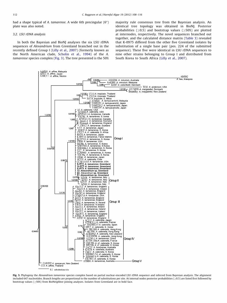
3.2. LSU rDNA analysis
In both the Bayesian and BioNJ analyses the six LSU rDNAsequences of Alexandrium from Greenland branched out in therecently defined Group I (Lilly et al., 2007) (formerly known asthe North American clade, Scholin et al., 1994) of the A.
tamarense species complex (Fig. 3). The tree presented is the 50%
Fig. 3. Phylogeny the Alexandrium tamarense species complex based on partial nuclear
included 647 nucleotides. Branch lengths are proportional to the number of substitution
bootstrap values (�50%) from BioNeighbor-joining analyses. Isolates from Greenland a
majority rule consensus tree from the Bayesian analysis. Anidentical tree topology was obtained in BioNJ. Posteriorprobabilities (�0.5) and bootstrap values (�50%) are plottedat internodes, respectively. The novel sequences branched outtogether, and the calculated distance matrix (Table 3) revealedthat K-0975 differed from the other five Greenland isolates bysubstitution of a single base pair (pos. 224 of the submittedsequence). These five were identical in LSU rDNA sequences tonine other strains belonging to Group I and distributed fromSouth Korea to South Africa (Lilly et al., 2007).
-encoded LSU rDNA sequence and inferred from Bayesian analysis. The alignment
s per site. At internal nodes posterior probabilities (�0.5) are listed first followed by
re in bold face.
Table 3Absolute distance matrix of 647 base pairs from the domain D1 to D2 of the LSU
rDNA gene. Numbers indicate that K-0975 has 1 base pair substitution compared to
the other five isolates.
K-0973 K-0975 K-0974 A1 D2 E1
K-0973 –
K-0975 1 –
K-0974 0 1 –
A1 0 1 0 –
D2 0 1 0 0 –
E1 0 1 0 0 0 –
C. Baggesen et al. / Harmful Algae 19 (2012) 108–116 113
3.3. Toxin composition
All A. tamarense isolates analyzed from Greenland containedsaxitoxin or derivatives thereof (Fig. 4, Table 4) and werecharacterized by high percentages of the gonyautoxins GTX1/GTX4. Although the epimers GTX1 and GTX4, and GTX2 and GTX3,were analytically separated, they are presented (Fig. 4) as epimericpairs due to facile interconversion resulting from thermodynamicequilibrium. The isolates K-0973, K-0974, and K-0975 exhibited asimilar toxin profile (but not virtually identical molar composition)composed of high GTX1/GTX4 (>60 mol%), with lesser proportionsof GTX2/GTX3, neosaxitoxin (NEO) and STX (Fig. 4). No trace ofeither N-sulfocarbamoyl (B1/2, C1–C4) or decarbamoyl (dcSTX,dcNEO, dcGTX1–dcGTX4) toxins were detected in these isolates.All isolates were analyzed separately at least twice fromexponentially growing cultures because in the first round thenumber of cells extracted was not reliably counted and thus onlythe molar percentage of the toxins was obtained. Isolate K-0974was analyzed more thoroughly than the others as STX and NEOwere close to the detection limit. After increasing the number ofextracted cells for this isolate, NEO was detected again, whereasSTX was not. The cell toxicity of the three isolates, calculated asSTXeq cell�1 according to toxicity factors given in Oshima (1995),ranged from 10.3 to 16.8 pg STXeq cell�1.
The identification of PSP toxins in our isolates of A. tamarense
from Greenland based on LC-FD (i.e. Fig. 4 and Table 4) wasconfirmed unambiguously by liquid chromatography with tandemmass spectrometry (Krock et al., 2007). The two methods revealedquantitative differences in PSP toxin content per cell among the
Fig. 4. Toxin composition of Alexandrium tamarense from the west coast of Greenla
isolates and LC-FD and LC–MS/MS independently verified thepresence of the principal toxins GTX4, GTX1, GTX3 and NEO.
4. Discussion
Based on the overall morphological characteristics and platetabulations of the three isolates from Greenland they clearlybelong to the A. tamarense morphotype. The ventral pore on thefirst apical (10) plate is also present in A. minutum but the shape ofthe sixth precingular (600) as well as the sa plate are tamarensoid.Although the general size and shape of the cells are more similar toA. tamarense than A. minutum these characters are variable (Balech,1995) and thus can only be used as a first guide. Length (l) andwidth (w) as well as the l/w ratio were quite stable within theisolates but one isolate (K-0973) produced somewhat larger cells(Table 2). The species A. ostenfeldii often found in North Atlantic,North Sea and Scandinavian coastal waters is ruled out as anaffiliation for any of the Greenland isolates by the absence of thecharacteristic large globose cell shape and the lack of a largekidney-shaped ventral pore at the margin of the 10 plate.
Large differences in size may be attributed to different stages inthe life cycle – vegetative cells, gametes, planozygotes (Balech,1995). However, the size variation within the strains is quite low,indicating that the cultures likely consist almost exclusively ofvegetative cells.
The Alexandrium isolates from Greenland examined by molec-ular phylogenetic characteristics in this investigation all emergedin a clade previously known as the North American clade (Scholinet al., 1994), as part of the A. tamarense species complex within thenewly defined Group 1 (Lilly et al., 2007), for lack of a better term.The known distribution of the strains in this group extends fromthe northeast Atlantic westward around the Americas to thenorthwest Pacific. Not surprisingly the strains from Greenlandbelong to this group, and we now confirm that toxigenic membersof this clade occur in arctic waters. Whether the species is a newarrival in the Arctic, either due to natural or human mediateddispersal, or if the late discovery reflects a paucity of observationsis unknown, but we are not aware of A. tamarense having beenobserved previously in Greenland waters.
Numerous previous investigations of PSP toxin variation amongAlexandrium species and populations (reviewed by Anderson et al.,
nd expressed as mol% of total toxins. Error bars represent standard deviations.
Table 4PSP toxin concentration and composition of Alexandrium tamarense cultures from Greenland determined by LC-FD. Numbers in brackets are standard deviations.
a Combined epimer pairs are: GTX1 + GTX4, GTX2 + GTX3.b Data from Cembella et al. (1987).c n = 4.d n = 6.e n = 7.f n = 9.
C. Baggesen et al. / Harmful Algae 19 (2012) 108–116114
1994; Cembella, 1998; Alpermann et al., 2010) have indicated thattoxin profiles are genetically determined and stable enough(within limits of physiological variation under defined conditions)to serve as a phenotypic marker. The fact that Greenland isolate K-0974 produces more than 98 mol% of 1-N-hydroxy (R1 = –OH)toxins may also be helpful for elucidation of the biosyntheticpathway of these toxins. However, the toxin profiles of theGreenland isolates are rather unusual and atypical for A. tamarense.One unusual feature is the complete absence of N-sulfocarbamoylC1/C2 or B1/B2 toxins, which are usually present in most strains ofthe A. tamarense species complex, often in a high molar percentage(Cembella et al., 1987; Anderson et al., 1994; Persich et al., 2006;Krock et al., 2007; Orlova et al., 2007). The high molar percentage ofGTX1/GTX4 toxins (>60 mol%) and lesser amounts of onlycarbamoyl toxins, including GTX2/3, NEO or STX, plus the absenceof decarbamoyl derivatives are more typical of strains of A.
minutum (Franco et al., 1994; Hwang and Lu, 2000; Carreto et al.,2001; Hansen et al., 2003; Chou et al., 2004; Pitcher et al., 2007).The toxin profile of K-0974 with the almost exclusive production ofGTX1/GTX4 (>95 mol%) is similar to that reported from strainsNEPCC 253 from Laguna Obidos, Portugal and NEPCC 508 fromWhangarei, North Island, New Zealand and originally assigned tothe NEP Culture Collection as members of the A. tamarense speciescomplex (see Table 4) (Cembella et al., 1987). One small differenceis the detection of NEO in K-0974, whereas this component isabsent from the Portuguese and New Zealand isolates.
No LSU rDNA sequences or other molecular markers are availablefor these latter strains, but it is unlikely that they are closely relatedto K-0974. In any case, subsequent careful morphological analysis ofthecal plates of NEPCC 253 and NEPCC 508 (A. Cembella,unpublished observations) indicate that both strains belong tothe A. minutum sub-group. NEPCC 508 accords best with thedescription of A. angustitabulatum (unusually narrow 600 plate).
To our knowledge, previous molecular data on Alexandrium
phylogenetic affiliations from high latitude oceans are limited to
a single strain of A. tamarense (Group 1) of unknown toxicityfrom the Faroe Islands (Lilly et al., 2007). We show here thatAlexandrium from Greenland are toxic and provisionallyconclude that A. tamarense is likely the primary contributor toPSP toxicity in scallops in the Attu area. Alexandrium ostenfeldii,another potential PSP toxin producer, was also found in the area(Ø. Moestrup, personal observation), but, with the exception ofthe Baltic Sea, in northern Europe this species has never beenknown to produce dense blooms. Furthermore, isolated strainsfrom the North Sea and North Atlantic tend to produce themacrocyclic imine toxins spirolides and only little (if any) PSPtoxins (MacKinnon et al., 2006). Alexandrium minutum ofunknown toxicity has been found in the Disko Bay area furthernorth (Jensen and Veland, 2006) and although not seen in theAttu area it could be present cryptically and contribute to PSPtoxicity in scallops.
Concerns have been expressed that rising global temperaturescould lead to a northward range extension and/or increase inendemic HABs in arctic areas. This could include blooms ofAlexandrium spp. along the Greenland coast. We noted that theGreenland isolates grew very slowly in culture when incubated at4 8C, approximately the ambient sea temperature of their naturalhabitat, but shifted up growth rates dramatically at highertemperatures (i.e. 10 8C). Since PSP toxin cell quota is generallypositively correlated with growth rate in Alexandrium spp.(reviewed in Cembella, 1998), any major rise in sea temperatureoffers the possibility of both higher magnitude toxic blooms andincreased cell potency. Under present circumstances in Greenland,this also provokes the question of how the current Alexandrium
populations generate enough toxins to cause toxicity in thescallops, even in some cases beyond the regulatory limit. We arenot certain that under ambient nutrient and light regimes (e.g.,long day length in summer) in nature, that the low growth rates weachieved in culture at low temperatures are representative.Furthermore, under low temperatures the reduced metabolic
C. Baggesen et al. / Harmful Algae 19 (2012) 108–116 115
rates in bivalve molluscs would be expected to cause scallopsto retain the toxins for longer periods (Bricelj and Shumway,1998).
5. Conclusions
The LSU sequences clearly place the isolates from Greenlandof A. tamarense within Group 1 of the A. tamarense speciescomplex as defined by Lilly et al. (2007). One of the sixsequences differed from the others by a single substitution,indicating one large homogeneous population of A. tamarense
along the west coast of Greenland. Further genetic assays,microsatellite or amplified fragment length polymorphisms(AFLP), will be helpful in elucidating further the populationstructure of the A. tamarense species complex from Greenland.The toxin profiles of the three cultured strains, with large molarpercentages of GTX1/GTX4, are closer to the toxin profile of A.
minutum than to that of members of the A. tamarense speciescomplex. The latter group is usually characterized by a highpercentage of N-sulfocarbamoyl (C1/C2) toxins. Additionalstrains of the A. tamarense species complex from Greenland aswell as other areas in the Arctic should be established todetermine whether the unusual toxin profiles are a commonfeature of arctic strains or if they represent a local or regionalanomaly. Natural blooms of members of the Group I clade of A.
tamarense, represented by the three strains established here,must be considered as the most likely agents for PSP toxinaccumulation in the scallops from western Greenland, but notoxin profiles are available from the contaminated bivalves orother putatively toxic Alexandrium species from this region.Therefore, an effort should be made to obtain A. ostenfeldii and A.
minutum in culture, as these species have also been observedalong the west coast of Greenland.
Acknowledgements
We thank the ATI School in Maniitsoq for arranging the visit ofØM to Greenland and the sampling cruise. Helle Siegstad,(Greenland Institute of Natural Resources, Nuuk) and Bjarne RingThorbjørnsen (Danish Food and Veterinary Administration, Viborg)generously provided access to data. Lene Christiansen (Depart-ment of Biology) provided laboratory assistance and AnnegretMuller (AWI, Bremerhaven) performed toxin chromatographicanalysis.[SS]
References
Alpermann, T.J., Tillmann, U., Beszteri, B., Cembella, A.D., John, U., 2010. Phenotypicvariation and genotypic diversity in a planktonic population of the toxigenicmarine dinoflagellate Alexandrium tamarense (Dinophyceae). Journal of Phycol-ogy 46, 18–32.
Anderson, D.M., Kulis, D.M., Doucette, G.J., Gallagher, J.C., Balech, E.T., 1994. Bioge-ography of toxic dinoflagellates in the genus Alexandrium from the northeasternUnited States and Canada. Marine Biology 120, 467–478.
Anderson, D.M., Alpermann, T.J., Cembella, A.D., Collos, Y., Masseret, E., Montresor,M., 2012. The globally distributed genus Alexandrium: multifaceted roles inmarine ecosystems and impacts on human health. Harmful Algae 14, 10–35.
Anonymous, 2003. Udenrigshandel 2002. Grønlands Statistik.Anonymous, 2004. Status for kammusling ved Vestgrønland 2003. Grønlands
occurrence, transfer kinetics, and biotransformation. Reviews in FisheriesScience 6, 315–383.
Balech, E., 1995. The genus Alexandrium Halim (Dinoflagellata). Sherkin IslandMarine Station, Sherkin Island, Ireland, 151 pp.
Carreto, J.I., Carignan, M.O., Montoya, N.G., 2001. Comparative studies on mycos-porine-like amino acids, paralytic shellfish toxins and, pigment profiles of thetoxic dinoflagellates Alexandrium tamarense, A. catenella and A. minutum. MarineEcology – Progress Series 223, 49–60.
Cembella, A.D., 1998. Ecophysiology and metabolism of paralytic shellfish toxins inmarine microalgae. In: Anderson, D.M., Cembella, A.D., Hallegraeff, G.M. (Eds.),
Physiological Ecology of Harmful Algal Blooms, vol. 41. NATO-Advanced StudyInstitute Series, Springer-Verlag, Heidelberg, pp. 381–404.
Cembella, A.D., Taylor, F.J.R., Therriault, J.C., 1988. Cladistic analysis of electropho-retic variants within the toxic dinoflagellate genus Protogonyaulax. BotanicaMarina 31, 39–51.
Cembella, A.D., Sullivan, J.J., Boyer, G.L., Taylor, F.J.R., Andersen, R.J., 1987. Variationin paralytic shellfish toxin composition within the Protogonyaulax tamarensis/catenella species complex; red tide dinoflagellates. Biochemical Systematicsand Ecology 15, 171–186.
Cembella, A.D., Quilliam, M.A., Lewis, N.I., Bauder, A.G., Dell’ Aversano, C., Thomas,K., Carver, C., Jellett, J., Cusack, R.R., 2002. The toxigenic marine dinoflagellateAlexandrium tamarense as the probable cause of enhanced mortality of cagedsalmon in Nova Scotia. Harmful Algae 1, 313–325.
Chou, H.N., Chen, Y.M., Chen, C.Y., 2004. Variety of PSP toxins in four culture strainsof Alexandrium minutum collected from southern Taiwan. Toxicon 43, 337–340.
Clark, R.F., Williams, S.R., Nordt, S.P., Manoguerra, A.S., 1999. A review of selectedseafood poisonings. Undersea and Hyperbaric Medicine 26, 175–184.
Daugbjerg, N., Hansen, G., Larsen, J., Moestrup, Ø., 2000. Phylogeny of some of themajor genera of dinoflagellates based on ultrastructure and partial LSU rDNAsequence data, including the erection of three new genera of unarmoureddinoflagellates. Phycologia 39, 302–317.
Diener, M., Erler, K., Hiller, S., Christian, B., Luckas, B., 2006. Determination ofparalytic shellfish poisoning (PSP) toxins in dietary supplements by applicationof a new HPLC/FD method. European Food Research and Technology 224,147–151.
Diener, M., Erler, K., Christian, B., Luckas, B., 2007. Application of a new zwitterionichydrophilic interaction chromatography column for determination of paralyticshellfish poisoning toxins. Journal of Separation Science 30, 1821–1826.
Franco, J.M., Fernandez, P., Reguera, B., 1994. Toxin profiles of natural populationsand cultures of Alexandrium minutum Halim from Galician (Spain) coastalwaters. Journal of Applied Phycology 6, 275–279.
Garcia, E.G., 2006. The fishery for Iceland scallop (Chlamys islandica) in the North-east Atlantic. Advances in Marine Biology 51, 1–55.
Gascuel, O., 1997. BIONJ: An improved version of the NJ algorithm based on a simplemodel of sequence data. Molecular Biology and Evolution 14, 685–695.
Hall, T.A., 1999. BioEdit: a user-friendly biological sequence alignment editor andanalysis program for Windows 95/98/NT. Nucleic Acids Symposium Series 41,95–98.
Hallegraeff, G.M., 1993. A review of harmful algal blooms and their apparent globalincrease. Phycologia 32, 79–99.
Hansen, G., Daugbjerg, N., Franco, J.M., 2003. Morphology, toxin composition andLSU rDNA phylogeny of Alexandrium minutum (Dinophyceae) from Denmark,with some morphological observations on other European strains. HarmfulAlgae 2, 317–335.
Hwang, D.F., Lu, Y.H., 2000. Influence of environmental and nutritional factors ongrowth, toxicity, and toxin profile of dinoflagellate Alexandrium minutum.Toxicon 38, 1491–1503.
Jensen, M.H., Veland, I.R., 2006. A survey of marine dinoflagellates in the waterssurrounding Disko Island, Western Greenland. In: Arctic Biology Field Course,Arctic Station, University of Copenhagen, Qeqertarsuaq, pp. 5–85.
Kao, C.Y., Walker, S.E., 1982. Active groups of saxitoxin and tetrodotoxin as deducedfrom actions of saxitoxin analogs on frog muscle and squid axon. Journal ofPhysiology (London) 323, 619–637.
Kofoid, C.A., 1909. On Peridinium steini Jørgensen, with a note on the nomenclatureof the skeleton of the Peridinidae. Archiv Fur Protistenkunde 16, 25–47.
Krock, B., Seguel, C.G., Cembella, A.D., 2007. Toxin profile of Alexandrium catenellafrom the Chilean coast as determined by liquid chromatography with fluores-cence detection and liquid chromatography coupled with tandem mass spec-trometry. Harmful Algae 6, 734–744.
Larsen, N.H., Moestrup, Ø., Pedersen, P.M., 1994. Scandinavian Culture Centre forAlgae and Protozoa. 1994 Catalogue. Botanical Institute, University of Copen-hagen, Copenhagen, 51 pp.
Lilly, E.L., Halanych, K.M., Anderson, D.M., 2007. Species boundaries and globalbiogeography of the Alexandrium tamarense complex (Dinophyceae). Journal ofPhycology 43, 1329–1338.
Nunn, G.B., Theisen, B.F., Christensen, B., Arctander, P., 1996. Simplicity-correlatedsize growth of the nuclear 28S ribosomal RNA D3 expansion segment in thecrustacean order Isopoda. Journal of Molecular Evolution 42, 211–223.
Scholin, C.A., Herzog, M., Sogin, M., Anderson, D.M., 1994. Identification of group-and strain-specific genetic markers for globally distributed Alexandrium (Dino-phyceae). 2. Sequence analysis of a fragment of the LSU rRNA gene. Journal ofPhycology 30, 999–1011.
Sebastian, C.R., Etheridge, S.M., Cook, P.A., O’Ryan, C., Pitcher, G.C., 2005. Phyloge-netic analysis of toxic Alexandrium (Dinophyceae) isolates from South Africa:implications for the global phylogeography of the Alexandrium tamarensespecies complex. Phycologia 44, 49–60.
Steidinger, K.A., Tangen, K., 1997. Dinoflagellates. In: Thomas, C.R. (Ed.),Identifying Marine Phytoplankton. Academic Press, New York, pp. 387–584.
Tamura, K., Nei, M., 1993. Estimation of the number of nucleotide substitutions inthe control region of mitochondrial DNA in humans and chimpanzees. Molec-ular Biology and Evolution 10, 512–526.
Thompson, J.D., Higgins, D.G., Gibson, T.J., 1994. Clustal-W – improving the sensi-tivity of progressive multiple sequence alignment through sequence weighting,position specific gap penalties and weight matrix choice. Nucleic Acids Re-search 22, 4673–4680.