10996 Abstract. – OBJECTIVE: Aortic valve steno- sis (AS) presents a disease during which there are changes of the aortic valve structure that modi- fy the blood structure of patients. The aim of this study was to improve the effectiveness of differ- ential diagnostics of aortic stenosis and aortic re- gurgitation using molecular techniques on both mRNA (RT-PCR) and protein (biochip protein). PATIENTS AND METHODS: An experimental group (n = 58) consisting of patients with aortic valve stenosis (n = 26) and aortic regurgitation (AR, n = 32) was compared with a control group (n = 35). Both blood serum and valve tissue sam- ples were used for the determination of gene ex- pression specific genes related to inflammatory response (CRP, IL6, IL2R, IL6R, TNFR1, and 2) as well as genes and proteins involved in remodeling of the extracellular matrix (MMP9, TIMP, Emilin-1). RESULTS: We found that hsCRP and IL6 plas- ma levels of patients with AS were higher than both controls and patients with AR (mean 5.6 ng/ml). The differences between AS and AR were detected only in mRNA levels of MMP9 and TIMP where increased levels characteristic for AS were found (about 74%, p < 0.01 and 87%, p < 0.001 higher than AR). CONCLUSIONS: The achieved results could contribute to the improvement of early diagno- sis of selected cardiovascular disease in the fu- ture and improve the quality of patient’s life. Key Words: Aortic valve stenosis, Blood serum, Aortic tissue, Gene expression, RT-PCR. Introduction Aortic valve stenosis (AS) is the most common form of adult heart valve disease 1 , that is spe- cific by progressive aortic valve narrowing and secondary hypertrophy 2 . The earliest pathophys- iological stages are characterized by endothelial damage, lipid deposition, and inflammation. All that results in leaflet stiffening reduced sepa- ration and valve narrowing 3 . The biochemical mechanisms, such as inflammation, lipid infiltra- tion, extracellular matrix remodeling, and finally calcification in the wall of the aortic valve are ac- tivated during the aortic valve stenosis formation and progression 4 . The inflammatory process begins with disrup- tion of valve endothelium, followed by infiltra- tion of inflammatory cells, mainly macrophages and T-lymphocytes 5 . Apolipoproteins increase oxidized low density lipoproteins (LDL) level during the inflammatory process 6 . C-reactive protein (CRP), one of the basic inflammatory markers, occurs in the extracellular matrix of the damaged aortic valve 7 . Tumor necrosis factor-α (TNF-α) and interleukin-6 (IL6) have also been found in the extracellular matrix of the aortic valve, predominantly in areas of leukocytes and macrophage infiltration. This pro-inflammatory cytokine is secreted by activated macrophages during a bacterial infection and the extracellular matrix remodeling 8 . It is responsible for immune regulation, inflammation, and tissue remodeling 9 . The extracellular matrix remodeling of the aortic valve is also caused by the increased pro- duction of the matrix metalloproteinases (mainly MMP-2 and MMP9). These specific enzymes degrade collagen and elastin fibers 10 . The degradation of MMPs by endogenous tis- sue inhibitors of MMPs (TIMPs) decreases the European Review for Medical and Pharmacological Sciences 2019; 23: 10996-11003 P. URBAN 1 , M. RABAJDOVÁ 1 , I. ŠPAKOVÁ 1 , F. SABOL 2 , H. MIČKOVÁ 3 , K. LAKATOSOVÁ 1 , M. ZAVACKÁ 4 1 Department of Medical and Clinical Biochemistry, Pavol Jozef Šafárik University in Košice, Faculty of Medicine, Košice, Slovak Republic 2 Department of Cardiovascular Surgery, Pavol Jozef Šafárik University in Košice and VUSCH, Faculty of Medicine, Košice, Slovak Republic 3 Department of Medical Biology, Pavol Jozef Šafárik University in Košice, Faculty of Medicine, Košice, Slovak Republic 4 Department of Vascular Surgery, Pavol Jozef Šafárik University in Košice and VUSCH, Faculty of Medicine, Košice, Slovak Republic Corresponding Author: Martina Zavacká, MD; e-mail: [email protected]Molecular recognition of aortic valve stenosis and regurgitation
Transcript
10996
Abstract. – OBJECTIVE: Aortic valve steno-sis (AS) presents a disease during which there are changes of the aortic valve structure that modi-fy the blood structure of patients. The aim of this study was to improve the effectiveness of differ-ential diagnostics of aortic stenosis and aortic re-gurgitation using molecular techniques on both mRNA (RT-PCR) and protein (biochip protein).
PATIENTS AND METHODS: An experimental group (n = 58) consisting of patients with aortic valve stenosis (n = 26) and aortic regurgitation (AR, n = 32) was compared with a control group (n = 35). Both blood serum and valve tissue sam-ples were used for the determination of gene ex-pression specific genes related to inflammatory response (CRP, IL6, IL2R, IL6R, TNFR1, and 2) as well as genes and proteins involved in remodeling of the extracellular matrix (MMP9, TIMP, Emilin-1).
RESULTS: We found that hsCRP and IL6 plas-ma levels of patients with AS were higher than both controls and patients with AR (mean 5.6 ng/ml). The differences between AS and AR were detected only in mRNA levels of MMP9 and TIMP where increased levels characteristic for AS were found (about 74%, p < 0.01 and 87%, p < 0.001 higher than AR).
CONCLUSIONS: The achieved results could contribute to the improvement of early diagno-sis of selected cardiovascular disease in the fu-ture and improve the quality of patient’s life.Key Words:
Aortic valve stenosis (AS) is the most common form of adult heart valve disease1, that is spe-
cific by progressive aortic valve narrowing and secondary hypertrophy2. The earliest pathophys-iological stages are characterized by endothelial damage, lipid deposition, and inflammation. All that results in leaflet stiffening reduced sepa-ration and valve narrowing3. The biochemical mechanisms, such as inflammation, lipid infiltra-tion, extracellular matrix remodeling, and finally calcification in the wall of the aortic valve are ac-tivated during the aortic valve stenosis formation and progression4.
The inflammatory process begins with disrup-tion of valve endothelium, followed by infiltra-tion of inflammatory cells, mainly macrophages and T-lymphocytes5. Apolipoproteins increase oxidized low density lipoproteins (LDL) level during the inflammatory process6. C-reactive protein (CRP), one of the basic inflammatory markers, occurs in the extracellular matrix of the damaged aortic valve7. Tumor necrosis factor-α (TNF-α) and interleukin-6 (IL6) have also been found in the extracellular matrix of the aortic valve, predominantly in areas of leukocytes and macrophage infiltration. This pro-inflammatory cytokine is secreted by activated macrophages during a bacterial infection and the extracellular matrix remodeling8. It is responsible for immune regulation, inflammation, and tissue remodeling9.
The extracellular matrix remodeling of the aortic valve is also caused by the increased pro-duction of the matrix metalloproteinases (mainly MMP-2 and MMP9). These specific enzymes degrade collagen and elastin fibers10.
The degradation of MMPs by endogenous tis-sue inhibitors of MMPs (TIMPs) decreases the
European Review for Medical and Pharmacological Sciences 2019; 23: 10996-11003
P. URBAN1, M. RABAJDOVÁ1, I. ŠPAKOVÁ1, F. SABOL2, H. MIČKOVÁ3, K. LAKATOSOVÁ1, M. ZAVACKÁ4
1Department of Medical and Clinical Biochemistry, Pavol Jozef Šafárik University in Košice, Faculty of Medicine, Košice, Slovak Republic 2Department of Cardiovascular Surgery, Pavol Jozef Šafárik University in Košice and VUSCH, Faculty of Medicine, Košice, Slovak Republic3Department of Medical Biology, Pavol Jozef Šafárik University in Košice, Faculty of Medicine, Košice, Slovak Republic4Department of Vascular Surgery, Pavol Jozef Šafárik University in Košice and VUSCH, Faculty of Medicine, Košice, Slovak Republic
Corresponding Author: Martina Zavacká, MD; e-mail: [email protected]
Molecular recognition of aortic valve stenosis and regurgitation
Molecular recognition of aortic valve stenosis and regurgitation
10997
proteolysis of the extracellular matrix11. The im-balance between activities of MMPs and TIMPs leads to the destruction of the tissue structure. Biochemical processes then initiate inflammation and the formation of diseases on molecular level, such as abdominal aortic aneurysm, hyperten-sion, valvular diseases12.
Aortic regurgitation (AR) is characterized by regurgitation of blood from the aorta to the left ventricle (LV) during diastole. It is attributable to diverse congenital and acquired abnormalities of the aortic valve or the wall of the aortic root13. Acute severe AR may be difficult to recognize clinically and is often erroneously diagnosed as another acute condition, such as sepsis, pneumo-nia, or nonvalvular heart disease14.
AS and AR are a heterogeneous diseases with a complex pathophysiology involving the aortic valve and interconnected cardiac structures. For careful clinical assessment of AS or AR occur-rence, echocardiography that is currently used has functional limitations for stratification of risk patients with asymptomatic progression15.
The aim of this study was to improve the effec-tiveness of differential diagnostics of AS and AR using molecular technics (RT-PCR) and biochip protein analysis. A prompt and accurate diagno-sis of acute AS or AR is of great importance, as urgent or emergent aortic valve surgery could be life-saving.
Patients and Methods
Experimental DesignThe control group consists of healthy blood
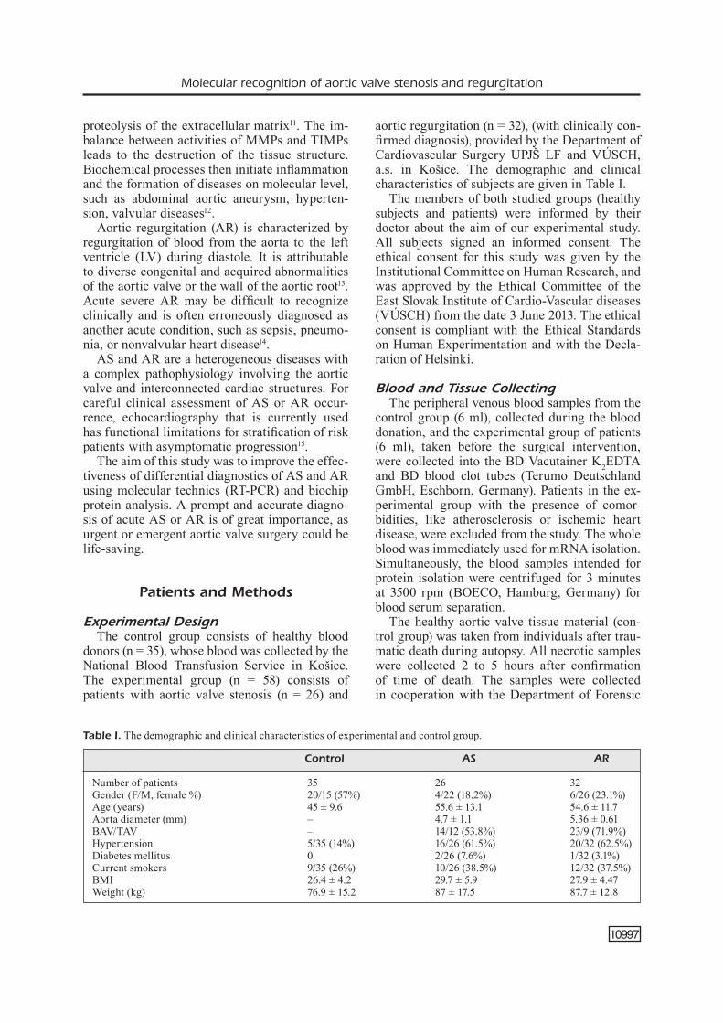
donors (n = 35), whose blood was collected by the National Blood Transfusion Service in Košice. The experimental group (n = 58) consists of patients with aortic valve stenosis (n = 26) and
aortic regurgitation (n = 32), (with clinically con-firmed diagnosis), provided by the Department of Cardiovascular Surgery UPJŠ LF and VÚSCH, a.s. in Košice. The demographic and clinical characteristics of subjects are given in Table I.
The members of both studied groups (healthy subjects and patients) were informed by their doctor about the aim of our experimental study. All subjects signed an informed consent. The ethical consent for this study was given by the Institutional Committee on Human Research, and was approved by the Ethical Committee of the East Slovak Institute of Cardio-Vascular diseases (VÚSCH) from the date 3 June 2013. The ethical consent is compliant with the Ethical Standards on Human Experimentation and with the Decla-ration of Helsinki.
Blood and Tissue CollectingThe peripheral venous blood samples from the
control group (6 ml), collected during the blood donation, and the experimental group of patients (6 ml), taken before the surgical intervention, were collected into the BD Vacutainer K2EDTA and BD blood clot tubes (Terumo Deutschland GmbH, Eschborn, Germany). Patients in the ex-perimental group with the presence of comor-bidities, like atherosclerosis or ischemic heart disease, were excluded from the study. The whole blood was immediately used for mRNA isolation. Simultaneously, the blood samples intended for protein isolation were centrifuged for 3 minutes at 3500 rpm (BOECO, Hamburg, Germany) for blood serum separation.
The healthy aortic valve tissue material (con-trol group) was taken from individuals after trau-matic death during autopsy. All necrotic samples were collected 2 to 5 hours after confirmation of time of death. The samples were collected in cooperation with the Department of Forensic
Table I. The demographic and clinical characteristics of experimental and control group.
P. Urban, M. Rabajdová, I. Špaková, F. Sabol, H. Mičková, K. Lakatosová, M. Zavacká
10998
Medicine UPJŠ LF. The cause of death was not related to cardio-vascular diseases. In all the relevant cases, no histopathologically significant change in myocardium had been reported (n = 10). Autopsy protocols were registered in all ten cases between years 2014 to 2016. For example, one case was a 40-year-old woman that died by falling on the sidewalk with subsequent aspira-tion of gastric content. In another case, the death of a 45-year-old woman was caused by drowning during delusional deterioration. Another case was a still-born child, due to the asphyxiation of the umbilical cord. Other cases were two 28-year-old men who died after falling from a motorcycle. The tissue samples from the patients of the ex-perimental group were collected during surgical remodeling of aorta or during the aortic valve replacement (AVR) surgery with a mechanical or biological prosthesis with or without replacement of the ascendant aorta. Other surgical interven-tions were mitral valve replacement (MVR) and off pump coronary revascularization (OPCAB). Surgery was done through median sternotomy with mild hypothermia using extracorporeal cir-culation. The cannulations were made in the as-cendant aorta and in the right atrium of the heart, and the cold blood cardioplegia was also used. Cardioanesthesia was performed by standard op-erating procedures. The tissue taken from both groups was frozen in liquid nitrogen. Subsequent-ly, all collected samples were stored in a freezer at temperature -71°C (New Brunswick Scientific, Enfield, CT, USA).
Isolation of RNA and RT-PCRThe commercial RNA isolation kit (RNeasy
Mini Kit, Qiagen, Hilden, Germany) was used for RNA isolation from blood and tissue samples. The isolated RNA was transcribed into cDNA by using kit RevertAid H minus First strand
cDNA Synthesis kit (Thermo Fisher Scientific, Waltham, MA, USA). The RT-PCR method was used to detect the changes in mRNA expres-sion levels of the selected specific genes related to inflammation (hsCRP, IL6), and degradation of extracellular matrix (Emilin-1, MMP9, and TIMP). Amplification of the specific genes ran for 35 cycles (94°C 5 min, 95°C 10 s and 58-62°C 20 s and 72°C 25 s), using appropriate specific primer sequences (Table II) in the thermocy-cler Rotor Gene Q-PCR thermocycler (Qiagene, Hilden, Germany).
Normalization of the results was performed by using housekeeping genes Gapdh, HPRT, and ETNK. Numerical quantification of changes in the expression of mRNA levels was evaluated by the comparative quantification and Ct value Q Rotor Gene Software (Qiagen, Hilden, Germa-ny). The difference between ΔCt of the studied gene and the control gene was calculated, then, subtracted between ΔCt of sample with unknown value and ΔCt of the calibrator. The final result was a multiple of the calibrator value.
Protein Analysis by Randox Biochip For the detection of the protein levels in the se-
rum and tissue samples of both the experimental and control groups, a Cytokine Array IV assay kit was used in combination with a biochip ana-lyzer (Evidence Investigator, Randox Laborato-ries Ltd., London, UK). The detection of proteins CRP, IL6, IL6R, hsCRP, MMP9, TNFR1, and TNFR2 started with the incubation of a sample with 200 μl of assay buffer for 1 h/37°C/370 rpm of 100 μl. After incubation, the procedure continued by decantation of the liquid and the washing of each well 2 times. The second incu-bation, using the same conditions, continued after adding conjugation buffer. After the second in-cubation, another decantation of liquid was done
Table II. List of used primer sequences.
Name Length of gene Loci Forward sequence Reverse sequence (bp)
Molecular recognition of aortic valve stenosis and regurgitation
10999
and each well was washed 4 times. A mixture of luminol-EV-70l together with hydrogen peroxide was added to each well and incubated for 2 min-utes. Visualization and calculation of the proteins levels (ng/ml) of each biomarker was performed using Evidence Investigation biochip software version 4 (Evidence Investigator, Randox Labo-ratories Ltd., London, UK).
Statistical Analysis The categorical variables were analyzed using
Pearson Chi-squared test, and continuous vari-ables with normally distributed values were an-alyzed using a Student’s t-test, whereas non-nor-mally distributed continuous data were analyzed with a Mann-Whitney U-test for two indepen-dent samples and Kruskal-Wallis test for more than two independent samples. Statistical and Spearman correlation analysis was processed by the program GraphPad InSTAT (GraphPad, San Diego, CA, USA) and IBM SPSS Statistics 22.0 (IBM Corp., Armonk, NY, USA).
Results
Gene Expression Changes on mRNA Level
For determination of progressive valve inflam-mation and pathological remodeling of the valve
tissues during valve stenosis and regurgitation, the expression levels of specific genes (CRP, IL6, MMP9, Emilin-1, and TIMP) were detected from tissue using the RT-PCR method. Inflammatory marker CRP showed significantly increased val-ues in both AS and AR in comparison with con-trols (about 284%, p < 0.01 and 372%, p < 0.001 higher) (Figure 1).
The expression of IL6 also showed significant differences in AS and AR against control (about 438%, p < 0.001 and 390%, p < 0.01 higher). Both genes, however, didn’t show any significant difference suitable for specific recognition of AS from AR.
The differences between AS and AR were detected only in mRNA levels of MMP9 and TIMP, where increased levels characteristic for AS were found (about 74%, p < 0.01 and 87%, p < 0.001 higher than AR). An expression of Emilin-1 showed decreased levels in comparison with the control group in both AS and AR (about 123.7%, p < 0.01 and 73%, p < 0.001).
From the obtained results we can assume that MMP9/TIMP ratio is higher in the AS group (1.98, p < 0.028) in comparison with the AR group (1.60, p < 0.05) and, therefore, we suggest that an imbalance between MMP and TIMP expression is responsible for the shift toward a proteolytic state of ECM mostly in patients with AS.
Figure 1. Gene expression on mRNA levels in tissue. The mRNA levels of all detected genes were compared to controls (C, n = 10 autopsy samples). All data are presented as average ± SD, **p < 0.01, ***p < 0.001 means statistical significance against the control group. A red asterisk means significant difference between AS (n = 26) and AR (n = 32).
P. Urban, M. Rabajdová, I. Špaková, F. Sabol, H. Mičková, K. Lakatosová, M. Zavacká
11000
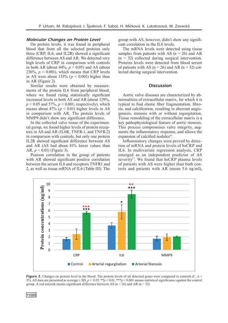
Molecular Changes on Protein LevelOn protein levels, it was found in peripheral
blood that from all the selected proteins only three (CRP, IL6, and IL2R) showed a significant difference between AS and AR. We detected very high levels of CRP in comparison with controls in both AR (about 44%, p > 0.05) and AS (about 236%, p < 0.001), which means that CRP levels in AS were about 133% (p < 0.001) higher than in AR (Figure 2).
Similar results were obtained by measure-ments of the protein IL6 from peripheral blood, where we found rising statistically significant increased levels in both AS and AR (about 129%, p < 0.05 and 57%, p < 0.001, respectively), which means about 47% (p < 0.01) higher levels in AS in comparison with AR. The protein levels of MMP9 didn’t show any significant difference.
In the collected valve tissue of the experimen-tal group, we found higher levels of protein recep-tors in AS and AR (IL6R, TNFR-1, and TNFR-2) in comparison with controls, but only one protein IL2R showed significant difference between AS and AR (AS had about 81% lower values than AR, p < 0.01) (Figure 3).
Pearson correlation in the group of patients with AR showed significant positive correlation between the serum IL6 and receptors TNFR1 and 2, as well as tissue mRNA of IL6 (Table III). The
group with AS, however, didn’t show any signifi-cant correlation in the IL6 levels.
The mRNA levels were detected using tissue samples from patients with AS (n = 26) and AR (n = 32) collected during surgical intervention. Proteins levels were detected from blood serum of patients with AS (n = 26) and AR (n = 32) col-lected during surgical intervention.
Discussion
Aortic valve diseases are characterized by ab-normalities of extracellular matrix, for which it is typical to find elastic fiber fragmentation, fibro-sis, and calcification, resulting in aberrant angio-genesis, stenosis with or without regurgitation. Tissue remodeling of the extracellular matrix is a key pathophysiological feature of aortic stenosis. This process compromises valve integrity, aug-ments the inflammatory response, and allows the expansion of calcified nodules16.
Inflammatory changes were proved by detec-tion of mRNA and protein levels of hsCRP and IL6. In multivariate regression analysis, CRP emerged as an independent predictor of AS severity17. We found that hsCRP plasma levels of patients with AS were higher than both con-trols and patients with AR (mean 5.6 ng/ml),
Figure 2. Changes on protein level in the blood. The protein levels of all detected genes were compared to controls (C, n = 35). All data are presented as average ± SD, p < 0.05, **p < 0.01, ***p < 0.001 means statistical significance against the control group. A red asterisk means significant difference between AS (n = 26) and AR (n = 32).
Molecular recognition of aortic valve stenosis and regurgitation
11001
which is supported by the results of Sharma et al18. They detected that plasmatic CRP concen-tration was significantly higher in patients with rapid AS progression (2.3 to 11.3 ng/ml). Pro-tein IL6 also showed increased levels than con-trol values in both groups AR and AS, specifi-cally higher in patients with AS in comparison with AR group (about 47%). High production of IL-6 is related to an osteogenic transition and mineralization of the aortic valve19. IL-6 also promotes endothelial-mesenchymal transition in the aortic valve20.
On the mRNA level, significant differenc-es were also detected in the gene expression of markers of extracellular matrix remodeling, like Emilin-1, MMP9, and TIMP. Emilin-1 is localized at the interface between elastin and microfibrils in the artery and undoubtedly op-erates to facilitate the function of elastin, which plays an important role in arterial structure
and function21. Emilin-1 regulates elastogenesis and inhibits TGF-β signaling. There are several studies, which deal with the role of Emilin-1 in aortic tissue of patients with cardiovascular diseases. Munjal et al21 in their study identi-fied Emilin-1 as extracellular matrix protein which is necessary for mature valve function and structure. Emilin-1 deficiency and the re-sulting elastic fiber assembly defects have been studied in the tissue of the aorta, showing that Emilin-1 deficiency causes TGF-β upreg-ulation22. We found that Emilin-1 expression is non-significantly lower in AS group than AR group (about 29%, p > 0.05). However, the mR-NA levels of MMP9 were significantly higher in the AS group in comparison with the AR group (about 74%, p < 0.01). Increased levels didn’t correlate with protein levels. MMP-9 is involved in tissue remodeling through the degradation of extracellular matrix substrates like collagen and
Figure 3. Changes on protein levels in the blood. The protein levels of all detected genes were compared to controls (C, n = 35). All data are presented as average ± SD, ** p < 0.01, ***p < 0.001 means statistical significance against the control group. A red asterisk means significant difference between AS (n = 26) and AR (n = 32).
Table III. Correlations of parameters in AR and AS with Pearson coefficients and p-values.
P. Urban, M. Rabajdová, I. Špaková, F. Sabol, H. Mičková, K. Lakatosová, M. Zavacká
11002
elastin23,24, but also involved in the conversion of cytokines and chemokines into active forms process proteins, like intercellular adhesion mol-ecule-1 (ICAM-1)25, and release proangiogenic factor VEGF-A26,27. TIMP-1 is an endogenous inhibitor of MMP-9, whose low physiological level is disturbed under pathologic conditions, which results in excessive matrix degradation with subsequent vascular growth28,29. The mR-NA levels of TIMP were significantly elevated in AS group in comparison with AR group of patients (about 87%, p < 0.001), which suggests the increase of extracellular matrix degradation during AR. Interleukin receptors on protein levels showed the difference between AS and AR only in the levels of IL2 receptor, where we found about 81% lower values than AR (p < 0.01). Until now no data existed comparing the soluble protein of IL2R concentrations and the difference between AS and AR. Spearman correlation showed positive correlation between mRNA for IL6 in the valve tissue and its protein in blood of patients with AR (p = 0.02), which can help improve differential diagnostics. There may be some possible limitations in this study. The usage of health aortic valve tissue material (control group), that was taken from individuals during the autopsy in two to five hours after the traumatic death still showed relevant viability proved by the protein levels of CRP and IL6 that were found in range for heathy tissue samples. However, the small number of collected control samples suggests that our data should be inter-preted with caution.
Conclusions
Cardiovascular diseases are one of the most common causes of death in the world. The study on the mechanism of formation of cardiovascular diseases presents motivation for application of new laboratory investigative methods. Finding new diagnostic biomarkers specific for aortic stenosis and regurgitation during early diagnosis can help not only in prevention but also during capture in the early stage of the disease and the subsequent successful treatment. This study described the differences in gene expression of inflammatory markers in blood and valve tissue on both mRNA and protein levels. We found that there are significant molecular changes specific for aortic stenosis that showed increased (IL6, CRP, MMP9, TIMP) or decreased (Emilin-1,
IL2R) expression levels than aortic regurgitation samples. The achieved results could contribute to the improvement of early diagnosis of selected cardiovascular disease in the future and improve the quality of patient’s life.
Conflict of InterestThe Authors declare that they have no conflict of interests.
AcknowledgementsThis work was supported by the Slovak Academy of Science and the Slovak Grant Agency for Science under the con-tracts VEGA 1/0873/16, Medipark II. ITMS 313011D103.
References
1) Benjamin ej, Virani SS, Callaway Cw, ChamBerlain am, Chang ar, Cheng S, ChiuVe Se, CuShman m, Delling Fn, Deo r, De Ferranti SD, FerguSon jF, For-nage m, gilleSpie C, iSaSi Cr, jiménez mC, jorDan lC, juDD Se, laCklanD D, liChtman jh, liSaBeth l, liu S, longeneCker Ct, lutSey pl, maCkey jS, matChar DB, matSuShita k, muSSolino me, naSir k, o’Fla-herty m, palaniappan lp, panDey a, panDey Dk, reeVeS mj, ritChey mD, roDriguez Cj, roth ga, roSamonD wD, SampSon uka, Satou gm, Shah Sh, Spartano nl, tirSChwell Dl, tSao Cw, VoekS jh, willey jz, wilkinS jt, wu jh, alger hm, wong SS, muntner p; ameriCan heart aSSoCiation CounCil on epiDemiol-ogy anD preVention StatiStiCS Committee anD Stroke StatiStiCS SuBCommittee. Heart disease and stroke statistics—2018 update: a report from the Amer-ican Heart Association. Circulation 2018; 137: e67-e492.
2) DweCk mr, Boon na, newBy De. Calcific aortic stenosis: a disease of the valve and the myocar-dium. J Am Coll Cardiol 2012; 60: 1854-1863.
3) yarBrough wm, mukherjee r, ikonomiDiS jS, zile mr, Spinale Fg. Myocardial remodeling with aortic ste-nosis and after aortic valve replacement: mecha-nisms and future prognostic implications. J Tho-rac Cardiov Surg 2012; 143: 656-664.
4) natarajan D, prenDergaSt B. Aortic stenosis--patho-genesis, prediction of progression, and percuta-neous intervention. J R Coll Physicians Edinb 2017; 47: 172-175.
5) Steiner i, krBal l, rozkoS t, harrer j, laCo j. Calcific aortic valve stenosis: Immunohistochemical anal-ysis of inflammatory infiltrate. Pathol Res Pract 2012; 208: 231-234.
6) FantuS D, awan z, SeiDah ng, geneSt j. Aortic cal-cification: novel insights from familial hypercho-lesterolemia and potential role for the low-densi-ty lipoprotein receptor. Atherosclerosis 2013; 226: 9-15.
Molecular recognition of aortic valve stenosis and regurgitation
11003
7) anzai t. Inflammatory mechanisms of cardiovas-cular remodeling. Circ J 2018; 82: 629-635.
8) Dahal S, huang p, murray Bt, mahler gj. Endo-thelial to mesenchymal transformation is induced by altered extracellular matrix in aortic valve en-dothelial cells. J Biomed Mater Res A 2017; 105: 2729-2741.
9) yu z, Seya k, Daitoku k, motomura S, FukuDa i, Fu-rukawa k. Tumor necrosis factor-α accelerates the calcification of human aortic valve interstitial cells obtained from patients with calcific aortic valve stenosis via the BMP2-Dlx5 pathway. J Pharma-col Exp Ther 2011; 337: 16-23.
10) olSzowSka m. Pathogenesis and pathophysiology of aortic valve stenosis in adults. Pol Arch Med Wewn 2011; 121: 409-413.
11) linDman Br, Bonow ro, otto Cm. Current man-agement of calcific aortic stenosis. Circ Res 2013; 113: 223-237.
12) wang X, khalil ra. Matrix metalloproteinases, vascular remodeling, and vascular disease. Adv Pharmacol 2018; 81: 241-330.
13) hamirani yS, Dietl Ca, VoyleS w, peralta m, Begay D, raizaDa V. Acute aortic regurgitation. Circula-tion 2012; 126: 1121-1126.
14) nakamura e, nakamura k, niina k, kazuShi k, iShii h, mD ai, kuBo h, Satoru i. Acute aortic regurgitation resulting from dehiscence of the aortic valve com-missures. Ann Thorac Cardiovasc Surg 2010; 16: 294-296.
15) iShii h, nakamura k, nagahama h, matSuyama m, en-Do g, niShimura m. Two dehiscences of the aor-tic valve commissure and cusp with progressive acute aortic regurgitation. Ann Vasc Dis 2015; 8: 43-45.
16) Chang ry, Chen CC, hSu wp, hSiao pC, tSai hl, hSiao pg, wu jD, guo hr. Nontraumatic avulsion of aortic valve commissure as a cause of acute aortic valve regurgitation: a case report. Medicine (Baltimore) 2016; 95: e5053.
17) Fertin m, DuBoiS e, BelliarD a, amouyel p, pinet F, BauterS C. Usefulness of circulating biomarkers for the prediction of left ventricular remodeling af-ter myocardial infarction. Am J Cardiol 2012; 110: 277-283.
18) Sharma g, Shetkar S, BhaSin a, ramakriShnan l, june-ja r, naik n, roy a, ramakriShnan S, BhargaVa B, Bahl Vk. High sensitive c-reactive protein and in-terleukin 6 in atrial fibrillation with rheumatic mi-tral stenosis from indian cohort. Indian Heart J 2017; 69: 505-511.
19) el huSSeini D, Boulanger mC, mahmut a, BouCha-reB r, laFlamme mh, Fournier D, piBarot p, BoSSe y,
mathieu p. P2Y2 receptor represses IL-6 expres-sion by valve interstitial cells through Akt: Impli-cation for calcific aortic valve disease. J Mol Cell Cardiol 2014; 72: 146-156.
20) mahler gj, Farrar ej, ButCher jt. Inflammatory cy-tokines promote mesenchymal transformation in embryonic and adult valve endothelial cells. Arte-rioscler Thromb Vasc Biol 2013; 33: 121-130.
21) munjal C, jegga ag, opoka am, StoiloV i, norriS ra, thomaS Cj, Smith jm, meCham rp, BreSSan gm, hinton rB. Inhibition of MAPK-Erk pathway in vivo attenuates aortic valve disease processes in Emi-lin1-deficient mouse model. Physiol Rep 2017; 5. pii: e13152.
22) zaVaCka m, FrankoViCoVa m, poBehoVa j, zaVaCky p. The impact of the angioplasty of the renal artery and cold ischemia time in kidney transplantation on graft function. Bratisl Lek Listy 2018; 119: 416-420.
23) ranDell a, DaneShtalaB n. Elastin microfibril inter-face-located protein 1, transforming growth factor beta, and implications on cardiovascular compli-cations. J Am Soc Hypertens 2017; 11: 437-448.
24) kapelouzou a, tSoureliS l, kaklamaniS l, DegianniS D, kogerakiS n, CokkinoS DV. Serum and tissue bio-markers in aortic stenosis. Glob Cardiol Sci Pract 2015; 2015: 49. doi: 10.5339/gcsp.2015.49. eCol-lection 2015.
25) Cauwe B, opDenakker g. Intracellular substrate cleavage: a novel dimension in the biochemistry, biology and pathology of matrix metalloprotein-ases. Crit Rev Biochem Mol Biol 2010; 45: 351-423.
26) ChriStoFFerSSon g, walDen t, SanDBerg m, opDenak-ker g, CarlSSon po, phillipSon m. Matrix metallo-proteinase-9 is essential for physiological be-ta cell function and islet vascularization in adult mice. Am J Pathol 2015; 185: 1094-1103.
27) wei t, zhang h, Cetin n, miller e, moak t, Suen jy, riChter gt. Elevated expression of matrix metallo-proteinase-9 not matrix metalloproteinase-2 con-tributes to progression of extracranial arteriove-nous malformation. Sci Rep 2016; 6: 24378.
28) hou XD, Ding F, wang Xk, liu Xg, yi k, zhang p, you t. Concomitant mitral valve replacement and tricuspid valvuloplasty for severe mitral steno-sis. Eur Rev Med Pharmacol Sci 2017; 21: 3436-3440.
29) aleSSanDri n, tuFano F, petraSSi m, aleSSanDri C, Di CriStoFano C, Della roCCa C, gallo p. Atrial fibril-lation in pure rheumatic mitral valvular disease is expression of an atrial histological change. Eur Rev Med Pharmacol Sci 2009; 13: 431-442.