METHODS published: 08 August 2017 doi: 10.3389/fphys.2017.00490 Frontiers in Physiology | www.frontiersin.org 1 August 2017 | Volume 8 | Article 490 Edited by: Graziano Fiorito, Stazione Zoologica Anton Dohrn, Italy Reviewed by: Radka Symonova, University of Innsbruck, Austria Mauro Mandrioli, University of Modena and Reggio Emilia, Italy Pamela Imperadore, Association for Cephalopod Research – CephRes, Italy *Correspondence: Ciro Rivera-Casas criverac@fiu.edu Rodrigo Gonzalez-Romero [email protected]Juan Ausio [email protected]Jose M. Eirin-Lopez jeirinlo@fiu.edu † These authors have contributed equally to this work. Specialty section: This article was submitted to Invertebrate Physiology, a section of the journal Frontiers in Physiology Received: 31 March 2017 Accepted: 26 June 2017 Published: 08 August 2017 Citation: Rivera-Casas C, Gonzalez-Romero R, Garduño RA, Cheema MS, Ausio J and Eirin-Lopez JM (2017) Molecular and Biochemical Methods Useful for the Epigenetic Characterization of Chromatin-Associated Proteins in Bivalve Molluscs. Front. Physiol. 8:490. doi: 10.3389/fphys.2017.00490 Molecular and Biochemical Methods Useful for the Epigenetic Characterization of Chromatin-Associated Proteins in Bivalve Molluscs Ciro Rivera-Casas 1 * † , Rodrigo Gonzalez-Romero 1 * † , Rafael A. Garduño 2 , Manjinder S. Cheema 3 , Juan Ausio 3 * † and Jose M. Eirin-Lopez 1 * † 1 Environmental Epigenetics Group, Department of Biological Sciences, Florida International University, North Miami, FL, United States, 2 Department of Microbiology and Immunology, Dalhousie University, Halifax, NS, Canada, 3 Department of Biochemistry and Microbiology, University of Victoria, Victoria, BC, Canada Bivalve molluscs constitute a ubiquitous taxonomic group playing key functions in virtually all ecosystems, and encompassing critical commercial relevance. Along with a sessile and filter-feeding lifestyle in most cases, these characteristics make bivalves model sentinel organisms routinely used for environmental monitoring studies in aquatic habitats. The study of epigenetic mechanisms linking environmental exposure and specific physiological responses (i.e., environmental epigenetics) stands out as a very innovative monitoring strategy, given the role of epigenetic modifications in acclimatization and adaptation. Furthermore, the heritable nature of many of those modifications constitutes a very promising avenue to explore the applicability of epigenetic conditioning and selection in management and restoration strategies. Chromatin provides a framework for the study of environmental epigenetic responses. Unfortunately, chromatin and epigenetic information are very limited in most non-traditional model organisms and even completely lacking in most environmentally and ecologically relevant organisms. The present work aims to provide a comprehensive and reproducible experimental workflow for the study of bivalve chromatin. First, a series of guidelines for the molecular isolation of genes encoding chromatin-associated proteins is provided, including information on primers suitable for conventional PCR, Rapid Amplification of cDNA Ends (RACE), genome walking and quantitative PCR (qPCR) experiments. This section is followed by the description of methods specifically developed for the analysis of histone and SNBP proteins in different bivalve tissues, including protein extraction, purification, separation and immunodetection. Lastly, information about available antibodies, their specificity and performance is also provided. The tools and protocols described here complement current epigenetic analyses (usually limited to DNA methylation) by incorporating the study of structural elements modulating chromatin dynamics. Keywords: epigenetics, bivalves, chromatin, histones, SNBPs, methods, environment
Transcript
METHODSpublished: 08 August 2017
doi: 10.3389/fphys.2017.00490
Frontiers in Physiology | www.frontiersin.org 1 August 2017 | Volume 8 | Article 490
Molecular and Biochemical MethodsUseful for the EpigeneticCharacterization ofChromatin-Associated Proteins inBivalve MolluscsCiro Rivera-Casas 1*†, Rodrigo Gonzalez-Romero 1*†, Rafael A. Garduño 2,
Manjinder S. Cheema 3, Juan Ausio 3*† and Jose M. Eirin-Lopez 1*†
1 Environmental Epigenetics Group, Department of Biological Sciences, Florida International University, North Miami, FL,
United States, 2Department of Microbiology and Immunology, Dalhousie University, Halifax, NS, Canada, 3Department of
Biochemistry and Microbiology, University of Victoria, Victoria, BC, Canada
Bivalve molluscs constitute a ubiquitous taxonomic group playing key functionsin virtually all ecosystems, and encompassing critical commercial relevance. Alongwith a sessile and filter-feeding lifestyle in most cases, these characteristics makebivalves model sentinel organisms routinely used for environmental monitoring studiesin aquatic habitats. The study of epigenetic mechanisms linking environmentalexposure and specific physiological responses (i.e., environmental epigenetics)stands out as a very innovative monitoring strategy, given the role of epigeneticmodifications in acclimatization and adaptation. Furthermore, the heritable nature ofmany of those modifications constitutes a very promising avenue to explore theapplicability of epigenetic conditioning and selection in management and restorationstrategies. Chromatin provides a framework for the study of environmental epigeneticresponses. Unfortunately, chromatin and epigenetic information are very limited in mostnon-traditional model organisms and even completely lacking in most environmentallyand ecologically relevant organisms. The present work aims to provide a comprehensiveand reproducible experimental workflow for the study of bivalve chromatin. First, aseries of guidelines for the molecular isolation of genes encoding chromatin-associatedproteins is provided, including information on primers suitable for conventional PCR,Rapid Amplification of cDNA Ends (RACE), genome walking and quantitative PCR(qPCR) experiments. This section is followed by the description of methods specificallydeveloped for the analysis of histone and SNBP proteins in different bivalve tissues,including protein extraction, purification, separation and immunodetection. Lastly,information about available antibodies, their specificity and performance is also provided.The tools and protocols described here complement current epigenetic analyses (usuallylimited to DNA methylation) by incorporating the study of structural elements modulatingchromatin dynamics.
Rivera-Casas et al. Chromatin Characterization in Bivalve Molluscs
INTRODUCTION
Epigenetics and the Structure of ChromatinIn eukaryotes, DNA is packaged and compacted within the cellnucleus thanks to its association with chromosomal proteins,constituting the chromatin fiber. The fundamental subunitof the chromatin, the nucleosome, consists of approximately145 bp of DNA wrapped around a protein core formed bysmall and highly basic proteins known as histones (H2A,H2B, H3, and H4 families) (Kornberg, 1974; van Holde,1988). Higher-order chromatin structures are formed by theincorporation of linker histones (H1 family) which bindto adjacent nucleosomes and linker-DNA, facilitating thecompaction of the chromatin fiber (Simpson, 1978). Histoneproteins can be classified in two groups based on structuraland functional considerations: canonical histones, which areincorporated to the DNA behind the replication fork; and histonevariants, a group of functionally specialized proteins that areincorporated independently of the DNA synthesis. The dynamicexchange of histones in nucleosomes genome-wide may resultin heritable (i.e., epigenetic) alterations in chromatin structure,regulating the accessibility of DNA for transcription, replicationand repair factors involved in DNA metabolism (Wanget al., 2007b). Additionally, the chemical post-translationalmodifications (PTMs) of histone tails (e.g., phosphorylation,acetylation, methylation, etc.) can also modulate local chromatinenvironments, contributing to the epigenetic regulation of cell’sresponses to environmental changes (Wang et al., 2007a). Thestructural complexity and diversity of chromatin components ismirrored by its configuration in different cell types, especiallyin the case of the male germinal line. There, histones arealmost completely replaced by smaller and even more basicproteins known as Sperm Nuclear Basic Proteins (SNBPs) (Ausioet al., 2007). These proteins can be divided into three types,evolutionary related to the linker histone H1, including: histone(H) type, protamine-like (PL) type, and protamine (P) type(Eirin-Lopez and Ausio, 2009). Overall, the study of germchromatin structure and function is indispensable to understandhow epigenetic marks are trans-generationally inherited.
Histones and Environmental EpigeneticResponsesEpigenetics constitutes the next frontier for understandinghow mechanisms of temporal and spatial control of geneactivity work during transient acclimatory responses andlong-term adaptations (Holliday, 1990; Feil and Fraga, 2012;Palumbi et al., 2014). To this end, it is fundamental toinvestigate the relationships between specific epigenetic marksand subsequent modifications in gene expression patterns, as wellas the environmental factors triggering those epigenetic marks
Abbreviations: 2D-PAGE, two-dimensional polyacrylamide gel electrophoresis;
rapid amplification of cDNA ends; RP-HPLC, reversed-phase HPLC; SNBP, sperm
nuclear basic protein.
(Cortessis et al., 2012). The study of the epigenetic mechanismsmediating exposure-response relationships constitutes the basisfor environmental epigenetic analyses (Baccarelli and Bollati,2009; Bollati and Baccarelli, 2010), providing informationabout how different environmental factors influence phenotypicvariation (Cortessis et al., 2012; Suarez-Ulloa et al., 2015;Etchegaray and Mostoslavsky, 2016) and a very innovative andpowerful tool to study adaptation (Etchegaray and Mostoslavsky,2016; Rey et al., 2016). Chromatin provides a framework forthe study of such environmental epigenetic responses (Alliset al., 2007), as demonstrated by the role of histone variantsin the epigenetic regulation of different processes. For instance,histones H2A.X, H2A.Z, macroH2A, and H3.3 are involvedin the maintenance of genomic integrity during exposure togenotoxic compounds (Rogakou et al., 1998; Xu C. et al., 2012;Xu Y. et al., 2012; Luijsterburg et al., 2016; Gonzalez-Romeroet al., 2017). Similarly, H2A.Z is involved in responses tothermal fluctuations (Kumar andWigge, 2010) and salicylic acid-dependent immunity (March-Diaz et al., 2008) in Arabidopsis.Also, macroH2A participates in the regulation of ribosomalgenes in response to seasonal changes in the carp Cyprinuscarpo (Araya et al., 2010). The role of histone variants duringepigenetic responses is best exemplified by the differentiation ofspecialized histones in different species, possibly helping themto cope with specific life conditions (Van Doninck et al., 2009;Rutowicz et al., 2015). Such diversification further supports thecontribution of histone variants to the adaptive evolution ofliving organisms (Talbert and Henikoff, 2014). Lastly, histoneproteins can contribute to environmental responses by way oftheir antimicrobial role, as the release of histones or fragmentsof histones to the extracellular medium contribute to defend thecell against pathogens such as bacteria or viruses (Poirier et al.,2014; Bachere et al., 2015).
Bivalve Molluscs as Emerging ModelOrganisms in Environmental EpigeneticsBivalve molluscs constitute a very important group ofinvertebrates present in a great variety of environments,encompassing both marine and freshwater species. In addition,their sessile and filter-feeding lifestyle makes them excellentsentinel organisms in environmental studies (Gosling, 2003;Suarez-Ulloa et al., 2015). That, combined with the availabilityof genome sequences for charismatic species such as the Pacificoyster Crassostrea gigas (Zhang et al., 2012), the Pearl oysterPinctada fucata (Takeuchi et al., 2012), and the MediterraneanmusselMytilus galloprovincialis (Murgarella et al., 2016), supportthese organisms as emerging model systems in environmentalepigenetics. On one hand, DNA methylation analyses have beenconducted in oysters, demonstrating the implication of thismechanism in the regulation of gene expression (Gavery andRoberts, 2010, 2013; Riviere et al., 2013; Olson and Roberts, 2014;Riviere, 2014; Wang et al., 2014; Li et al., 2015; Saint-Carlierand Riviere, 2015; Jiang et al., 2016; Tran et al., 2016) as wellas in responses to environmental stressors (Gonzalez-Romeroet al., 2017). On the other, our work has provided informationabout histone diversity and function in the somatic line within
Frontiers in Physiology | www.frontiersin.org 2 August 2017 | Volume 8 | Article 490
Rivera-Casas et al. Chromatin Characterization in Bivalve Molluscs
this group (Eirin-Lopez et al., 2002, 2004a; Gonzalez-Romeroet al., 2008, 2009, 2012a,b), including the discovery of histonevariants such as macroH2A (Rivera-Casas et al., 2016a) orH2A.Z.2 (Rivera-Casas et al., 2016b). In the case of the germinalline, the structural and compositional heterogeneity in thesperm chromatin of bivalves has been elucidated (Ausio,1986), including the evolutionary mechanisms leading to thedifferentiation of SNBPs from somatic histone H1 (Ausio, 1999;Eirin-Lopez et al., 2002, 2004a,b, 2006a,b; Gonzalez-Romeroet al., 2009).
Chromatin components are remarkably conserved amongeukaryotic organisms. However, while the basic methodologiesfor their study can be applied to a great variety of species,certain considerations should be made when working withbivalve molluscs. Here, we present a series of protocols suitablefor the genetic and biochemical characterization of chromatin-associated proteins of bivalve molluscs. The workflow describedhere includes guidelines for the isolation and characterization ofhistone and SNBP genes, as well as protocols for the extraction,purification and analyses of their protein products. Overall, thiswork aims to constitute a useful reference methodological toolfor researchers interested in the study of chromatin in bivalvemolluscs, fostering environmental epigenetic analyses in thisgroup.
EXPERIMENTAL METHODS
Histone and SNBP Gene Isolation inBivalvesThe increasing availability of “omic” data, especially in agreat diversity of non-model organisms, is currently facilitatingthe development of genetic analyses in ecologically andenvironmentally relevant organisms. Bivalves are not anexception to this trend, with the complete genome of the Pacificoyster, Crassostrea gigas (Zhang et al., 2012) and draft or low-coverage genomes of other species such as the Pearl oyster,Pinctada fucata (Takeuchi et al., 2012) and the Mediterraneanmussel,Mytilus galloprovincialis (Murgarella et al., 2016) alreadyavailable, as well as genome projects from other species suchas the Eastern oyster, Crassostrea virginica (Gomez-Chiarriet al., 2015). In addition, multiple transcriptomes from otherbivalve species are also available facilitating gene and proteindiscovery as well as structure and functional studies (e.g., asof March 2017, 225 public SRA BioProjects encompassing 15different orders and 112 bivalve species). However, many of thoseresources are not yet available in most non-model bivalves, stillrequiring traditional methods to obtain genomic sequences. Thepresent section provides information on gene sequence isolationand amplification for the most widely studied histone variants(H2A.X, H2A.Z, macroH2A, and H3.3) and SNBPs, facilitatingthe characterization of new sequences in bivalve species for whichgenome information is lacking.
Histone Variants
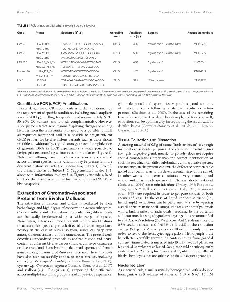
The design of PCR and qPCR primers for histone variants is quiteoften limited by the high level of similarity among histones withina given family (e.g., H2A). Consequently, it is recommended
to incorporate at least part of divergent untranslated regions(UTR, usually specific from different variants) into primerdesign, minimizing unspecific amplifications. On the contrary,the use of primers annealing in coding regions is a goodstrategy for amplification of histone sequences in unexploredspecies, either through RACE or genome walking experiments,given the higher conservation of these regions. Accordingly,the design of such primers requires a more careful sequenceanalysis in order to avoid targeting regions where variantsand canonical histones are similar, as the latter are far moreabundant and tend to amplify easily in PCR experiments. Forprimer design we recommend using the Primer-BLAST tool (Yeet al., 2012), which includes the software Primer3 (Untergasseret al., 2012) and BLAST searches in selected databases toavoid regions that can cause non-specific amplifications. Table 1shows primers designed using this software to amplify histonevariants in bivalves annealing in UTR regions, following theindications discussed above (note that some of these primerscan be efficiently used in multiple species). It is importantto note that species belonging to the genus Mytilus possessat least two different H2A.Z isoforms with specialized roles[H2A.Z.1, which is structurally and functionally equivalentto H2A.Z from other bivalve species, and H2A.Z.2, whichis has been specifically identified in Mytilus (Rivera-Casaset al., 2016b)] and that some species (i.e., C. gigas) haveevolved additional H2A.X genes (unpublished work), whichwill have to be considered when performing PCR experiments.In addition, primers for canonical histones as well as primersannealing in coding regions suitable for RACE or genomewalking experiments are indicated in Supplementary Tables 1, 2,respectively.
As a general rule, these primers have been successfullyemployed in PCR experiments at a final concentration of0.2µM. For each pair of primers, the annealing temperaturesare indicated in the tables, as well as the amplicon lengththat will serve to calculate the extension time employed in theamplification of each histone gene. For the complete setup of thePCR reaction and thermal profile, we recommend following theTaq DNA polymerase manufacturer’s guidelines.
Sperm Nuclear Basic Proteins
SNBPs include a diverse group of proteins, including bivalvemollusc Protamine-Like (PL) proteins. However, all of themshare a common evolutionary origin that can be tracedback to histone H1 (Eirin-Lopez et al., 2006b). Nonetheless,SNBPs are much more divergent than histones (the PLcontent varies extensively even among related species). Thepresent work provides information on accession numbers fortwo PL-I sequence isoforms (AY626224 and AY626225) fromthe surf clam Spisula solidissima, as well as PL-II/PL-IV(DQ305038) and PL-III (DQ305039) sequences from the musselM. californianus. In the case of Mytilus, the PL-II/PL-IVprecursor is post-translationally cleaved resulting in two differentprotein byproducts, PL-II∗ and PL-IV. In addition, primersemployed by previous reports in genome walking experimentsfor the characterization of these SNBPs (Lewis et al., 2004; Eirin-Lopez et al., 2006b) are indicated in Supplementary Table 2.
Frontiers in Physiology | www.frontiersin.org 3 August 2017 | Volume 8 | Article 490
H3.3 H3.3Fw2 TGAAGAAGAATAAGTCGTGAACCG 59◦C 523 Chlamys varia MF152785
H3.3Rv2 TGACTTGCATGATCTGTAGAAATTG
*Primers were originally designed to amplify the indicated histone variants in M. galloprovincialis and successfully employed in other Mytilus species and C. varia using less stringent
PCR conditions. Accession numbers for H2A.X, H2A.Z, and H3.3 correspond to C. varia sequences, submitted to GenBank as part of this work.
Quantitative PCR (qPCR) Amplifications
Primer design for qPCR experiments is further constrained bythe requirement of specific conditions, including small ampliconsizes (<200 bp), melting temperatures of approximately 60◦C,50–60% GC content, and low self-complementarity. However,since primers target gene regions displaying divergence amonghistones from the same family, it is not always possible to fulfillall requisites mentioned. Still, it is possible to design efficientqPCR primers for bivalve histone variants such as those shownin Table 2. Additionally, a good strategy to avoid amplificationof genomic DNA in qPCR experiments is, when possible, todesign primers annealing at intron/exon boundaries (Figure 1).Note that, although such positions are generally conservedacross different species, some variation may be present in moredivergent histone variants (i.e., macroH2A, Figure 1). Overall,the primers shown in Tables 1, 2, Supplementary Tables 1, 2,along with information displayed in Figure 1, provide a headstart for the characterization of histone variants and SNBPs inbivalve species.
Extraction of Chromatin-AssociatedProteins from Bivalve MolluscsThe extraction of histones and SNBPs is facilitated by theirbasic charge and high degree of conservation across eukaryotes.Consequently, standard isolation protocols using diluted acidscan be easily implemented in a wide range of species.Nonetheless, extraction procedures still require modificationsto account for specific particularities of different organisms,notably in the case of nuclei isolation, which can vary evenamong different tissues from the same species. The present workdescribes standardized protocols to analyze histone and SNBPcontent in different bivalve tissues (muscle, gill, hepatopancreasor digestive gland, hemolymph, male gonad, sperm, and femalegonad), using the mussel Mytilus as a reference. These protocolshave also been successfully applied to other bivalves, includingclams (e.g., Venerupis decussatus; Gonzalez-Romero et al., 2008),oysters (e.g., Crassostrea virginica; Gonzalez-Romero et al., 2017)and scallops (e.g., Chlamys varia), supporting their efficiencyacross multiple taxonomic groups. Based on previous experience,
gill, male gonad and sperm tissues produce good amountsof histone proteins following a standard acidic extractionprotocol (Shechter et al., 2007). In the case of the remainingtissues (muscle, digestive gland, hemolymph, and female gonad),extractions can be optimized by incorporating the modificationsdetailed below (Gonzalez-Romero et al., 2012b, 2017; Rivera-Casas et al., 2016a,b).
Tissue Collection and Dissection
A starting material of 0.5 g of tissue (fresh or frozen) is enoughfor most experimental purposes. The collection of solid tissues(i.e., gills, digestive gland, muscle, or gonads) does not requirespecial considerations other than the correct identification ofsuch tissues, which can differ substantially among bivalve species.For instance, in the present context, the difference between malegonad and sperm refers to the developmental stage of the gonad.In other words, the sperm constitutes a very mature gonadwhose content is mostly sperm cells. Thermal shock treatment(Soria et al., 2010), serotonin injections (Braley, 1985; Fong et al.,1996) or 0.5 M KCl injections (Breese et al., 1963; Beaumontet al., 1988) are required in order to get pure extracts of bothsperm and eggs. In the case of liquid connective tissue (i.e.,hemolymph), extractions can be performed in vivo by openinga small aperture in the shell using a lime (or a grinder if you workwith a high number of individuals), reaching to the posterioradductor muscle using a hypodermic syringe. It is recommendedto add Alsever’s solution (2.05% glucose, 0.42% sodium chloride,0.8% sodium citrate, and 0.055% citric acid) to the extractionsyringe (500µL of Alsever per every 10 mL of hemolymph) inorder to avoid the hemocytes aggregation. Hemolymph mustbe collected carefully (preventing contamination from gonadalcontent), immediately transferred into 15mL tubes and placed onice until all samples are collected. Samples should be subsequentlycentrifuged at 250 × g for 5 min at 4◦C, obtaining a pellet ofbivalve hemocytes that are suitable for the subsequent processes.
Nuclei Isolation
As a general rule, tissue is initially homogenized with a douncehomogenizer in 5 volumes of Buffer A (0.15 M NaCl, 10 mM
Frontiers in Physiology | www.frontiersin.org 4 August 2017 | Volume 8 | Article 490
FIGURE 1 | Schematic representation of bivalve histone gene structure and primer annealing positions. Histone genes H2A, H2A.Z.1, H2A.Z.2, H2A.X, macroH2A,H3, and H3.3 are represented using the mussel Mytilus as model species. Variations in length and intron/exon composition can be present in other bivalve species,such as the case of macroH2A in the oyster Crassostrea. Colored boxes represent coding exons, red uncolored boxes represent exons from UTR regions, and blacklines represent introns (length not proportional to actual size). Details of PCR primers annealing in UTR regions (including name and annealing positions, see Table 1)are indicated above the corresponding histones. Details corresponding to qPCR primers (see Table 2) are indicated below each gene for the mussel Mytiluscalifornianus (blue) and the Eastern oyster Crassostrea virginica (green).
Tris-HCl [pH 7.5], 0.5% Triton X-100) mixed with proteaseinhibitors 1/100 (v/v) (e.g., complete protease inhibitor cocktail[Roche Applied Science]). The homogenate is then placed onice for 10 min (breaking cellular membranes), and centrifugedat 4,000 × g for 10 min at 4◦C. In order to maximize yieldand quality, this protocol should be modified depending onthe tissue source as follows: In the case of digestive gland andmuscle tissue, it is recommended to repeat homogenization stepsat least once, as the insoluble materials contained in fat cells,plus the shape and composition of muscle cells, hamper cellmembrane break and cell disaggregation, respectively. In the caseof hemocytes, these steps should be repeated up to three times toobtain higher protein yields, due to the small size of these cellsand their tendency to form aggregates obstructing disruption of
cell membranes. Lastly, in the case of female gonad, a doubleamount of protease inhibitors should be used while skipping theincubation on ice for 10 min, in order to avoid the degradationof histone proteins as a result of the high content in proteolyticenzymes of this tissue.
Upon centrifugation, the pellet containing the nuclei issubjected to a second round of homogenization in 5 volumes ofbuffer without detergent, Buffer B (0.1 M KCl, 50 mM Tris-HCl[pH 7.5], 1mMMgCl2), adding amix of protease inhibitors 1/100(v/v). The homogenate is then incubated on ice for 10 min andcentrifuged at 4,000 × g for 10 min at 4◦C. Once more, specificmodifications are required for different tissue types. For digestivegland and muscle, prior to the homogenization in Buffer B, itis recommended to remove insoluble materials (present in high
Frontiers in Physiology | www.frontiersin.org 5 August 2017 | Volume 8 | Article 490
Rivera-Casas et al. Chromatin Characterization in Bivalve Molluscs
amounts in both tissues) by filtering nuclear extracts through asterilized cheesecloth soaked in Buffer B. In the case of femalegonad, doubling the amount of protease inhibitors and skip the
incubation on ice for 10 min is recommended, in order to avoidthe degradation of histone proteins by proteases. Overall, thenuclear fraction obtained at this point can be used in downstream
FIGURE 2 | Electrophoretic analysis of histone and SNBP acid extractions from different tissues of the mussel Mytilus californianus. SDS-PAGE of histone extractsfrom hepatopancreas (1), muscle (2), male gonad (3), hemolymph (4), and gill (5), and histone and SNBP extracts from female gonad (6) and sperm (7). CM, chickenerythrocyte histones used as molecular marker. Band corresponding to PL-II* in sperm tissue was identified based on our previous work (Gonzalez-Romero et al.,2012b). For each tissue, pellet resulting from the protein acid extraction (from 1g of starting material) was resuspended in 100µL of ultrapure-water, diluted in thesame amount of SDS Sample Buffer and loaded 1, 2 and 4µL in the SDS gel. Gradients in the upper part of the gel indicate the increase in the relative concentrationof the proteins loaded in each lane.
A
B C
FIGURE 3 | Histone fractionation in bivalves. (A) RP-HPLC chromatogram of 1mg of histones from male gonad of the variegated scallop Chlamys varia using a VydacC18 column (300 Å pore diameter). Peaks indicate the elution of the five histone families. (B) Acetonitrile gradient employed in this experiment. (C) SDS-PAGEseparation of selected fractions corresponding to each peak. CM, chicken erythrocyte histones used as molecular marker.
Frontiers in Physiology | www.frontiersin.org 6 August 2017 | Volume 8 | Article 490
Rivera-Casas et al. Chromatin Characterization in Bivalve Molluscs
experiments such as Micrococcal Nuclease (MNase) chromatinfractionation (Rivera-Casas et al., 2016a).
Histone and SNBP Extraction and Precipitation
The pelleted nuclear fraction constitutes the starting materialfor the extraction of bivalve chromosomal proteins. Accordingly,the supernatant resulting from treatment with Buffer B isdiscarded, the pellet resuspended in 2.5 volumes of 0.6 N HClusing a dounce homogenizer or by pipetting (both histones andSNBPs are very soluble in diluted acids such as 0.6 N HCl,due to their high basic amino acid content), and subsequentlycentrifuged at 8,200 × g for 10 min at 4◦C. The resultingsupernatant will be then transferred to fresh tube with 6 volumesof acetone, mixing the solutions by inverting the tubes 6-8times, and incubating the samples at −20◦C for at least 1 h.The solution will turn cloudy after a few minutes due to theprecipitation of histones and SNBPs in acetone. In the case oftissues producing low protein yields (i.e., digestive gland, muscleor hemolymph), it is recommended that acetone incubation isextended overnight. Upon protein precipitation, samples arepelleted by centrifuging at 10,000 × g for 10 min at 4◦C,discarding the supernatant and carefully washing the pellet withacetone in order to remove rests of acid. Samples are thensubject to an additional centrifugation, carefully discarding thesupernatant and air-drying the histone pellet for 20–30 min.Alternatively, a vacuum concentrator can be employed for 3–5 min (especially in the case of larger pellets). Lastly, proteinsare dissolved in a variable volume of ultrapure water (dependingon pellet size, 100µL is appropriate in most cases), being readyto be used immediately or stored at −80◦C. The integrity andconcentration of histones and SNBPs can be assessed throughprotein separation in sodium dodecyl sulfate polyacrylamidegel electrophoresis (SDS-PAGE) and/or by using western blotexperiments (see sections below).
The protein yield resulting from extractions will varydepending on the source tissue (Figure 2). For instance, proteinextractions from gills provide the best results among somatictissues in terms of purity and amount of protein. Digestivegland and hemolymph also provide relatively pure extracts whenusing the specific protocol modifications discussed previously.On the contrary, protein extracts from muscle tissues usuallycontain unidentified proteins in higher amount than histoneproteins, reducing purity. In the case of germinal tissues, spermextractions provide the highest amount of histones, due to thesmall size of the sperm cells. It is important to note that bivalvesperm chromatin, in addition to histones, is mostly composedby protamine-like proteins (increasing the compaction of DNA),constituting the most abundant proteins in SDS-PAGE gels (seeband corresponding to mussel PL-II∗ in Figure 2). Nonetheless,it is recommended to run acid-urea (AU) gels in order todiscriminate the different PL types (see next section for details).In the case of male gonads is easy to obtain a good histoneextract with no special considerations. However, depending onthe developmental stage, the presence of PL proteins from spermcells can be difficult to avoid. Lastly, it is important to avoiddegradation of histone proteins when working with female gonadtissue. For that purpose, the implementation of the protocol
outlined above yields a very good amount of histones withoutnoticeable degradation.
Histone and SNBP Fractionation inBivalvesChromosomal proteins can be easily purified usingchromatographic approaches. More specifically, histonesand SNBP extractions can be further purified using ReversePhase High Performance Liquid Chromatography (RP-HPLC)(as described in Rocchini et al., 1995; Ausio and Moore,1998; Shechter et al., 2007; Gonzalez-Romero et al., 2012b)and the resulting products can be employed in additionalexperiments such as nucleosome reconstitutions. The mobilephase (acetonitrile) gradient more often employed in separations,along with the corresponding chromatograms for histones andSNBPs are shown in Figures 3, 4, respectively. For detailedguidelines about RP-HPLC experiments for histone purification,see the work from Cheema and Ausio (2017).
For analytical purposes, acid-extracted histones and SNBPscan be separated and analyzed using polyacrylamide gelelectrophoresis under denaturing conditions such as SDS-PAGE (15% acrylamide, 0.4% bis-acrylamide), AU-PAGE (15%acrylamide, 0.1% bis-acrylamide, 5% acetic acid, 2.5 M urea),triton AU(AUT)-PAGE (10% acrylamide, 0.5% bis-acrylamide,
A
B
FIGURE 4 | SNBP fractionation in bivalves. (A) RP-HPLC chromatogram of1mg of histones and SNBPs from Mytilus male gonad HCl extract using aVydac C18 column (300 Å pore diameter). Peaks corresponding to thedifferent SNBP components are indicated in the figure. The acetonitrilegradient employed in this experiment is indicated by a straight line (0–60%). (B)AU-PAGE separation of selected fractions corresponding to each peak. SS,starting sample; CM, chicken erythrocyte histones used as molecular marker.
Frontiers in Physiology | www.frontiersin.org 7 August 2017 | Volume 8 | Article 490
Rivera-Casas et al. Chromatin Characterization in Bivalve Molluscs
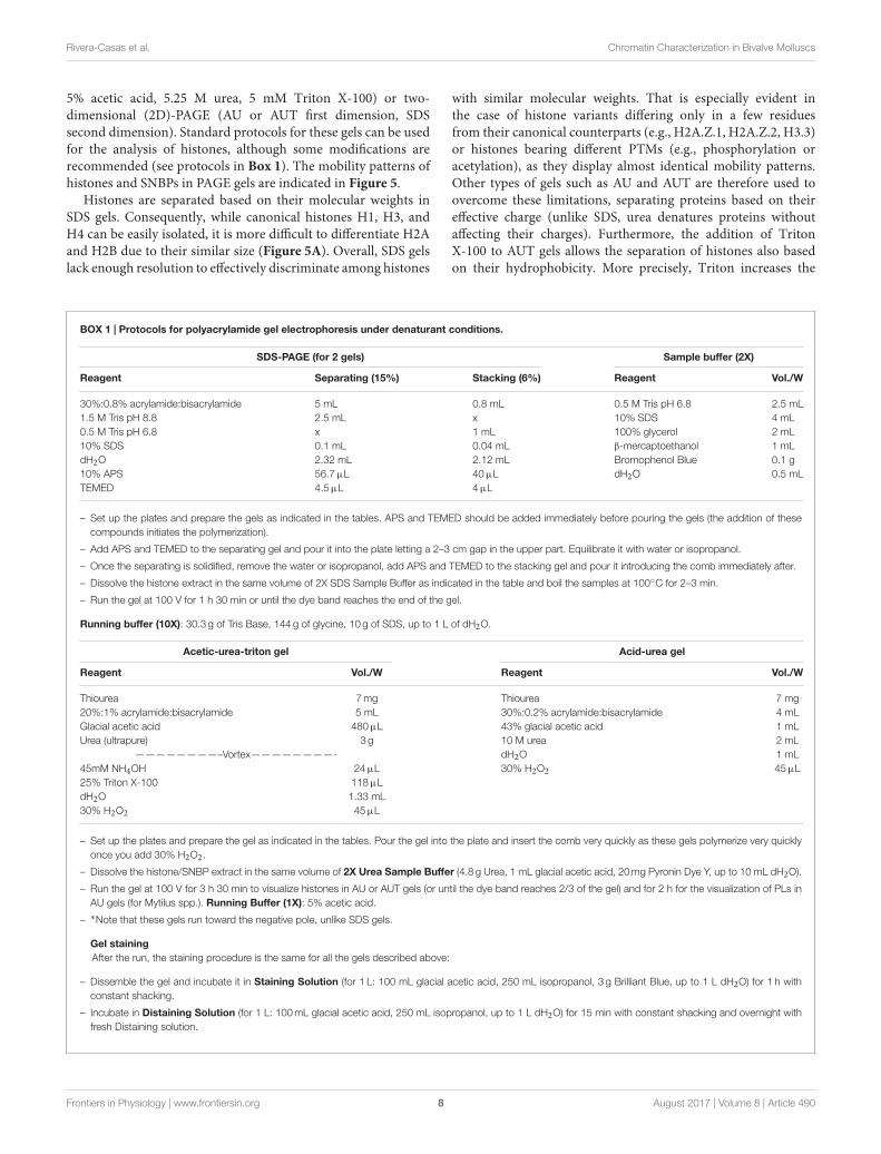
5% acetic acid, 5.25 M urea, 5 mM Triton X-100) or two-dimensional (2D)-PAGE (AU or AUT first dimension, SDSsecond dimension). Standard protocols for these gels can be usedfor the analysis of histones, although some modifications arerecommended (see protocols in Box 1). The mobility patterns ofhistones and SNBPs in PAGE gels are indicated in Figure 5.
Histones are separated based on their molecular weights inSDS gels. Consequently, while canonical histones H1, H3, andH4 can be easily isolated, it is more difficult to differentiate H2Aand H2B due to their similar size (Figure 5A). Overall, SDS gelslack enough resolution to effectively discriminate among histones
with similar molecular weights. That is especially evident inthe case of histone variants differing only in a few residuesfrom their canonical counterparts (e.g., H2A.Z.1, H2A.Z.2, H3.3)or histones bearing different PTMs (e.g., phosphorylation oracetylation), as they display almost identical mobility patterns.Other types of gels such as AU and AUT are therefore used toovercome these limitations, separating proteins based on theireffective charge (unlike SDS, urea denatures proteins withoutaffecting their charges). Furthermore, the addition of TritonX-100 to AUT gels allows the separation of histones also basedon their hydrophobicity. More precisely, Triton increases the
BOX 1 | Protocols for polyacrylamide gel electrophoresis under denaturant conditions.
30%:0.8% acrylamide:bisacrylamide 5 mL 0.8 mL 0.5 M Tris pH 6.8 2.5 mL1.5 M Tris pH 8.8 2.5 mL x 10% SDS 4 mL0.5 M Tris pH 6.8 x 1 mL 100% glycerol 2 mL10% SDS 0.1 mL 0.04 mL β-mercaptoethanol 1 mLdH2O 2.32 mL 2.12 mL Bromophenol Blue 0.1 g10% APS 56.7µL 40µL dH2O 0.5 mLTEMED 4.5µL 4µL
– Set up the plates and prepare the gels as indicated in the tables. APS and TEMED should be added immediately before pouring the gels (the addition of thesecompounds initiates the polymerization).
– Add APS and TEMED to the separating gel and pour it into the plate letting a 2–3 cm gap in the upper part. Equilibrate it with water or isopropanol.
– Once the separating is solidified, remove the water or isopropanol, add APS and TEMED to the stacking gel and pour it introducing the comb immediately after.
– Dissolve the histone extract in the same volume of 2X SDS Sample Buffer as indicated in the table and boil the samples at 100◦C for 2–3 min.
– Run the gel at 100 V for 1 h 30 min or until the dye band reaches the end of the gel.
Running buffer (10X): 30.3 g of Tris Base, 144 g of glycine, 10 g of SDS, up to 1 L of dH2O.
Acetic-urea-triton gel Acid-urea gel
Reagent Vol./W Reagent Vol./W
Thiourea 7mg Thiourea 7 mg20%:1% acrylamide:bisacrylamide 5 mL 30%:0.2% acrylamide:bisacrylamide 4 mLGlacial acetic acid 480µL 43% glacial acetic acid 1 mLUrea (ultrapure) 3 g 10 M urea 2 mL
– Set up the plates and prepare the gel as indicated in the tables. Pour the gel into the plate and insert the comb very quickly as these gels polymerize very quicklyonce you add 30% H2O2.
– Dissolve the histone/SNBP extract in the same volume of 2X Urea Sample Buffer (4.8 g Urea, 1 mL glacial acetic acid, 20mg Pyronin Dye Y, up to 10 mL dH2O).
– Run the gel at 100 V for 3 h 30 min to visualize histones in AU or AUT gels (or until the dye band reaches 2/3 of the gel) and for 2 h for the visualization of PLs inAU gels (for Mytilus spp.). Running Buffer (1X): 5% acetic acid.
– ∗Note that these gels run toward the negative pole, unlike SDS gels.
Gel staining
After the run, the staining procedure is the same for all the gels described above:
– Dissemble the gel and incubate it in Staining Solution (for 1 L: 100 mL glacial acetic acid, 250 mL isopropanol, 3 g Brilliant Blue, up to 1 L dH2O) for 1 h withconstant shacking.
– Incubate in Distaining Solution (for 1 L: 100mL glacial acetic acid, 250 mL isopropanol, up to 1 L dH2O) for 15 min with constant shacking and overnight withfresh Distaining solution.
Frontiers in Physiology | www.frontiersin.org 8 August 2017 | Volume 8 | Article 490
Rivera-Casas et al. Chromatin Characterization in Bivalve Molluscs
A B
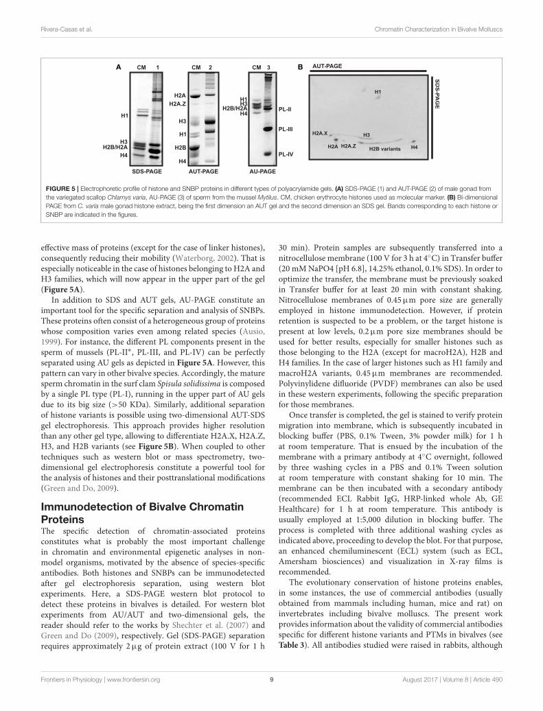
FIGURE 5 | Electrophoretic profile of histone and SNBP proteins in different types of polyacrylamide gels. (A) SDS-PAGE (1) and AUT-PAGE (2) of male gonad fromthe variegated scallop Chlamys varia, AU-PAGE (3) of sperm from the mussel Mytilus. CM, chicken erythrocyte histones used as molecular marker. (B) Bi-dimensionalPAGE from C. varia male gonad histone extract, being the first dimension an AUT gel and the second dimension an SDS gel. Bands corresponding to each histone orSNBP are indicated in the figures.
effective mass of proteins (except for the case of linker histones),consequently reducing their mobility (Waterborg, 2002). That isespecially noticeable in the case of histones belonging to H2A andH3 families, which will now appear in the upper part of the gel(Figure 5A).
In addition to SDS and AUT gels, AU-PAGE constitute animportant tool for the specific separation and analysis of SNBPs.These proteins often consist of a heterogeneous group of proteinswhose composition varies even among related species (Ausio,1999). For instance, the different PL components present in thesperm of mussels (PL-II∗, PL-III, and PL-IV) can be perfectlyseparated using AU gels as depicted in Figure 5A. However, thispattern can vary in other bivalve species. Accordingly, the maturesperm chromatin in the surf clam Spisula solidissima is composedby a single PL type (PL-I), running in the upper part of AU gelsdue to its big size (>50 KDa). Similarly, additional separationof histone variants is possible using two-dimensional AUT-SDSgel electrophoresis. This approach provides higher resolutionthan any other gel type, allowing to differentiate H2A.X, H2A.Z,H3, and H2B variants (see Figure 5B). When coupled to othertechniques such as western blot or mass spectrometry, two-dimensional gel electrophoresis constitute a powerful tool forthe analysis of histones and their posttranslational modifications(Green and Do, 2009).
Immunodetection of Bivalve ChromatinProteinsThe specific detection of chromatin-associated proteinsconstitutes what is probably the most important challengein chromatin and environmental epigenetic analyses in non-model organisms, motivated by the absence of species-specificantibodies. Both histones and SNBPs can be immunodetectedafter gel electrophoresis separation, using western blotexperiments. Here, a SDS-PAGE western blot protocol todetect these proteins in bivalves is detailed. For western blotexperiments from AU/AUT and two-dimensional gels, thereader should refer to the works by Shechter et al. (2007) andGreen and Do (2009), respectively. Gel (SDS-PAGE) separationrequires approximately 2µg of protein extract (100 V for 1 h
30 min). Protein samples are subsequently transferred into anitrocellulose membrane (100 V for 3 h at 4◦C) in Transfer buffer(20 mMNaPO4 [pH 6.8], 14.25% ethanol, 0.1% SDS). In order tooptimize the transfer, the membrane must be previously soakedin Transfer buffer for at least 20 min with constant shaking.Nitrocellulose membranes of 0.45µm pore size are generallyemployed in histone immunodetection. However, if proteinretention is suspected to be a problem, or the target histone ispresent at low levels, 0.2µm pore size membranes should beused for better results, especially for smaller histones such asthose belonging to the H2A (except for macroH2A), H2B andH4 families. In the case of larger histones such as H1 family andmacroH2A variants, 0.45µm membranes are recommended.Polyvinylidene difluoride (PVDF) membranes can also be usedin these western experiments, following the specific preparationfor those membranes.
Once transfer is completed, the gel is stained to verify proteinmigration into membrane, which is subsequently incubated inblocking buffer (PBS, 0.1% Tween, 3% powder milk) for 1 hat room temperature. That is ensued by the incubation of themembrane with a primary antibody at 4◦C overnight, followedby three washing cycles in a PBS and 0.1% Tween solutionat room temperature with constant shaking for 10 min. Themembrane can be then incubated with a secondary antibody(recommended ECL Rabbit IgG, HRP-linked whole Ab, GEHealthcare) for 1 h at room temperature. This antibody isusually employed at 1:5,000 dilution in blocking buffer. Theprocess is completed with three additional washing cycles asindicated above, proceeding to develop the blot. For that purpose,an enhanced chemiluminescent (ECL) system (such as ECL,Amersham biosciences) and visualization in X-ray films isrecommended.
The evolutionary conservation of histone proteins enables,in some instances, the use of commercial antibodies (usuallyobtained from mammals including human, mice and rat) oninvertebrates including bivalve molluscs. The present workprovides information about the validity of commercial antibodiesspecific for different histone variants and PTMs in bivalves (seeTable 3). All antibodies studied were raised in rabbits, although
Frontiers in Physiology | www.frontiersin.org 9 August 2017 | Volume 8 | Article 490
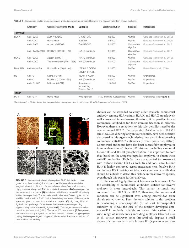
PL-II* Anti-PL-II* Home-Made Whole protein 1:400 (Immuno-fluorescence) Mytilus Unpublished (see Figure 6)
The asterisk (*) in PL-II indicates that this protein is a cleavage product from the larger PL-II/PL-IV precursor (Carlos et al., 1993).
FIGURE 6 | Immuno-histochemical analysis of PL-II* distribution in malegonad from the mussel Mytilus trossulus. (A) Hematoxylin-eosin stainedlongitudinal section of the tip of a seminiferous tubule from a M. trossulushighly mature male gonad. The bar = 400 micrometers. (B,C) correspond tothe same section shown in (A) but stained with histone H4 and PL-II* primaryantibodies respectively. The secondary antibodies were Oregon green for H4and Rhodamine red for PL-II*. Notice the relatively low levels of histone H4 inspermatocytes compared to spermatids and sperm. (D) High magnificationlight microscope image of a section of the same tissue correspondingapproximately to the square highlighted in (B). The images were obtained asdescribed in Casas et al. (1993). The bar = 200 micrometers. (E,F) representelectron microscopy images to show the three main different cell types presentduring the late spermiogenic stages of differentiation. The bars = 100 and 10micrometers, respectively.
choices can be extended to every other available commercialantibody. AmongH2A variants, H2A.X, andH2A.Z are relativelywell conserved in metazoans, therefore, it is possible to usecommercial antibodies for their immunodetection in bivalves.However, there are exceptions to this rule, best illustrated by thecase of mussel H2A.Z. Two separate H2A.Z variants (H2A.Z.1and H2A.Z.2), differing only in four residues, have been recentlydiscovered in this organism, hindering their discrimination usingcommercial anti-H2A.Z antibodies (Rivera-Casas et al., 2016b).Commercial antibodies have also been successfully employed inimmunodetection of bivalve H3 histones, including canonicalhistone H3 and H3S10 phosphorylation. It is important to notethat, based on the antigenic peptides employed to obtain theseanti-H3 antibodies (Table 3), they are expected to cross-reactwith histone variant H3.3 as well. In addition, since histoneH3.3 is highly conserved across metazoans (e.g., bivalve H3.3and human H3.3 proteins are identical), commercial antibodiesshould be suitable to detect this histone in most bivalve species,even though this awaits further analyses.
In the case of highly divergent histones such as macroH2Athe availability of commercial antibodies suitable for bivalvemolluscs is more improbable. This variant is much lessconserved than H2A.X or H2A.Z, therefore, the amino acidvariation can be significant even among macroH2As fromclosely related species. Thus, the only solution to this problemis developing a species-specific (or at least taxon-specific)antibody, as it was the case of the recently developed anti-macroH2A antibody suitable to detect this variant in awide range of invertebrates including molluscs (Rivera-Casaset al., 2016a). However, since this antibody displays a smalldegree of cross-reactivity with histone H1 (a phenomenon also
Frontiers in Physiology | www.frontiersin.org 10 August 2017 | Volume 8 | Article 490
Rivera-Casas et al. Chromatin Characterization in Bivalve Molluscs
BOX 2 | Immunohistochemistry (IHC) protocol.
Fixing and embedding the tissue
Dissect the gonads of mussel specimens and immerse them in primary fixative (4% freshly depolymerized paraformaldehyde in Millonig’s Phosphate Buffer [11.04 gof NaH2PO4 in a total of 200 ml of deionized water, pH 7.4]). One group of gonads is kept uncut for light/fluorescence microscopy. Another group is cut into long2-mm thick strips (it is important to create strips with different orientations). Fixation of both whole gonads and gonad strips is conducted for 1 h at room temperature.
Fixed specimens are then dehydrated in ethanol series (30% for 10 min—70% for 10 min—95% for 10 min—95% for 2 min − 100% for 5 min—100% for 5 min)using a volume of dehydrating solution of approximately 10-fold the volume of the tissue. Gonad strips are subsequently infiltrated with 50% LR-White (London ResinCo.) in 100% ethanol for 3 h, followed by pure LR-White resin overnight at room temperature. The whole gonads are treated with two steps of xylene for 1 h each atroom temperature, followed by 6 steps of melted pure paraffin kept at 60◦C for 20 min each. Gonads are then infiltrated in pure melted paraffin overnight.
Next day, LR-White embedded specimens are solidified in block molds at 60◦C in a vacuum oven, as oxygen inhibits the polymerization of the LR-White resin.Paraffin-embedded specimens are then solidified in molds at room temperature. Paraffin blocks are kept at 4◦C until sectioning.
Sectioning and mounting the sections
LR-White blocks are sectioned into 0.3–0.5µm-thick sections in an ultramicrotome; while paraffin blocks are sectioned into ribbons of approximately 5µm-thicksections in a histological microtome. Sections are subsequently mounted on glass slides, air dried and baked for 10 min at 50◦C. Paraffin sections are then de-paraffinized in Copling jars using the following step series: xylene for 5 min (2 times)—100% ethanol for 5 min (2 times)—70% ethanol for 5 min (2 times)—Millonig’sbuffer for 5 min.
Immunohistochemical staining
Immunolabeling steps are performed at room temperature in Coplin jars, with the exception of antibody incubations, for which small volumes (50–150µl, dependingon the size of the sections) are used to cover the sections and the slides are kept in a humid chamber to prevent drying of the antibody solution during incubation.Briefly, sections are blocked in 1% bovine serum albumin −1% casein in Millonig’s phosphate buffer for 1 h, washed once for 10 min in the same buffer, incubatedwith the primary antibody for 2 h, washed 3 times as before, incubated with the secondary antibody for 1 h, washed 3 times as before, blotted and mounted with No.1 coverslips in gelvatol mounting medium (0.35 g Gelvatol, 3 ml Millonig’s buffer, 1.5 ml glycerol) for observation. In this work, primary antibodies, house-made PL-II∗
and H4 were diluted 1:400 and 1:200, respectively, in Millonig’s phosphate buffer with 2% BSA; whereas commercial goat anti-rabbit IgG tagged with Rhodaminered (R6394) and Oregon green (O6381) from Molecular Probes (Eugene, OR) were diluted 1:200 in the same buffer.
Histological staining
After immunolabeling, sister sections are histologically stained to be used as reference for structural features of the immunolabeled sections. LR-White sections bakedon glass slides (as above) are stained in a single-step procedure, covering the sections with freshly filtered Richardson’s stain (prepared by mixing equal volumesof 1% azure II in deionized water, and 1% methylene blue in 1% borax), evaporating the stain to near dryness on a heating block at 50◦C, and rinsing in deionizedwater to remove excess stain. De-paraffinized hydrated sections on glass slides (as above) are conventionally stained with Hematoxylin-Eosin. Histologically stainedsections are permanently mounted with No. 1 coverslips in PermountTM, before observation.
described in other anti-macroH2A antibodies from mammals,due to the structural similarities between both proteins;Pehrson et al., 1997), its application for genome-wide analysessuch as chromatin immunoprecipitation (ChIP) is somewhatlimited. Similarly, although commercial anti-H2A.X and anti-gammaH2A.X antibodies are suitable for bivalves, a bivalve-specific antibody developed in mussel is available for this variant,targeting the peptide SQSQEF characteristic from MytilusH2A.X.
With the exception of an antibody for PL-II∗ in themussel Mytilus (Table 3), there have been no additionalantibodies developed for SNBPs. Figure 6 provides anexample for the immuno-histochemical use of this antibody.The protocol for the immunohistochemical procedure isdetailed in Box 2. Overall, the high degree of conservationof most histone proteins enables the possibility of usingcommercial antibodies in bivalve tissues, overcoming one ofthe greatest limitations faced when working with non-modelorganisms.
CONCLUSIONS
The study of the mechanisms mediating physiological responsesto environmental changes constitutes a key discipline tounderstand how climate change will affect organisms.Environmental epigenetics is at the center stage of such efforts,given the role of epigenetic modifications during the regulation
of gene activity, and their implications for acclimatization andadaptation under ever-changing environments. Unfortunately,epigenetic information for most non-model and ecologicallyrelevant organisms is very limited, with environmental epigeneticstudies almost exclusively focused on DNA methylation (leavingother mechanisms largely unexplored). However, it is now clearthat chromatin-associated proteins participate in organism-environment interactions in different capacities (e.g., regulationof gene expression, active role in defense against externalpathogens, etc.). By providing a description of experimentalmethods for studying chromatin-associated proteins, the presentwork aims to provide a reference for researchers interested instudying how DNA is organized and regulated in molluscs,a ubiquitous taxonomic group playing critical functions invirtually all ecosystems. By doing so, this work fosters a moreholistic approach to study the epigenetic mechanisms underlyingenvironmental responses in bivalve molluscs, ultimatelyimproving our understanding of their physiological responsesto climate change, their application as environmental sentinelorganisms as well as optimizing their management.
AUTHOR CONTRIBUTIONS
The concept of the work was conceived by JE and developed incollaboration with CR, RG-R and JA. All authors were involvedin the design and performance of the experiments as wellas in the analyses of the data. CR and JE wrote the article,
Frontiers in Physiology | www.frontiersin.org 11 August 2017 | Volume 8 | Article 490
Rivera-Casas et al. Chromatin Characterization in Bivalve Molluscs
with contribution from RG-R and JA. All authors reviewed themanuscript draft.
ACKNOWLEDGMENTS
This material is based upon work supported by the NationalScience Foundation under Grant No. HRD-1547798. ThisNSF Grant was awarded to Florida International Universityas part of the Centers of Research Excellence in Scienceand Technology (CREST) Program. This is contributionnumber 40 from the Marine Education and Research Centerin the Institute of Water and Environment at Florida
International University. Additional support was providedby start-up funds from the College of Arts, Sciences andEducation (CASE) at Florida International University (JE), theNatural Science and Engineering Research Council of Canada(NSERC) grant 43699-2012 (JA), and the Fundacion RamonAreces (CR).
SUPPLEMENTARY MATERIAL
The Supplementary Material for this article can be foundonline at: http://journal.frontiersin.org/article/10.3389/fphys.2017.00490/full#supplementary-material
REFERENCES
Allis, C. D., Jenuwein, T., and Reinberg, D. (2007). Epigenetics. NewYork, NY: Cold
Spring Harbor Laboratory Press.
Araya, I., Nardocci, G., Morales, J., Vera, M., Molina, A., and Alvarez, M.
(2010). MacroH2A subtypes contribute antagonistically to the transcriptional
regulation of the ribosomal cistron during seasonal acclimatization of the carp