Molecular Structure and Function of the Novel BrnT/BrnA Toxin-Antitoxin System of Brucella abortus * □ S Received for publication, December 8, 2011, and in revised form, February 3, 2012 Published, JBC Papers in Press, February 14, 2012, DOI 10.1074/jbc.M111.332163 Brook E. Heaton ‡§1 , Julien Herrou ¶1 , Anne E. Blackwell 2 , Vicki H. Wysocki , and Sean Crosson ‡§3 From the ‡ Committee on Microbiology and the ¶ Department of Biochemistry and Molecular Biology, University of Chicago, Chicago, Illinois 60637, the § Howard T. Ricketts Laboratory, Argonne National Laboratory, Argonne, Illinois 60439, and the Department of Chemistry and Biochemistry, University of Arizona, Tucson, Arizona 85721 Background: Toxin-antitoxin (TA) systems are broadly conserved in the bacterial kingdom. Results: Brucella abortus RNase toxin, BrnT, has a RelE-like fold and is neutralized by its antitoxin, BrnA. brnTA transcription is activated by a number of environmental stressors. Conclusion: BrnTA is a novel, stress-regulated TA system. Significance: This study structurally and functionally defines a novel member of the RelE toxin family. Type II toxin-antitoxin (TA) systems are expressed from two- gene operons that encode a cytoplasmic protein toxin and its cognate protein antitoxin. These gene cassettes are often pres- ent in multiple copies on bacterial chromosomes, where they have been reported to regulate stress adaptation and persistence during antimicrobial treatment. We have identified a novel type II TA cassette in the intracellular pathogen Brucella abor- tus that consists of the toxin gene, brnT, and its antitoxin, brnA. BrnT is coexpressed and forms a 2:2 tetrameric complex with BrnA, which neutralizes BrnT toxicity. The BrnT 2 -BrnA 2 tetramer binds its own promoter via BrnA, and autorepresses its expression; its transcription is strongly induced in B. abortus by various stressors encountered by the bacterial cell during infec- tion of a mammalian host. Although highly divergent at the pri- mary sequence level, an atomic resolution (1.1 A ˚ ) crystal struc- ture of BrnT reveals a secondary topology related to the RelE family of type II ribonuclease toxins. However, overall tertiary structural homology to other RelE family toxins is low. A func- tional characterization of BrnT by site-directed mutagenesis demonstrates a correspondence between its in vitro activity as a ribonuclease and control of bacteriostasis in vivo. We further present an analysis of the conserved and variable features of structure required for RNA scission in BrnT and the RelE toxin family. This structural investigation informs a model of the RelE-fold as an evolutionarily flexible scaffold that has been selected to bind structurally disparate antitoxins, and exhibit distinct toxin activities including RNA scission and DNA gyrase inhibition. The Gram-negative intracellular pathogen, Brucella abortus, naturally infects ruminant hosts where it causes both a chronic and an acute infection. In humans, infection is strictly zoonotic and results in a debilitating chronic disease known as undulant fever. B. abortus primarily resides in professional phagocytic cells and placental trophoblasts in its mammalian host (1, 2). While trafficking through the host system, B. abortus must adapt to a number of stressors including oxidative bursts from macrophages (3), low pH (4), antimicrobial peptides, and nutri- ent limitation (5). A handful of B. abortus proteins that mediate adaptation to host-generated stress are known (6). However, many putative stress response genes encoded in the genome remain entirely undefined. Among these are type II toxin-anti- toxin (TA) 4 systems. Type II TA systems consist of a stable toxin protein and a labile antitoxin protein that form a high-affinity, neutral com- plex (7). The genes encoding type II TAs were initially identified on plasmids, where they were demonstrated to promote stable plasmid inheritance (8, 9). Several distinct families of this type of TA system have since been identified on bacterial chromo- somes (10 –12) and are demonstrated to regulate stress survival (13, 14) and persister cell formation (15–17). As antitoxin pro- teins are generally degraded at a faster rate than toxins by the cellular proteolytic machinery (18 –20), the arrest of protein synthesis as a result of antibiotic insult, nutrient deprivation, or other stresses results in accumulation of free toxin in the cyto- sol. Upon release from their cognate antitoxins, type II toxins are known to act via one of several mechanisms: degradation of mRNA (21, 22), inhibition of the translating ribosome (23), inhibition of DNA gyrase (24 –26), degradation of tRNA fMet (27), or inhibition of peptidoglycan synthesis (28, 29). Depend- ing on the system and dosage, toxins may be bacteriostatic or bactericidal (30, 31). * This work was supported, in whole or in part, by National Institutes of Health Grant 1-U54-AI-057153 to the Region V “Great Lakes” Regional Center of Excellence in Biodefense and Emerging Infectious Diseases Consortium (GLRCE) from the NIAID (to S. C.), by a National Science Foundation MRI grant for development of the custom tandem mass spectrometer, and the Pacific Southwest Regional Center for Excellence Grant 1U54-AI-065359 (to V. H. W.). □ S This article contains supplemental Table S1 and Fig. S1. The atomic coordinates and structure factors (code 3U97) have been deposited in the Protein Data Bank, Research Collaboratory for Structural Bioinformatics, Rutgers University, New Brunswick, NJ (http://www.rcsb.org/). 1 Both authors contributed equally. 2 Supported by an National Science Foundation Graduate Research Fellowship. 3 To whom correspondence should be addressed: 929 East 57th St., GCIS W138, Chicago, IL 60637. Tel.: 773-834-1926; Fax: 773-702-0439; E-mail: [email protected]. 4 The abbreviations used are: TA, type II toxin-antitoxin; AUC, analytical ultra- centrifugation; CID, collision-induced dissociation; nEIS, nanoelectrospray ionization; IPTG, isopropyl 1-thio--D-galactopyranoside. THE JOURNAL OF BIOLOGICAL CHEMISTRY VOL. 287, NO. 15, pp. 12098 –12110, April 6, 2012 Published in the U.S.A. 12098 JOURNAL OF BIOLOGICAL CHEMISTRY VOLUME 287 • NUMBER 15 • APRIL 6, 2012 at Ohio State University Libraries-Columbus, on September 20, 2012 www.jbc.org Downloaded from http://www.jbc.org/content/suppl/2012/02/14/M111.332163.DC1.html Supplemental Material can be found at:
Transcript
Molecular Structure and Function of the Novel BrnT/BrnAToxin-Antitoxin System of Brucella abortus*□S
Received for publication, December 8, 2011, and in revised form, February 3, 2012 Published, JBC Papers in Press, February 14, 2012, DOI 10.1074/jbc.M111.332163
Brook E. Heaton‡§1, Julien Herrou¶1, Anne E. Blackwell�2, Vicki H. Wysocki�, and Sean Crosson‡§3
From the ‡Committee on Microbiology and the ¶Department of Biochemistry and Molecular Biology, University of Chicago,Chicago, Illinois 60637, the §Howard T. Ricketts Laboratory, Argonne National Laboratory, Argonne, Illinois 60439, and the�Department of Chemistry and Biochemistry, University of Arizona, Tucson, Arizona 85721
Background: Toxin-antitoxin (TA) systems are broadly conserved in the bacterial kingdom.Results: Brucella abortus RNase toxin, BrnT, has a RelE-like fold and is neutralized by its antitoxin, BrnA. brnTA transcriptionis activated by a number of environmental stressors.Conclusion: BrnTA is a novel, stress-regulated TA system.Significance: This study structurally and functionally defines a novel member of the RelE toxin family.
Type II toxin-antitoxin (TA) systems are expressed from two-gene operons that encode a cytoplasmic protein toxin and itscognate protein antitoxin. These gene cassettes are often pres-ent in multiple copies on bacterial chromosomes, where theyhave been reported to regulate stress adaptation andpersistenceduring antimicrobial treatment. We have identified a noveltype II TA cassette in the intracellular pathogen Brucella abor-tus that consists of the toxin gene, brnT, and its antitoxin, brnA.BrnT is coexpressed and forms a 2:2 tetrameric complex withBrnA, which neutralizes BrnT toxicity. The BrnT2-BrnA2tetramer binds its ownpromoter via BrnA, and autorepresses itsexpression; its transcription is strongly induced in B. abortus byvarious stressors encountered by the bacterial cell during infec-tion of a mammalian host. Although highly divergent at the pri-mary sequence level, an atomic resolution (1.1 A) crystal struc-ture of BrnT reveals a secondary topology related to the RelEfamily of type II ribonuclease toxins. However, overall tertiarystructural homology to other RelE family toxins is low. A func-tional characterization of BrnT by site-directed mutagenesisdemonstrates a correspondence between its in vitro activity as aribonuclease and control of bacteriostasis in vivo. We furtherpresent an analysis of the conserved and variable features ofstructure required for RNA scission in BrnT and the RelE toxinfamily. This structural investigation informs a model of theRelE-fold as an evolutionarily flexible scaffold that has beenselected to bind structurally disparate antitoxins, and exhibit
distinct toxin activities including RNA scission andDNA gyraseinhibition.
TheGram-negative intracellular pathogen,Brucella abortus,naturally infects ruminant hosts where it causes both a chronicand an acute infection. In humans, infection is strictly zoonoticand results in a debilitating chronic disease known as undulantfever. B. abortus primarily resides in professional phagocyticcells and placental trophoblasts in its mammalian host (1, 2).While trafficking through the host system, B. abortus mustadapt to a number of stressors including oxidative bursts frommacrophages (3), low pH (4), antimicrobial peptides, and nutri-ent limitation (5). A handful of B. abortus proteins thatmediateadaptation to host-generated stress are known (6). However,many putative stress response genes encoded in the genomeremain entirely undefined. Among these are type II toxin-anti-toxin (TA)4 systems.
Type II TA systems consist of a stable toxin protein and alabile antitoxin protein that form a high-affinity, neutral com-plex (7). The genes encoding type IITAswere initially identifiedon plasmids, where they were demonstrated to promote stableplasmid inheritance (8, 9). Several distinct families of this typeof TA system have since been identified on bacterial chromo-somes (10–12) and are demonstrated to regulate stress survival(13, 14) and persister cell formation (15–17). As antitoxin pro-teins are generally degraded at a faster rate than toxins by thecellular proteolytic machinery (18–20), the arrest of proteinsynthesis as a result of antibiotic insult, nutrient deprivation, orother stresses results in accumulation of free toxin in the cyto-sol. Upon release from their cognate antitoxins, type II toxinsare known to act via one of several mechanisms: degradation ofmRNA (21, 22), inhibition of the translating ribosome (23),inhibition of DNA gyrase (24–26), degradation of tRNAfMet
(27), or inhibition of peptidoglycan synthesis (28, 29). Depend-ing on the system and dosage, toxins may be bacteriostatic orbactericidal (30, 31).
* This work was supported, in whole or in part, by National Institutes of HealthGrant 1-U54-AI-057153 to the Region V “Great Lakes” Regional Center ofExcellence in Biodefense and Emerging Infectious Diseases Consortium(GLRCE) from the NIAID (to S. C.), by a National Science Foundation MRIgrant for development of the custom tandem mass spectrometer, and thePacific Southwest Regional Center for Excellence Grant 1U54-AI-065359(to V. H. W.).
□S This article contains supplemental Table S1 and Fig. S1.The atomic coordinates and structure factors (code 3U97) have been deposited in
the Protein Data Bank, Research Collaboratory for Structural Bioinformatics,Rutgers University, New Brunswick, NJ (http://www.rcsb.org/).
1 Both authors contributed equally.2 Supported by an National Science Foundation Graduate Research
Fellowship.3 To whom correspondence should be addressed: 929 East 57th St., GCIS
W138, Chicago, IL 60637. Tel.: 773-834-1926; Fax: 773-702-0439; E-mail:[email protected].
4 The abbreviations used are: TA, type II toxin-antitoxin; AUC, analytical ultra-centrifugation; CID, collision-induced dissociation; nEIS, nanoelectrosprayionization; IPTG, isopropyl 1-thio-�-D-galactopyranoside.
THE JOURNAL OF BIOLOGICAL CHEMISTRY VOL. 287, NO. 15, pp. 12098 –12110, April 6, 2012Published in the U.S.A.
12098 JOURNAL OF BIOLOGICAL CHEMISTRY VOLUME 287 • NUMBER 15 • APRIL 6, 2012
at Ohio S
tate University Libraries-C
olumbus, on S
eptember 20, 2012
ww
w.jbc.org
Dow
nloaded from
http://www.jbc.org/content/suppl/2012/02/14/M111.332163.DC1.html Supplemental Material can be found at:
We report a functional, biochemical, and structural char-acterization of a novel type II toxin-antitoxin system, BrnT-BrnA, from B. abortus. BrnT is a small (81 residues) ribonu-clease that is present in bacteria, archaea, bacteriophage, andplasmids. BrnT-BrnA forms a 2:2 tetrameric complex andautoregulates its expression, which is induced by a numberof stress insults. Our atomic resolution (1.1 Å) crystal struc-ture of BrnT revealed an overall secondary structural topol-ogy that is related to the RelE family of ribonuclease toxins(13). Indeed, the BrnT toxin cleaves RNA in vitro, and site-directed mutagenesis of the BrnT coding sequence has iden-tified a set of polar residues clustered in the �1–�5 region ofthe structure that are required for RNA scission in vitro andcontrol of bacteriostasis in vivo. We further define the con-served and variable features of structure required for RNAscission in BrnT and the RelE toxin family.
EXPERIMENTAL PROCEDURES
Bacterial Strains and Growth Conditions—Escherichia colistrains used for growth curves, protein purification, and in vivoprotein synthesis assays were E. coli Rosetta pLysS/DE3 withbab1_0993, bab1_0994, or bab1_0993-4 cloned into pET151/D-TOPO (Table 1). E. coli used for bacteriostasis experimentswere in Top 10 with bab1_0994 cloned into NdeI/KpnI sites ofpSRK-Km (32) and either pBAD 24 (33) as an empty vector con-trolorbab1_0993cloned intotheNdeI/HindIII siteofpBAD24.Cul-turesweregrownwith100�g/mlofampicillinand50�g/mlofkana-mycin. Cultures were induced with 0.5 mM IPTG and plated on LBagar�100�g/mlof ampicillin, 50�g/mlofkanamycin�0.2%arab-inose. B. abortus 2308 strains were grown in Brucella broth at 37 °C.brnT,brnA, andbrnTAexpressionstrainswereconstructed inawild-typeB. abortus 2308backgroundby introducing pSRK-Kmalone, orwith brnT, brnA, and brnTA cloned into NdeI/KpnI sites. Protein
expression inB. abortuswas inducedwith 1mM IPTG.An in-frame,unmarked brnTA deletion strain was made by cloning an upstreamfragment of brnT (primers used to amplify the fragments were: for-ward, 5�-GGATCCGCCACGGATTTCAAAATATC, reverse, 5�-GCGAGCCCAGACGAATAGGGCCGTATGAACGATG), and adownstream fragment of brnA (primers used to amplify the frag-mentswere: forward,5�-CATCGTTCATACGGCCCTATTCGTC-TGGGCTCGC, and reverse, 5�-GGTACCCGTATTGCATGTC-TGTCTCC). These two fragments were cloned into pNPTS138,which was subsequently mated into B. abortus 2308 usingE. coli S17-1. Strains were first mixed and plated without selec-tion overnight. Bacterial cells were then scraped and plated onSchadelar blood agar with kanamycin and nalidixic acid toselect for single B. abortus recombinants. Single colonies werethen picked and grown overnight in Brucella broth and thenplated the next day on Schadelar blood agar plates with 10%sucrose. Colonies that grew were screened by PCR to confirmthat the gene replacement had occurred and that thepNPTS138 backbone had been lost. The PbrnTA-lacZ pro-moter fusion was made cloning �500 nucleotides upstream ofthe brnT ATG start codon. Primers used were: forward, 5�-AAGCTTGAGCGGGCGATGATCTTCCGC, and reverse,5�-GGATCCAAAACGTATGTACAATAATTCGTCTG-GGC. This fragment was cloned into the HindIII/BamHIsites of pMR15. This transcriptional fusion plasmid wastransformed into B. abortus 2308 and B. abortus 2308�brnTA.Growth Assays—E. coli growth curves were done in LB� 100
�g/ml of ampicillin at 37 °C. Overnight cultures were diluted1:100 and grown for 2 h. Cultures were then diluted to an A600of 0.02. After 1 h of growth, cultures were induced with 0.5 mM
IPTG (final concentration). Time points were taken every 30min after induction. To enumerate viable colony forming units,10 �l of culture was removed, 1:10 dilutions were prepared inPBS, and cells were plated on LB� 100�g/ml of ampicillin. Forbacteriostasis experiments, the same protocol was followed asabove except cultures were plated on media without arabinoseor with 0.2% arabinose. Time points were taken at 30 and 60min after induction and every hour thereafter for 5 h. For B.abortus growth curves, overnight cultures were diluted to anA600 of 0.02 in Brucella broth � 50 �g/ml of kanamycin. Timepoints were taken every hour. To quantify viable colony form-ing units, 10 �l of culture was removed, 1:10 dilutions wereprepared in PBS, and cells were plated on Schadeler bloodagar � 50 �g/ml of kanamycin.Protein Purification—The same strains used to measure
E. coli growth were used for protein expression and purifica-tion. Protein expression for solution and mass spectrometryanalysis was induced with 1 mM IPTG. Cells were lysed by son-ication in 20 mM Tris, pH 7.6, 200 mM NaCl and centrifuged at15,000 � g for 30 min to remove cell debris. Soluble recombi-nant proteins were affinity purified by using Ni2� ChelatingResin (GE Healthcare) using a 50–500 mM imidazole gradient.Further purification was carried out on a Superdex 75 sizeexclusion column. Purity was confirmed by 14% PAGE. Expres-sion and purification for crystallization was carried out as fol-lows: a 50-ml overnight LBmedium culture supplementedwith100 �g/ml of ampicillin was used to inoculate 2 liters of LB �
TABLE 1Strains
Strain Source
E. coli strainsE. coli Rosetta (DE3) pLysS NovagenRosetta/pET151-brnA This studyRosetta/pET151-brnT This studyE. coli Top 10 InvitrogenTop 10/pSRK-km-brnT pBAD 24-brnA This studyTop 10/pSRK-km-brnT pBAD 24 This studyRosetta/pET151-brnTA This studyRosetta/pET151-brnT D6A This studyRosetta/pET151-brnT E7A This studyRosetta/pET151-brnT K9A This studyRosetta/pET151-brnT K16A This studyRosetta/pET151-brnT H17A This studyRosetta/pET151-brnT H25A This studyRosetta/pET151-brnT R41A This studyRosetta/pET151-brnT R72A This studyRosetta/pET151-brnT K77A This studyRosetta/pET151-brnT E78A This studyRosetta/pET151-brnT R79A This study
Brucella abortus strainsB. abortus 2308 R. M. Roop II2308�brnTA This study2308/pMR15 This study2308/pMR15-PbrnTA This study2308�brnTA/pMR15 This study2308�brnTA/pMR15-PbrnTA This study2308/pSRK-km This study2308/pSRK-km-brnA This study2308/pSRK-km-brnT This study2308/pSRK-km-brnTA This study
Characterization of BrnTA Type II Toxin-Antitoxin System
APRIL 6, 2012 • VOLUME 287 • NUMBER 15 JOURNAL OF BIOLOGICAL CHEMISTRY 12099
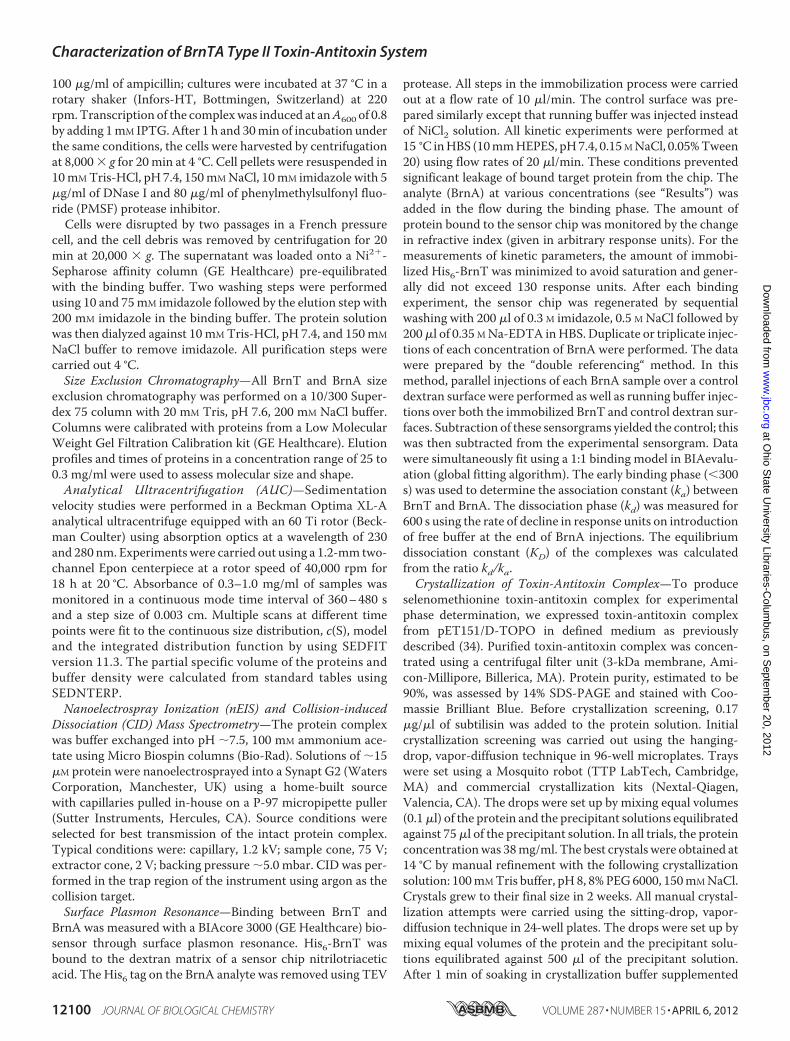
100 �g/ml of ampicillin; cultures were incubated at 37 °C in arotary shaker (Infors-HT, Bottmingen, Switzerland) at 220rpm.Transcription of the complexwas induced at anA600 of 0.8by adding 1mM IPTG.After 1 h and 30min of incubation underthe same conditions, the cells were harvested by centrifugationat 8,000 � g for 20 min at 4 °C. Cell pellets were resuspended in10mMTris-HCl, pH 7.4, 150mMNaCl, 10mM imidazole with 5�g/ml of DNase I and 80 �g/ml of phenylmethylsulfonyl fluo-ride (PMSF) protease inhibitor.Cells were disrupted by two passages in a French pressure
cell, and the cell debris was removed by centrifugation for 20min at 20,000 � g. The supernatant was loaded onto a Ni2�-Sepharose affinity column (GE Healthcare) pre-equilibratedwith the binding buffer. Two washing steps were performedusing 10 and 75mM imidazole followed by the elution step with200 mM imidazole in the binding buffer. The protein solutionwas then dialyzed against 10 mMTris-HCl, pH 7.4, and 150mM
NaCl buffer to remove imidazole. All purification steps werecarried out 4 °C.Size Exclusion Chromatography—All BrnT and BrnA size
exclusion chromatography was performed on a 10/300 Super-dex 75 column with 20 mM Tris, pH 7.6, 200 mM NaCl buffer.Columns were calibrated with proteins from a Low MolecularWeight Gel Filtration Calibration kit (GE Healthcare). Elutionprofiles and times of proteins in a concentration range of 25 to0.3 mg/ml were used to assess molecular size and shape.Analytical Ultracentrifugation (AUC)—Sedimentation
velocity studies were performed in a Beckman Optima XL-Aanalytical ultracentrifuge equipped with an 60 Ti rotor (Beck-man Coulter) using absorption optics at a wavelength of 230and 280 nm. Experimentswere carried out using a 1.2-mm two-channel Epon centerpiece at a rotor speed of 40,000 rpm for18 h at 20 °C. Absorbance of 0.3–1.0 mg/ml of samples wasmonitored in a continuous mode time interval of 360–480 sand a step size of 0.003 cm. Multiple scans at different timepoints were fit to the continuous size distribution, c(S), modeland the integrated distribution function by using SEDFITversion 11.3. The partial specific volume of the proteins andbuffer density were calculated from standard tables usingSEDNTERP.Nanoelectrospray Ionization (nEIS) and Collision-induced
Dissociation (CID) Mass Spectrometry—The protein complexwas buffer exchanged into pH �7.5, 100 mM ammonium ace-tate using Micro Biospin columns (Bio-Rad). Solutions of �15�M protein were nanoelectrosprayed into a Synapt G2 (WatersCorporation, Manchester, UK) using a home-built sourcewith capillaries pulled in-house on a P-97 micropipette puller(Sutter Instruments, Hercules, CA). Source conditions wereselected for best transmission of the intact protein complex.Typical conditions were: capillary, 1.2 kV; sample cone, 75 V;extractor cone, 2 V; backing pressure �5.0 mbar. CID was per-formed in the trap region of the instrument using argon as thecollision target.Surface Plasmon Resonance—Binding between BrnT and
BrnA was measured with a BIAcore 3000 (GE Healthcare) bio-sensor through surface plasmon resonance. His6-BrnT wasbound to the dextran matrix of a sensor chip nitrilotriaceticacid. The His6 tag on the BrnA analyte was removed using TEV
protease. All steps in the immobilization process were carriedout at a flow rate of 10 �l/min. The control surface was pre-pared similarly except that running buffer was injected insteadof NiCl2 solution. All kinetic experiments were performed at15 °C inHBS (10mmHEPES, pH7.4, 0.15MNaCl, 0.05%Tween20) using flow rates of 20 �l/min. These conditions preventedsignificant leakage of bound target protein from the chip. Theanalyte (BrnA) at various concentrations (see “Results”) wasadded in the flow during the binding phase. The amount ofprotein bound to the sensor chip was monitored by the changein refractive index (given in arbitrary response units). For themeasurements of kinetic parameters, the amount of immobi-lized His6-BrnT was minimized to avoid saturation and gener-ally did not exceed 130 response units. After each bindingexperiment, the sensor chip was regenerated by sequentialwashing with 200 �l of 0.3 M imidazole, 0.5 M NaCl followed by200�l of 0.35 MNa-EDTA inHBS. Duplicate or triplicate injec-tions of each concentration of BrnA were performed. The datawere prepared by the “double referencing“ method. In thismethod, parallel injections of each BrnA sample over a controldextran surface were performed as well as running buffer injec-tions over both the immobilized BrnT and control dextran sur-faces. Subtraction of these sensorgrams yielded the control; thiswas then subtracted from the experimental sensorgram. Datawere simultaneously fit using a 1:1 binding model in BIAevalu-ation (global fitting algorithm). The early binding phase (�300s) was used to determine the association constant (ka) betweenBrnT and BrnA. The dissociation phase (kd) was measured for600 s using the rate of decline in response units on introductionof free buffer at the end of BrnA injections. The equilibriumdissociation constant (KD) of the complexes was calculatedfrom the ratio kd/ka.Crystallization of Toxin-Antitoxin Complex—To produce
selenomethionine toxin-antitoxin complex for experimentalphase determination, we expressed toxin-antitoxin complexfrom pET151/D-TOPO in defined medium as previouslydescribed (34). Purified toxin-antitoxin complex was concen-trated using a centrifugal filter unit (3-kDa membrane, Ami-con-Millipore, Billerica, MA). Protein purity, estimated to be90%, was assessed by 14% SDS-PAGE and stained with Coo-massie Brilliant Blue. Before crystallization screening, 0.17�g/�l of subtilisin was added to the protein solution. Initialcrystallization screening was carried out using the hanging-drop, vapor-diffusion technique in 96-well microplates. Trayswere set using a Mosquito robot (TTP LabTech, Cambridge,MA) and commercial crystallization kits (Nextal-Qiagen,Valencia, CA). The drops were set up by mixing equal volumes(0.1�l) of the protein and the precipitant solutions equilibratedagainst 75�l of the precipitant solution. In all trials, the proteinconcentrationwas 38mg/ml. The best crystals were obtained at14 °C by manual refinement with the following crystallizationsolution: 100mMTris buffer, pH 8, 8%PEG6000, 150mMNaCl.Crystals grew to their final size in 2 weeks. All manual crystal-lization attempts were carried using the sitting-drop, vapor-diffusion technique in 24-well plates. The drops were set up bymixing equal volumes of the protein and the precipitant solu-tions equilibrated against 500 �l of the precipitant solution.After 1 min of soaking in crystallization buffer supplemented
Characterization of BrnTA Type II Toxin-Antitoxin System
12100 JOURNAL OF BIOLOGICAL CHEMISTRY VOLUME 287 • NUMBER 15 • APRIL 6, 2012
with 20% of glycerol and 5 mM �-mercaptoethanol, crystalswere flash-frozen directly at 100 K in liquid nitrogen. Incorpo-ration of selenomethionine in the crystal was confirmed bymeasuring x-ray fluorescence.Crystallographic Data Collection and Processing—Crystal
diffraction data were collected at a temperature of 100 K onbeamline 21-ID-D (LS-CAT, Advanced Photon Source,Argonne, IL) using a MARMosaic 300 detector and an oscilla-tion range of 1 degree. Diffraction images were reduced usingthe HKL 2000 suite (35).Phasing and Refinement—Diffraction from a single sel-
enomethionine-BrnT protein crystal was measured at anenergy of 12.66 keV (0.979 Å), and the structure of BrnT wasphased from the resulting 1.4-Å dataset by selenium single-wavelength anomalous dispersion (36). Three selenium siteswere located within the asymmetric unit using the Autosol sin-gle-wavelength anomalous dispersion routine in Phenix (37).An initial structure was built into these maps and refined to anRfree of 21%. This structure was then used as a molecularreplacement searchmodel to phase a 1.1-Ånative BrnTdataset.BrnTwas further refined against the 1.1-Å data to anRwork of
14.0% and an Rfree of 15.8% using phenix.refine in the Phenixsoftware suite. Manual model building, solvent addition, andrefinement of this structure was conducted iteratively usingCoot (38) and phenix.refine (see Table 3).Sequence Alignment and Protein Visualization Methods—
Protein sequence alignments were carried out in ClustalW2.BrnT ribbon structure rendering, electrostatic potential sur-faces calculation (by using the generate-vacuum electrostaticsfunction), and visualization of the hydrophobic surfaces(according to the Eisenberg hydrophobicity scale) (39) wereperformed with PyMOL. Visualization of the conserved resi-dues in the structure was performed by using the Consurfserver (40). Predicted residues involved in RNA binding havebeen predicted with three different programs: RNABindR (41),BindN (42), and PiRaNhA (43).Protein Synthesis Assay—E. coli Rosetta pLysS/DE3 with
pET151/D�bab1_0993, pET151/D�bab1_0994, or pET151/D�bab1_0993-4 were grown to an A600 0.5 and then inducedwith IPTG to a final concentration of 0.5 mM for 30 min. Cellswere washed twice by centrifuging at 7,000 � g for 1 min andresuspended in 1 ml of M9 medium � all amino acids butmethionine and cysteine. [35S]Methionine and cysteine wereadded to media for 1 min and then protein synthesis wasstopped by adding 75 �l of ice-cold 100% TCA followed by astandardTCAprecipitation. Sampleswere then resuspended insample buffer (0.5 M Tris, 4% SDS, pH 8.0), boiled for 10 min,and 5 �l were added to scintillation fluid and scintillationcounted.In Vitro RNase Assay—In vitro lacZ RNA transcript was
made with pET151/D � lacZ and the MEGAscript high yieldtranscription kit (Ambion) according to the manufacturer’sprotocol. RNAwas incubatedwithwater, buffer, 8, 4, 2, 1, or 0.5�M BrnT, 16 �M BrnA, or 4 �M BrnT � 16 �M BrnA for 20 minat room temperature. All proteins were purified with a Ni2�
affinity column and a Superdex 75 size exclusion column. Sam-ples were loaded in 1% TAE-agarose gel. In the RNase assay
withmutant proteins, 0.6�MBrnTwas incubatedwith RNA for30 min at 37 °C.Electrophoretic Mobility Shift Assay—Fifty nucleotide oligos
were purchased from IDT (Coralville, IA) (bab1_0994, �50forward, 5�-GTACGATTATTATTGCGAGCCCAGACGAA-TTATTGTACATACGTTTTATG, bab1_0994, �50 reverse,5�-CATAAAACGTATGTACAATAATTCGTCTGGGCTC-GCAATAATAATCGTAC).Oligoswere labeledwith 32P usingT4 polynucleotide kinase (Fermentas) by incubating the reac-tion at 37 °C for 20 min. Unincorporated nucleotides wereremoved using Micro Bio-spin 6 chromatography columns(Bio-Rad) and oligoswere annealed bymixing and incubating at80 °C for 20 min, then letting them cool to room temperatureovernight. Binding reactions consisted of 1 �l of 20 nM 32P-annealed oligo, 20 ng of salmon spermDNA, 4�l of 5� bindingbuffer (100 mM Tris, pH 8.0, 250 mM NaCl, 25% glycerol, 250ng/�l of BSA), 13 �l of H2O, and 1 �l of protein at concentra-tion of interest. The binding reaction took place for 30 minat room temperature and then 4 �l of 80% glycerol was addedto each sample. Samples were loaded in a 12% polyacryl-amide native gel and run at 140 V for 2 h. The gel was placedon a phosphorscreen overnight and imaged with a Typhoonscanner.
�-Galactosidase Transcription Reporter Assay—Transcrip-tional activity of the brnTA promoter was assayed using aPbrnTA-lacZ promoter fusion plasmid incorporated in wild-type B. abortus or �brnTA. Overnight cultures of both strainswere diluted toA600 0.05. Cultures were grown for 3 h and thenthe A600 was measured. �-Galactosidase activity was deter-mined as previously described (44).Stress Assays and Quantification of Transcription by
RT-qPCR—B. abortus 2308 (1 � 108 cfu/ml) was stressed witheither 200 �g/ml of chloramphenicol versus an equal volume of95% ethanol as a control, 44 versus 37 °C, 5 mM H2O2 versus anequal volume of H2O, or grown in Brucella broth and exposedto pH4.0 versus 7.0. All stress assayswere carried out for 30minwith the exception of low pH, which was done for 3 h. RNAwasharvested using TRIzol. CustomTaqMan primers were used tomeasure brnT transcript: forward, 5�-TCATCGCCGTCATT-TTCAAG, reverse, 5�-TGCTGAACGCATGGAGATCA, andprobe, 5�-CGGTTGGTTCGGAAGCCCTCTCC. RNA wasreverse transcribed for 30 min at 50 °C followed by an inactiva-tion step, 95 °C for 6 min. The cDNAwas then amplified for 50cycles of 95 °C for 15 s, 60 °C for 30 s, and 72 °C for 15 s. B.abortus RNA levels were normalized to 16 S ribosomal RNA:forward, 5�-CCTTACGGGCTGGGCTAC, reverse: 5�-TGCT-CGCTGCCCACTGT, probe, 5�-ACGTGCTACAATGGTGG.All assays were performed on an ABI 7300 system and analyzedwith SDS 1.3 software (Applied Biosystems).
RESULTS
BrnTA Is a Novel Toxin-Antitoxin System—Using a combi-nation of bioinformatic metrics, Makarova and colleagues (45)predicted four type II TA systems in the B. abortus 2308genome. Of these bab1_0993 and bab1_0994, encoding a puta-tive antitoxin and toxin, respectively, have an unusual genearrangement and physicochemical properties as comparedwith typical type II systems (31). Atypical features include: 1)
Characterization of BrnTA Type II Toxin-Antitoxin System
APRIL 6, 2012 • VOLUME 287 • NUMBER 15 JOURNAL OF BIOLOGICAL CHEMISTRY 12101
the putative toxin gene, bab1_0994, precedes the antitoxin,bab1_0993, in the operon (Fig. 1A); generally the antitoxin geneis positioned upstream of the toxin, 2) the predicted toxin andantitoxin proteins have equivalent theoretical isoelectric pointsof 9.5; toxins are typically basic and antitoxins acidic, and 3) thepredicted antitoxin is larger than the toxin, and has a C-termi-nal (rather than N-terminal) ribbon-helix-helix DNA bindingdomain.To test if bab1_0993–0994 encodes a bona fide TA sys-
tem, we expressed the predicted toxin, antitoxin, and toxin-antitoxin operon heterologously in E. coli from an induciblepromoter. In strains expressing the toxin alone, bacterialgrowth as measured by visible optical density ceased within
1 h of induction (Fig. 2A). Expression of antitoxin alone didnot significantly attenuate growth relative to wild-type.Coordinate expression of the antitoxin significantly reducedtoxicity of the toxin (Fig. 2A). When viable colony formingunits were plated at time points after induction of toxin,antitoxin, or toxin-antitoxin operon (Fig. 2B), expression ofthe toxin, but not the antitoxin or full TA operon, resulted inan apparent �3 log loss of viable cfu after 1 h, and an over 4log loss after 2 h.An E. coli strain that has toxin under the control of the lac
promoter and antitoxin under control of the ara promoter,exhibits the same apparent decrease in “viability” in the pres-ence of IPTG and absence of arabinose (Fig. 2E). However, if we
FIGURE 1. The brnTA operon. A, the brnT and brnA genes overlap by 1 bp. B, clustal multiple sequence alignment of B. abortus BrnT toxin and homologous toxinsequences from 10 other Gram-negative species including several human pathogens. Ba, B. abortus (GI: 82615932); Ec, E. coli (GI: 309704811); Yp, Yersinia pestis(GI: 22126312); Ypt, Yersinia pseudotuberculosis (GI: 170024520); Bpp, Bordatella parapertussis (GI: 33596571); Nm, Neisseria meningitidis (GI: 161870004); Hi,Haemophilus influenzae (GI: 270670382); Bp, Burkholderia pseudomallei (GI: 217424545); Vc, Vibrio cholerae (GI: 15601097); Cb, Coxiella burnetii (GI: 154705785);Cc, Caulobacter crescentus (GI: 16124744). Boxes shaded black are amino acids identical in 40% of sequences shown. Boxes shaded gray are similar in 40% ofsequences.
FIGURE 2. B. abortus BrnTA is a toxin-antitoxin system. A, antitoxin (brnA, blue), toxin (brnT, red), or brnTA operon (green) were expressed in E. coli and opticaldensity at 600 nm (A600) was measured at 30-min intervals. B, starting at the time of induction (marked with black arrows) colony forming units were quantifiedon LB agar. C, growth curves of B. abortus 2308 � pSRK-Km (empty vector control, black), pSRK-brnT (red), pSRK-brnA (blue), or pSRK-brnTA (green). A660 wasmeasured every hour; expression of genes from pSRK was induced after 4 h (black arrow). D, B. abortus viable colony forming units after induction of geneexpression were enumerated by plating on Schaedler blood agar. E, E. coli Plac-brnT � Para-brnA or Plac-brnT � Para empty vector control (EVC) were grownin the presence or absence of 0.5 mM IPTG for increasing amounts of time. Cells were then plated on LB agar containing ampicillin and kanamycin � 0.2%arabinose. Solid lines indicate the presence of arabinose in agar; dashed lines indicate agar without arabinose. � or � indicates induction of specific protein.Error bars represent S.D.
Characterization of BrnTA Type II Toxin-Antitoxin System
12102 JOURNAL OF BIOLOGICAL CHEMISTRY VOLUME 287 • NUMBER 15 • APRIL 6, 2012
induce toxin and plate on solid medium containing 0.2% arab-inose at time points up to 5 h after induction, it is evident thatexpression of toxin does not result in a decrease in viability, butrather a decrease in culturability (in this timeperiod). Inductionof bab1_0994 thus leads to a bacteriostatic state, and subse-quent induction of the antitoxin is able to neutralize the toxinand rescue cells from stasis.To confirm bab1_0993–0994 acts in the same manner in B.
abortus 2308, we performed similar experiments in which weectopically expressed toxin, antitoxin, or TA operon from aninducible promoter. The results of these experiments in B.abortus were consistent with what we observed in E. coli:expression of the toxin resulted in arrest of bacterial growth anda decrease in culturability, and co-expression of the antitoxinneutralized these effects (Fig. 2, C and D). Together these dataindicate that BAB1_0993–0994 functions as a toxin-antitoxinsystem and that expression of the toxin, BAB1_0994, isbacteriostatic.A search of the BAB1_0994 primary sequence against the
Pfam 25.0 data base shows that this toxin is not limited to B.abortus. BAB1_0994 is described by the profile hiddenMarkovmodel of domain family DUF497 in Pfam 25.0 (E � 2e-11) (46).This hidden Markov model apprehends 499 sequences from317 species spanning archaea, plasmids, bacteriophage, andbacteria, several of which are human pathogens (Fig. 1B). Ourstudy, for the first time, ascribes a function to this domain fam-ily. Based ondata thatwewill present anddiscuss further below,we have named this conserved toxin, BrnT and its cognate anti-toxin, BrnA.BrnT-BrnAToxin-Antitoxin SystemForms aHighAffinity 2:2
Tetrameric Complex—To test if there is a physical associationbetweenBrnT andBrnA,we co-expressedHis6-BrnT andBrnAfrom a single plasmid. His6-BrnT and BrnA were co-purified
fromNi2�-chelating Sepharose resin. Resolution of this proteincomplex by SDS-PAGE revealed a ladder of protein bands atweights larger than monomeric toxin or antitoxin (Fig. 3A).Analysis of each of the five bands on the gel by mass spectrom-etry showed that four were composed solely of BrnT and onewas BrnA (supplemental Table S1). BrnT has no cysteines andformed these unusual oligomers under denaturing condi-tions (as assessed by SDS-PAGE) in both the presence orabsence of the reductant �-mercaptoethanol, suggesting it isnot a result of oxidative bond formation. We do not cur-rently understand why BrnT forms higher order oligomersunder denaturing conditions.To characterize the molecular weight, oligomeric state, and
structure of the BrnT-BrnA complex under native conditions,we used AUC and nEIS-MS. Sedimentation velocity analysis ofthe purified BrnT-BrnA complex revealed one major 2.9 � 0.3S particle (Fig. 3A). Although a molecular weight distributionassociated with this particle can be calculated from a distribu-tion of Lamm equation solutions, it requires knowledge of theshape and other parameters that affect its hydrodynamic prop-erties (47). Given a molecular weight, one can thus draw con-clusions about the shape and folded state of a protein complexfrom the sedimentation velocity data. As such, we directlydetermined the molecular weight of the BrnT-BrnA complexby nEIS-MS. The data clearly show that the purified proteincomplex exists primarily as a species ofmass 48,906Da (Fig. 3B,top); this is the exact predictedmolecularmass of a 2:2 antitoxinto toxin tetramer. The 15� tetramer species (m/z 3261) wasisolated and dissociated via CID, resulting in complementarymonomer and trimer product ions (A and AT2, T and A2T; Fig.3B, bottom) that confirm the precursor ionwas anA2T2 hetero-tetramer. Further analysis of theCIDdata revealed a low level ofBrnA-BrnT heterodimer and BrnA2 homodimer. BrnA and
FIGURE 3. BrnTA forms a tetramer consisting of two antitoxins and two toxins. A, c(S) distribution calculated from sedimentation velocity measurementsof the purified BrnTA complex. BrnT-BrnA sediments as a single 2.9 � 0.3 S species (root mean square deviation 0.006). Resolution of this purified complexby 14% SDS-PAGE reveals multiple Coomassie-stained bands; mass spectrometry of tryptic peptides from these excised bands demonstrates that they containeither BrnT or BrnA, labeled as A or T to the right of each band. B, top, nESI-MS of purified BrnA-BrnT identifies a major complex with a molecular weight thatmatches a tetramer with 2:2 stoichiometry. B, bottom, 70-V CID spectrum of the 15� tetramer ion species reveals monomeric and trimeric product ions,supporting a model in which the precursor is a 2:2 tetramer. C, size exclusion chromatography of BrnT (red) and BrnA (blue) and SDS-PAGE of major peaks. D, c(S)distribution from AUC of BrnT (red) and BrnA (blue), BrnT is 13.2 kDa, root mean square deviation 0.022; frictional ratio 1.2. BrnA is 29.3 kDa, root meansquare deviation 0.006, frictional ratio 1.8.
Characterization of BrnTA Type II Toxin-Antitoxin System
APRIL 6, 2012 • VOLUME 287 • NUMBER 15 JOURNAL OF BIOLOGICAL CHEMISTRY 12103
BrnT monomers are approximately equally abundant (at lowlevels) in these spectra. There is no evidence of BrnT2 dimerby CID. There is a significantly greater abundance of theT/A2T pair than the A/AT2 pair, suggesting that it is moredifficult to release the antitoxin than the toxin from the com-plex. Based on these mass spectrometry data, we propose amodel in which dimers of antitoxin recruit two individualtoxin monomers to form a tetrameric complex consisting oftwo BrnA proteins and two BrnT proteins (Fig. 3B, inset).Based on the presence of a small amount of AT2 trimer afterCID, we think toxins may interact after formation of thetetrameric complex.We additionally analyzed BrnT and BrnA individually by size
exclusion chromatography andAUC.Weobserved that BrnT ismonomeric across a range of concentrations and that BrnAsediments as a likely dimer of high frictional ratio (Fig. 3, C andD). These data support our nEIS-MS results where we observemonomers of both toxin and antitoxin, and dimers of antitoxin,but no toxin dimers. Given a complexmolecularmass of 48,906Da, we reanalyzed our sedimentation velocity data to assess thehydrodynamic properties of theTAcomplex. Froma fit of thesedata we determined that a frictional ratio of 1.6 is consistentwith a 48.9-kDa 2.9 S particle. This frictional ratio is higher thantypical globular proteins (1.1–1.3) and suggests that a portion ofthe BrnT2-BrnA2 complex is loosely structured or random coil(48). Based on the fit frictional ratio of BrnA2, we propose thatthe BrnA antitoxin has disordered, unstructured regions thatcontribute to the high apparent frictional ratio. This is consis-tent with the high relative susceptibility of BrnA to proteolysisin vitro (see our description of BrnT crystallization below), ashas been reported for other antitoxins.We further measured the affinity of the BrnT-BrnA complex
by surface plasmon resonance. His6-BrnT was immobilized ona nickel-nitrilotriacetic acid chip, and BrnA flowed at concen-trations ranging from 17 to 500 nM. BrnA binds BrnT withassociation and dissociation rate constants of 2.44 � 105 M�1
s�1 and 1.63 � 10�4 s�1, respectively. The ratio of the dissoci-ation to association rate constant yields an equilibrium affinityof 670 pM (Table 2).Crystal Structure of BrnT Reveals a Fold Topologically
Related to RelE Toxin Family—Having defined BrnTA as a tox-in-antitoxin system that forms a direct physical association, wenext sought to characterize the molecular structure of thisnovel TA system. Although purified the BrnT-BrnA complexfailed to yield crystals, incubation of BrnT-BrnA with a lowconcentration of subtilisin in the crystallization drop (see“Experimental Procedures”) produced orthorhombic crystalsof space group I4, with cell dimensions a and b 74.4 Å andc 29.6 Å. The structure was solved from a single selenome-thionine crystal phased by single-wavelength anomalous dis-persion. This crystal contained onemolecule of BrnT per asym-
metric unit; the protein structure was refined to a final Rwork of0.140 and an Rfree of 0.158 (Table 3).
Full-length BrnT (PDB ID 3U97) contains 81 residues; elec-tron density for the five C-terminal residues is not visible,which is likely due to disorder or to proteolysis of this segmentof the structure. BrnT is topologically related to the RelE familyof bacterial RNase toxins at the level of secondary structure,althoughoverall the tertiary structural homology toRelE and itsstructural homologs (e.g. YoeB and MqsR) is low (DALIZ-scores �5.3). BrnT is composed of a 4-stranded antiparallel�-scaffold (�2-�3-�4-�5); �5 is flanked by a single parallel �strand (�1) (Fig. 4). Two short�-helices (�2 and�3) are dockedon a hydrophobic face of the �-scaffold, and helix �1 interacts
FIGURE 4. Structure of BrnT. A, ribbon structure of BrnT. �-Strands, pink;�-helices, light blue. B, electrostatic surface map of BrnT; basic, blue; acidic, red.C, conservation surface map of BrnT reveals the structural position of residuesthat are conserved among BrnT homologs (DUF497 in the Conserved DomainDatabase). Coordinates of B. abortus BrnT have been deposited in the ProteinData Bank (PDB ID 3U97).
TABLE 2BrnT-BrnA binding measured by surface plasmon resonance
TABLE 3Crystallographic data and refinement statistics
aRmerge hkli�Ii-�I��/hkliIi, for all data � �3.b Experimental phases were determined by SAD using the anomalous signal fromselenium. Total figure of merit values are based on experimental phase informa-tion (prior) for all reflections.
c Rcryst hkl �Fobs� � �Fcalc�/hkl�Fobs�.d Rfree uses 1906 total reflections for cross-validation.e rmsd, root mean square deviation.
Characterization of BrnTA Type II Toxin-Antitoxin System
12104 JOURNAL OF BIOLOGICAL CHEMISTRY VOLUME 287 • NUMBER 15 • APRIL 6, 2012
directly with �5. The most conserved region of the BrnT pri-mary sequence corresponds to the solvent-exposed face of the�-scaffold and surface of helix�1 that interacts with the�-scaf-fold. This surface contains a highly positive basic patch thatresembles a functional site for RNA interaction and cleavage(Fig. 4). Experiments in which we test this hypothesis are dis-cussed below.BrnT Is a Ribonuclease—As discussed above, type II toxins
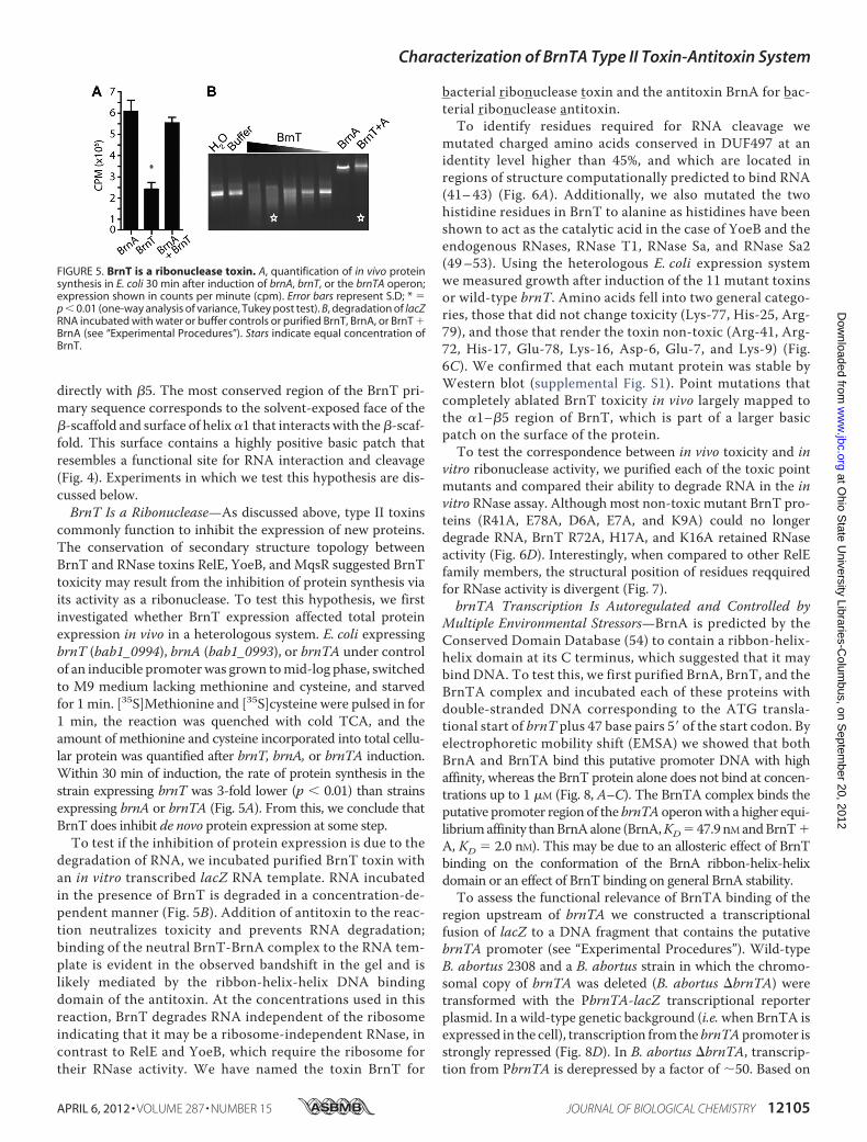
commonly function to inhibit the expression of new proteins.The conservation of secondary structure topology betweenBrnT and RNase toxins RelE, YoeB, and MqsR suggested BrnTtoxicity may result from the inhibition of protein synthesis viaits activity as a ribonuclease. To test this hypothesis, we firstinvestigated whether BrnT expression affected total proteinexpression in vivo in a heterologous system. E. coli expressingbrnT (bab1_0994), brnA (bab1_0993), or brnTA under controlof an inducible promoterwas grown tomid-log phase, switchedto M9 medium lacking methionine and cysteine, and starvedfor 1 min. [35S]Methionine and [35S]cysteine were pulsed in for1 min, the reaction was quenched with cold TCA, and theamount of methionine and cysteine incorporated into total cellu-lar protein was quantified after brnT, brnA, or brnTA induction.Within 30 min of induction, the rate of protein synthesis in thestrain expressing brnT was 3-fold lower (p � 0.01) than strainsexpressing brnA or brnTA (Fig. 5A). From this, we conclude thatBrnT does inhibit de novo protein expression at some step.To test if the inhibition of protein expression is due to the
degradation of RNA, we incubated purified BrnT toxin withan in vitro transcribed lacZ RNA template. RNA incubatedin the presence of BrnT is degraded in a concentration-de-pendent manner (Fig. 5B). Addition of antitoxin to the reac-tion neutralizes toxicity and prevents RNA degradation;binding of the neutral BrnT-BrnA complex to the RNA tem-plate is evident in the observed bandshift in the gel and islikely mediated by the ribbon-helix-helix DNA bindingdomain of the antitoxin. At the concentrations used in thisreaction, BrnT degrades RNA independent of the ribosomeindicating that it may be a ribosome-independent RNase, incontrast to RelE and YoeB, which require the ribosome fortheir RNase activity. We have named the toxin BrnT for
bacterial ribonuclease toxin and the antitoxin BrnA for bac-terial ribonuclease antitoxin.To identify residues required for RNA cleavage we
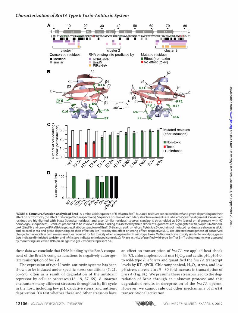
mutated charged amino acids conserved in DUF497 at anidentity level higher than 45%, and which are located inregions of structure computationally predicted to bind RNA(41–43) (Fig. 6A). Additionally, we also mutated the twohistidine residues in BrnT to alanine as histidines have beenshown to act as the catalytic acid in the case of YoeB and theendogenous RNases, RNase T1, RNase Sa, and RNase Sa2(49–53). Using the heterologous E. coli expression systemwe measured growth after induction of the 11 mutant toxinsor wild-type brnT. Amino acids fell into two general catego-ries, those that did not change toxicity (Lys-77, His-25, Arg-79), and those that render the toxin non-toxic (Arg-41, Arg-72, His-17, Glu-78, Lys-16, Asp-6, Glu-7, and Lys-9) (Fig.6C). We confirmed that each mutant protein was stable byWestern blot (supplemental Fig. S1). Point mutations thatcompletely ablated BrnT toxicity in vivo largely mapped tothe �1–�5 region of BrnT, which is part of a larger basicpatch on the surface of the protein.To test the correspondence between in vivo toxicity and in
vitro ribonuclease activity, we purified each of the toxic pointmutants and compared their ability to degrade RNA in the invitro RNase assay. Although most non-toxic mutant BrnT pro-teins (R41A, E78A, D6A, E7A, and K9A) could no longerdegrade RNA, BrnT R72A, H17A, and K16A retained RNaseactivity (Fig. 6D). Interestingly, when compared to other RelEfamily members, the structural position of residues reqquiredfor RNase activity is divergent (Fig. 7).brnTA Transcription Is Autoregulated and Controlled by
Multiple Environmental Stressors—BrnA is predicted by theConserved Domain Database (54) to contain a ribbon-helix-helix domain at its C terminus, which suggested that it maybind DNA. To test this, we first purified BrnA, BrnT, and theBrnTA complex and incubated each of these proteins withdouble-stranded DNA corresponding to the ATG transla-tional start of brnT plus 47 base pairs 5� of the start codon. Byelectrophoretic mobility shift (EMSA) we showed that bothBrnA and BrnTA bind this putative promoter DNA with highaffinity, whereas the BrnT protein alone does not bind at concen-trations up to 1 �M (Fig. 8, A–C). The BrnTA complex binds theputative promoter region of the brnTA operonwith a higher equi-libriumaffinity thanBrnAalone (BrnA,KD47.9nMandBrnT�A, KD 2.0 nM). This may be due to an allosteric effect of BrnTbinding on the conformation of the BrnA ribbon-helix-helixdomain or an effect of BrnT binding on general BrnA stability.To assess the functional relevance of BrnTA binding of the
region upstream of brnTA we constructed a transcriptionalfusion of lacZ to a DNA fragment that contains the putativebrnTA promoter (see “Experimental Procedures”). Wild-typeB. abortus 2308 and a B. abortus strain in which the chromo-somal copy of brnTA was deleted (B. abortus �brnTA) weretransformed with the PbrnTA-lacZ transcriptional reporterplasmid. In a wild-type genetic background (i.e.when BrnTA isexpressed in the cell), transcription from the brnTApromoter isstrongly repressed (Fig. 8D). In B. abortus �brnTA, transcrip-tion from PbrnTA is derepressed by a factor of �50. Based on
FIGURE 5. BrnT is a ribonuclease toxin. A, quantification of in vivo proteinsynthesis in E. coli 30 min after induction of brnA, brnT, or the brnTA operon;expression shown in counts per minute (cpm). Error bars represent S.D; * p � 0.01 (one-way analysis of variance, Tukey post test). B, degradation of lacZRNA incubated with water or buffer controls or purified BrnT, BrnA, or BrnT �BrnA (see “Experimental Procedures”). Stars indicate equal concentration ofBrnT.
Characterization of BrnTA Type II Toxin-Antitoxin System
APRIL 6, 2012 • VOLUME 287 • NUMBER 15 JOURNAL OF BIOLOGICAL CHEMISTRY 12105
these data we conclude that DNA binding by the BrnA compo-nent of the BrnTA complex functions to negatively autoregu-late transcription of brnTA.The expression of type II toxin-antitoxin systems has been
shown to be induced under specific stress conditions (7, 21,55–57), often as a result of degradation of the antitoxinrepressor by cellular proteases (18, 19, 57–59). B. abortusencounters many different stressors throughout its life cyclein the host, including low pH, oxidative stress, and nutrientdeprivation. To test whether these and other stressors have
an effect on transcription of brnTA we applied heat shock(44 °C), chloramphenicol, 5 mMH2O2, and acidic pH, pH 4.0,to wild-type B. abortus and quantified the brnTA transcriptlevels by RT-qPCR. Chloramphenicol, H2O2 stress, and lowpH stress all result in a 9–80-fold increase in transcription ofbrnTA (Fig. 8E). We presume these stressors lead to the deg-radation of BrnA through an unknown protease and thisdegradation results in derepression of the brnTA operon.However, we cannot rule out other mechanisms of brnTAtranscriptional activation.
FIGURE 6. Structure/function analysis of BrnT. A, amino acid sequence of B. abortus BrnT. Mutated residues are colored in red and green depending on theireffect on BrnT toxicity (no effect or strong effect, respectively). Sequence position of secondary structure elements are labeled above the alignment. Conservedresidues are highlighted with black (identical residues) and gray (similar residues) squares; shading is thresholded at 50% (based on alignment with 97homologous sequences). Residues predicted to be involved in RNA binding as assessed by three different algorithms are highlighted with purple (RNABindR),pink (BindN), and orange (PiRaNhA) squares. B, ribbon structure of BrnT. �-Strands, pink; �-helices, light blue. Side chains of mutated residues are shown as sticksand colored in red and green depending on their effect on BrnT toxicity (no effect or strong effect, respectively). C, site-directed mutagenesis of conservedcharged amino acids in BrnT reveals residues required for full toxicity when compared with wild-type toxin. Red bars indicate toxicity similar to wild-type, greenbars indicate diminished toxicity, and white bars indicate uninduced controls. D, RNase activity of purified wild-type BrnT or BrnT point mutants was assessedby monitoring uncleaved RNA on an agarose gel. Error bars represent S.D.
Characterization of BrnTA Type II Toxin-Antitoxin System
12106 JOURNAL OF BIOLOGICAL CHEMISTRY VOLUME 287 • NUMBER 15 • APRIL 6, 2012
Homeostatic Regulation and Stoichiometry—We have pre-sented evidence thatB. abortusBrnTA constitutes a novel, con-served ribonuclease toxin-antitoxin system. brnT expressionresults in cessation of bacterial growth that can be rescued bysubsequent expression of brnA, even 5 h after BrnT induction.This effect on bacterial cell physiology is similar to what hasbeen reported for the RelE and MazF toxins (60). BrnTA is aheterotetramer consisting of two molecules of BrnA and twomolecules of BrnT. brnTA is organized with the brnT toxingene preceding brnA antitoxin on B. abortus chromosome 1. Ithas been postulated that the canonical genetic organization of
type II TA systems (i.e. antitoxin preceding toxin in the operon)ensures that there is an excess of antitoxin protein, as the geneexpression level is often proportional to the gene order on apolycistronicmessage (10, 61, 62). However, there are a handfulof systems, such as BrnTA, in which toxin precedes antitoxin(e.g. mqsRA, higBA, and hicAB) (61, 63–65). This raises thequestion of whether there are additional regulatory mecha-nisms that ensure proper toxin:antitoxin ratios in the cell. In thecase of BrnTA, regulation may occur at the level of translation,as there is an apparent Shine-Dalgarno sequence upstream ofthe BrnA translation start, but no clear Shine-Dalgarnoupstream of BrnT. Thus, BrnT and BrnA expression may be
FIGURE 7. Structural comparison between BrnT and members of the RelE toxin family. For each toxin (BrnT, RelE, YoeB, and MqsR) a secondary structurediagram and a surface rendered model (white) are shown. Residues that are known to be required for RNA cleavage in each of these structures are highlightedin red. PDB codes of each structure are in parentheses.
FIGURE 8. BrnA negatively autoregulates the brnTA operon; brnTA transcription is activated by multiple stressors. Purified antitoxin (A) and TA complex(B), but not toxin alone (C) bind to 32P-labeled probe corresponding to the 50 nucleotides upstream of the brnT start codon, and cause a gel shift in EMSA.D, �-galactosidase assay of wild-type B. abortus or B. abortus �brnTA carrying a PbrnTA-lacZ promoter fusion plasmid. E, TaqMan assay quantifying brnTtranscript in wild-type B. abortus in various stress conditions including 5 mM H2O2 in Gerhardt’s minimal media, heat shock (44 °C), pH 4.0, and 200 �g/ml ofchloramphenicol. Each sample is normalized to 16 S RNA and then compared with a relevant non-stressed control. Error bars represent S.D.
Characterization of BrnTA Type II Toxin-Antitoxin System
APRIL 6, 2012 • VOLUME 287 • NUMBER 15 JOURNAL OF BIOLOGICAL CHEMISTRY 12107
affected by mRNA-ribosome pairing efficiency near the toxinand antitoxin translation start sites. Such a mechanism couldensure an excess of antitoxin relative to toxin under normalgrowth conditions.Structure and RNA Cleavage—BrnT is related to the RelE
family of RNase toxins in terms of its secondary topology. How-ever, the overall structure of BrnT differs from known RelEfamily members in several respects. BrnT lacks the C-terminalhelix that spans the solvent-exposed �-sheet of RelE. Addition-ally, �2–�3 that are docked across the hydrophobic face of the�-sheet are much smaller than the two corresponding helicesfound in RelE and YoeB. Moreover, the �-sheet of BrnT has 5strands in contrast to the 4 of RelE and the 6 of MqsR (Fig. 7).Despite these structural differences, the function of BrnT, sim-ilar to other RelE family members, is conserved as it rapidlyattenuates protein synthesis in vivo. Similar to the toxins RelE,YoeB, andMqsR, BrnTmost likely attenuates protein synthesisvia its activity as a ribonuclease.Our functional analysis of BrnTby site-directedmutagenesis identified several amino acids thatare required for RNA degradation. Surprisingly, not all of theamino acids required for toxicity in vivo are required for BrnTRNase activity in vitro under our assay conditions. Mutation ofresidues Arg-72, His-17, and Lys-16 to alanine blocks BrnTtoxicity in cells, but does not completely abrogate RNA cleav-age. It is not known whether mutation of these residues affectsKm or Vmax of the RNA scission reaction; clearly under theconditions we have assayed in vitro RNA cleavage can stilloccur.Scission of RNA by RelE family toxins requires specific gen-
eral acid and general base residues, as well as residues requiredfor orientation of nucleotides and stabilization of the transitionstate (51). The structural position of putative general acid andbase residues that we have identified in BrnT by site-directedmutagenesis do not correspond to those described for otherRelE family ribonucleases (Fig. 7). Our structural analysis isthus consistent with the emerging picture of the RelE fold as ascaffold onwhich particular residues required for RNA scissionare plastic; the position and identity of these residues may con-fer different properties/specificities to this family of ribonu-cleases (13).Environmental Regulation and Function—B. abortus brnA is
a member of COG3514 in the Conserved Domain Database.Based on secondary structure prediction, BrnA consists of 3�-helices and a C-terminal ribbon-helix-helix DNA bindingdomain. Like other toxin-antitoxin systems (66, 67), BrnA neg-atively autoregulates the brnTA operon and has higher affinityfor the DNA operator when complexed with BrnT. In responseto various environmental stressors, such as low pH and oxida-tive stress, we have shown that transcription of the brnTAoperon is up-regulated in B. abortus. B. abortus encountersthese stressors throughout the course of an infection, and tox-in-antitoxin systems such as BrnTA may facilitate persistenceof this bacterium within hostile host niches. We do not cur-rently know if regulated proteolysis of BrnA provides a mecha-nism by which expression of brnTA is up-regulated duringstress.An intriguing hypothesis is that BrnTA and the three other
TA systems in B. abortus underpin persistence and recurrent
infection in those individuals that contract Brucellosis. Indeed,TA systems such as HigBA and MqsRA can contribute to theestablishment of persister cells (17) and confer the ability ofbacteria to enter a dormant state during antibiotic treatment(16). Persistence is particularly problematic in Brucella spp.infection; even after rigorous antibiotic treatment, relapse ratesrange from 3 to 30% (2). Future studies on the functional role ofthis system and how it contributes to the fitness of B. abortusduring infectionmay informnew strategies for the treatment ofBrucellosis.
Acknowledgments—We thank Elena Solomaha for help with AUCand surface plasmon resonance experiments and Dick Winant at theStanford PAN facility for help with mass spectrometry protein finger-printing. The Advanced Photon Source is supported by the Depart-ment of Energy Office of Basic Energy Sciences (contract DE-AC02-06CH11357). LS-CAT is supported by the Michigan EconomicDevelopment Corp. and Michigan Technology Tri-Corridor Grant085P1000817. We also thank Aretha Fiebig and Nicholas Heaton forcritical discussions and comments on the manuscript and MartyRoop at East Carolina University for plasmids and B. abortus 2308.
REFERENCES1. Anderson, T. D., Meador, V. P., and Cheville, N. F. (1986) Pathogenesis of
placentitis in the goat inoculated with Brucella abortus. I. Gross and his-tologic lesions. Vet. Pathol. 23, 219–226
2. Moreno, E., andMoriyon, I. (2006) inThe Prokaryotes:AHandbook on theBiology of Bacteria (Dworkin, M., Falkow, S., Rosenberg, E., Schleifer,K. H., and Stackebrandt, E., eds) 3rd Ed., pp. 315–456, Springer, NewYork
3. Jiang, X., Leonard, B., Benson, R., and Baldwin, C. L. (1993) Macrophagecontrol of Brucella abortus. Role of reactive oxygen intermediates andnitric oxide. Cell. Immunol. 151, 309–319
4. Porte, F., Liautard, J. P., and Kohler, S. (1999) Early acidification of phago-somes containing Brucella suis is essential for intracellular survival inmurine macrophages. Infect. Immun 67, 4041–4047
5. Kohler, S., Michaux-Charachon, S., Porte, F., Ramuz, M., and Liautard,J. P. (2003) What is the nature of the replicative niche of a stealthy bugnamed Brucella? Trends Microbiol. 11, 215–219
6. Roop, R.M., 2nd, Gaines, J.M., Anderson, E. S., Caswell, C. C., andMartin,D. W. (2009) Survival of the fittest. How Brucella strains adapt to theirintracellular niche in the host.Med. Microbiol Immunol. 198, 221–238
7. Gerdes, K., Christensen, S. K., and Løbner-Olesen, A. (2005) Prokaryotictoxin-antitoxin stress response loci. Nat. Rev. Microbiol. 3, 371–382
8. Gerdes, K., Larsen, J. E., andMolin, S. (1985) Stable inheritance of plasmidR1 requires two different loci. J. Bacteriol 161, 292–298
9. Jaffe, A., Ogura, T., and Hiraga, S. (1985) Effects of the ccd function of theF plasmid on bacterial growth. J. Bacteriol. 163, 841–849
10. Pandey, D. P., and Gerdes, K. (2005) Toxin-antitoxin loci are highly abun-dant in free-living but lost from host-associated prokaryotes. Nucleic Ac-ids Res. 33, 966–976
11. Van Melderen, L., and Saavedra De Bast, M. (2009) Bacterial toxin-anti-toxin systems. More than selfish entities? PLoS Genet. 5, e1000437
12. Moyed, H. S., and Bertrand, K. P. (1983) hipA, a newly recognized gene ofEscherichia coli K-12 that affects frequency of persistence after inhibitionof murein synthesis. J. Bacteriol. 155, 768–775
13. Blower, T. R., Salmond, G. P., and Luisi, B. F. (2011) Balancing at survival’sedge. The structure and adaptive benefits of prokaryotic toxin-antitoxinpartners. Curr. Opin. Struct. Biol. 21, 109–118
14. Buts, L., Lah, J., Dao-Thi, M. H., Wyns, L., and Loris, R. (2005) Toxin-antitoxin modules as bacterial metabolic stress managers. TrendsBiochem. Sci. 30, 672–679
15. Keren, I., Shah, D., Spoering, A., Kaldalu, N., and Lewis, K. (2004) Special-ized persister cells and the mechanism of multidrug tolerance in Esche-richia coli. J. Bacteriol 186, 8172–8180
Characterization of BrnTA Type II Toxin-Antitoxin System
12108 JOURNAL OF BIOLOGICAL CHEMISTRY VOLUME 287 • NUMBER 15 • APRIL 6, 2012
16. Maisonneuve, E., Shakespeare, L. J., Jørgensen, M. G., and Gerdes, K.(2011) Bacterial persistence by RNA endonucleases. Proc. Natl. Acad. Sci.U.S.A. 108, 13206–13211
17. Wang, X., andWood, T. K. (2011) Toxin-antitoxin systems influence bio-film and persister cell formation and the general stress response. Appl.Environ. Microbiol. 77, 5577–5583
18. Tsuchimoto, S., Nishimura, Y., and Ohtsubo, E. (1992) The stable main-tenance system pem of plasmid R100. Degradation of PemI protein mayallow PemK protein to inhibit cell growth. J. Bacteriol. 174, 4205–4211
19. Van Melderen, L., Bernard, P., and Couturier, M. (1994) Lon-dependentproteolysis of CcdA is the key control for activation of CcdB in plasmid-free segregant bacteria.Mol. Microbiol. 11, 1151–1157
20. Lehnherr, H., and Yarmolinsky, M. B. (1995) Addiction protein Phd ofplasmid prophage P1 is a substrate of the ClpXP serine protease of Esch-erichia coli. Proc. Natl. Acad. Sci. U.S.A. 92, 3274–3277
21. Christensen, S. K., Pedersen, K., Hansen, F. G., and Gerdes, K. (2003)Toxin-antitoxin loci as stress-response elements. ChpAK/MazF andChpBK cleave translated RNAs and are counteracted by tmRNA. J. Mol.Biol. 332, 809–819
22. Pedersen, K., Zavialov, A. V., Pavlov, M. Y., Elf, J., Gerdes, K., and Ehren-berg, M. (2003) The bacterial toxin RelE displays codon-specific cleavageof mRNAs in the ribosomal A site. Cell 112, 131–140
23. Schumacher, M. A., Piro, K. M., Xu, W., Hansen, S., Lewis, K., and Bren-nan, R. G. (2009) Molecular mechanisms of HipA-mediated multidrugtolerance and its neutralization by HipB. Science 323, 396–401
24. Jiang, Y., Pogliano, J., Helinski, D. R., and Konieczny, I. (2002) ParE toxinencoded by the broad-host range plasmid RK2 is an inhibitor of Esche-richia coli gyrase.Mol. Microbiol. 44, 971–979
25. Maki, S., Takiguchi, S., Miki, T., and Horiuchi, T. (1992) Modulation ofDNA supercoiling activity of Escherichia coli DNA gyrase by F plasmidproteins. Antagonistic actions of LetA (CcdA) and LetD (CcdB) proteins.J. Biol. Chem. 267, 12244–12251
26. Yuan, J., Yamaichi, Y., andWaldor,M. K. (2011) The three vibrio choleraechromosome II-encoded ParE toxins degrade chromosome I followingloss of chromosome II. J. Bacteriol. 193, 611–619
27. Winther, K. S., and Gerdes, K. (2011) Enteric virulence associated proteinVapC inhibits translation by cleavage of initiator tRNA. Proc. Natl. Acad.Sci. U.S.A. 108, 7403–7407
28. Mutschler, H., Gebhardt, M., Shoeman, R. L., and Meinhart, A. (2011) Anovel mechanism of programmed cell death in bacteria by toxin-antitoxinsystems corrupts peptidoglycan synthesis. PLoS Biol. 9, e1001033
29. Lioy, V. S., Martín, M. T., Camacho, A. G., Lurz, R., Antelmann, H.,Hecker, M., Hitchin, E., Ridge, Y., Wells, J. M., and Alonso, J. C. (2006)pSM19035-encoded � toxin induces stasis followed by death in a subpop-ulation of cells.Microbiology 152, 2365–2379
30. Van Melderen, L. (2010) Toxin-antitoxin systems. Why so many, whatfor? Curr. Opin. Microbiol. 13, 781–785
31. Hayes, F., and Van Melderen, L. (2011) Toxins-antitoxins: Diversity, evo-lution, and function. Crit. Rev. Biochem. Mol. Biol. 46, 386–408
32. Khan, S. R., Gaines, J., Roop, R. M., 2nd, and Farrand, S. K. (2008) Broad-host range expression vectors with tightly regulated promoters and theiruse to examine the influence of TraR and TraM expression on Ti plasmidquorum sensing. Appl. Environ. Microbiol. 74, 5053–5062
33. Guzman, L. M., Belin, D., Carson, M. J., and Beckwith, J. (1995) Tightregulation, modulation, and high-level expression by vectors containingthe arabinose PBAD promoter. J. Bacteriol. 177, 4121–4130
34. Doublie, S. (1997) Preparation of selenomethionyl proteins for phase de-termination.Methods Enzymol. 276, 523–530
35. Otwinowski, Z., andMinor,W. (1997)Processing of X-ray diffraction datacollected in oscillation mode.Methods Enzymol. 276, 307–326
37. Adams, P.D., Afonine, P. V., Bunkoczi, G., Chen,V. B., Davis, I.W., Echols,N., Headd, J. J., Hung, L. W., Kapral, G. J., Grosse-Kunstleve, R. W., Mc-Coy, A. J., Moriarty, N. W., Oeffner, R., Read, R. J., Richardson, D. C.,Richardson, J. S., Terwilliger, T. C., and Zwart, P. H. (2010) PHENIX, acomprehensive Python-based system for macromolecular structure solu-tion. Acta Crystallogr. D 66, 213–221
38. Emsley, P., Lohkamp, B., Scott,W. G., and Cowtan, K. (2010) Features anddevelopment of Coot. Acta Crystallogr. D 66, 486–501
39. Eisenberg D., Schwarz, E., Komaromy, M., andWall, R. (1984) Analysis ofmembrane and surface protein sequences with the hydrophobic momentplot. J. Mol. Biol. 179, 125–142
40. Ashkenazy, H., Erez, E., Martz, E., Pupko, T., and Ben-Tal, N. (2010) Con-Surf 2010, calculating evolutionary conservation in sequence and struc-ture of proteins and nucleic acids. Nucleic Acids Res. 38,W529–533
41. Terribilini,M., Sander, J. D., Lee, J. H., Zaback, P., Jernigan, R. L., Honavar,V., and Dobbs, D. (2007) RNABindR, a server for analyzing and predictingRNA-binding sites in proteins. Nucleic Acids Res. 35,W578–584
42. Wang, L., and Brown, S. J. (2006) BindN, a web-based tool for efficientprediction of DNA and RNA binding sites in amino acid sequences. Nu-cleic Acids Res. 34,W243–248
43. Murakami, Y., Spriggs, R. V., Nakamura,H., and Jones, S. (2010) PiRaNhA,a server for the computational prediction of RNA-binding residues inprotein sequences. Nucleic Acids Res. 38,W412–416
44. Miller, J. H. (1972) Experiments in Molecular Genetics, Cold Spring Har-bor Laboratory, Cold Spring Harbor, NY
45. Makarova, K. S., Wolf, Y. I., and Koonin, E. V. (2009) Comprehensivecomparative-genomic analysis of type 2 toxin-antitoxin systems and re-lated mobile stress response systems in prokaryotes. Biol. Direct 4, 19
46. Finn, R. D.,Mistry, J., Tate, J., Coggill, P., Heger, A., Pollington, J. E., Gavin,O. L., Gunasekaran, P., Ceric, G., Forslund, K., Holm, L., Sonnhammer,E. L., Eddy, S. R., and Bateman, A. (2010) The Pfam protein families data-base. Nucleic Acids Res. 38, D211–222
47. Schuck, P. (2000) Size-distribution analysis of macromolecules by sedi-mentation velocity ultracentrifugation and lamm equationmodeling. Bio-phys. J. 78, 1606–1619
48. Tanford, C., Kawahara, K., Lapanje, S., Hooker, T.M., Jr., Zarlengo,M. H.,Salahuddin, A., Aune, K. C., and Takagi, T. (1967) Proteins as randomcoils. 3. Optical rotatory dispersion in 6 M guanidine hydrochloride. J. Am.Chem. Soc. 89, 5023–5029
49. Kamada, K., and Hanaoka, F. (2005) Conformational change in the catalyticsite of the ribonuclease YoeB toxin by YefM antitoxin.Mol. Cell 19, 497–509
50. Bauerova-Hlinkova, V., Dvorsky, R., Perecko, D., Povazanec, F., and Sev-cík, J. (2009) Structure of RNase Sa2 complexes with mononucleotides.New aspects of catalytic reaction and substrate recognition. FEBS J. 276,4156–4168
51. Heinemann, U., and Saenger, W. (1983) Crystallographic study of mech-anism of ribonuclease T1-catalyzed specific RNA hydrolysis. J. Biomol.Struct. Dyn. 1, 523–538
52. Yakovlev, G. I., Mitkevich, V. A., Shaw, K. L., Trevino, S., Newsom, S.,Pace, C. N., andMakarov, A. A. (2003) Contribution of active site residuesto the activity and thermal stability of ribonuclease Sa. Protein Sci. 12,2367–2373
53. Sevcik, J., Zegers, I., Wyns, L., Dauter, Z., and Wilson, K. S. (1993) Com-plex of ribonuclease Sa with a cyclic nucleotide and a proposed model forthe reaction intermediate. Eur. J. Biochem. 216, 301–305
54. Marchler-Bauer, A., Lu, S., Anderson, J. B., Chitsaz, F., Derbyshire, M. K.,DeWeese-Scott, C., Fong, J. H., Geer, L. Y., Geer, R. C., Gonzales, N. R.,Gwadz, M., Hurwitz, D. I., Jackson, J. D., Ke, Z., Lanczycki, C. J., Lu, F.,Marchler, G. H., Mullokandov, M., Omelchenko, M. V., Robertson, C. L.,Song, J. S., Thanki, N., Yamashita, R. A., Zhang, D., Zhang, N., Zheng, C.,and Bryant, S. H. (2011) CDD, a Conserved Domain Database for thefunctional annotation of proteins. Nucleic Acids Res. 39, D225–229
55. Fiebig, A., Castro Rojas, C. M., Siegal-Gaskins, D., and Crosson, S. (2010)Interaction specificity, toxicity, and regulation of a paralogous set of ParE/RelE-family toxin-antitoxin systems.Mol. Microbiol. 77, 236–251
56. Hazan, R., Sat, B., and Engelberg-Kulka, H. (2004) Escherichia colimazEF-mediated cell death is triggered by various stressful conditions. J. Bacteriol.186, 3663–3669
57. Christensen, S. K., Mikkelsen, M., Pedersen, K., and Gerdes, K. (2001)RelE, a global inhibitor of translation, is activated during nutritional stress.Proc. Natl. Acad. Sci. U.S.A. 98, 14328–14333
58. Aizenman, E., Engelberg-Kulka, H., and Glaser, G. (1996) An Escherichiacoli chromosomal “addictionmodule” regulated by guanosine 3�,5�-bispy-rophosphate. A model for programmed bacterial cell death. Proc. Natl.
Characterization of BrnTA Type II Toxin-Antitoxin System
APRIL 6, 2012 • VOLUME 287 • NUMBER 15 JOURNAL OF BIOLOGICAL CHEMISTRY 12109
Acad. Sci. U.S.A. 93, 6059–606359. Donegan, N. P., Thompson, E. T., Fu, Z., and Cheung, A. L. (2010) Pro-
teolytic regulation of toxin-antitoxin systems by ClpPC in Staphylococcusaureus. J. Bacteriol 192, 1416–1422
60. Pedersen, K., Christensen, S. K., and Gerdes, K. (2002) Rapid inductionand reversal of a bacteriostatic condition by controlled expression of tox-ins and antitoxins.Mol. Microbiol 45, 501–510
61. Leplae, R., Geeraerts, D., Hallez, R., Guglielmini, J., Dreze, P., and VanMelderen, L. (2011) Diversity of bacterial type II toxin-antitoxin systems.A comprehensive search and functional analysis of novel families.NucleicAcids Res. 39, 5513–5525
62. Yamaguchi, Y., and Inouye, M. (2011) Regulation of growth and death inEscherichia coli by toxin-antitoxin systems. Nat. Rev. Microbiol. 9,779–790
63. Brown, B. L., Grigoriu, S., Kim, Y., Arruda, J. M., Davenport, A., Wood,T. K., Peti, W., and Page, R. (2009) Three-dimensional structure of the
MqsR�MqsA complex. A novel TA pair comprised of a toxin homolo-gous to RelE and an antitoxin with unique properties. PLoS Pathog. 5,e1000706
64. Jørgensen, M. G., Pandey, D. P., Jaskolska, M., and Gerdes, K. (2009)HicA of Escherichia coli defines a novel family of translation-indepen-dent mRNA interferases in bacteria and archaea. J. Bacteriol. 191,1191–1199
65. Tian, Q. B., Hayashi, T.,Murata, T., andTerawaki, Y. (1996)Gene productidentification and promoter analysis of high locus of plasmid Rts1.Biochem. Biophys. Res. Commun. 225, 679–684
66. Kedzierska, B., Lian, L. Y., andHayes, F. (2007)Toxin-antitoxin regulation.Bimodal interaction of YefM-YoeB with paired DNA palindromes exertstranscriptional autorepression. Nucleic Acids Res. 35, 325–339
67. Bailey, S. E., and Hayes, F. (2009) Influence of operator site geometry ontranscriptional control by the YefM-YoeB toxin-antitoxin complex. J.Bacteriol. 191, 762–772
Characterization of BrnTA Type II Toxin-Antitoxin System
12110 JOURNAL OF BIOLOGICAL CHEMISTRY VOLUME 287 • NUMBER 15 • APRIL 6, 2012