1 Monitoring nutrition in the ICU 1 2 Mette M Berger 1 , Annika Reintam-Blaser 2,3 3 Philip C. Calder 4 , Michael Casaer 5 , Michael J. Hiesmayr 6 , Konstantin Mayer 7 , 4 Juan Carlos Montejo 8 , Claude Pichard 9 , Jean-Charles Preiser 10 , Arthur R.H. van 5 Zanten 11 , Stephan C. Bischoff 12 , Pierre Singer 13 6 7 1. Service of Adult Intensive Care and Burns, Lausanne University hospital - CHUV, Lausanne, 8 Switzerland 9 2. Department of Anaesthesiology and Intensive Care, University of tartu, Tartu, Estonia 10 3. Department of Intensive Care Medicine, Lucern Cantonal Hospital, Lucerne, Switzerland 11 4. Human Development and Health Academic Unit, Faculty of Medicine, University of Southampton, 12 and NIHR Southampton Biomedical Research Centre, University Hospital Southampton NHS 13 Foundation Trust, Southampton SO16 6YD, United Kingdom 14 5. Clinical Department and Laboratory of Intensive Care Medicine, KU Leuven, Herestraat 49, B- 15 3000 Leuven, Belgium. [email protected]16 6. Division Cardiac-, Thoracic-, Vascular Anaesthesia and Intensive Care, Medical University 17 Vienna, Waehringerguertel 18-20, A-1090 Vienna, Austria. [email protected]18 7. Universitätsklinikum Gießen Medizinische, Klinikstr. 33, 35392 Gießen, Germany. 19 [email protected]20 8. Intensive Care Department. Universitary Hospital 12 de Octubre; Surgery Department, Facultad 21 de Medicina, Universidad Complutense de Madrid; Instituto de Investigación Sanitaria Hospital 12 22 de Octubre (imas12), Madrid, España. [email protected]23 9. Clinical Nutrition, Geneva University Hospital, Geneva, Switzerland . [email protected]24 10. Department of Intensive Care, Erasme University Hospital, Université Libre de Bruxelles, Belgium 25 11. Department of Intensive Care, Gelderse Vallei Hospital, Willy Brandtlaan 10,6716 RP Ede, the 26 Netherlands. [email protected]27 12. Department of Nutritional Medicine/Prevention, University of Hohenheim, Fruwirthstrasse 12, 28 70593 Stuttgart, Germany [email protected]29 13. Department of General intensive Care and Institute for Nutrition Research, Rabin Medical Center, 30 Beilinson Hospital, Sackler School of Medicine, Tel Aviv University, Israel. 31 [email protected]32 33 Corresponding author: Mette M Berger, Service of intensive care medicine and Burns 34 Lausanne University hospital (CHUV-BH08.612), Rue du Bugnon 46, 1011 35 Lausanne, Switzerland, Tel +41 21 31 42 095 36 Mail: [email protected]37 38 Word count abstract: n=151 39 Word count: n= 5903 40 References n=104 41
Transcript
1
Monitoring nutrition in the ICU 1
2
Mette M Berger 1, Annika Reintam-Blaser 2,3 3
Philip C. Calder 4, Michael Casaer 5, Michael J. Hiesmayr 6, Konstantin Mayer 7, 4
Juan Carlos Montejo 8, Claude Pichard 9, Jean-Charles Preiser 10, Arthur R.H. van 5
Zanten 11, Stephan C. Bischoff 12, Pierre Singer 13 6
7
1. Service of Adult Intensive Care and Burns, Lausanne University hospital - CHUV, Lausanne, 8Switzerland 9
2. Department of Anaesthesiology and Intensive Care, University of tartu, Tartu, Estonia 103. Department of Intensive Care Medicine, Lucern Cantonal Hospital, Lucerne, Switzerland 114. Human Development and Health Academic Unit, Faculty of Medicine, University of Southampton,12
and NIHR Southampton Biomedical Research Centre, University Hospital Southampton NHS 13Foundation Trust, Southampton SO16 6YD, United Kingdom 14
5. Clinical Department and Laboratory of Intensive Care Medicine, KU Leuven, Herestraat 49, B-153000 Leuven, Belgium. [email protected] 16
6. Division Cardiac-, Thoracic-, Vascular Anaesthesia and Intensive Care, Medical University 17Vienna, Waehringerguertel 18-20, A-1090 Vienna, Austria. [email protected] 18
8. Intensive Care Department. Universitary Hospital 12 de Octubre; Surgery Department, Facultad 21de Medicina, Universidad Complutense de Madrid; Instituto de Investigación Sanitaria Hospital 12 22de Octubre (imas12), Madrid, España. [email protected] 23
9. Clinical Nutrition, Geneva University Hospital, Geneva, Switzerland . [email protected] 2410. Department of Intensive Care, Erasme University Hospital, Université Libre de Bruxelles, Belgium 2511. Department of Intensive Care, Gelderse Vallei Hospital, Willy Brandtlaan 10,6716 RP Ede, the 26
Netherlands. [email protected] 2712. Department of Nutritional Medicine/Prevention, University of Hohenheim, Fruwirthstrasse 12, 28
70593 Stuttgart, Germany [email protected] 2913. Department of General intensive Care and Institute for Nutrition Research, Rabin Medical Center, 30
Beilinson Hospital, Sackler School of Medicine, Tel Aviv University, Israel. [email protected] 32
33
Corresponding author: Mette M Berger, Service of intensive care medicine and Burns 34
Lausanne University hospital (CHUV-BH08.612), Rue du Bugnon 46, 1011 35
Phosphate is the major intracellular anion necessary for many biological processes 361
especially for ATP regeneration from ADP but also for glycolysis, intracellular 362
buffering and building of cell membranes. Hypophosphatemia is clinically 363
associated with decreased cardiac function and arrhythmias as well as ventilatory 364
insufficiency. Low and high phosphate values are both associated with excess 365
mortality following a U-shaped curve form 45 (Figure 1a). Hyperphosphatemia 366
mainly occurs with renal failure and may lead to hypocalcemia causing 367
hypotension. Hypophosphatemia may be induced or aggravated by administration 368
of insulin to achieve tight glucose control, and may be an indicator of a refeeding 369
syndrome caused by entry of phosphate from the extra- to the intracellular 370
compartment. Hypophosphatemia is also frequently caused by continuous renal 371
replacement therapy (CRRT). Hypophosphatemia typically has two peaks in ICU 372
patients. The first peak of frequency is during the first 12 hours after ICU admission 373
even in the absence of any nutrition and the second 3-5 days after the start of 374
artificial nutrition 33, 53. While levels <0.3 mmol/l are considered a concern outside 375
of the ICU, values <0.6 mmol/l should be of concern in the ICU as shown by Figure 376
1a. 377
Sampling routines should include the risk profile (starvation, use of diuretics, 378
alcohol abuse): we suggest an early measurement 6 - 12 hours after admission, 379
and thereafter daily for the first week. Daily measurement can be decreased to 380
twice weekly if patients are stabilised, the nutrition target is stable and no CRRT is 381
in use 33, 53. For details, please see the upcoming ESPEN guidelines about 382
refeeding. 383
Overlooking the rapid development of severe hypophosphatemia may lead to 384
death after initiation of feeding, as patients admitted to the ICU are often 385
malnourished either before or during admission to the hospital. Missed 386
dyselectrolytemia might explain the dramatic increase in early mortality associated 387
with intensive feeding in the INTACT trial including patients with acute lung injury 388
and not fed for 6-8 days prior to the intervention 54, 55. Even when meticulously 389
monitoring and providing electrolytes, full early feeding may increase mortality in 390
patients with an early phosphate decrease upon initiation of feeding 33. Two 391
13
publications suggest that the harm by full early feeding in such patients goes 392
beyond dyselectrolytemia 56, 57. 393
394
4.3. Other electrolytes: potassium, sodium, chloride and magnesium 395
Fluid and electrolyte balance is often poorly understood, and given limited 396
attention, while inappropriate prescribing can cause increased morbidity and 397
mortality 58. All these electrolyte abnormalities are important to be detected, 398
corrected and further monitored as they are associated with subsequent organ 399
failure 59. 400
401
Potassium: Potassium is the most abundant monovalent intracellular cation and is 402
the main contributor to the electro-chemical gradient across the cell membrane. A 403
potassium < 3 mmol/l is considered to be severely low in adults. The most severe 404
features are cardiac arrhythmias, but many other systems are also affected. 405
Gastrointestinal symptoms include ileus and constipation, the kidney has impaired 406
concentration capacity, compensation of metabolic alkalosis is delayed, neuro-407
muscular function is impaired but also endocrine function is affected with impaired 408
glucose tolerance. While both hyper- and hypokalemia can be life-threatening 409
because of cardiac arrhythmias, only hypokalemia is nowadays related to a severe 410
nutritional complication, namely the refeeding syndrome, whereas hyperkalemia is 411
frequently associated with acute and chronic renal failure (Figure 1.B). Potassium 412
should be part of standard monitoring in all critically ill patients. 413
Hypokalemia may be induced or aggravated by administration of insulin to achieve 414
tight glucose control (particularly dangerous if blood glucose levels are guided by 415
point of care glucometers not measuring potassium simultaneously, rather than 416
blood gas analyzers) 60. Increased potassium losses through the GI tract may lead 417
to severe hypokalemia; this may occur in a state of paralytic ileus, not only with 418
diarrhoea. 419
Sodium: Sodium is the major extracellular cation, is associated with volume 420
regulation and is one of the most tightly regulated electrolytes in humans. Both 421
hypo- and hypernatremia occur in the ICU and are associated with poor outcome 422
14
(Fig.1.C). Hyponatremia occurs in the context of fluid overload 61, while 423
hypernatremia has multiple etiologies 59 including being of nutritional origin. 424
Chloride: Chloride is the major extracellular anion, and is associated with sodium 425
and acid-base disturbances. Patients with large drainage of gastric fluid may loose 426
chloride and develop hypochloremic alkalosis. Accumulation of unmeasured anions 427
such as ketones, citrate or acetate should be suspected in patients with an 428
increased anion gap. 429
Magnesium: Hypomagnesemia may occur along with the refeeding syndrome, 430
and may trigger or aggravate arrhythmias. Hypermagnesemia may occur in with 431
the context of renal failure. 432
Normal values of K and Mg help preserve bowel motility, whereas low values may 433
contribute to development of paralytic ileus. 434
435
4.4. Liver function tests (AST, ALT): 436
There are multiple reasons for alterations of liver function tests in critically ill 437
patients, mainly sepsis and shock, but this may also reflect incipient overfeeding. 438
Grau et al. showed that administration of energy exceeding 26-28 kcal/kg/day by 439
any route was associated with liver dysfunction (defined as cholestasis or more 440
than 10% increase in liver enzymes, bilirubin or INR from previously normal values) 44162. These data support the regular monitoring of liver function, but particularly 442
cytolysis tests, to assist in early detection of possible overfeeding 62. Recently, 443
alpha-glutathione S-transferase (alpha-GST) has been suggested to be an even 444
more sensitive marker of liver function and should possibly be included in the 445
monitoring in the future 63, 64. In children with long-term PN increases in liver 446
enzymes and cholestasis where found to be reversible when LCT based fat 447
solutions were substituted by fat solutions providing omega-3 fatty acids 65. 448
449
4.5. Triglycerides 450
Hypertriglyceridemia in the ICU is associated with sepsis, administration of 451
Due to the losses with the effluents of small water soluble molecules, prolonged 537
need for CRRT (i.e. more than 2 weeks) will cause the loss of significant amounts 538
of essential micronutrients, resulting in severe acute depletion. Deficiencies will 539
need to be replaced to prevent metabolic complications. These acute deficiencies 540
go undetected if not systematically searched for, and may be responsible for life 541
threatening complications. 542
Among vitamins, thiamine and ascorbic acid are also lost in large amounts in the 543
effluents. Carnitine is also lost which may produce severe alterations of lipid and 544
energy metabolism at the mitochondrial level 80. While all trace elements are lost, 545
copper losses are particularly elevated and important 81, and may lead to life-546
threatening cardiac, immune and wound healing complications 82. The biochemical 547
consequences of the losses start appearing after 2 weeks of CRRT, and analytical 548
18
invstigations should be considered in case of cardiac, pressure sore and wound 549
healing deterioration. 550
551
4.11.2. Major burns 552
Another condition exposing to acute micronutrient deficiencies is major burns (i.e. 553
those exceeding 20% body surface): these are associated with large exudative 554
losses that contain significant amounts of Cu, Se, and Zn. Early i.v. repletion has 555
become a recognized strategy as it results in reduction of infectious complications 556
and improved wound healing 83, 84: repletion is recommended by American and 557
European societies 85. In the absence of a systematic repletion strategy, a weekly 558
determination of these elements should occur at least in patients with burns 559
exceeding 40% of body surface. In major burns, it has been shown that such 560
investigations will enable the detection of pathologically low values 86. 561
562
4.11.3: Prolonged EN 563
Enteral feeding solutions ensure the provision of recommended daily intakes (RDI) 564
of micronutrients provided more than 1500 kcal are delivered per day. As to PN, 565
the multi-component trace element and vitamin solutions, produced in a “one size 566
fits all” form, usually cover the daily recommended intakes of adult subjects. 567
Specific conditions with additional needs are discussed below. 568
Several studies have shown that in patients needing EN lasting for 6 months and 569
more, trace element deficiencies may develop, in particular of Cu and Se, leading 570
to repeated infections. Measurement of blood levels might contribute to the 571
differential diagnosis of clinical deteriorarion. 572
573
5. Monitoring energy expenditure and body compostion 574
5.1. Indirect calorimetry - Energy needs 575
Energy expenditure (EE) may be highly variable and change during the course of 576
critical illness 87, 88, therefore requiring re-evaluation of prescribed energy targets, 577
with monitoring the patient’s evolution. As predicted (calculated) energy targets are 578
highly inaccurate, particularly in obese patients 89, 90. Zijlstra et al. showed a large 579
19
variation in EE between patients, but no wide variations within individual patients 580
over the course of a day 91. On the other hand, Kreymann et al. showed that in 581
patients with septic shock, the EE changes between the different phases of 582
disease may be quite large 88. 583
Measurement of EE should be performed at least in patients requiring intensive 584
care for more than a week. A single indirect calorimetry study is therefore not 585
sufficient: calorimetry should be repeated in patients staying for longer periods due 586
to the decrease in lean body mass such as is the case in chronic critically ill 587
patients (>21 days in ICU) 6. 588
Some energy delivery deficit in the acute phase (first 72 hours) of critical illness is 589
probably desirable to accommodate the endogenous energy production and avoid 590
overfeeding from the sum of exogenous plus endogenous substrates 92, 93. But the 591
extrinsic deficit, i.e. from feeding as opposed to endogenous production, should 592
remain moderate. In the course of illness, monitoring of the ratio of provided to 593
prescribed calories and protein is important to trigger immediate measures 594
optimizing provision and minimizing unnecessary interruptions in nutrition to avoid 595
further continuing deficit. Three studies (2 observational studies 94, 95 and one 596
randomized trial 92) indicate that the cumulative extrinsic energy balance after ICU 597
admission beyond which energy-deficit related complications start increasing, is 598
around -4000 kcal (or -50 kcal/kg). In a large observational study, definining their 599
high-risk ICU patients on the basis of the NUTRIC score which combines 600
APACHEII and SOFA scores, reaching EN >80% of target was associated with 601
lowest mortality, whereas no such correlation was found in the low-risk patients 96. 602
603
5.2. Body composition: Bioimpedance analysis (BIA) and phase angle 604
BIA enables the determination of fat, and fat-free components of the body, but fluid 605
resuscitation complicates the analysis particularly of the fat free mass. Recently it 606
was shown that the calculation of the phase-angle might be more useful than 607
complete body composition 97, as it reflects fat-free mass and cellular integrity. 608
Loss of the lean body mass was associated with worse prognosis in chronic 609
diseases and in critical illness as shown by this recent multicentric trial including 610
931 patients. There is still no information as to how frequent such determination 611
20
should be, but it might also be useful to observe the evolution of the fat mass, 612
especially in the chronic critically ill. 613
Muscle mass determination by ultrasound and CT-scanner at the 3rd lumbar level 614
(L3) 98, although very useful for diagnosis of sarcopenia in cancer patients 99, has 615
not yet been validated as a monitoring tool for nutrition in critical illness. This is 616
also the case for dynamometry which requires alert patients 100. 617
618
6. Conclusion 619
Clinical nutrition is an important part of critical care. Artificial nutrition has evolved 620
from a support tool into a therapy that requires close attention and monitoring. As 621
with any therapeutic strategy, only appropriate monitoring allows achieving safety 622
and desired effect, especially in the most vulnerable patients such as the old, frail 623
and malnourished patients. As effects of nutritional interventions are often hidden 624
or delayed, standardization of monitoring becomes even more important.The use 625
of a defined monitoring strategy involving SOPs and relevant laboratory tests is a 626
further step into individualisation of nutritional therapy, and improving the definition 627
of research goals.Importantly, we are still missing tools to determine the magnitude 628
of endogenous glucose production, particularly in the early phase of acute illness: 629
a similar gap also exits for indicators of protein and lipid metabolism. Research is 630
warranted in this area. 631
632
21
Legends to the figure 633
Figure 1: Association between minimum (blue) and maximum (red) serum 634
electrolyte concentrations during the ICU stay and hospital mortality in 635
6323 patients after major cardiothoracic surgery (34% women, median 636
age 66 years, length of ICU stay 4 days) treated in the cardiothoracic 637
ICU of the Medical University Vienna between 1999 and 2015. 638
A: Serum phosphate 639
B: Serum potassium 640
C: Serum sodium 641
642
22
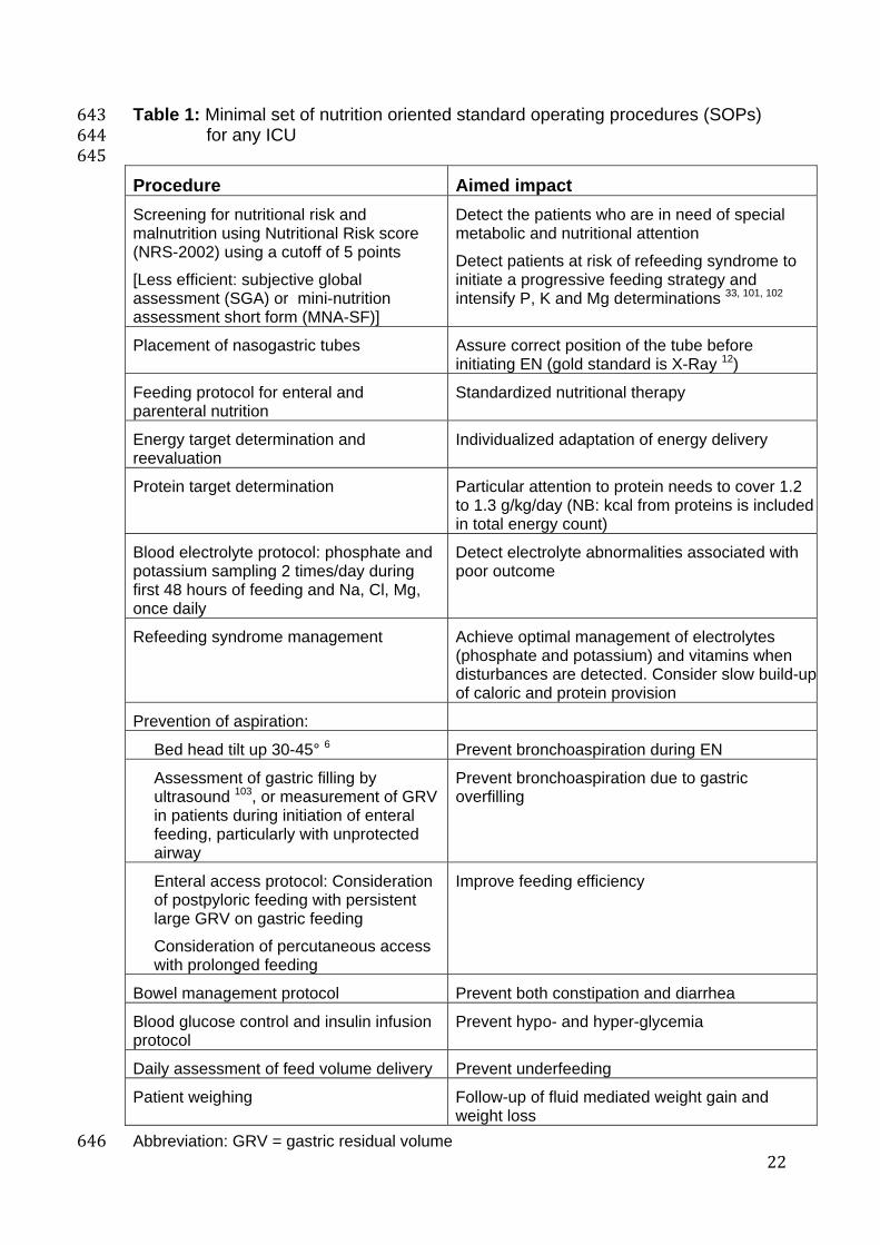
Table 1: Minimal set of nutrition oriented standard operating procedures (SOPs) 643for any ICU 644
645
Procedure Aimed impact
Screening for nutritional risk and malnutrition using Nutritional Risk score (NRS-2002) using a cutoff of 5 points
[Less efficient: subjective global assessment (SGA) or mini-nutrition assessment short form (MNA-SF)]
Detect the patients who are in need of special metabolic and nutritional attention
Detect patients at risk of refeeding syndrome to initiate a progressive feeding strategy and intensify P, K and Mg determinations 33, 101, 102
Placement of nasogastric tubes Assure correct position of the tube before initiating EN (gold standard is X-Ray 12)
Feeding protocol for enteral and parenteral nutrition
Standardized nutritional therapy
Energy target determination and reevaluation
Individualized adaptation of energy delivery
Protein target determination Particular attention to protein needs to cover 1.2 to 1.3 g/kg/day (NB: kcal from proteins is included in total energy count)
Blood electrolyte protocol: phosphate and potassium sampling 2 times/day during first 48 hours of feeding and Na, Cl, Mg, once daily
Detect electrolyte abnormalities associated with poor outcome
Refeeding syndrome management Achieve optimal management of electrolytes (phosphate and potassium) and vitamins when disturbances are detected. Consider slow build-upof caloric and protein provision
Prevention of aspiration:
Bed head tilt up 30-45° 6 Prevent bronchoaspiration during EN
Assessment of gastric filling by ultrasound 103, or measurement of GRV in patients during initiation of enteral feeding, particularly with unprotected airway
Prevent bronchoaspiration due to gastric overfilling
Enteral access protocol: Consideration of postpyloric feeding with persistent large GRV on gastric feeding
Consideration of percutaneous access with prolonged feeding
Improve feeding efficiency
Bowel management protocol Prevent both constipation and diarrhea
Blood glucose control and insulin infusion protocol
Prevent hypo- and hyper-glycemia
Daily assessment of feed volume delivery Prevent underfeeding
Patient weighing Follow-up of fluid mediated weight gain and weight loss
frequency, cost, and relative cost: the latter enables comparison 650between countries and is based on the Swiss average ICU day cost 651(4000 CHF/day) *. 652
653Variable Frequency Relative
cost index
Glucose First 24 hr of ICU admission /feeding : every 4-6 hrs Later: at least 2 times daily
0.6 ‰
Phosphate Within first 6-12 hr of admission Later: once a day
0.8 ‰
Potassium First 24 hr of ICU admission /feeding : every 6 hr with blood gases
0.7 ‰
Sodium, Chloride, Magnesium
Once daily 0.6 and 2.1 ‰
Liver tests: AST, ALT
Twice weekly 2 ‰
Triglycerides 66 Twice weekly 0.7 ‰
Prealbumin Once weekly 5 ‰
Glutamine In selected cases (renal remplacement therapy, burns, PN without glutamine)
3 ‰
Trace elements: Cu, Se, Zn
In selected cases (such as e.g. burns, addressed in the text)
11, 26 and 17 ‰
Urea – blood 3 times weekly 0.6 ‰
Urea – urine 6-hr urine collection once weekly in absence of renal failure
0.7 ‰
Ammonium In case of unexplained worsening of consciousness state 44
10 ‰
Carnitine Considering the limited availability and cost, to be done only in presence of unexplained rapid muscle catabolism and hyperlactatemia 80 with adequate protein supply
51 ‰
654Based on Swiss prices 104 on 1.1.2018 (1 CHF = 0.85 €) 655*: an approach comparable to the “Big Mac Index” which is an informal way of 656measuring the purchasing power parity between currencies, first introduced by the 657Economist (https://www.economist.com/content/big-mac-index) 658 659 660
24
References 6611. Kahn JM, Le T, Angus DC, Cox CE, Hough CL, White DB et al. The epidemiology of chronic critical 662
illness in the United States*. Critical care medicine 2015; 43:282-87. 6632. Arabi YM, Casaer MP, Chapman M, Heyland DK, Ichai C, Marik PE et al. The intensive care 664
medicine research agenda in nutrition and metabolism. Intensive care medicine 2017; 43:1239-56. 6653. Ferrie S, Tsang E. Monitoring nutrition in critical illness: What can we use? Nutr Clin Pract e-pub 666
2017 May 1: doi: 10.1177/0884533617706312 6674. Kipnis E, Ramsingh D, Bhargava M, Dincer E, Cannesson M, Broccard A et al. Monitoring in the 668
intensive care. Critical care research and practice 2012; 2012:473507. 6695. Kavanagh BP. The GRADE system for rating clinical guidelines. PLoS Med 2009; 6:e1000094. 6706. Taylor BE, McClave SA, Martindale RG, Warren MM, Johnson DR, Braunschweig C et al. Guidelines 671
for the provision and assessment of nutrition support therapy in the adult critically ill patient: Society 672of Critical Care Medicine (SCCM) and American Society for Parenteral and Enteral Nutrition 673(A.S.P.E.N.). Critical care med 2016; 44:390-438. 674
7. Jenkins B, Calder PC, Marino LV. Evaluation of implementation of fasting guidelines for enterally fed 675critical care patients. Clinical nutrition 2018; in press: 676
8. Reintam Blaser A, Starkopf J, Alhazzani W, Berger MM, Casaer MP, Deane AM et al. Early enteral 677nutrition in critically ill patients: ESICM clinical practice guidelines. Intensive care med 2017; 43:380-67898. 679
9. Krenitsky J. Adjusted body weight, pro: evidence to support the use of adjusted body weight in 680calculating calorie requirements. Nutrition in clinical practice : 2005; 20:468-73. 681
10. Reintam Blaser A., Malbrain M. L., Starkopf J., Fruhwald S., Jakob S. M., De Waele J. et al. 682Gastrointestinal function in intensive care patients: terminology, definitions and management. 683Recommendations of the ESICM Working Group on Abdominal Problems. Intensive care med 2012; 68438:384-94. 685
11. Reignier J, Lascarrou JB. Residual gastric volume and risk of ventilator-associated pneumonia--reply. 686JAMA 2013; 309:2090-1. 687
12. McFarland A. A cost utility analysis of the clinical algorithm for nasogastric tube placement 688confirmation in adult hospital patients. J Adv Nurs 2017; 73:201-16. 689
13. Reintam A, Parm P, Kitus R, Kern H, Starkopf J. Primary and secondary intra-abdominal 690hypertension--different impact on ICU outcome. Intensive care med 2008; 34:1624-31. 691
14. Kirkpatrick AW, Roberts DJ, Jaeschke R, De Waele JJ, De Keulenaer BL, Duchesne J et al. 692Methodological background and strategy for the 2012-2013 updated consensus definitions and 693clinical practice guidelines from the abdominal compartment society. Anaesthesiol Intensive Ther 6942015; 47 Spec No:s63-77. 695
16. Mathur SK, Singh P. Transoesophageal echocardiography related complications. Indian J Anaesth 6992009; 53:567-74. 700
17. Macht M, Wimbish T, Clark BJ, Benson AB, Burnham EL, Williams A et al. Diagnosis and treatment 701of post-extubation dysphagia: results from a national survey. Journal of critical care 2012; 27:578-86. 702
18. Sallum RA, Duarte AF, Cecconello I. Analytic review of dysphagia scales. Arq Bras Cir Dig 2012; 70325:279-82. 704
19. Suntrup S, Meisel A, Dziewas R, Ende F, Reichmann H, Heuschmann P et al. [Dysphagia diagnostics 705and therapy of acute stroke: federal survey of certified stroke units]. Nervenarzt 2012; 83:1619-24. 706
20. Blumenstein I, Shastri YM, Stein J. Gastroenteric tube feeding: techniques, problems and solutions. 707World J gastroenterol : WJG 2014; 20:8505-24. 708
21. Berger MM, Revelly JP, Wasserfallen JB, Schmid A, Bouvry S, Cayeux MC et al. Impact of a 709computerized information system on quality of nutritional support in the ICU. Nutrition 2006; 22:221-71029. 711
22. Berger MM. How to prescribe nutritional support using computers. World Rev Nutr Diet 2013; 105:32-71242. 713
23. Bousie E, van Blokland D, van Zanten ARH. Effects of implementation of a computerized nutritional 714protocol in mechanically ventilated critically ill patients: A single-centre before and after study. Clin 715Nutr ESPEN 2016; 11:e47-e54. 716
25
24. Bousie E, van Blokland D, Lammers HJ, van Zanten A. R. Relevance of non-nutritional calories in 717mechanically ventilated critically ill patients. Eur J Clin Nutr 2016; 70:1443-50. 718
25. Charrière M, Ridley E, Hastings J , Bianchet O , Scheinkestel C, Berger MM. Propofol sedation 719substantially increases the caloric and lipid intake in critically ill patients. Nutrition 2017; 42:64-68. 720
26. Alberda C, Gramlich L, Jones N, Jeejeebhoy K, Day AG, Dhaliwal R et al. The relationship between 721nutritional intake and clinical outcomes in critically ill patients: results of an international multicenter 722observational study. Intensive care med 2009; 35:1728-37. 723
27. Kurmis R, Heath K, Ooi S, Munn Z, Forbes S, Young V et al. A prospective multi-center audit of 724nutrition support parameters following burn injury. Journal of burn care & research : official publication 725of the American Burn Association 2015; 36:471-77. 726
28. Dupertuis YM, Kossovsky MP, Kyle UG, Raguso CA, Genton L, Pichard C. Food intake in 1707 727hospitalised patients: a prospective comprehensive hospital survey. Clin nutr 2003; 22:115-23. 728
29. van Bokhorst-de van der Schueren M. A., Roosemalen M. M., Weijs P. J., Langius J. A. High waste 729contributes to low food intake in hospitalized patients. Nutrition in clinical practice : 2012; 27:274-80. 730
30. McClave SA, Lowen CC, Kleber MJ, Nicholson JF, Jimmerson SC, McConnel JW et al. Are patients 731fed appropriately according to their caloric requirements? JPEN 1998; 22:375-81. 732
31. Reid C. Frequency of under- and overfeeding in mechanically ventilated ICU patients: causes and 733possible consequences. J human nutrition and dietetics : 2006; 19:13-22. 734
32. Allingstrup M. J., Kondrup J., Wiis J., Claudius C., Pedersen U. G., Hein-Rasmussen R. et al. Early 735goal-directed nutrition versus standard of care in adult intensive care patients: the single-centre, 736randomised, outcome assessor-blinded EAT-ICU trial. Intensive care medicine 2017; 43:1637-47. 737
33. Doig GS, Simpson F, Heighes PT, Bellomo R, Chesher D, Caterson ID et al. Restricted versus 738continued standard caloric intake during the management of refeeding syndrome in critically ill adults: 739a randomised, parallel-group, multicentre, single-blind controlled trial. Lancet Respir Med 2015; 7403:943-52. 741
34. Preiser JC, van Zanten ARH, Berger MM, Biolo G, Casaer M, Doig G et al. Metabolic and nutritional 742support of critically ill patients: consensus and controversies. Critical care 2015; 19:35. 743
35. Raiten DJ, Sakr Ashour FA, Ross AC, Meydani SN, Dawson HD, Stephensen CB et al. Inflammation 744and Nutritional Science for Programs/Policies and Interpretation of Research Evidence (INSPIRE). 745The Journal of nutrition 2015; 145:1039S-108S. 746
36. Ishibashi N, Plank LD, Sando K, Hill GL. Optimal protein requirement during the first 2 weeks after 747the onset of critical illness. Critical care medicine 1998; 26:1529-35. 748
37. Berger MM, Soguel L, Charrière M, Theriault B, Pralong F, Schaller MD. Impact of the reduction of 749the recommended energy target in the ICU on protein delivery and clinical outcomes. Clin Nutr 2017; 75036 281–87 751
38. Rooyackers O, Kouchek-Zadeh R, Tjader I, Norberg A, Klaude M, Wernerman J. Whole body protein 752turnover in critically ill patients with multiple organ failure. Clinical nutrition 2015; 34:95-100. 753
39. Singer P, Hiesmayr M, Biolo G, Felbinger TW, Berger MM, C Goeters et al. Pragmatic approach to 754nutrition in the ICU: Expert opinion regarding which calorie protein target. Clinical nutrition 2014; 75533:246-51. 756
40. Hermans G, Casaer MP, Clerckx B, Guiza F, Vanhullebusch T, Derde S et al. Effect of tolerating 757macronutrient deficit on the development of intensive-care unit acquired weakness: a subanalysis of 758the EPaNIC trial. Lancet Respir Med 2013; 1:621-29. 759
41. Weijs PJ, Looijaard WG, Beishuizen A, Girbes AR, Oudemans-van Straaten HM. Early high protein 760intake is associated with low mortality and energy overfeeding with high mortality in non-septic 761mechanically ventilated critically ill patients. Critical care 2014; 18:701. 762
42. Doig GS, Simpson F, Bellomo R, Heighes PT, Sweetman EA, Chesher D et al. Intravenous amino 763acid therapy for kidney function in critically ill patients: a randomized controlled trial. Intensive care 764medicine 2015; 41:1197-208. 765
43. Allingstrup MJ, Kondrup J, Wiis J, Claudius C, Pedersen UG, Hein-Rasmussen R et al. Early 766goal-directed nutrition versus standard of care in adult intensive care patients: the single-767centre, randomised, outcome assessor-blinded EAT-ICU trial. Intensive care med 2017; 76843:1637-47: 769
45. Calabrese EJ, Baldwin LA. U-shaped dose-responses in biology, toxicology, and public health. Annu 772Rev Public Health 2001; 22:15-33. 773
46. Finfer S, Wernerman J, Preiser JC, Cass T, Desaive T, Hovorka R et al. Clinical review: Consensus 774recommendations on measurement of blood glucose and reporting glycemic control in critically ill 775adults. Critical care 2013; 17:229. 776
47. Krinsley JS, Chase JG, Gunst J, Martensson J, Schultz MJ, Taccone FS et al. Continuous glucose 777monitoring in the ICU: clinical considerations and consensus. Critical care 2017; 21:197. 778
48. Preiser J. C., Devos P., Ruiz-Santana S., Melot C., Annane D., Groeneveld J. et al. A prospective 779randomised multi-centre controlled trial on tight glucose control by intensive insulin therapy in adult 780intensive care units: the Glucontrol study. Intensive Care Med 2009; 35:1738-48. 781
49. Brunkhorst F. M., Engel C., Bloos F., Meier-Hellmann A., Ragaller M., Weiler N. et al. Intensive 782insulin therapy and pentastarch resuscitation in severe sepsis. New Engl J med 2008; 358:125-39. 783
50. Investigators Nice-Sugar Study, Finfer S., Chittock D. R., Su S. Y., Blair D., Foster D. et al. Intensive 784versus conventional glucose control in critically ill patients. N Engl J Med 2009; 360:1283-97. 785
51. Krinsley J. S., Preiser J. C. Time in blood glucose range 70 to 140 mg/dl >80% is strongly associated 786with increased survival in non-diabetic critically ill adults. Crit Care 2015; 19:179. 787
52. Dungan KM, Braithwaite SS, Preiser JC. Stress hyperglycaemia. Lancet 2009; 373:1798-807. 78853. Mehanna HM, Moledina J, Travis J. Refeeding syndrome: what it is, and how to prevent and treat it. 789
Bmj 2008; 336:1495-98. 79054. Braunschweig CA, Sheean PM, Peterson SJ, Gomez Perez S, Freels S, Lateef O et al. Intensive 791
Nutrition in Acute Lung Injury: A Clinical Trial (INTACT). JPEN Journal of parenteral and enteral 792nutrition 2015; 39:13-20. 793
55. Berger MM, Pichard C. Understanding the causes of death in INTACT by Braunschweig et al. JPEN 794Journal of parenteral and enteral nutrition 2015; 39:144. 795
56. Olthof LE, Koekkoek Wack, van Setten C, Kars JCN, van Blokland D, van Zanten ARH. Impact of 796caloric intake in critically ill patients with, and without, refeeding syndrome: A retrospective study. 797Clinical nutrition 2017 798
57. Koekkoek WAC, Van Zanten ARH. Is refeeding syndrome relevant for critically ill patients? Curr Opin 799Clin Nutr Metab Care 2017; 21:130-37. 800
58. Lobo D. N. Fluid, electrolytes and nutrition: physiological and clinical aspects. Proc Nutr Soc 2004; 80163:453-66. 802
59. Guadagni M., Biolo G. Effects of inflammation and/or inactivity on the need for dietary protein. 803Current opinion in clinical nutrition and metabolic care 2009; 12:617-22. 804
60. Tran NK, Godwin ZR, Bockhold JC, Passerini AG, Cheng J, Ingemason M. Clinical impact of sample 805interference on intensive insulin therapy in severely burned patients: a pilot study. J burn care rese: 8062014; 35:72-79. 807
61. Almond CS, Shin AY, Fortescue EB, Mannix R.C, Wypij D, Binstadt BA et al. Hyponatremia among 808runners in the Boston Marathon. The New England journal of medicine 2005; 352:1550-56. 809
62. Grau T, Bonet A, Rubio M, Mateo D, Farre M, Acosta JA et al. Liver dysfunction associated with 810artificial nutrition in critically ill patients. Critical care 2007; 11:R10. 811
63. Piper SN, Rohm KD, Boldt J, Odermatt B, Maleck WH, Suttner SW. Hepatocellular integrity in 812patients requiring parenteral nutrition: comparison of structured MCT/LCT vs. a standard MCT/LCT 813emulsion and a LCT emulsion. Eur J Anaesthesiol 2008; 25:557-65. 814
64. Fivez T, Kerklaan D, Verbruggen S, Vanhorebeek I, Verstraete S, Tibboel D et al. Impact of 815withholding early parenteral nutrition completing enteral nutrition in pediatric critically ill patients 816(PEPaNIC trial): study protocol for a randomized controlled trial. Trials 2015; 16:202. 817
65. Puder M., Valim C., Meisel J. A., Le H. D., de Meijer V. E., Robinson E. M. et al. Parenteral fish oil 818improves outcomes in patients with parenteral nutrition-associated liver injury. Ann surg 2009; 819250:395-402. 820
66. Devaud JC, Berger MM, Pannatier A, Marques-Vidal P, Tappy L, Rodondi N et al. 821Hypertriglyceridemia: a potential side effect of propofol sedation in critical illness. Intensive care 822medicine 2012; 38:1990-98. 823
67. Kruger PS. Forget glucose: what about lipids in critical illness? Critical care and resuscitation : 2009; 8241:305-09. 825
27
68. Dickerson RN, Medling TL, Smith AC, Maish GO, 3rd, Croce MA, Minard G et al. Hypocaloric, high-826protein nutrition therapy in older vs younger critically ill patients with obesity. JPEN Journal of 827parenteral and enteral nutrition 2013; 37:342-51. 828
69. Oddoye EA, Margen S. Nitrogen balance studies in humans: long-term effect of high nitrogen intake 829on nitogen accretion. J Nutrition 1979; 109:363-77. 830
70. Thiessen SE, Derde S, Derese I, Dufour T, Vega CA, Langouche L et al. Role of Glucagon in 831Catabolism and Muscle Wasting of Critical Illness and Modulation by Nutrition. Am J resp crit care 832med 2017; 196:1131-43. 833
71. Vincent J. L., Russell J. A., Jacob M., Martin G., Guidet B., Wernerman J. et al. Albumin 834administration in the acutely ill: what is new and where next? Critical care 2014; 18:231. 835
72. Davis CJ, Sowa D, Keim KS, Kinnare K, Peterson S. The use of prealbumin and C-reactive protein 836for monitoring nutrition support in adult patients receiving enteral nutrition in an urban medical center. 837JPEN Journal of parenteral and enteral nutrition 2012; 36:197-204. 838
73. Delliere S, Cynober L. Is transthyretin a good marker of nutritional status? Clinical nutrition 2017; 83936:364-70. 840
74. Rodas PC, Rooyackers O, Hebert C, Norberg A, Wernerman J. Glutamine and glutathione at ICU 841admission in relation to outcome. Clinical science 2012; 122:591-7. 842
75. Pettersson L, Ryden S, Smedberg M, Tjader I, Rooyackers O, Wernerman J. Validation of a point-of-843care instrument for bedside glutamine screening in the intensive care unit. Clinical nutrition 2017; 84436:186-90. 845
76. Scheinkestel CD, Adams F, Mahony L, Bailey M, Davies AR, Nyulasi I et al. Impact of increasing 846parenteral protein loads on amino acid levels and balance in critically ill anuric patients on continuous 847renal replacement therapy. Nutrition 2003; 19:733-40. 848
77. Bahri S, Zerrouk N, Aussel C, Moinard C, Crenn P, Curis E et al. Citrulline: from metabolism to 849therapeutic use. Nutrition 2013; 29:479-84. 850
78. Poole A, Deane A, Summers M, Fletcher J, Chapman M. The relationship between fasting plasma 851citrulline concentration and small intestinal function in the critically ill. Critical care 2015; 19:16. 852
79. Voth M, Holzberger S, Auner B, Henrich D, Marzi I, Relja B. I-FABP and L-FABP are early markers 853for abdominal injury with limited prognostic value for secondary organ failures in the post-traumatic 854course. Clinical chemistry and laboratory medicine : CCLM / FESCC 2015; 53:771-80. 855
80. Bonafé L, Berger MM, Que YA, Mechanick JI. Carnitine deficiency in chronic critical illness Curr opin 856clin nutr metab care 2014; 17:200-09. 857
81. Berger MM, Shenkin A, Revelly JP, Roberts E, Cayeux MC, Baines M et al. Copper, selenium, zinc 858and thiamine balances during continuous venovenous hemodiafiltration in critically ill patients. Am J 859Clin Nutr 2004; 80:410-16. 860
82. Ben-Hamouda N, Charrière M, Voirol P, Berger MM. Massive copper and selenium losses cause life-861threatening deficiencies during prolonged continuous renal replacement. Nutrition 2017; 34:71-75. 862
83. Berger MM, Eggimann P, Heyland DK, Chioléro RL, Revelly JP, Day A et al. Reduction of nosocomial 863pneumonia after major burns by trace element supplementation: aggregation of two randomised 864trials. Critical care 2006; 10:e-pub 2 Nov. 865
84. Berger MM, Baines M, Raffoul W, Benathan M, Chiolero RL, Reeves C et al. Trace element 866supplements after major burns modulate antioxidant status and clinical course by way of increased 867tissue trace element concentration. Am J Clin Nutr 2007; 85:1293-300. 868
85. Rousseau AF, Losser MR, Ichai C, Berger MM. ESPEN endorsed recommendations: Nutritional 869therapy in major burns. Clin nutr 2013; 32:497-502. 870
86. Gagnon G, Voirol P, Soguel L, Boulat O, Berger MM. Trace element monitoring in the ICU: Quality 871and economic impact of a change in sampling practice. Clin nutr2014; 34:422-27. 872
87. Cunningham JJ, Hegarty MT, Meara PA, Burke JF. Measured and predicted calorie requirements of 873adults during recovery from severe burn trauma. Am J Clin Nutr 1989; 49:404-08. 874
88. Kreymann G, Grosser S, Buggisch P, Gottschall C, Matthaei S, Greten H. Oxygen consumption and 875resting metabolic rate in sepsis, sepsis syndrome, and septic shock. Critical care med 1993; 21:1012-87619. 877
89. Kross EK, Sena M, Schmidt K, Stapleton RD. A comparison of predictive equations of energy 878expenditure and measured energy expenditure in critically ill patients. J crit care 2012; 27:321 e5-12. 879
28
90. De Waele E, Opsomer T, Honore PM, Diltoer M, Mattens S, Huyghens L et al. Measured versus 880calculated resting energy expenditure in critically ill adult patients. Do mathematics match the gold 881standard? Minerva Anesthesiol 2015; 81:272-82. 882
91. Zijlstra N, ten Dam SM, Hulshof PJ, Ram C, Hiemstra G, de Roos NM. 24-hour indirect calorimetry in 883mechanically ventilated critically ill patients. Nutrition Clin Pract 2007; 22:250-55. 884
92. Heidegger CP, Berger MM, Graf S, Zingg W, Darmon P, Costanza MC et al. Optimisation of energy 885provision with supplemental parenteral nutrition in critically ill patients: a randomised controlled 886clinical trial. Lancet 2013; 381:385-93. 887
93. Casaer MP, Van den Berghe G. Nutrition in the acute phase of critical illness. The New England 888journal of medicine 2014; 370:1227-36. 889
94. Villet S, Chioléro RL, Bollmann MD, Revelly JP, Cayeux MC, Delarue J et al. Negative impact of 890hypocaloric feeding and energy balance on clinical outcome in ICU patients. Clinical nutrition 2005; 89124:502-09. 892
95. Dvir D, Cohen J, Singer P. Computerized energy balance and complications in critically ill patients: 893An observational study. Clin nutr 2006; 25:37-44. 894
96. Rahman A, Hasan RM, Agarwala R, Martin C, Day AG, Heyland DK. Identifying critically-ill patients 895who will benefit most from nutritional therapy: Further validation of the "modified NUTRIC" nutritional 896risk assessment tool. Clin nutr 2016; 35:158-62. 897
97. Thibault R, Makhlouf AM, Mulliez A, Cristina Gonzalez MC, Kekstas G, Kozjek NR et al. Fat-free 898mass at admission predicts 28-day mortality in intensive care unit patients: the international 899prospective observational study Phase angle project. Intensive care med 2016; 42:1445-53. 900
98. Looijaard WG, Dekker IM, Stapel SN, Girbes AR, Twisk JW, Oudemans-van Straaten HM et al. 901Skeletal muscle quality as assessed by CT-derived skeletal muscle density is associated with 6-902month mortality in mechanically ventilated critically ill patients. Critical care 2016; 20:386. 903
99. Ni Bhuachalla EB, Daly LE, Power DG, Cushen SJ, MacEneaney P, Ryan AM. Computed 904tomography diagnosed cachexia and sarcopenia in 725 oncology patients: is nutritional screening 905capturing hidden malnutrition? J Cachexia Sarcopenia Muscle 2018; 9:295-305. 906
100. Baldwin CE, Paratz JD, Bersten AD. Muscle strength assessment in critically ill patients with 907handheld dynamometry: an investigation of reliability, minimal detectable change, and time to peak 908force generation. J crit care 2013; 28:77-86. 909
101. Crook MA. Refeeding syndrome: problems with definition and management. Nutrition 2014; 30:1448-91055. 911
102. Friedli N, Stanga Z, Sobotka L, Culkin A, Kondrup J, Laviano A et al. Revisiting the refeeding 912syndrome: Results of a systematic review. Nutrition 2017; 35:151-60. 913
103. Gilja O. H., Hausken T., degaard S., Berstad A. Gastric emptying measured by ultrasonography. 914World J gastroenterol : WJG 1999; 5:93-94. 915
104. OFSP, Office Fédéral des assurances sociales. Liste des Analyses. Confédération Suisse 2017; 916Bern:https://www.bag.admin.ch/...tarife/.../Liste%20des%20analyses%20du%2001.01.17. 917