28
Case Report Prenatal Ultrasound Diagnosis Chang Bing Show Chwan Memorial Hospital Li-hsun Chen Morbidly Adherent Placenta
| Date post: | 15-Jul-2015 |
| Category: |
Health & Medicine |
| Upload: | - |
| View: | 77 times |
| Download: | 0 times |

Case Report
Prenatal Ultrasound Diagnosis
Chang Bing Show ChwanMemorial Hospital
Li-hsun Chen
Morbidly
Adherent
Placenta

Miss Chen35Y/o, negative for major systemic disease and past history.Habits of smoking and no alcohol drinking.G3P1A1
2
Case 1
G1: scheduled cesarean section for malpresentation (2012 Oct.) G2: artificial abortion with RU486 (2013 Mar.)G3: LMP 20140510 EDC 20150213Regular antenatal visit our clinic.
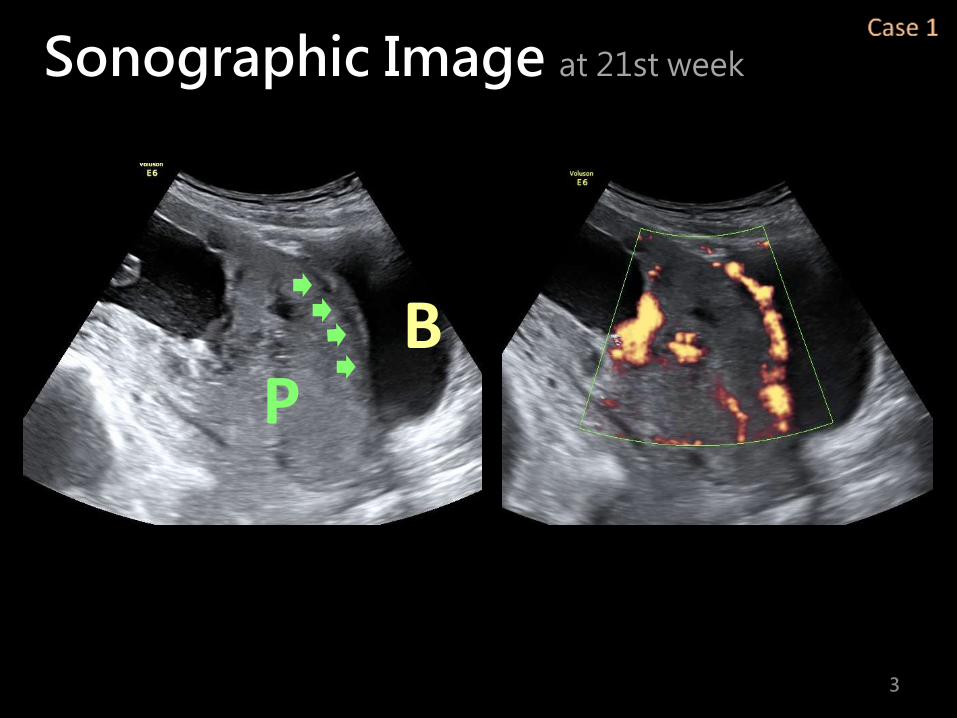
Sonographic Image at 21st week
3
BP

Sonographic Image at 21st week
4
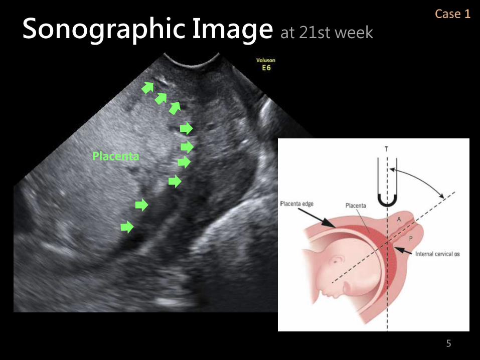
Sonographic Image at 21st week
5
Placenta

Sonographic Image at 21st week
6
Pregnancy at 32nd week,she got massive vaginal bleeding and admitted intothe ward.
7
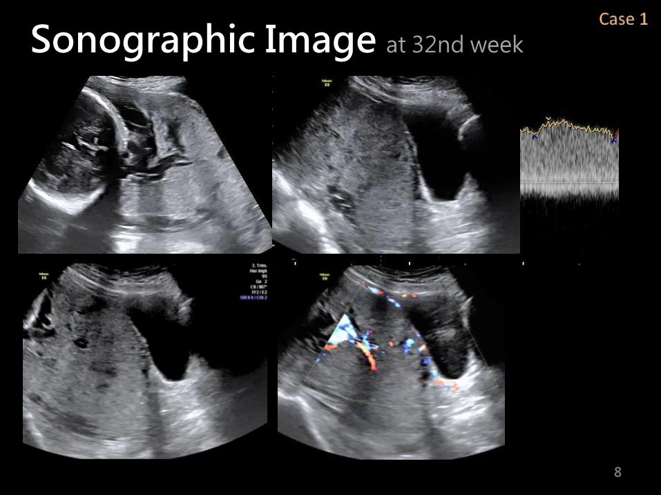
Sonographic Image at 32nd week
8

3D Sonographic Image at 32nd week
9
BP
Placenta previa with MAP

10
Four days later, she was transferred to the terminal center for delivery and placed balloon catheterization before cesarean section.
Leave the placenta in situ

Miss Ke39Y/o, negative for major systemic disease and past history.G1P0
11
Case 2
LMP 201405102 EDC 20150214Regular antenatal visit our clinic.

Sonographic Image at 32nd week
12

Sonographic Image at 32nd week
13
Placenta previa without MAP

14
Pregnancy at 34th week,she got massive vaginal bleeding and caserean section .

Morbidly Adherent Placenta

16


2D ultrasonography
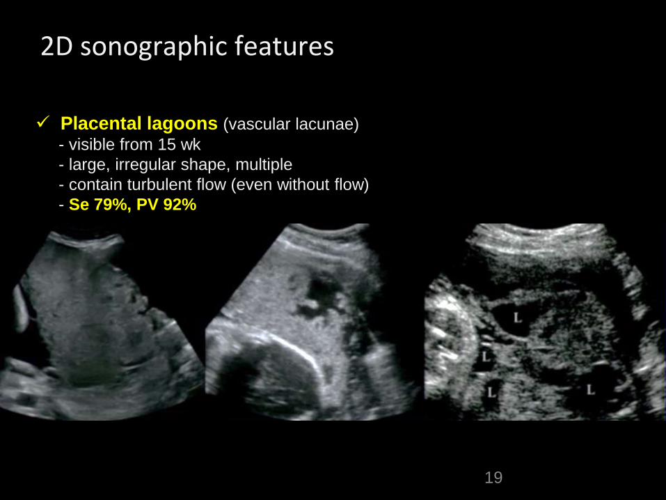
2D sonographic features
19
Placental lagoons (vascular lacunae)
- visible from 15 wk
- large, irregular shape, multiple
- contain turbulent flow (even without flow)
- Se 79%, PV 92%

2D sonographic features
20
Placental lagoons (vascular lacunae)
- visible from 15 wk
- large, irregular shape, multiple
- contain turbulent flow (even without flow)
- Se 79%, PV 92%
Loss of retroplacental hypoecoic area
- reflects loss of basal decidua and retroplacental vascular bed
- Se, 50%, PV 55%

2D sonographic features
21
Placental lagoons (vascular lacunae)
- visible from 15 wk
- large, irrrgular shape, multiple
- contain turbulent flow (even without flow)
- Se 79%, PV 92%
Disruption utero-vesical serosa
Extrauterine placental parenchyma
Loss of retroplacental hypoecoic area
- reflects loss of basal decidua and retroplacental vascular bed
- Se, 50%, PV 52%
Progressive thinning of retroplacental myometrium
- If < 1mm
- Se 85%, PV 80%

22
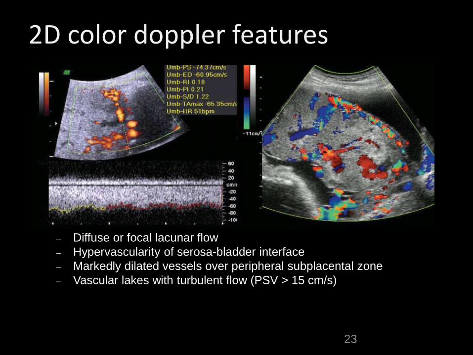
2D color doppler features
Diffuse or focal lacunar flow
Hypervascularity of serosa-bladder interface
Markedly dilated vessels over peripheral subplacental zone
23
2D color doppler features
Diffuse or focal lacunar flow
Hypervascularity of serosa-bladder interface
Markedly dilated vessels over peripheral subplacental zone
Vascular lakes with turbulent flow (PSV > 15 cm/s)

24
Irregular intraplacental vascularization with tortuous confluent vessels crossing placental width.
Hypervascularityof uterine serosa–bladder wall interface.
3D ultrasonography
25
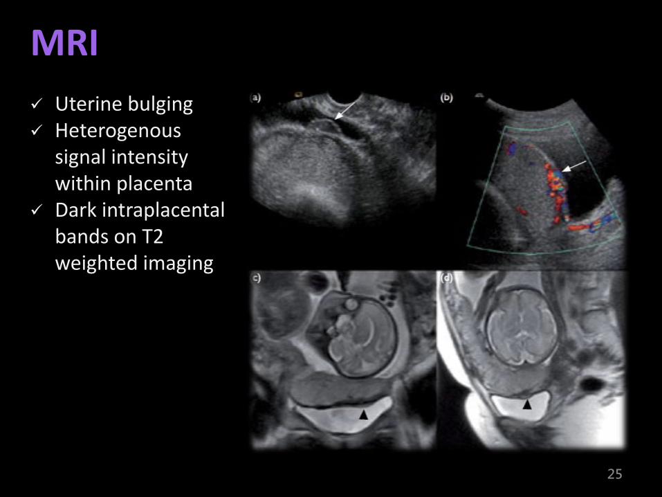
Uterine bulging Heterogenous
signal intensity within placenta
Dark intraplacentalbands on T2 weighted imaging
MRI

Conclusion
TAS+ Doppler
TVS
MRI

Thanks
for your attentions

Chang Bing Show ChwanMemorial Hospital
Li-hsun Chen
Thanks
for your attentions


![REPORTS OF ORIGINAL INVESTIGATIONS · morbidly adherent placenta was one per 731 births (95% confidence interval [CI], 1/632 to 1/866) during the periods 2008 and 2011.1 The United](https://static.documents.pub/doc/80x56/5f39b1b1ef6dd30fe67533f1/reports-of-original-investigations-morbidly-adherent-placenta-was-one-per-731-births.jpg)
