77
Morphologic Patterns Defect in leukocyte function Complement deficiency Systemic manifestation
| Date post: | 13-Dec-2015 |
| Category: |
Documents |
| Upload: | charlotte-gray |
| View: | 218 times |
| Download: | 1 times |
Morphologic PatternsDefect in leukocyte function
Complement deficiencySystemic manifestation

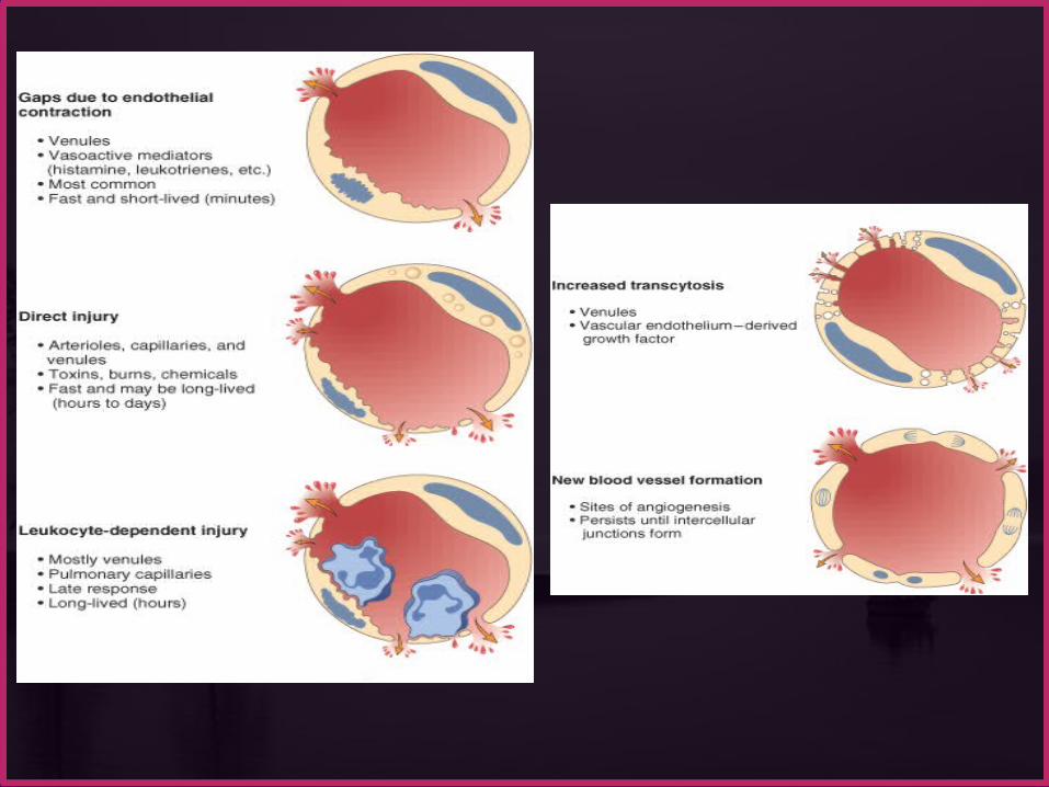


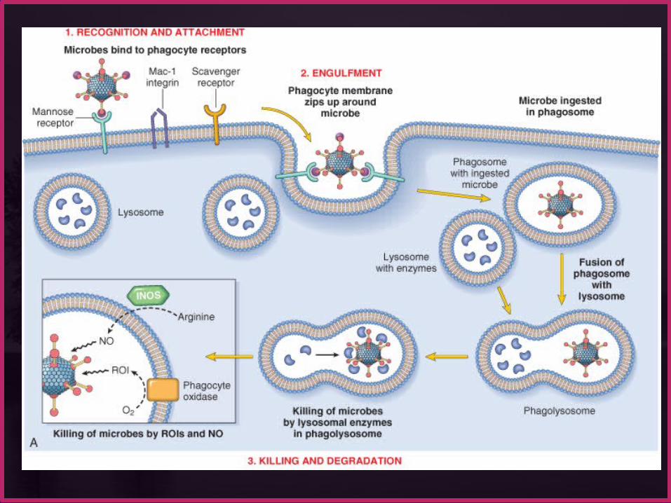
Acute InflammationCELLULAR EVENTS:
LEUKOCYTE EXTRAVASATION AND PHAGOCYTOSIS
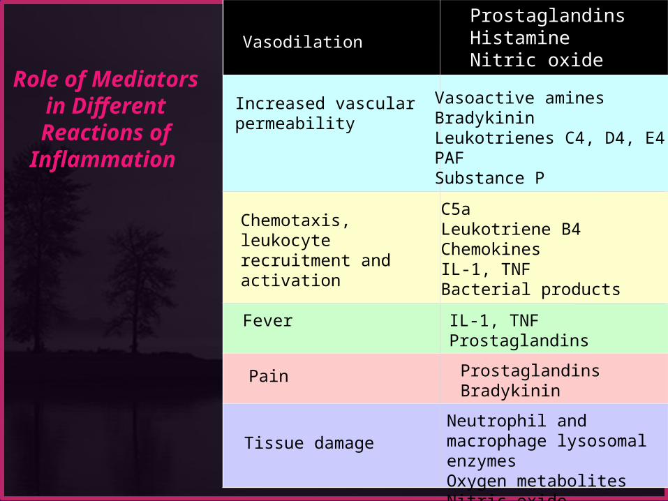
Role of Mediators in Different
Reactions of Inflammation
Vasodilation
ProstaglandinsHistamineNitric oxide
Increased vascular permeability
Vasoactive aminesBradykininLeukotrienes C4, D4, E4PAFSubstance P
Chemotaxis, leukocyte recruitment and activation
C5aLeukotriene B4ChemokinesIL-1, TNFBacterial products
Fever IL-1, TNFProstaglandins
Pain ProstaglandinsBradykinin
Tissue damageNeutrophil and macrophage lysosomal enzymesOxygen metabolitesNitric oxide
Patterns of Acute Inflammation Outcomes of Acute Inflammation Patterns of chronic Inflammation Defect in leukocyte function Complement deficiency Systemic manifestation

Patterns of Acute Inflammation Outcomes of Acute Inflammation Patterns of chronic Inflammation Defect in leukocyte function Complement deficiency Systemic manifestation

Several types of inflammation vary in their morphology and clinical correlates. Why? The severity of the reaction specific cause the particular tissue site involved
SEROUS INFLAMMATION FIBRINOUS INFLAMMATION SUPPURATIVE OR PURULENT
INFLAMMATION ULCERS

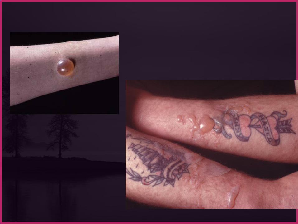
SEROUS INFLAMMATION: Serous inflammation is marked by the
outpouring of a thin fluid e.g. the skin blister resulting from a burn or
viral infection represents a large accumulation of serous fluid

FIBRINOUS INFLAMMATION more severe injuries and more greater vascular
permeability, larger molecules such as fibrinogen pass the vascular barrier, and fibrin is formed and deposited
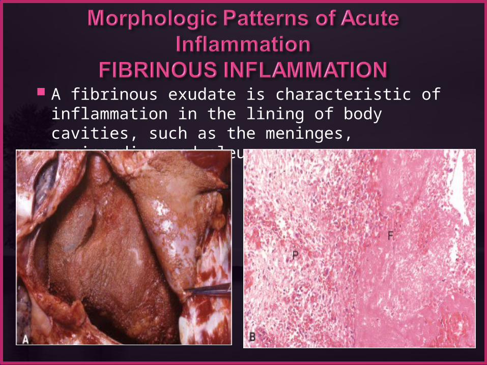
A fibrinous exudate is characteristic of inflammation in the lining of body cavities, such as the meninges, pericardium and pleura

FIBRINOUS INFLAMMATION Fibrinous exudates may be removed by fibrinolysis But when the fibrin is not removed, it may stimulate
the ingrowth of fibroblasts and blood vessels and
thus lead to scarring (organization)
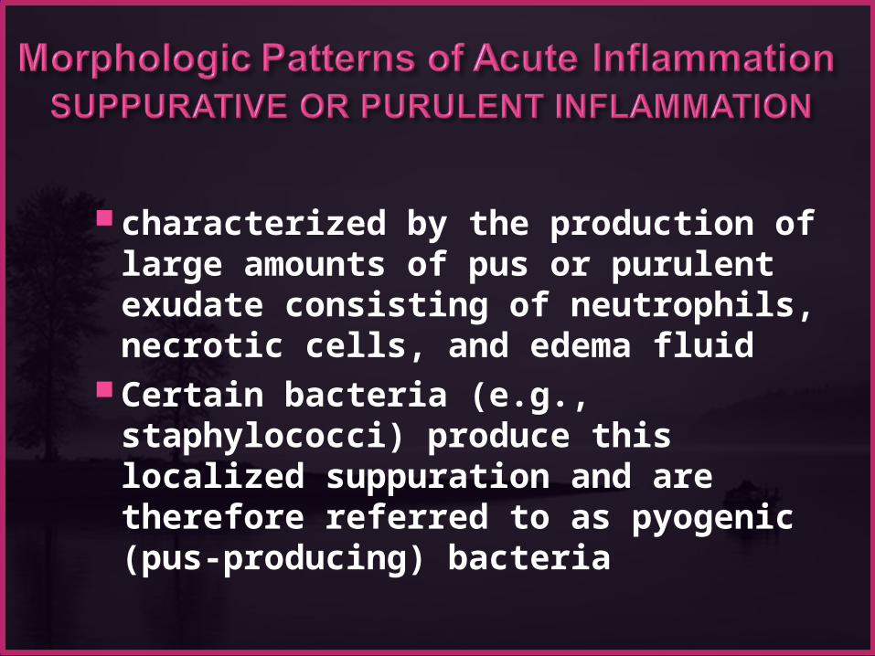
characterized by the production of large amounts of pus or purulent exudate consisting of neutrophils, necrotic cells, and edema fluid
Certain bacteria (e.g., staphylococci) produce this localized suppuration and are therefore referred to as pyogenic (pus-producing) bacteria

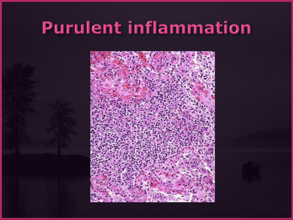
Suppurative inflammation. A, A subcutaneous bacterial abscess with collections of pus. B, The abscess contains neutrophils, edema fluid, and cellular debris.

Abscesses : localized collections of purulent inflammatory tissue caused by suppuration buried in a tissue, an organ, or a confined space
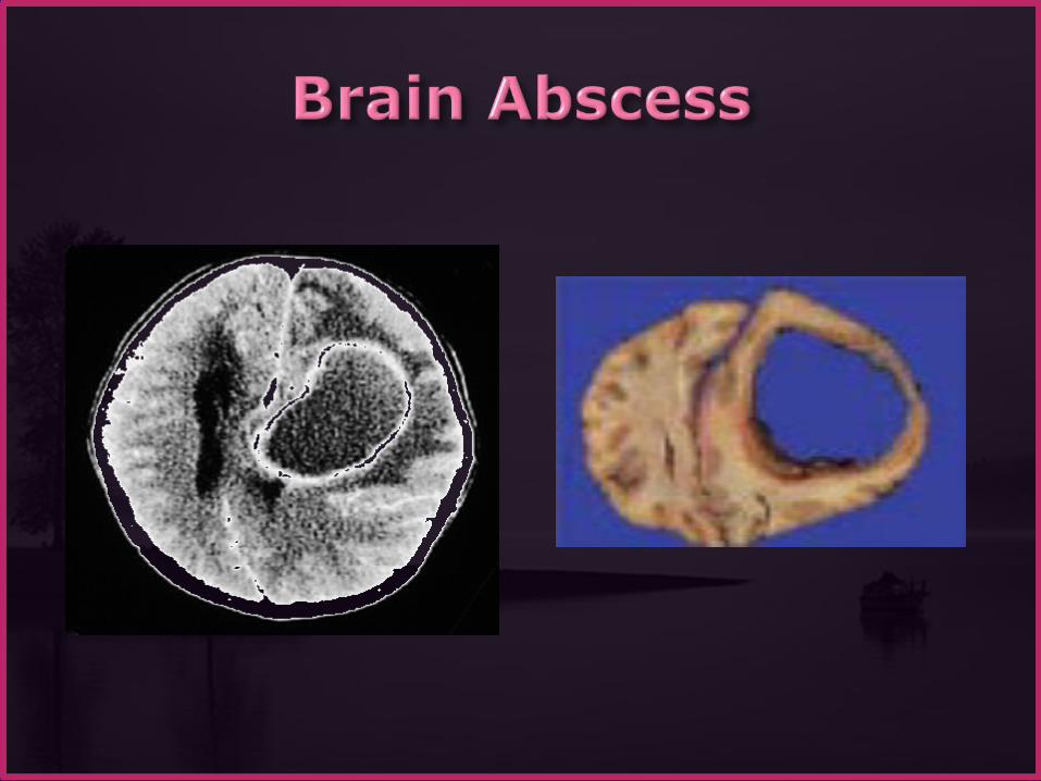


Localized liquefactive necrosis liver abscess
Removal of the dead tissue leaves behind a scar
ULCERS
An ulcer is a local defect of the surface of an organ or tissue that is produced by the sloughing (shedding) of inflammatory necrotic tissue
ULCERSencountered in:
1) inflammatory necrosis of the mucosa of the mouth, stomach, intestines, or genitourinary tract
2) subcutaneous inflammation of the lower extremities in older persons who have circulatory disturbances
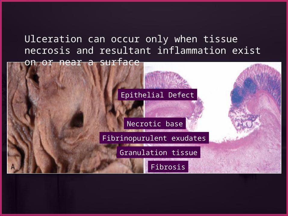
Ulceration can occur only when tissue necrosis and resultant inflammation exist on or near a surface
Epithelial Defect
Fibrinopurulent exudates
Granulation tissue
Fibrosis
Necrotic base
Patterns of Acute Inflammation Outcomes of Acute Inflammation Patterns of chronic Inflammation Defect in leukocyte function Complement deficiency Systemic manifestation

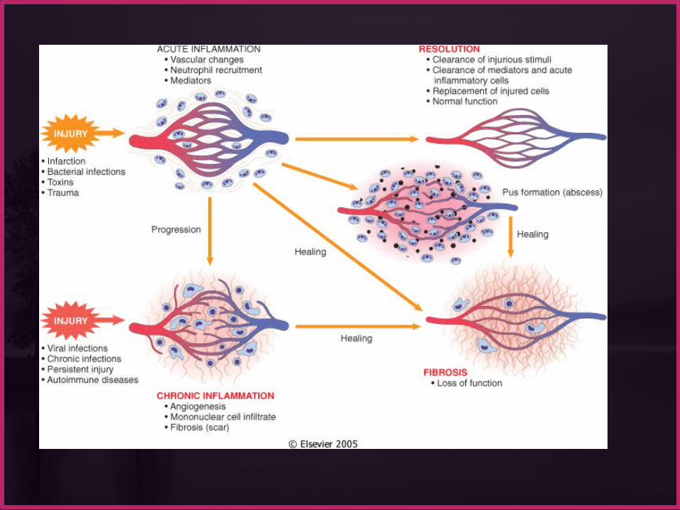
Acute inflammation may have one of the four outcomes:
Complete resolution Healing by connective tissue replacement (fibrosis) Progression of the tissue response to chronic
inflammation
Abcess formation
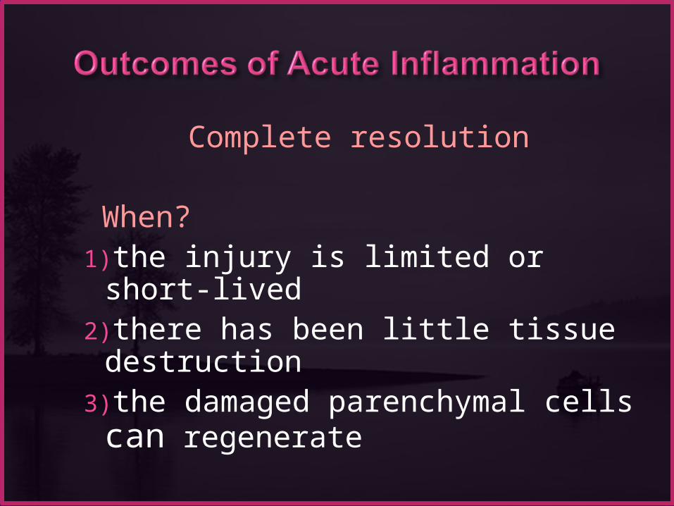
Complete resolution
When?1)the injury is limited or short-lived 2)there has been little tissue destruction
3)the damaged parenchymal cells can regenerate

Complete resolution
Mechanism: Neutralization and removal of chemical mediators
Normalization of vascular permeability
halting of leukocyte emigration
Clearance of edema (lymphatic drainage) , inflammatory cells and necrotic debris (macrophages).
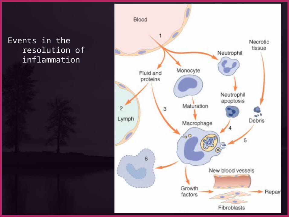
Events in the resolution of inflammation

Healing by connective tissue replacement (fibrosis):
This occurs after substantial tissue destruction the inflammatory injury involves tissues that are incapable of
regeneration there is abundant fibrin exudation.
The destroyed tissue is resaorbed and eventually replaced by fibrosis.
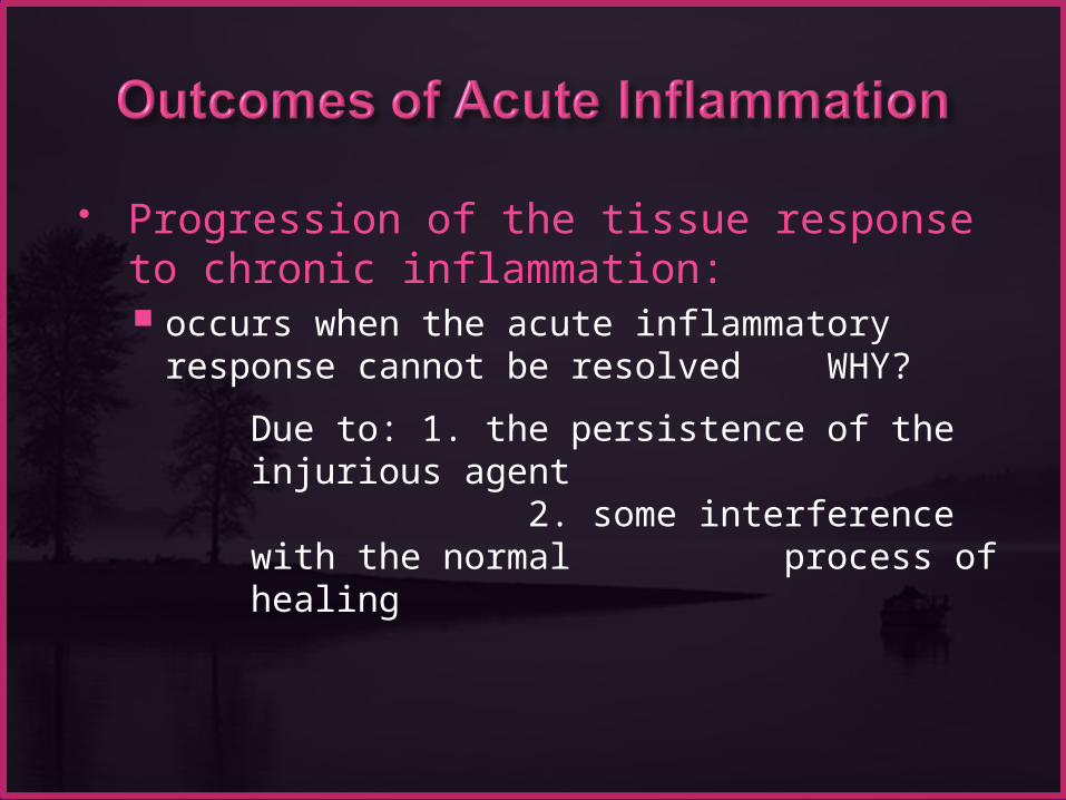
Progression of the tissue response to chronic inflammation: occurs when the acute inflammatory response
cannot be resolved WHY?
Due to: 1. the persistence of the injurious agent 2. some interference with the normal
process of healing

A 36-year-old man has had midepigastric abdominal pain for the past 3 months. An upper gastrointestinal endoscopy shows a 2-cm, sharply demarcated, shallow ulceration of the gastric antrum. A biopsy specimen of the ulcer base shows angiogenesis, fibrosis, and mononuclear cell infiltrates with lymphocytes, macrophages, and plasma cells. Which of the following terms best describes this pathologic process?
(A) Acute inflammation (B) Serous inflammation (C) Granulomatous inflammation (D) Fibrinous inflammation (E) Chronic inflammation
Patterns of Acute Inflammation Outcomes of Acute Inflammation Patterns of chronic Inflammation Defect in leukocyte function Complement deficiency Systemic manifestation

inflammation of prolonged duration weeks or months Mixture of active inflammation, tissue destruction, and
attempts at repair it may follow:
1. acute inflammation
2. begins insidiously,
as a low-grade,
often asymptomatic
response.
This is the cause of tissue damage in some of the most
common and disabling human diseases, such as rheumatoid
arthritis, atherosclerosis, tuberculosis, and chronic lung
diseases
or

1. Viral infection
2. Persistent infections by certain microorganisms, e.g. tubercle bacilli, Treponema pallidum, fungi, and parasites.
3. Prolonged exposure to potentially toxic agents, either exogenous or endogenous
e.g. of exogenous agent is particulate silica, when inhaled for prolonged periods, results in silicosis
e.g. of endogenous agent is atherosclerosis (a chronic inflammatory process of the arterial wall induced by endogenous toxic plasma lipid components)
4. Autoimmunity: immune reactions develop against the individual's own tissues
In these diseases, autoantigens evoke immune reaction that results in chronic tissue damage and inflammation e.g. rheumatoid arthritis and lupus erythematosus
1. Infiltration with mononuclear cells include Macrophages Lymphocytes Plasma cells Eosinophils
2. Tissue destruction induced by the persistent offending agent or by the
inflammatory cells.
3. Healing by connective tissue replacement of damaged tissue,
accomplished by proliferation of small blood vessels (angiogenesis) and, in particular, fibrosis
MONONUCLEAR CELL INFILTRATION Macrophages
the dominant cellular player in chronic inflammation The mononuclear phagocyte system (sometimes called
reticuloendothelial system) consists of closely related cells of bone marrow origin, including blood monocytes and tissue macrophages

mononuclear phagocyte system
–monocytes begin to emigrate into extravascular tissues quite early in acute inflammation and within 48 hours they may constitute the predominant cell type

Macrophages may be activated by a variety of stimuli, including cytokines (e.g., IFN-γ) secreted by sensitized T lymphocytes
and by NK cells bacterial endotoxins other chemical mediators
Activation results in increased cell size increased levels of lysosomal enzymes more active metabolism greater ability to phagocytose and kill ingested microbes.
Activated macrophages secrete a wide variety of biologically active products that, if unchecked, result in the tissue injury and fibrosis

.
Products of macrophages
1.Acid and neutral proteases
2.Chemotactic factors
3.Reactive oxygen metabolites
4.Complement components
5. Coagulation factors
6.Growth promoting factors for fibroblasts, blood vessels and myeloid progenitor cells
7.Cytokines : IL-1, TNF
8.Other biologic active agents ( PAF, interferon, AA metabolites) to eliminate injurious agents such as
microbes to initiate the process of repair It is responsible for much of the tissue injury in chronic inflammation
Function?!!..

The roles of activated macrophages in chronic inflammation.
Acute &
Chronic inflam. persist

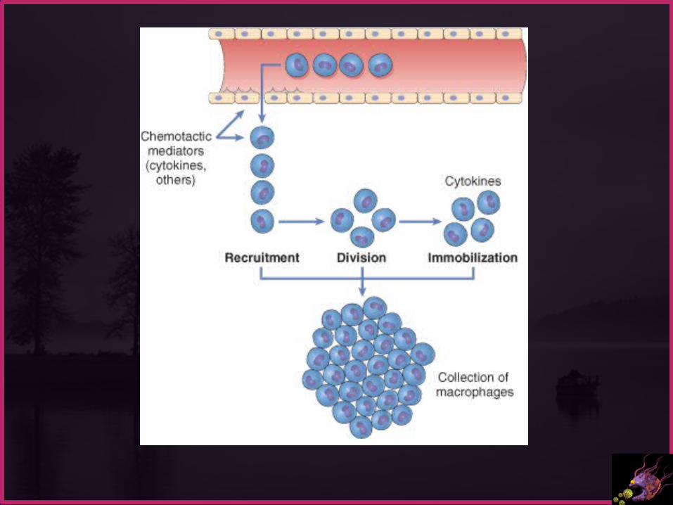
In chronic inflammation, macrophage accumulation persists, this is mediated by different mechanisms:
1. Recruitment of monocytes from the circulation, which results from the expression of adhesion molecules and chemotactic factors
2. Local proliferation of macrophages after their emigration from the bloodstream
3. Immobilization of macrophages within the site of inflammation

Lymphocytes Both T & B Lymphocytes migrates into inflammation
site
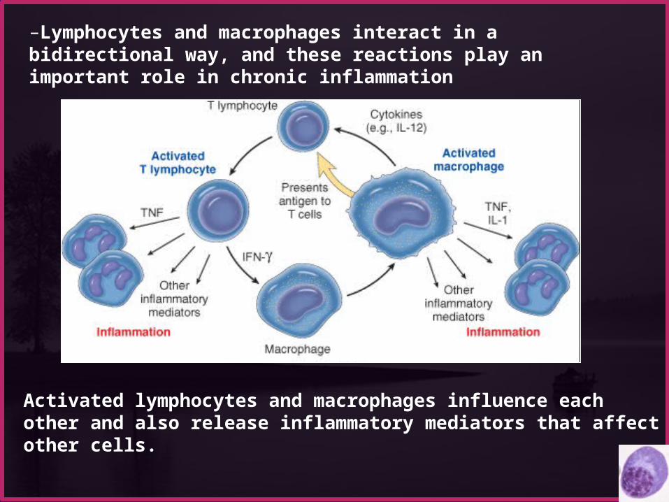
Activated lymphocytes and macrophages influence each other and also release inflammatory mediators that affect other cells.
–Lymphocytes and macrophages interact in a bidirectional way, and these reactions play an important role in chronic inflammation

•Eosinophils are abundant in immune reactions mediated by IgE and in parasitic infections
• respond to chemotactic agents derived largely from mast cells• Granules contain major basic protein: toxic to parasites and lead to lysis of mammalian epithelial cells

Mast cells are widely distributed in connective tissues express on their surface the receptor that binds the
Fc portion of IgE antibody , the cells degranulate and release mediators, such as
histamine and products of AA oxidation

GRANULOMATOUS INFLAMMATION Granulomatous inflammation is a distinctive
pattern of chronic inflammatory reaction characterized by focal accumulations of activated macrophages, which often develop an epithelial-like (epithelioid) appearance

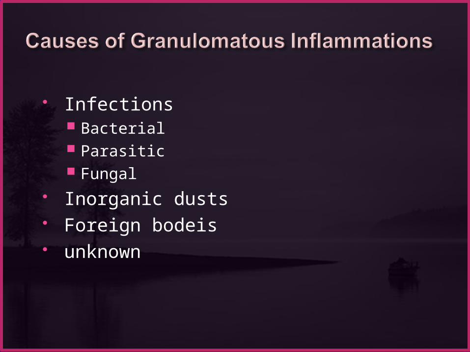
Infections Bacterial Parasitic Fungal
Inorganic dusts Foreign bodeis unknown
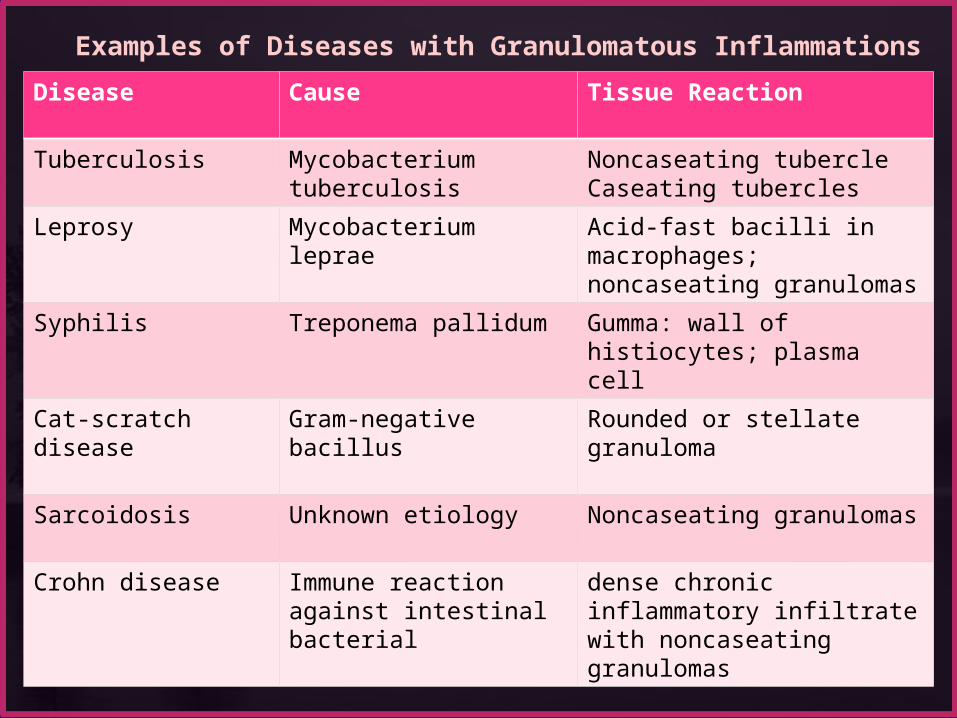
Disease Cause Tissue Reaction
Tuberculosis Mycobacterium tuberculosis
Noncaseating tubercleCaseating tubercles
Leprosy Mycobacterium leprae Acid-fast bacilli in macrophages; noncaseating granulomas
Syphilis Treponema pallidum Gumma: wall of histiocytes; plasma cell
Cat-scratch disease Gram-negative bacillus Rounded or stellate granuloma
Sarcoidosis Unknown etiology Noncaseating granulomas
Crohn disease Immune reaction against intestinal bacterial
dense chronic inflammatory infiltrate with noncaseating granulomas
Examples of Diseases with Granulomatous Inflammations
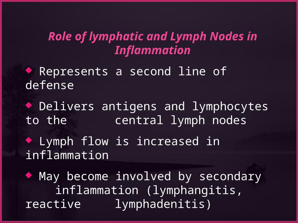
Role of lymphatic and Lymph Nodes in Inflammation
Represents a second line of defense
Delivers antigens and lymphocytes to the central lymph nodes
Lymph flow is increased in inflammation
May become involved by secondary inflammation (lymphangitis, reactive lymphadenitis)
Patterns of Acute Inflammation Outcomes of Acute Inflammation Patterns of chronic Inflammation
Defect in leukocyte function Complement deficiency Systemic manifestation
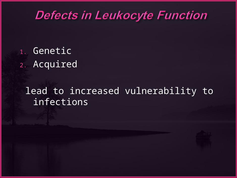
1. Genetic
2. Acquired
lead to increased vulnerability to infections
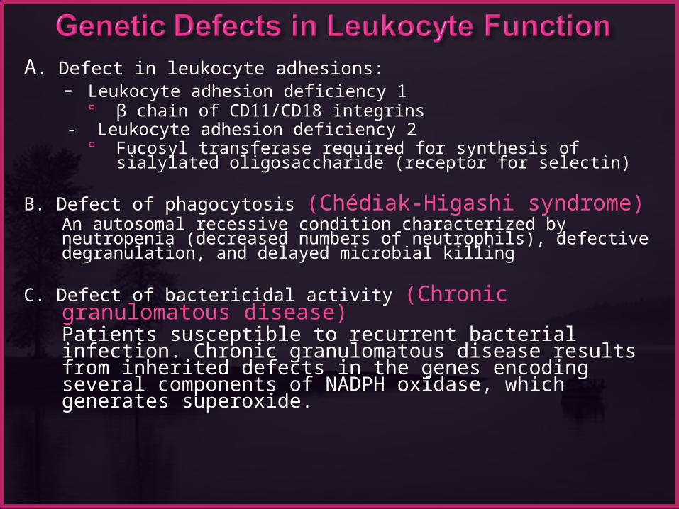
A. Defect in leukocyte adhesions:- Leukocyte adhesion deficiency 1
β chain of CD11/CD18 integrins - Leukocyte adhesion deficiency 2
Fucosyl transferase required for synthesis of sialylated oligosaccharide (receptor for selectin)
B. Defect of phagocytosis (Chédiak-Higashi syndrome) An autosomal recessive condition characterized by neutropenia (decreased numbers of neutrophils), defective degranulation, and delayed microbial killing
C. Defect of bactericidal activity (Chronic granulomatous disease)Patients susceptible to recurrent bacterial infection. Chronic granulomatous disease results from inherited defects in the genes encoding several components of NADPH oxidase, which generates superoxide.
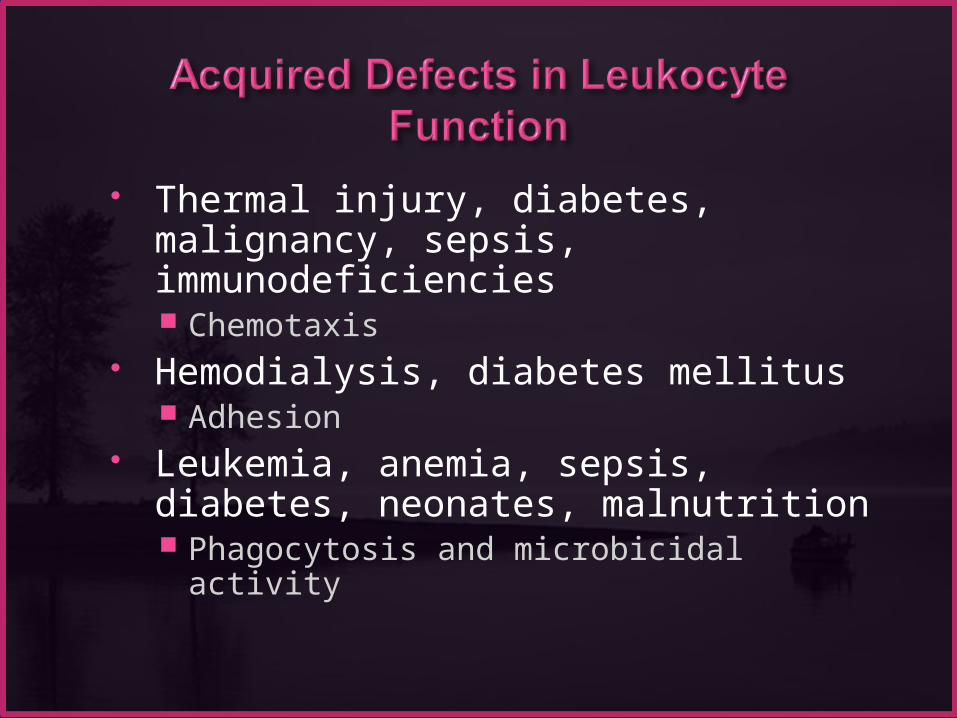
Thermal injury, diabetes, malignancy, sepsis, immunodeficiencies Chemotaxis
Hemodialysis, diabetes mellitus Adhesion
Leukemia, anemia, sepsis, diabetes, neonates, malnutrition Phagocytosis and microbicidal activity
Patterns of Acute Inflammation Outcomes of Acute Inflammation Patterns of chronic Inflammation Defect in leukocyte function
Complement deficiency Systemic manifestation


Complement deficiencyComplement deficiencyhereditary deficiency of C3 results in an increased susceptibility to infection with pyogenic bacteria.
Inherited deficiencies of C1q, C2, and C4 increase the risk of immune complex-mediated disease (e.g., SLE)
Deficiencies of the late components of the classical complement pathway (C5-C8) result in recurrent infections by Neisseria

hereditary deficiency of complement components, especially C3 (critical for both the classical and alternative pathways), results in an increased susceptibility to infection with pyogenic bacteria.
Inherited deficiencies of C1q, C2, and C4 do not make individuals susceptible to infections, but they do increase the risk of immune complex-mediated disease (e.g., SLE), possibly by impairing the clearance of apoptotic cells or of antigen-antibody complexes from the circulation.
Deficiencies of the late components of the classical complement pathway (C5-C8) result in recurrent infections by Neisseria (gonococci, meningococci) but not by other microbes.

Lack of the regulatory protein C1 inhibitor allows C1 activation, with the generation of down-stream vasoactive complement mediators
The result is hereditary angioedema, characterized by recurrent episodes of localized edema affecting the skin and/or mucous membranes.
Patterns of Acute Inflammation Outcomes of Acute Inflammation Patterns of chronic Inflammation Defect in leukocyte function Complement deficiency
Systemic manifestation
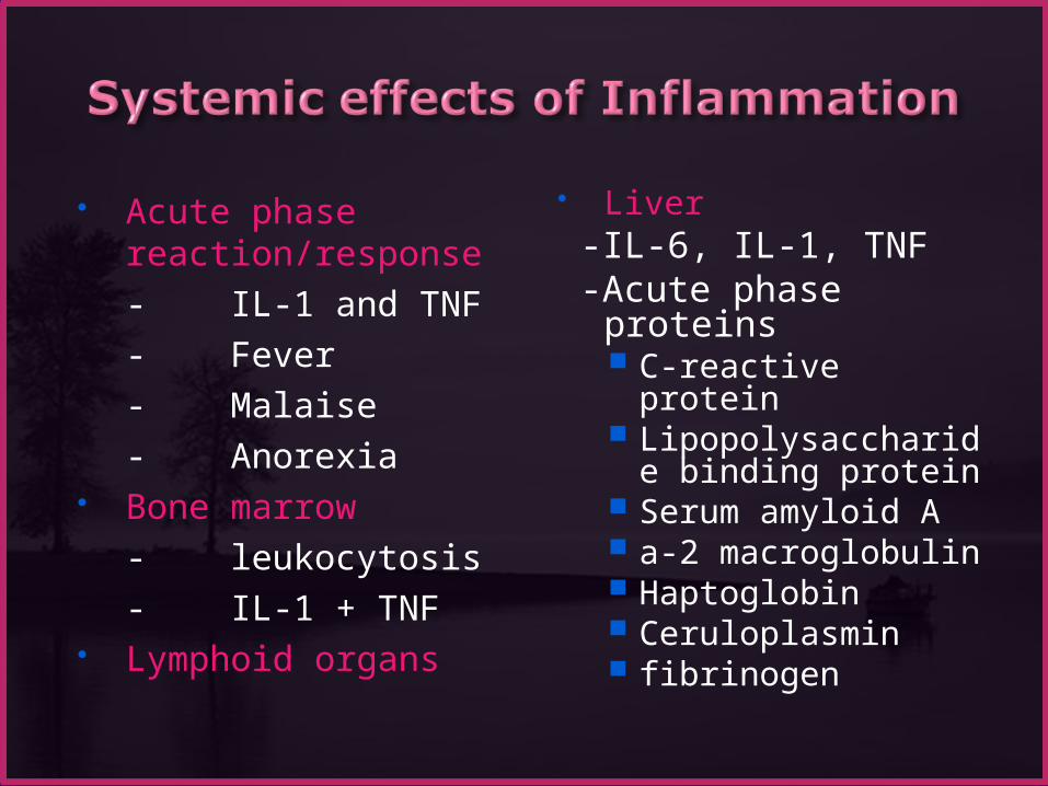
Acute phase reaction/response- IL-1 and TNF- Fever- Malaise- Anorexia
Bone marrow- leukocytosis- IL-1 + TNF
Lymphoid organs
Liver -IL-6, IL-1, TNF -Acute phase
proteins C-reactive protein Lipopolysaccharide
binding protein Serum amyloid A a-2 macroglobulin Haptoglobin Ceruloplasmin fibrinogen
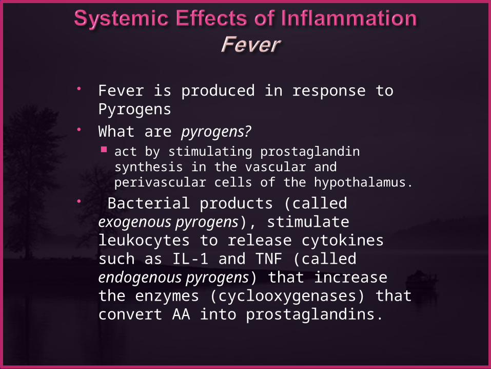
Fever is produced in response to Pyrogens What are pyrogens?
act by stimulating prostaglandin synthesis in the vascular and perivascular cells of the hypothalamus.
Bacterial products (called exogenous pyrogens), stimulate leukocytes to release cytokines such as IL-1 and TNF (called endogenous pyrogens) that increase the enzymes (cyclooxygenases) that convert AA into prostaglandins.

•In the hypothalamus, the prostaglandins, especially PGE2,
stimulate the production of neurotransmitters such as cyclic AMP, which function to reset the temperature set-point at a higher level.
•NSAIDs, including aspirin , reduce fever by inhibiting cyclooxygenase and thus blocking prostaglandin synthesis.
•fever may induce heat shock proteins that enhance lymphocyte responses to microbial antigens.
Fever
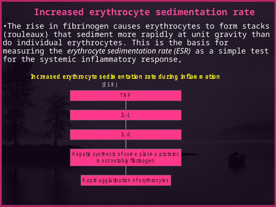
In c reased erythro cyte sedim en tatio n ra te du ring inflam m ation(E S R )
R a p id a gg lu tin a tio n o f e ryth rocytes
H e p atic syn th es is o f so m e p la sm a p ro te insm o st n o ta b ly f ib rin og en
IL -6
IL -1
T N F
•The rise in fibrinogen causes erythrocytes to form stacks (rouleaux) that sediment more rapidly at unit gravity than do individual erythrocytes. This is the basis for measuring the erythrocyte sedimentation rate (ESR) as a simple test for the systemic inflammatory response,
Increased erythrocyte sedimentation rate
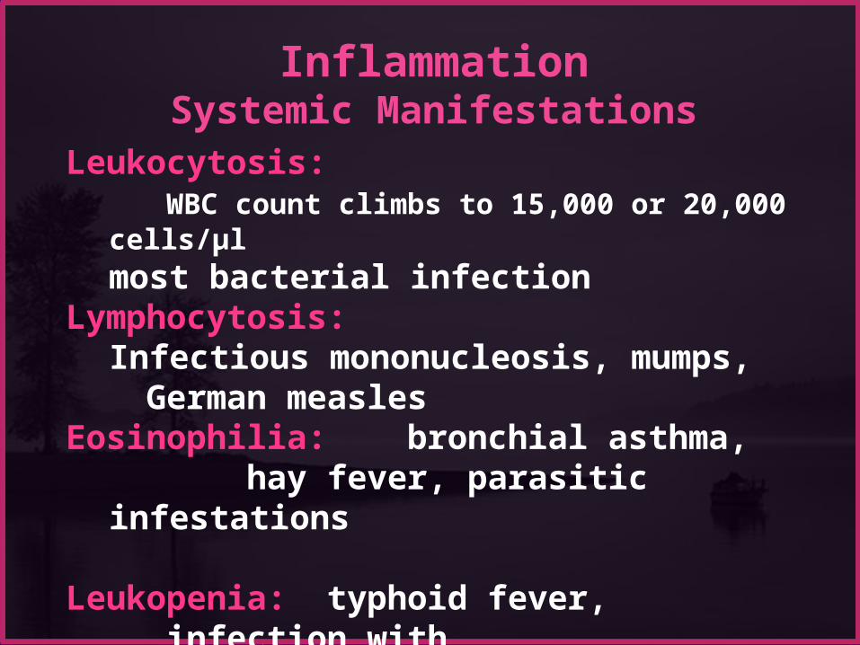
Inflammation
Systemic ManifestationsLeukocytosis: WBC count climbs to 15,000 or 20,000 cells/μl
most bacterial infectionLymphocytosis:
Infectious mononucleosis, mumps, German measlesEosinophilia: bronchial asthma, hay fever, parasitic infestations
Leukopenia: typhoid fever, infection with rickettsiae/protozoa


An experiment introduces bacteria into a perfused tissue preparation. Leukocytes then leave the vasculature and migrate to the site of bacterial inoculation. The movement of these leukocytes is most likely to be mediated by which of the following substances?
(A) Bradykinin (B) Chemokines (C) Histamine (D) Prostaglandins (E) Complement C3a

A 32-year-old woman has had a chronic cough with fever for the past month. On physical examination, she has a temperature of 37.5°C, and on auscultation of the chest, crackles are heard in all lung fields. A chest radiograph shows many small, ill-defined nodular opacities in all lung fields. A transbronchial biopsy specimen shows interstitial infiltrates with lymphocytes, plasma cells, and epithelioid macrophages. Which of the following infectious agents is the most likely cause of this appearance?
(A) Staphylococcus aureus (B) Plasmodium falciparurn (C) Candida albi cans (D) Mycobacteriurn tuberculosis (E) Klebsiella pneumo nine (F) Cytomegalovirus


Time 4-6 hours to 3-5 days
Vascular involvement Active hyperemia Edema, occ.fibrin
thrombi Neutrophils Cardinal signs of
inflammation Lymphatics
Role to remove exudate Can lead to
inflammation. Lymphangitis Lymphadenitis


