Triant and Pearson Genome Biology (2015) 16:99 DOI 10.1186/s13059-015-0656-7
RESEARCH Open Access
Most partial domains in proteins are alignmentand annotation artifactsDeborah A Triant and William R Pearson*
Abstract
Background: Protein domains are commonly used to assess the functional roles and evolutionary relationships ofproteins and protein families. Here, we use the Pfam protein family database to examine a set of candidate partialdomains. Pfam protein domains are often thought of as evolutionarily indivisible, structurally compact, units fromwhich larger functional proteins are assembled; however, almost 4% of Pfam27 PfamA domains are shorter than 50%of their family model length, suggesting that more than half of the domain is missing at those locations. To betterunderstand the structural nature of partial domains in proteins, we examined 30,961 partial domain regions from 136domain families contained in a representative subset of PfamA domains (RefProtDom2 or RPD2).
Results: We characterized three types of apparent partial domains: split domains, bounded partials, and unboundedpartials. We find that bounded partial domains are over-represented in eukaryotes and in lower quality proteinpredictions, suggesting that they often result from inaccurate genome assemblies or gene models. We also find that alarge percentage of unbounded partial domains produce long alignments, which suggests that their annotation as apartial is an alignment artifact; yet some can be found as partials in other sequence contexts.
Conclusions: Partial domains are largely the result of alignment and annotation artifacts and should be viewed withcaution. The presence of partial domain annotations in proteins should raise the concern that the prediction of theprotein’s gene may be incomplete. In general, protein domains can be considered the structural building blocks ofproteins.
BackgroundThe discovery of evolutionarily mobile protein domainsin the early 1980s, shortly after the recognition of eukary-otic splicing, revolutionized our understanding of pro-tein structure. Before the discovery of the exon-shuffleddomains in the EGF receptor [1,2], most proteins (globins,cytochrome c, serine proteases, etc.) were understood tobe globally similar single-domain proteins. While proteinslike calmodulin were known to contain repeated domains,the structural implications of modular proteins were notfully appreciated until clearly homologous domains wereseen in different sequence contexts.Today, domains are central to our understanding of the
structure, evolution, and functional roles of proteins andprotein families. Protein domain assignments using Pfam[3], InterPro [4], and other domain annotation resourcesare widely used to infer protein evolutionary relationships,
*Correspondence: [email protected] of Biochemistry and Molecular Genetics, University of Virginia,Box 800733, Charlottesville, VA 22908, USA
because it is often the protein domain, rather than the pro-tein as a whole, that is conserved over evolution. Evolu-tionarily conserved, structurally compact protein domainsare often found in very different sequence contexts, andonly by subdividing a protein into its constituent domainscan one understand its evolutionary history.Some protein domains have clearly understood func-
tions [5]. For example, protein kinase domains are cat-alytic modules with well-defined roles; other domainsdirect protein–protein interactions, target other proteinmodifications or play critical roles in binding and sig-nal recognition (e.g., SH2, SH3, or EF-hand Ca-binding).Identification of these domains helps identify the biologi-cal function of the protein containing them.The evolutionary, structural, and functional roles of
domains suggest that domains are the indivisible build-ing blocks from which larger modular proteins are built.Thus, we were surprised to find that 5% to 10% of pro-tein domain annotations in the Pfam protein domaindatabase suggest that only a fraction of the domain is
Triant and Pearson Genome Biology (2015) 16:99 Page 2 of 12
present in the protein. These partial protein domainscan cause problems with iterative profile-based similaritysearches [6]. Restricting PSI-BLAST searches to librariesof proteins with full-length Pfam protein domains dra-matically reduces position-specific scoringmatrix (PSSM)corruption, and improves PSI-BLAST specificity and sen-sitivity [6]. Because PSSM contamination is often causedby the extension of a homologous alignment into a non-homologous neighboring sequence, alignment to a partialPfam domain might corrupt a PSSM by nucleating a non-homologous alignment across the part of the domain thatwas missing from the partial domain location. However, ifdomains are indivisible then the nature of partial domainsis puzzling. Do the boundaries of partial domains corre-spond to structurally distinct regions, or are they bothevolutionarily mobile and structurally diverse? Are thesepartial domains authentic structural units or possibleannotation artifacts?To investigate the nature of these partial protein
domains, we used the Pfam database, which uses hid-den Markov models (HMMs) to scan UniProt proteinsequences and classify conserved domain regions [3].Pfam has been widely used to characterize the dynam-ics of protein domain coverage [7], compare sequence andstructure [8], and predict erroneous protein sequences [9].However, profile HMMs do not always detect full-lengthdomains, evenwhen they are present. Sometimes, only themost conserved part domain aligns with the HMM, lead-ing to annotation errors [10]. To examine in detail a set ofprotein domains representative of the Pfam database, weused domain annotations from the RPD2 protein database[11] that appeared to include less than 50% of the proteindomain family model length.
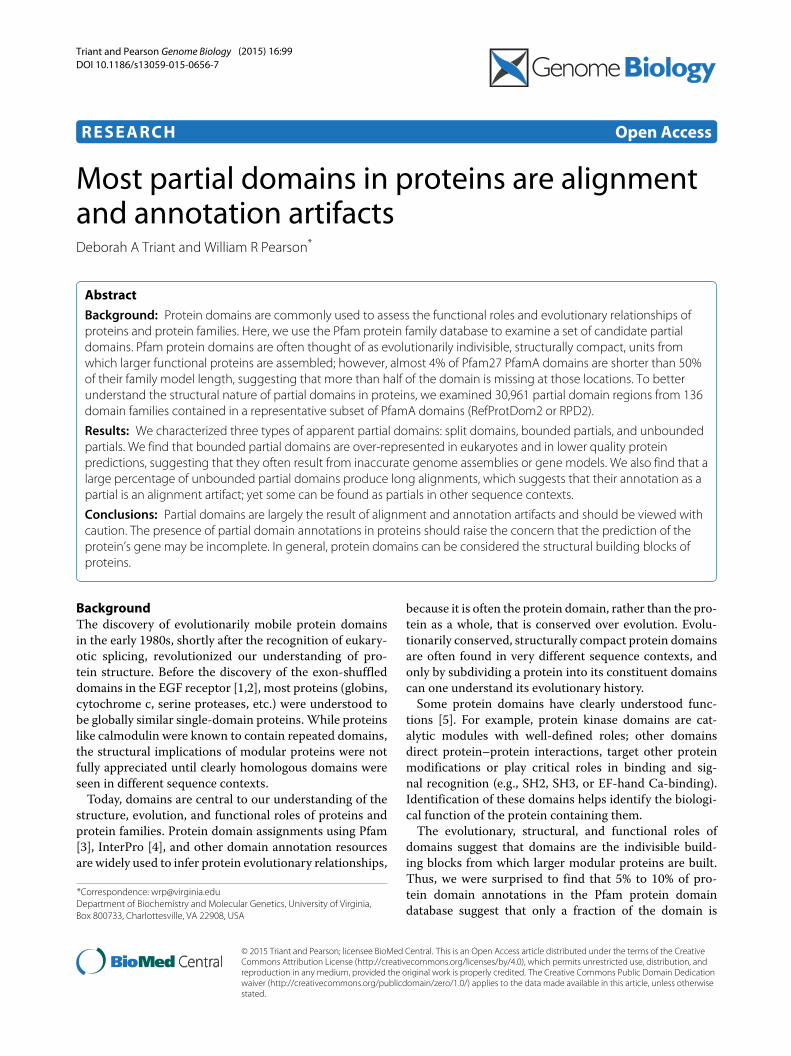
Results and discussionProtein domain lengthsTo characterize partial domains in proteins, we exam-ined 136 domain families from Pfam27 (the RDP2subset, see Methods). We chose Pfam because it isthe largest contributor to the InterPro compendium ofprotein domain databases (Pfam annotates more than40 million sequences of the 42 million sequences inUniProt/InterPro; the next most comprehensive annota-tion source covers about half as many). Pfam providesboth a model_length parameter, which can be thoughtof as the characteristic length of the Pfam domain fam-ily, and the model_start and model_end coordinates,which we used to calculate the coverage, or partial-ness ofthe domain in the sequence.Figure 1 shows the fraction of Pfam27 and RPD2
domains (A) and sequences (B) that contain at leastone domain at different fractional coverage of themodel_length characteristic domain length. Morethan 80% of Pfam27 domain mappings cover 90% or moreof the domain model_length (Figure 1), consistent withthe view that most Pfam domains are discrete-lengthstructurally compact building blocks. Likewise, 75% ofthe sequences annotated by Pfam contain domains thatare ≥90% of the family’s model_length. While veryshort partial domain instance alignments that cover<20%of model length are quite rare (about 0.5% of all domainsand non-fragment sequences in Pfam27), the numbers arelarge (134,676) because there are more than 24 millionPfam27 domains and 15million Pfam27 sequences. In thisreport, we focus on domain annotations where 50% ormore of the Pfam HMM, which defines the Pfam family,is missing at the domain annotation on the protein. In the
0.0001
0.001
0.01
0.1
1
dom
ain
frac
tion
fraction model length
Pfam27
Pf27>200
RPD2
0.0001
0.001
0.01
0.1
1
0 0.2 0.4 0.6 0.8 1 0 0.2 0.4 0.6 0.8 1
sequ
ence
frac
tion
fraction model length
A B
Figure 1 Distribution of partial domain lengths, Pfam27 and RPD2. (A) Cumulative fraction of domains versus fractional domain length. Cumulativefractions are shown for Pfam27 domains found in proteins marked as not fragments (24 million domains in total, of which 945,100 are <50% ofmodel length, blue squares) and the RPD2 domains in Pfam27 (290,148 domains, 30,030 <50% of model length, red circles). Also shown are Pfam27domains from families with more than 200 match states (6.9 million domains, 658,089 <50% partials, blue diamonds). (B) Cumulative number ofsequences with increasing domain length. Cumulative fractions for Pfam27 sequences (16 million sequences, 820,000 with <50% partials, bluesquares) and RPD2 sequences (274,000 total, 27,000 with a domain <50% of model length, red circles). Blue diamonds show sequences containingdomains with model length >200 match states (6.3 million sequences, 557,941 <50% partials).
Triant and Pearson Genome Biology (2015) 16:99 Page 3 of 12
15 million sequences in Pfam27, there are 945,100 partialdomains that are <50% of the model length in 820,720different sequences. The numbers of partial domains inFigure 1 exclude proteins annotated as fragments; includ-ing protein fragments increases the number of <50%partials from 0.95 to 2.0 million.In reporting the instances of partial domains in Pfam
protein annotations, we distinguish Pfam families – theset of largely distinct protein domains that are associ-ated with different Pfam hidden Markov models (HMMs)[12] – from Pfam domains – the annotation of a domain,or partial domain, at a particular location in a protein.Because a single protein can be annotated to contain mul-tiple Pfam domains from the same family (Figure 2B),we report both the number of Pfam domains (individ-ual HMM mappings to a sequence) and Pfam sequences.When the biological domain has been split into multi-ple parts by the HMM alignment process (Figure 2B), the
Pfam sequence count is a more conservative estimate ofthe number of partial domains.While Pfam provides a very comprehensive annotation
of domains in proteins, it is difficult to present statis-tics for representative Pfam families because of the widerange of family model lengths (maximum 2,208, minimum7, median 134), family sizes (maximum 363,409, mini-mum 2,median 333), and number of sequences containinga particular Pfam family (maximum 313,128, minimum1, median 315). Thus, we focused on the RPD2 subsetof Pfam27. RPD2 Pfam families have at least 100 mem-bers, with no more than 5,000 sequences for any domain.RPD2 also requires that the Pfam family have at least200 match states; distantly related partial domains withshortermodel_lengths can be difficult to detect. RPD2also limits families from Pfam clans. Pfam clans [12] areused to capture Pfam families that, while homologous, areso evolutionarily diverse that the members of the family
100 200 300B3J323sequence
PF01544model_start:4 model_end:292
seq_start:27 seq_end:312
PF01544 model
400 600 800P35724sequence
6 66
499 566
62 148
639 757
PF01544146 292
803 965
PF01544 model
400 600 800P43553sequence
PF015446 210
451 683
PF01544187 289
667 799
PF01544 model
103 187
end-bounded
100Q7U9V6sequence
PF01544167 292
1 130
100 200 300 400Q9S9N4sequence
PF01544190 289
302 438
complete domain
split domain
bounded partial
un-bounded partialPF01544? 1891
100 200 300E9GP80sequence
PF084545 73
PF00183396 525
183 301
domain-bounded
A
B
C
D
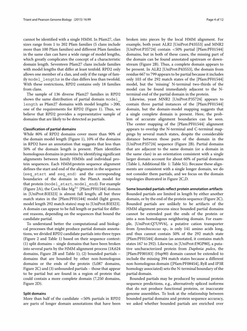
Figure 2 Complete, bounded, and unbounded partial domains. A complete domain, and three types of partial Pfam27 domain mappings.(A) Annotation of the complete [Pfam:PF01544] domain in Bacillus anthracis CorA protein [UniProt:B3J323]. The full length of the Pfam27 domain isshown in light green, as are the coordinates of the aligned domain in the [Pfam:PF01544] model (model_start, model_end) and [UniProt:B3J323]protein sequence (seq_start, seq_end). (B) Split domains. Annotation of [Pfam:PF01544] domains in yeast ALR2 [UniProt:P43553] and MNR2[UniProt:P35724]. (C) Partial domains bounded by the ends of the sequence [UniProt:Q7U9V6] or other domains [UniProt:E9GP80]. (D) Anunbounded partial domain in [UniProt:Q9S9N4].
Triant and Pearson Genome Biology (2015) 16:99 Page 4 of 12
cannot be identified with a single HMM. In Pfam27, clansizes range from 1 to 202 Pfam families (5 clans includemore than 100 Pfam families) and different Pfam familiesin the same clan can have a wide range of model lengths,which greatly complicates the concept of a characteristicdomain length. Seventeen Pfam27 clans include familieswith model lengths that differ at least tenfold. RPD2 onlyallows one member of a clan, and only if the range of fam-ily model_lengths in the clan differs less than twofold.With these restrictions, RPD2 contains only 18 familiesfrom clans.The sample of 136 diverse Pfam27 families in RPD2
shows the same distribution of partial domain model_length as Pfam27 domains with model lengths >200,one of the requirements for RPD2 (Figure 1). Thus, webelieve that RPD2 provides a representative sample ofdomains that are likely to be detected as partials.
Classification of partial domainsWhile 80% of RPD2 domains cover more than 90% ofthe domain model length (Figure 1), 10% of the domainsin RPD2 have an annotation that suggests that less than50% of the domain length is present. Pfam identifieshomologous domains in proteins by identifying significantalignments between family HMMs and individual pro-tein sequences. Each HMM:protein sequence alignmentdefines the start and end of the alignment in the sequence(seq_start and seq_end) and the correspondingboundaries of the domain in the PfamA model forthat protein (model_start, model_end). For example(Figure 2A), the CorA-like Mg2+ [Pfam:PF01544] domainin [UniProt:B3J323] is almost full length; all but threematch states in the [Pfam:PF01544] model (light green,model length 292 match states) map to [UniProt:B3J323].A domain can appear to be full length or partial for differ-ent reasons, depending on the sequences that bound thecandidate partial.To understand better the computational and biologi-
cal processes that might produce partial domain annota-tions, we divided RPD2 candidate partials into three types(Figure 2 and Table 1) based on their sequence context:(1) split domains – single domains that have been brokeninto several parts by the HMM alignment process (18,624domains, Figure 2B and Table 1); (2) bounded partials –domains that are bounded by other non-homologousdomains or the ends of the protein (5,087 domains,Figure 2C) and (3) unbounded partials – those that appearto be partial but are found in a region of protein thatcould contain a more complete domain (7,250 domains,Figure 2D).
Split domainsMore than half of the candidate <50% partials in RPD2are parts of longer domain annotations that have been
broken into pieces by the local HMM alignment. Forexample, both yeast ALR2 [UniProt:P43553] and MNR2[UniProt:P35724] contain <50% partial [Pfam:PF01544]domains, but in both of these cases, the missing part ofthe domain can be found annotated upstream or down-stream (Figure 2B). Thus, a complete domain appears tobe present. In ALR2 [UniProt:P43553], the domain fromresidue 667 to 799 appears to be partial because it includesonly 103 of the 292 match states of the [Pfam:PF01544]model, but the ‘missing’ N-terminal two-thirds of themodel can be found immediately adjacent to the N-terminal end of the partial domain in the protein.Likewise, yeast MNR2 [UniProt:P35724] appears to
contain three partial instances of the [Pfam:PF01544]domain, but the domain-model mapping suggests thata single complete domain is present. Here, the prob-lem of accurate alignment boundaries can be seen.The center mapping of the [Pfam:PF01544] alignmentappears to overlap the N-terminal and C-terminal map-pings by several match states, despite the considerabledistance between those parts of the domain in the[UniProt:P35724] sequence (Figure 2B). Partial domainsthat are adjacent to the same domain (or a domain inthe same clan) in an orientation consistent with a singlelarger domain account for about 60% of partial domains(Table 1, Additional file 1: Table S1). Because these align-ments are consistent with a single longer domain, we donot consider them partials, and we focus on the domaintopologies illustrated in Figure 2C,D.
Some bounded partials reflect protein annotation artifactsBounded partials are limited in length by either anotherdomain, or by the end of the protein sequence (Figure 2C).Bounded partials are unlikely to be artifacts of theHMM alignment process since bounded partial domainscannot be extended past the ends of the protein orinto a non-homologous neighboring domain. For exam-ple, [UniProt:Q7U9V6], a putative cation transporterfrom Synechococcus sp., is only 141 amino acids long,and thus cannot contain 50% of the 292 match state[Pfam:PF01544] domain (as annotated, it contains matchstates 167 to 292). Likewise, in [UniProt:E9GP80], a puta-tive uncharacterized protein from Daphnia pulex, the[Pfam:PF00183] (Hsp90) domain cannot be extended toinclude the missing 394 match states because a differentnon-homologous domain ([Pfam:PF08454], RyR and IP3Rhomology associated) sets the N-terminal boundary of thepartial domain.Bounded partials may be produced by unusual protein
sequence predictions, e.g., alternatively spliced isoformsthat do not produce functional proteins, or inaccurategenome assemblies. To look at the relationship betweenbounded partial domains and protein sequence accuracy,we asked whether bounded partials are enriched over
Triant and Pearson Genome Biology (2015) 16:99 Page 5 of 12
Table 1 Partial domains in RPD2
Sequences % Domains %
Total 270,776 100.0 290,148 100.0
50% partials 25,116 9.33 30,961 10.72
Split 13,090 4.84 18,624 6.42
Bounded 4,953 1.88 5,087 1.80
Unbounded 7,073 2.62 7,250 2.50
Putative partial 2,118 0.78 2,156 0.74
Minimum 1st quartile Median Mean 3rd quartile Maximum PfamAa
aThe Pfam27 family that produced the maximum percentage of partial domains in the corresponding partial category.
non-partial sequences in organisms that contain introns(eukaryotes), and thus might be splicing or assembly arti-facts. We examined the 94 RPD2 Pfam27 families with 10or more bounded 50% partial domains and asked whetherthe bounded partial-containing proteins in these familieswere more likely to come from eukaryotes than RPD2proteins that do not contain partial domains. If boundedpartial domains are due to inaccurate gene models, weexpect the errors more frequently in eukaryotes. We per-formed Fisher’s exact test on each of the 94 families, andthen calculated the false-discovery rate (q value) to iden-tify families that are significantly enriched for eukaryoticbounded partials (see Methods). When the non-partialand bounded-partial sequence sets were divided intoeukaryotic/non-eukaryotic sets, 47 of the 94 bounded par-tial sets were enriched for eukaryotic sequences at a qvalue (false-discovery rate) of <0.05, and 34 at <0.01.We conclude that many bounded partial domains resultfrom inaccurate gene models that produce incompleteproteins.In addition, we examined the relative abundance of
bounded partial domains from very carefully annotatedgenomes (human, mouse, and Drosophila) in reviewedproteins from SwissProt with an Ensembl gene model.During the Swissprot review process, multiple alterna-tively spliced transcripts with different accession numbersin the TREMBL division of UniProt are merged into
a single accession and labeled as isoforms [13]. Only4 of the 161 human, mouse, and Drosophila proteins(114 from human) in the RPD2 bounded partial cate-gory have been reviewed by SwissProt, and one of thosedoes not have an Ensembl gene model. In contrast 848of 2,893 non-partial RPD2 proteins from human, mouse,and Drosophila have been reviewed and 790 have anEnsembl gene model. Bounded partial domain proteinsfrom human, mouse, and Drosophila are dramaticallyenriched in unreviewed sequences lacking Ensembl genemodels (P < 10−15, Fisher’s exact test). Since boundedpartials are rarely found in carefully annotated full-lengthproteins from these organisms, we believe that many arelikely to be incomplete splice isoforms or other annotationartifacts.
Unbounded partialsExcluding split domains, a majority of the candidate par-tial domains belong to the unbounded partial category(Figure 2D). These domains are annotated as partial,but could contain a full-length domain, because there isroom for the missing part of the domain in the sequence.Thus, if the [Pfam:PF01544] HMM is projected onto the[UniProt:Q9S9N4] sequence from Figure 2D, one obtainsa sequence starting at residue 112, or 330 residues thatcould map to the 292 match state [Pfam:PF01544] model.When that sequence [UniProt:Q9S9N4:112-] is compared
Triant and Pearson Genome Biology (2015) 16:99 Page 6 of 12
to the sequences in RPD2 Pfam27 using SSEARCHwith Blosum62, 156 of the 4,760 sequences containing[Pfam:PF01544] in RPD2 have E() < 10−6, and all ofthose alignments contain an annotated [Pfam:PF01544]domain. However, all but two of the 10−6 homologs alignover more than 200 amino acids, and more than 75%align over more than 250 residues, close to the 292 matchstates in the [Pfam:PF01544] model. The three majortypes of alignments are shown in Figure 3. In about 40%of the homologs (67/156), the alignments are long, butthe domains annotated on the proteins are much shorter(Figure 3A); these proteins are most closely related tothe [UniProt:Q9S9N4] query. For most of the more dis-tant homologs (86/156), the alignment is still long, butthe aligned sequence is also annotated as containing a
full-length [Pfam:PF01544] domain (Figure 3C). In twocases ([UniProt:A5BS21], E() < 10−16, Figure 3D,E and[UniProt:B7FG10], E() < 10−20, not shown), both theannotated domains and the alignments are short. Forthe [UniProt:B7FG10] alignment, the alignment is shortbecause the protein is short and contains a boundedpartial.If [Pfam:PF01544] contains a true partial domain that
can be found in different protein contexts, then weexpect that the short alignment with [UniProt:A5BS21],seen in Figure 3B, would reflect novel sequencecontext, rather than incomplete alignment. A searchwith the full [UniProt:A5BS21] sequence suggests that[UniProt:A5BS21] does contain an evolutionarily mobilesub-domain. Most of the [UniProt:A5BS21] alignments
Q9S9N4
A5BS21
Q9S9N4 – two short domains; long alignment
Q9S9N4 – two short domains; short alignment
Q9S9N4 – one short, one long domain; long alignment
A5BS21 – two short domains; long alignment
A5BS21 – one long, one short domain; short alignment
Q9S9N4
P87149
100 200
A5BS21
Q9LJN2
200 300 400
PF01544166 276
PF01544189 282
100 200
A5BS21
200 300 400
B4FQF3
PF01544166 276
PF0154485 284
Q9S9N4
F2EH86
200 300 400
100 200 300PF01544192 288
PF01544190 289
200 300 400
100 200PF01544166 276
PF01544190 289
200 300 400
100 200 300 400PF01544110 286
PF01544190 289
A
B
C
D
E
Figure 3 Longer alignments with extended partial domains. Two proteins annotated with partial [Pfam:PF01544] domains, [UniProt:Q9S9N4:112-411]and [UniProt:A5BS21], were compared to the RPD2 proteins using SSEARCH36 (BLOSUM62 scoring matrix, gap open/extend −11/−1). (A), (B), and(C) show three representative alignments from the 156 RPD2 sequences sharing statistically significant similarity (E() < 10−6) with [UniProt:Q9S9N4](residues 112 to 411). Lines indicate the protein sequences; open trapezoidal boxes show the projection of the alignments onto the sequences.Shaded boxes map the [Pfam:PF01544] domains annotated on the proteins. The numbers in the shaded boxes report model start and endcoordinates from Pfam27. (A) One ([UniProt:Q9S9N4:UniProt:F2EH86]) of the 67 longer alignments (>200 amino acids) between proteins with shortdomain annotations (<200 residues). Non-self-alignments in this category ranged from 26% to 99% identical with 10−10 < E() < 10−157.(B) Alignment of [UniProt:Q9S9N4] with [UniProt:A5BS21], a short alignment (150 residues) between two much longer proteins. (C) One alignment([UniProt:Q9S9N4:UniProt:P87149]) representative of the 86 long alignments (>200 residues, E() < 10−6) to proteins with >50% partial [Pfam:PF01544]domain annotations. (D) One ([UniProt:A5BS21:UniProt:Q9LJN2]) of the five non-self-alignments>200 amino acids between proteins with [Pfam:PF01544]domain annotations <200 amino acids (51% identity, E() < 10−54). (E) A short alignment (156 residues, 37% identity, E() < 10−15, [UniProt:A5BS21:UniProt:B4FQF3]) where one protein is annotated to contain ≥200 matches to [Pfam:PF01544].
Triant and Pearson Genome Biology (2015) 16:99 Page 7 of 12
involve partial domains that produce short alignments likeFigure 3B (alignments with 10−25<E()<10−6 and 29% to48% identity), but [UniProt:A5BS21] sometimes produceslong alignments with more closely related sequences thatare annotated as having partial [Pfam:PF01544] domains(Figure 3D, five non-self-alignments with 10−54 <
E() < 10−39, 44% to 51% identity). Moreover, some-times [UniProt:A5BS21] produces short alignments withdistantly related proteins with longer [Pfam:PF01544]domains (Figure 3E). While many short alignments andpartial [Pfam:PF01544] annotations reflect incompletealignments caused by long evolutionary distances, theinstances of short alignments at modest evolutionary dis-tances (>40% identity) suggest that the C-terminal halfof [Pfam:PF01544] contains an evolutionarily mobile sub-domain. Below, we show that part of [Pfam:PF01544] ishomologous to a smaller CATH structural domain.
Putative partialsTo identify candidate true partial domains from theset of unbounded partials (Figure 2D), we generatedthe projected full-length domain regions from candi-date unbounded proteins and compared the candidatefull-length domains to RPD2 proteins. Using the logicdescribed above for [Pfam:PF01544] partials, we soughtexamples like [UniProt:A5BS21] that have some longdomain homologs but also many short domain homologs.Putative partial domains met two criteria: (a) the extendedquery found ten or more homologs with E() < 10−6 and(b) at least 25% of the homolog alignments were <50% ofthe family model length. These Pfam families are countedas putative partials in Table 1. In total, 48 of the RPD2families had more than 10 queries that met these cri-teria (22 had more than 25 queries). These extendedqueries that met criteria (a) and (b) were then comparedto sequences with known structures to identify putativepartial domains.About one quarter of our candidate partials map to the
Pfam model in a way that leaves room for a much morecomplete Pfam domain (unbounded partial, Figure 2D).Searches with those extended sequences show that mostof them produce long alignments. Thus, those domainannotations are partial because of the inability of the Pfammodel to capture the entire homologous region for verydistant domain homologs (Figure 3). However, about 30%of these extended sequences produced short alignments,suggesting an alternative sequence context (putative par-tials), and in the two families with the most abundantputative partials, alignments were consistent with a com-pact structural domain (e.g. Figure 4B). These sequences,as well as some bounded sequences, were used to iden-tify Pfam27 domains that have been divided into smallerstructural units by the CATH structure database [14](Table 2).
2H12
2NZW
2ZXC
A
B
C
Figure 4 Structural partials. (A) The structure of [PDB:1H12] withsecondary structures for CATH domain 1.10.580.10 (58 to 274, 385 to410) highlighted in red (helix) and yellow (strand), and secondarystructures for the CATH domain 1.10.230.10 (275 to 384) that alignswith the partial [Pfam:PF00285] region of [UniProt:Q98FC2],highlighted in cyan (helix) and salmon (coil). (B) The structure of2NZW, the most abundant putative partial ([Pfam:PF00852]), withresidues 180 to 330 highlighted. (C) The structure of 2ZXC, arepresentative of the PF04734 PfamA family. The putative partialanalysis identifies a shorter alignment in the first 200 residues of thePfamA family, and VAST+ annotates a domain from residues 1 to 292,which are highlighted.
Triant and Pearson Genome Biology (2015) 16:99 Page 8 of 12
Table 2 RPD2 partial domains corresponding to CATH structural domains
Family Pfam CATH CATH length Accession
(Pfam) length class (1st to 3rd quartile) UniProt/PDB
aThe CATH:1.20.58.340 domain is annotated to two adjacent locations covered by PF01544 in [PDB:2BBJ|A].
Some partial domains result from shorter structuraldomainsTo identify compact sub-domains that might account forRPD2 partial annotations, we compared bounded andcandidate partials from the unbounded searches to thesequences in the PDB structure collection [15]. From the57 RPD2 Pfam27 families that appeared to have a signifi-cant number of bounded or unbounded partial domains,we identified 11 that contain multiple CATH structuraldomains (Table 2) that are also annotated by VAST+ [16].Table 2 summarizes the clearest examples of structurallycompact partial RPD2 Pfam domains.Many of the examples in Table 2 are straightforward;
a Pfam domain aligns with a structure containing multi-ple CATH structural domains, and each CATH domainis a contiguous sequence. For example, [UniProt:Q98FC2]appears to largely contain a single unbounded partialdomain from the C-terminal half of the [Pfam:PF00285](citrate synthase) model (Additional file 2: Figure S1). Butthe alignment of [UniProt:Q98FC2] with [PDB:2H12|A]shows that the structure comprises two CATH domains,the shorter of which (1.10.230.10) is homologous to theC-terminal portion of [Pfam:PF00285]. Figure 4 highlightsthe structurally compact region of [PDB:2H12|A] thatcorresponds to the [Pfam:PF00285] partial domain.
There are 56 PDB structures containing theCATH:1.10.230.10 domain in the current version ofCATH, with a mean length of 109 residues and thefirst and third quartiles ranging from 104 to 115. Thus,the CATH:1.10.230.10 domain is much shorter thanthe 356 match states of the [Pfam:PF00285] domain,and may explain the Pfam partial domain. In othercases, e.g., [Pfam:PF00118] (TCP-1/cpn60 chaperonin)/[UniProt:F0YNJ0] vs [PDB:1A6D|B], the Pfam domainaligns with portions of structures with multiple CATHdomains, some of which are discontinuous in sequence.In addition to structural partials that could be identified
by comparing Pfam and CATH domain annotations, weexamined the most abundant putative partials, including[Pfam:PF00852] (histone deacetylase, Table 1). Searcheswith 142 projected domains from [Pfam:PF00852] againstsequences from PDB show two alignment patterns. Halfof the alignments were largely full-length alignment, whilethe other half aligned to only the C-terminal region ofthe protein, with starting points ranging from 160 to232 (mean 212). 2NZW chain A, the only homolog tothe [Pfam:PF00852] proteins in PDB, is not annotatedby CATH. 2NZW|A is annotated by VAST+ [16], whichdescribes a structural domain that aligns with the partial[Pfam:PF00852] domain in [Uniprot:O87156] (Additional
Triant and Pearson Genome Biology (2015) 16:99 Page 9 of 12
file 3: Figure S3). The compact nature of the PF000852partial is shown in Figure 4B.For the second most abundant putative partial,
[Pfam:PF04734] (neutral/alkaline non-lysomal cerami-dase), about 10% of the domains meet the putativepartial criterion. Its closest homolog in PDB is 2ZXC,where it aligns with the N-terminal third of the protein,an α/β/β/α sandwich in the middle third of the pro-tein structure that is structurally compact (Figure 4C).Although CATH does not annotate this structure,VAST+ assigns a domain that aligns almost exactlywith the PF04734 partial (Additional file 3: Figure S3).The third most abundant putative partial fami-ly ([Pfam:PF03598]/CO dehydrogenase/acetyl-CoA syn-thase complex beta subunit) has about 3.5% of domainsin this category, and the abundance of putative par-tial domains decreases gradually to slightly less than1% of domains at the third quartile. [Pfam:PF03598] ishomologous to several proteins with CATH and VAST+domain annotations, which divide the [Pfam:PF03598]domain into three parts, and one of those parts,CATH 3.30.1650.10, was detected independently in[Uniprot:B8FZT4] (Additional file 4: Figure S2).Thus, the three most abundant putative partial domains
correspond to structurally compact regions based onCATH and VAST+ annotations. Five of the ten mostabundant putative partials appear to be structural partials,based on shorter domains found in CATH or VAST. Fourof the remaining abundant putative partials Pfam mod-els align a single long CATH or VAST+ domain. In onecase ([Pfam:PF02738]), the domain appears to align withrepeats of the same structural domain.We compared our hierarchical strategy for identifying
evolutionarily mobile structural partials – identificationof putative partial Pfam domains followed by confirma-tion using CATH – with the simpler strategy of lookingat CATH domain content in the protein structures anno-tated by Pfam. Of the 136 Pfam families in RPD2, 107are mapped in Pfam to PDB structures that are anno-tated by CATH. We found 64 of those Pfam families mapto PDB structures with only one CATH domain, so theycannot be examples of structural partials. In total, 43RPD2 Pfam families map to PDB structures containingtwo ormore different CATHdomains; these Pfam familiesmight be composed of evolutionarily mobile, structurallycompact, sub-domains. But only 11 of those 43 Pfam fam-ilies were confirmed to be both evolutionarily mobile andstructurally compact. In 15 cases, either the CATH sub-domain was not consistent with the evolutionarily mobilesequence, or there was inconsistency between the CATHand the VAST annotation. For another 17 Pfam families,although CATH annotates multiple structural domains,we found very little evidence for evolutionary mobility(fewer than ten putative partials).
Our PDB/CATH/VAST+ searches found 11 Pfam fam-ilies comprising shorter CATH and VAST+ structuraldomains, and another 7 Pfam families that do not havehomologs annotated in CATH, but contain multipleVAST+ domains (Table 2, Additional file 2: Figure S1,Additional file 4: Figure S2, Additional file 3: Figure S3).The combination of evolutionary mobility (the puta-tive partial domain in different sequence contexts) andstructural compactness suggests that these Pfam domainscould be subdivided into shorter, independent domains.
ConclusionsWe have tested the hypothesis that Pfam27 domains arelargely structurally compact protein building blocks bycharacterizing the 10% of Pfam domains in the RPD2database that appear to be shorter than <50% of thecharacteristic domain length (the Pfam model length).RPD2 protein domains are representative of Pfam27 sincethey have a distribution of partial domain annotationlengths that is almost indistinguishable from all Pfam27domains with model lengths greater than 200match states(Figure 1). However, RPD2 reduces the extreme differ-ences in domain abundance and model length variation.We believe that the RPD2 family subset accurately repre-sents partial domains in Pfam27.Our results suggest that, with a small number of excep-
tions, Pfam27 domains are compact structural buildingblocks. Only about 15% of the Pfam domains in RPD2appear to have genuine structurally compact, evolution-arily mobile partial domains, suggesting that most Pfamdomains do not comprise smaller structural units. Indeed,most apparent partial domains are likely to be alignment,annotation, and sequence assembly artifacts, rather thansmaller sub-domains.The distributions of the three types of candidate partial
domains – split domains, and bounded and unboundedpartials – differ widely for the different Pfam families inRPD2 (Table 1).Partial domain annotations can be counted in two
ways – by the number of sequences containing the partialor the number of partial domains – but the distributionof partial annotation counts is similar for both measures.About 60% of candidate partial domains are split domains,including the family with the largest number of candi-date partials, [Pfam:PF00209] (sodium:neurotransmittersymporter), where 77.8% of the domain annotations arepartial, but 72.5% of those partials are split domains. Thefamily-specific nature of partial domains is illustrated bythe difference between the extremes (minimum and max-imum in Table 1) and the quartiles. There is no partialtype that is found in every Pfam family (the minimum isalways 0.00), and the maximum percentage is usually ten-fold higher than the third quartile percentage. Moreover,the Pfam family with the largest fraction of unbounded
Triant and Pearson Genome Biology (2015) 16:99 Page 10 of 12
partials ([Pfam:PF04734]) is not the same as the onewith the largest number of putative unbounded partials([Pfam:PF00852]). These differences in the types and fre-quencies of different partial domains reflect interactionsbetween the sensitivity of the HMM domain model, thedistribution of domains at different evolutionary dis-tances, and the sequence sampling of the databases usedto construct the models.Partial domain annotations are well recognized in Pfam
[10]; they are represented as jagged edges in Pfam’sgraphical presentation. But, because the model_start/model_end information is available only through Pfam’sXML or MySQL interfaces, the graphical Pfam presen-tation makes it difficult to distinguish split domains(Figure 2B) from a repeated set of N- or C-terminal par-tial domains. Mapping the domain to the HMM allows usto infer that 60% of 50% partials can be made complete bycombining split domain partials.About one sixth of candidate partial domains can-
not be extended to produce larger regions, becausethey are bounded by the end of the protein or byother non-homologous domains (Figure 2C). While someof these bounded partial domains are legitimate par-tials, some bounded partial domains are likely to beprotein assembly artifacts, while others may be alterna-tively spliced isoforms. Light and Elofsson [17] exam-ined how alternatively spliced isoforms can producestructurally incomplete, non-functional proteins. Indeed,of the 114 human proteins with bounded domains inRPD2, only three, [UniProt:A8MTL9] ([Pfam:PF00079],serpin), [UniProt:Q5T2L2] ([Pfam:PF00248], aldo-ketoreductase), and [UniProt:P0C7U1] ([Pfam:PF04734]),were both annotated as reviewed and had an Ensemblgene model. Both [Pfam:PF00079] and [Pfam:PF04734]appear to contain genuine partials (Table 2 and discus-sion below), and the gene encoding [UniProt:Q5T2L2]([Ensembl:ENSG00000264006]) is annotated to producetwo alternative transcripts with longer coding sequences,both of which are annotated as CDS (CoDing Sequence)incomplete.About one quarter of our candidate partials map to the
Pfam model in a way that leaves room for a much morecomplete Pfam domain (unbounded partial, Figure 2D).Searches with those extended sequences show that mostof them produce long alignments; thus, those domainannotations are partial because of the inability of thePfam model to capture the entire homologous regionfor very distant domain homologs (Figure 3). However,about 30% of these extended sequences produced shortalignments, suggesting an alternative sequence context(putative partials), and in the two families with the mostabundant putative partials, alignments were consistentwith a compact structural domain (e.g., Figure 4B). Thesesequences, as well as some bounded sequences, were
used to identify Pfam27 domains that have been dividedinto smaller structural units by CATH (Table 2). Thesesearches found 11 Pfam families annotated to containmultiple CATH and VAST+ structural domains. Exam-ination of VAST+ domain annotations on RPD2:PDBhomologs revealed seven additional structural partialsthat were not annotated by CATH, including the twomostabundant putative partial Pfam families, [Pfam:PF00852]and [Pfam:PF04734].Structural compactness is not sufficient by itself to
explain evolutionarily mobile structural partials. Thereare many examples of compact structural domains inCATH that do not exist in isolation and are not evolu-tionarily mobile. For example, CATH annotates two struc-turally similar half-domains (CATH:2.40.10.10) on humantrypsin-1 ([UniprotKB:P07477]), while Pfam annotatesa single trypsin domain. Since no proteins contain asingle CATH half-domain, we would not consider it astructural partial, and we have excluded proteins withrepeated structural domains from the list of candidatestructural partials in Table 2 and Additional file 2: FigureS1, Additional file 4: Figure S2, and Additional file 3:Figure S3. To be considered a structural partial, a domainmust have a compact structural domain and be found indifferent sequence contexts.We emphasize that these are conservative estimates.
Our 18 examples of structural partials excluded caseswhere CATH and VAST+ disagreed, and other caseswhere the partial domain topologies annotated on thestructure were intermingled because different parts of thesame structural domain were assigned to non-contiguousregions of the sequence domain annotated by Pfam. How-ever, because the VAST+ structural annotations do notinclude the domain-based homology classification pro-vided by CATH, we have less confidence that VAST+domains can be found in different structural contexts.We are very confident, however, that most of the can-didate partials, and about half of the putative par-tials, are not genuine structural partials. They are morelikely the result of sequence annotation and assemblyerrors.In a companion paper, Prakash and Bateman describe
a second mechanism that can produce partial domainsin proteins: domain atrophy [18]. Prakash and Batemanavoid some of the partial domain artifacts that we encoun-tered by focusing on bounded partials in proteins withUniProt evidence code 1, thus avoiding gene annotationerrors. Both our approaches are conservative. Some of theputative and bounded partials that we found but were notsupported by multiple CATH domains may be examplesof domain atrophy, and some partials initially ascribedto domain atrophy were later understood to be struc-tural partials where a Pfam domain comprises multiplestructural domains.
Triant and Pearson Genome Biology (2015) 16:99 Page 11 of 12
We suggest that genuine structural partials can beinferred based on two criteria: (1) evolutionary mobil-ity, where the same domain is found in different proteincontexts, and (2) structural compactness. However, allstructurally compact domains are probably not evolu-tionarily mobile and there are examples of evolutionarilymobile domains that are not structurally compact. More-over, in the absence of clear structural data, it is difficultto know whether a domain is in a novel protein context,because even the most sensitive sequence-based compar-isons can fail to detect structural homologs. The novel-context question is muddied further by the possibility thatsome protein sequences were inferred frommisassembledgenes and genomes. This study shows that most of thecandidates we initially characterized as partials are arti-facts of partial alignment, splice isoforms, and incorrectlyassembled proteins.Accurate domain identification and boundary charac-
terization can dramatically improve protein annotation[4,5]. Incomplete domain alignments can be detected withreverse sequence searches rather than an HMM align-ment. Partial domains in incorrectly assembled proteinspresent a greater challenge, because tracing a proteinback to its original gene model can be time-consuming.It was reassuring to find that the two reviewed humanproteins with bounded domains that are complete inEnsembl probably contain structural partials. Our resultssuggest that gene models and protein predictions thatproduce partial domains should be reviewed carefully; itis likely that many of those gene models and proteinsare incomplete. Conversely, the incorporation of proteindomain models should improve current gene annotationstrategies.The concept of proteins built from conserved domain
building blocks has fundamentally transformed ourunderstanding of protein evolution, folding, and func-tion. However, our ability to identify and accurately bounddomain building blocks is hampered by the technicalproblems inherent in identifying distant homologs and byinaccurate protein sets. A mixture of model-based (HMMand PSSM) and pairwise-alignment methods can providemore reliable domain identification. Our results suggestthat partial protein domains should be viewed with suspi-cion; most protein domains appear to have a characteristiclength.
MethodsProtein domain setsPfam family models, protein sequences, and mappingsof curated pfamA domains to protein sequences anddomainmodels were obtained from theMySQL tables dis-tributed with Pfam version 27 [3], without modification.Our dataset was selected from the RPD2 subset of 136Pfam26 pfamA families. The RPD2 database Pfam families
meet three criteria: (1) model length (model length≥200);(2) diversity (found in at least two of three kingdoms oflife) and (3) abundance (examples in ≥100 proteins) asdescribed in [11]. In addition, RPD2 families must comefrom Pfam clans whose model lengths differ no more thantwofold. Pfam clans are a way of representing distant evo-lutionary relationships in very diverse families for which asingle Pfam HMM cannot capture all the members of thefamily [12]. Because they can contain many different Pfamfamilies, the families in Pfam clans can have a wide rangeof model lengths, which can complicate the detection ofpartial domains.For this study, the RPD2 families and sequences were
projected onto the current Pfam27 domain set, causingsome Pfam26 sequences no longer in UniProt [19] to bedropped from the dataset. Likewise, domain coordinatesand model lengths were updated to Pfam27.
Identifying partials: domain andmodel boundariesDomain boundaries in the RPD2 protein set wereassigned using the pfamA_reg_full_significanttable in the Pfam27 mySQL distribution, using seq_start and seq_end to determine the coordinates inthe protein sequence, and model_start and model_end to determine the mapping of the protein sequenceto the domain model. Only pfamA_reg_full_significant domains with the in_full flag setwere used in the analysis, and only sequences with theis_fragment field set to zero were used.The model_length field from the PfamA table was
used to determine the full domain length. Partial domainsthat covered less than 50% of the model_lengthwere identified using the relationship model_end −model_start + 1 < 0.5 model_length. The num-ber of partial domains changes very little if seq_endand seq_start are used in place of model_end andmodel_start.
Sequence similarity searchesCandidate extended domains (unbounded partials) werecompared to the RPD2 protein sequence database usingthe SSEARCH program (version 36.3.6) [20], using theBLOSUM62 scoring matrix with −11/−1 gap-open/gap-extend penalties. SSEARCH version 36.3.6 allows analignment score to be subdivided based on domainannotations. Alignment annotation was produced usingthe pfamA_reg_full_significant table for RPD2sequences.Candidate partial domains were compared to the PDB
database of sequences with known structures [15], usingthe CATH protein structure classification database [14]and VAST+ [16] to annotate structural domains. TheFASTA web site [21] was used to compare domain anno-tations produced in alignments of Pfam27 annotated
Triant and Pearson Genome Biology (2015) 16:99 Page 12 of 12
RDP2 sequences with CATH and VAST+ annotated PDBsequences.
Enrichment analysisIn addition to identifying sequences with partial domains,RPD2 sequences that lacked <50% partial domains wereidentified. These sequences, and sequences from RPD2families with more than ten end-bound partials, weredivided into eukaryotic and non-eukaryotic groups basedon their National Center for Biotechnology Information(NCBI) taxon_id. Eukaryotic sequence enrichment wascalculated using the fisher.test() function from theR statistical analysis package. The R qvalue() functionwas used to estimate the number of families that were sig-nificantly enriched for eukaryotic end-bounded partials.We also used the NCBI taxon_id to identify proteinsfrom very well-annotated genomes (human, mouse, andDrosophila) and identified the subset of RPD2 proteinsthat have an Ensembl gene model [22].
Additional files
Additional file 1: Distribution of partial domain types for each of theRPD2 Pfam27 families. The Pfam27 PfamA family accession, total numberof domains in RPD2, total number of < 50% partials, total number ofsequences with at least one < 50% partial, and numbers of split domains(see text), bounded domains, unbounded domains, and putative partialdomains is shown for each of the 136 Pfam27 PfamA families in RPD2.
Additional file 2: Alignments of sequences with RPD2/Pfam27domains that are found in different structural contexts. For eachRPD2/PfamA family, several Uniprot proteins containing the same PfamAcandidate structural partial are shown aligned with the sequence of aprotein structure containing multiple CATH or VAST domains. The upper(or lower) solid horizontal line depicts the indicated protein sequence. Theboxes above the line represent Pfam27 domain annotations on thesequence, with the model-start and model-end coordinates inside theboxes. The boxes below the second horizontal line show the locations ofCATH or VAST domains on the structure used in the alignment.
Additional file 3: See legend for Additional file 2.
Additional file 4: See legend for Additional file 2.
AbbreviationsHMM: Hidden Markov model; NCBI: National Center for BiotechnologyInformation; PSSM: position-specific scoring matrix; RPD2: RefProtDom2.
Competing interestsThe authors declare that they have no competing interests.
Authors’ contributionsDT carried out the initial bioinformatic analysis of partial domains, developedthe partial domain classification shown in Figure 2, and implementedsimilarity searching strategies for unbounded domains (Figure 3). WPconceived of the study, and participated in the bioinformatics and statisticalanalyses. Both authors drafted and approved the manuscript.
AcknowledgementsThis work was supported by a grant from the National Library of Medicine(grant number R01-LM04969). We thank Royden Clark and Fitz Elliot fortechnical assistance.
Received: 20 November 2014 Accepted: 15 April 2015
References1. Russell DW, Schneider WJ, Yamamoto T, Luskey L, Brown MS, Goldstein
JL. Domain map of the LDL receptor: sequence homology with theepidermal growth factor precursor. Cell. 1984;37:577–85.
2. Sudhof TC, Goldstein JL, BrownMS, Russell DW. The LDL receptor gene: amosaic of exons shared with different proteins. Science. 1985;228:815–22.
3. Punta M, Coggill PC, Eberhardt RY, Mistry J, Tate J, Boursnell C, et al. ThePfam protein families database. Nucleic Acids Res. 2012;40:290–301.
4. Hunter S, Jones P, Mitchell A, Apweiler R, Attwood TK, Bateman A, et al.Interpro in 2011: new developments in the family and domain predictiondatabase. Nucleic Acids Res. 2012;40:306–12.
5. Forslund K, Sonnhammer ELL. Predicting protein function from domaincontent,. Bioinformatics. 2008;24:1681–7.
6. Gonzalez MW, Pearson WR. RefProtDom: a protein database withimproved domain boundaries and homology relationships.Bioinformatics. 2010;26:2361.
7. Rekapalli B, Wuichet K, Peterson GD, Zhulin IB. Dynamics of domaincoverage of the protein sequence universe. BMC Genomics. 2012;13:634.
8. Xu Q, Dunbrack Jr RL. Assignment of protein sequences to existingdomain and family classification systems: Pfam and the PDB.Bioinformatics. 2012;28:2763–72.
9. Nagy A, Hegyi H, Farkas K, Tordai H, Kozma E, Bányai L, et al.Identification and correction of abnormal, incomplete and mispredictedproteins in public databases. BMC Bioinformatics. 2008;9:353.
10. Finn RD, Mistry J, Tate J, Coggill P, Heger A, Pollington JE, et al. ThePfam protein families database. Nucleic Acids Res. 2010;38:211–12.
12. Finn RD, Mistry J, Schuster-Böckler B, Griffiths-Jones S, Hollich V,Lassmann T, et al. Pfam: clans, web tools and services. Nucleic Acids Res.2006;34:247–51.
13. UniProt. What is the canonical sequence? Are all isoforms described inone entry? www.uniprot.org/faq/30.
14. Sillitoe I, Cuff AL, Dessailly BH, Dawson NL, Furnham N, Lee D, et al.New functional families (FunFams) in CATH to improve the mapping ofconserved functional sites to 3D structures. Nucleic Acids Res.2013;41:490–8.
15. Rose PW, Bi C, BluhmWF, Christie CH, Dimitropoulos D, Dutta S, et al.The RCSB Protein Data Bank: new resources for research and education.Nucleic Acids Res. 2013;41:475–82.
17. Light S, Elofsson A. The impact of splicing on protein domainarchitecture,. Curr Op Struct Biol. 2013;23:451–8.
18. Prakash A, Bateman A. Domain atrophy creates rare cases of functionalpartial protein domains. Genome Biol. 2015;16:88.
19. UniProt Consortium: Activities at the universal protein resource (UniProt).Nucleic Acids Res. 2014;42:191–8.
20. Pearson WR. Searching protein sequence libraries: comparison of thesensitivity and selectivity of the Smith–Waterman and FASTA algorithms.Genomics. 1991;11:635–50.
21. FASTA sequence comparison at the University of Virginia. fasta.bioch.virginia.edu/fasta_www2.