Motion Correction in Dual Gated Cardiac PET using Mass-Preserving Image Registration Fabian Gigengack * , Lars Ruthotto † , Martin Burger ‡ , Carsten H. Wolters § , Xiaoyi Jiang ¶ and Klaus P. Sch¨afers k November 9, 2011 Abstract Respiratory and cardiac motion leads to image degradation in positron emission tomography (PET) studies of the human heart. In this paper we present a novel approach to motion correction based on dual gating and mass-preserving hyperelastic image registration. Thereby, we account for intensity modulations caused by the highly non-rigid cardiac motion. This leads to accurate and realistic motion estimates which are quantitatively validated on software phantom data and carried over to clinically relevant data using a hardware phantom. For patient data, the proposed method is first evaluated in a high statistic (20 minutes scans) dual gating study of 21 patients. It is shown that the proposed approach properly corrects PET images for dual - cardiac as well as respiratory - motion. In a second study the list mode data of the same patients is cropped to a scan time reasonable for clinical practice (3 minutes). This low statistic study not only shows the clinical applicability of our method but also demonstrates its robustness against noise obtained by hyperelastic regularization. Keywords: motion correction, mass-preservation, dual gating, image regis- tration, hyperelastic regularization, PET * F. Gigengack ([email protected]) is with the European Institute for Molecular Imaging (EIMI) and the Dept. of Mathematics and Computer Science, University of M¨ unster, Germany. This work was partly funded by the Deutsche Forschungsgemeinschaft, SFB 656 MoBil (projects B2 and B3) and projects BU2327/2-1, JU445/5-1 and WO1425/1-1. Asterisk indicates corresponding author. † L. Ruthotto is with the Institute of Mathematics and Image Computing (MIC), University of L¨ ubeck, Germany ‡ M. Burger is with the Institute for Computational and Applied Mathematics, University of M¨ unster, Germany § C.H. Wolters is with the Institute for Biomagnetism and Biosignalanalysis, University of M¨ unster, Germany ¶ X. Jiang is with the Dept. of Mathematics and Computer Science, University of M¨ unster, Germany k K. P. Sch¨afers is with the European Institute for Molecular Imaging (EIMI), University of M¨ unster, Germany 1
Transcript
Motion Correction in Dual Gated Cardiac PET
using Mass-Preserving Image Registration
Fabian Gigengack∗, Lars Ruthotto†, Martin Burger‡,Carsten H. Wolters§, Xiaoyi Jiang¶ and Klaus P. Schafers‖
November 9, 2011
Abstract
Respiratory and cardiac motion leads to image degradation in positronemission tomography (PET) studies of the human heart. In this paper wepresent a novel approach to motion correction based on dual gating andmass-preserving hyperelastic image registration. Thereby, we account forintensity modulations caused by the highly non-rigid cardiac motion. Thisleads to accurate and realistic motion estimates which are quantitativelyvalidated on software phantom data and carried over to clinically relevantdata using a hardware phantom. For patient data, the proposed methodis first evaluated in a high statistic (20 minutes scans) dual gating studyof 21 patients. It is shown that the proposed approach properly correctsPET images for dual - cardiac as well as respiratory - motion. In a secondstudy the list mode data of the same patients is cropped to a scan timereasonable for clinical practice (3 minutes). This low statistic study notonly shows the clinical applicability of our method but also demonstratesits robustness against noise obtained by hyperelastic regularization.
∗F. Gigengack ([email protected]) is with the European Institute forMolecular Imaging (EIMI) and the Dept. of Mathematics and Computer Science, Universityof Munster, Germany. This work was partly funded by the Deutsche Forschungsgemeinschaft,SFB 656 MoBil (projects B2 and B3) and projects BU2327/2-1, JU445/5-1 and WO1425/1-1.Asterisk indicates corresponding author.†L. Ruthotto is with the Institute of Mathematics and Image Computing (MIC), University
of Lubeck, Germany‡M. Burger is with the Institute for Computational and Applied Mathematics, University
of Munster, Germany§C.H. Wolters is with the Institute for Biomagnetism and Biosignalanalysis, University of
Munster, Germany¶X. Jiang is with the Dept. of Mathematics and Computer Science, University of Munster,
Germany‖K. P. Schafers is with the European Institute for Molecular Imaging (EIMI), University
of Munster, Germany
1
1 Introduction
Positron emission tomography (PET) requires relatively long image acquisitiontimes in the range of minutes. Hence, motion affects the spatial localization ofthe emission events and degrades the images. In thoracic PET both respiratoryand cardiac motion lead to spatially blurred images. In the literature, maxi-mal displacements of 23 mm (average 15− 20 mm) for respiratory [53], and even42 mm (average 8− 23 mm) for cardiac motion [61] are reported. The consid-eration of motion becomes more and more important, given the ever improvingspatial resolutions in modern scanning systems (currently about 4− 5 mm fullwidth at half maximum (FWHM)).
In contrast to PET images, CT images hardly suffer from motion as they areusually acquired during breath holding and can be corrected for cardiac motionby prospective electrocardiogram (ECG) triggering. In the presence of severemotion, computed tomography (CT) based attenuation correction can thus leadto considerable image artifacts [15, 7, 47, 48, 23].
To reduce motion artifacts in PET, gating based techniques were foundapplicable [38]. Gating is the decomposition of the whole dataset into partsthat represent different breathing and/or cardiac phases [11]. After gating eachsingle gate shows less motion, however, suffers from a relatively low signal tonoise ratio (SNR) as only a small portion of all available events is contained.The fact that images contain both cardiac and respiratory motion motivates thereduction of both types by means of dual gating [43].
The impact of motion and gating on image quality is illustrated in Fig. 1.A coronal slice of the human heart (20 minutes 18F-FDG PET scan withoutattenuation correction) is shown. In (a), a reconstruction of the whole datasetwithout gating can be seen. Respiratory and cardiac motion causes an obviousblurring of the heart contour. In contrast, a single phase out of the respiratoryand cardiac cycle (dual gating with five respiratory and five cardiac gates) isshown in (b). The blurring is considerably reduced. This phenomenon is furtherillustrated with line profiles in (d). The maximum peaks of the dash-dottedprofile (no gating) are clearly lower compared to the dashed profile (single gate).Furthermore, motion leads to an apparently higher blood pool activity in theimage without gating compared to the gated and motion corrected image. Apreview of the result after applying our method (Vampire) is given in (c).
1.1 Related Work: Motion Correction in Cardiac PET
To reduce motion in cardiac PET and its effects on further analysis, severalapproaches were proposed recently. Almost all approaches have in commonthat motion is estimated on basis of the PET data instead of, e.g., gated CTimages, in order to keep the radiation burden as low as possible. They can beclassified into four groups [6]:
1. Averaging of aligned images
2
(a) No gating (b) Single dual gate
(c) Our result (Vampire)
No gatingSingle dual gateVAMPIRE result
(d) Line profiles
Figure 1: Coronal slices of reconstructions from (a) whole data, (b) one singlerespiratory and cardiac phase, and (c) after applying our motion correctionmethod that combines the advantages of both images in (a) and (b): reducedmotion and a low noise level. This is further illustrated with line profiles (d).
2. Re-reconstruction using a time-varying system matrix
3. Event rebinning
4. Joint reconstruction of image and motion
1) After gating the data, each gate is reconstructed individually and alignedto one assigned reference gate. To overcome the problem of low SNRs, thealigned images are averaged (summed) afterwards. In the context of respiratorymotion correction, Dawood et al. [15] propose an optical flow based approachand Bai et al. [6, 4, 5] use a regularized B-spline approach with a Markov randomfield regularizer. In the context of cardiac motion correction, Klein et al. [31, 29]propose a technique using 3D optical flow and by modeling the myocardium asan elastic membrane. In their approach intensity modulations caused by cardiacmotion (see 1.2) are not addressed.
2) Similar to 1), gating is applied and each individually reconstructed gateis aligned to one assigned reference gate. The obtained motion information isincorporated into a subsequent re-reconstruction of the whole dataset. The linesof response are adjusted according to the motion field which implies time-varyingsystem matrices, see [36, 20] for respiratory motion correction approaches.
3) The acquired list mode data is gated and each gate is reconstructed.Motion is estimated based on these reconstructed images. The initial list modedata is rebinned in a subsequent step by applying the transformation gainedfrom the motion estimation step [35]. As only affine transformations are allowed,
3
(a) Systole (b) Diastole
SystoleDiastole
(c) Line profiles
Figure 2: Coronal slices of the systole (a) and diastole (b) and correspondingline profiles (c) are shown for one patient. The solid line belongs to the systoleand the dashed line to the diastole. It can be observed that the maximum peaksin these line profiles vary a lot due to the PVE.
such methods can correct for respiratory motion (which is primarily locally rigid)to a certain extent but not for non-linear cardiac motion.
4) In joint reconstruction, motion is estimated simultaneously to the recon-struction of the image [8, 10, 52, 26, 41]. An objective function is optimized intwo arguments: image and motion. Hence, only one image with the full statis-tic is reconstructed. A drawback of these approaches are the relatively highcomputational costs.
1.2 Mass-Preservation
Tissue compression and the partial volume effect (PVE) lead to intensity mod-ulations [42]. Especially for relatively small structures like the myocardium thetrue uptake values are affected by the PVE. An example is given in Fig. 2 wherea systolic and diastolic slice (same respiratory phase) of a dual gated 3D datasetand line profiles are shown. Especially the maximum intensity values of the twoheart phases indicate that corresponding points can differ in intensity.
To overcome the intensity artifacts, the mass-preserving property of PETimages has to be identified. In gating, all gates (for any gating approach) areformed over the same time interval. Hence, the total amount of radioactivity ineach phase is approximately equal. In other words, in any respiratory and/orcardiac gate no radioactivity can be lost or added apart from some minor changesat the edges of the field of view, which is referred to as mass-preservation in thefollowing.
1.3 Related Work: Mass-Preservation
The approaches discussed in 1.1 do not discuss the intensity modulation problemexplicitly. Apart from [31, 29, 41], even all of the above methods treat only res-piratory motion. However, mass-preserving transformation models were alreadyapplied to different kinds of medical data, including CT, single photon emissioncomputed tomography (SPECT), and magnetic resonance imaging (MRI).
4
CT: A mass-preserving registration framework for CT lung images with anadditional mapping of the Hounsfield units to density values was presented in[62]. Yin et al. applied successfully a mass-preserving multilevel B-spline ap-proach in a multiresolution formulation. Apart from a displacement constraintwhich ensures bijectivity, they do not mention any regularization of the B-splinetransformation like, e.g., in [6].
SPECT: An expansion ratio was incorporated into the objective function in[42, 59]. The expansion ratio is the ratio of volumetric change and guaranteesthe preservation of radioactivity respectively mass.
MRI: Recently, mass-preserving transformations were successfully appliedto reduce image degradation in MRI due to field inhomogeneities [50, 46, 12].In these works, mislocalizations are restricted along one line and have reversedeffects in two reference scans.
For PET, we developed the Variational Algorithm for Mass-Preserving Im-age REgistration (Vampire) which was firstly described by us in [50, 22]. Ascardiac motion is one component of dual motion, the present paper can be under-stood as an extension of the cardiac motion correction proposed in [22]. Apartfrom the changed data basis (cardiac vs. dual gating; enlarged patient number),the extension consists of an extensive validation (software and hardware phan-tom) and the incorporation of hyperelastic regularization. The hyperelasticityfeatures diffeomorphic transformations which becomes especially important forimages with low statistics as occurring in dual gating scenarios with their highnumber of gates. The idea of Vampire (i.e., mass-preserving image registra-tion) is, in addition to deforming the template volume, the multiplication bythe transformation’s Jacobian determinant det(∇y). Motivated by the integra-tion by substitution theorem for multiple variables, the Jacobian determinantexpresses the volumetric change due to the transformation y. In the followingwe refer to det(∇y) as the Jacobian map. The comparison with other methodsis not discussed in the following, since this was already done for standard imageregistration in [22].
Thielemans et al. [58] also proposed to incorporate the Jacobian determinantinto the registration functional, but with a significantly different numerical re-alization, i.e., the determinant is approximated by det(∇y) ≈ 1+tr(∇d), wherey(x) = x+d(x). For small deformations the approximation is certainly sufficient.Due to the relatively large local deformations in cardiac PET as discussed be-fore, one can be faced with large deformation vectors. During the developmentof our methods we also implemented the approximation and observed problemswith large motion especially during optimization. Therefore, it is preferable tochoose the exact calculation. In our approach, the full Jacobian determinant isdiscretized.
The background of applying mass-preservation transformation models inPET imaging varies slightly from the approaches for CT and MRI. In CT, thealgorithms compensate for tissue compression only and in MRI for field inhomo-geneities. In PET and SPECT, mass-preservation also compensates for tissuecompression, but in addition allows the matching of corresponding points withPVE disturbed intensities. It should be noted that we do not correct for the
5
PVE with our method. Approaches that correct for the PVE could be appliedin advance to potentially improve the motion estimation. The partial volumecorrection can be applied during reconstruction [1] (resolution recovery) or post-reconstruction [9] (filtering). However, in these approaches thin structures stillsuffer from tissue fraction due to the limited resolution and further, the recoveryof resolution typically results in an elevated amount of noise.
Recently, Dawood et al. [39] extended their optical flow method [15] by amass-preserving constraint using a continuity equation. Their results achievedfor cardiac motion correction are well in line with our previous work [22] andfurther motivate the extension of our method to dual gated PET in this paper todiminish motion to a higher extent. Technically, optical flow and image registra-tion approaches provide significantly different approaches to motion estimation.We choose the image registration framework as it simplifies the incorporationof physically meaningful regularization like hyperelasticity.
For completeness, the work of Klein [27, 28] should be mentioned. A forwarddeformation mapping is utilized which assures the preservation of mass. As sucha transformation model is not necessarily surjective, a sampling of each pointin the reference domain is not guaranteed. Furthermore, a complicated energyfunction with numerous local minima due to the forward deformation mappingis reported.
1.4 Related Work: Diffeomorphic Registration
In medical image registration, diffeomorphic transformations are imperative asthey are smooth, invertible, orientation preserving, free of foldings, etc. Re-cently, several approaches were developed to ensure diffeomorphisms [2, 3, 45,49]. Inspired by the Jacobian determinant in Vampire, we derived a new dis-cretization of a hyperelastic regularizer [18] that is now integrated in Fair. Itdirectly controls the volumetric change which in our case corresponds to theintensity modulations. Although it is a key feature of Vampire, it can alsobe used for any other registration task. The main ideas of this regularizer aresummarized in Sec. 2.2.
1.5 Our Method
In this paper we present an image registration based motion correction approachfor dual gated PET. Hence, respiratory and cardiac motion are reduced signifi-cantly by the following strategy:
1. Dual gating
2. Mass-preserving motion estimation (Vampire)
3. Averaging of the aligned images
The key contribution is the novel image registration approach Vampire thatis used for accurate motion estimation. Vampire incorporates the identified
6
mass-preserving property of PET into a variational formulation of the imageregistration problem. Due to the mass-preservation, Vampire depends - evenmore than standard registration approaches - on the invertibility of the trans-formation, which is required to model the physiological motion of body tissue.In our application we further deal with noisy data with large and nonlinear in-herent deformations. These requirements complicate proper regularization andmotivate the restriction to diffeomorphic registration schemes. The hyperelasticregularizer in Fair was used for Vampire.
The accuracy and efficiency of the derived method is validated on phantomdata and is applied to real patient data in two group studies. It is shown thatthe proposed method enhances the images even for short acquisition times.
2 Materials and Methods
The idea behind mass-preserving registration is described first in this section.Subsequently, the phantom and human patient data and dual gating will beaddressed. Methods for evaluation of our findings are discussed at the end ofthis section.
2.1 VAMPIRE - Variational Algorithm for Mass-Preser-ving Image REgistration
For motion correction, a template image T : Ω → R is registered onto anassigned reference image R : Ω → R, where Ω ⊂ Rd is the image domain andd the dimension (in our case d = 3). This yields a transformation y : Rd → Rd
representing point-to-point correspondences between T and R. To find y, thefollowing functional has to be minimized
minyD(M(T , y), R) + α S(y) . (1)
Here D denotes the distance functional and M the transformation model. S isthe regularization functional. D measures the dissimilarity between the trans-formed template image and the fixed reference image. Since both images, i.e.,T and R, are of the same modality in our case, we can use the sum-of-squareddifferences (SSD) as a distance functional D.
Definition 1 (DSSD) The sum of squared differences DSSD of two images Tand R is defined as
DSSD(T ,R) :=1
2
∫Ω
(T (x)−R(x))2dx . (2)
Using the SSD measure in the distance functional entails the assumptionof similar intensities at corresponding points. However, this assumption doesnot hold for dual gated PET as discussed in connection with Fig. 2. Thus, thestandard image registration model Mstd(T , y) := T y = T (y) needs somemodification to express this feature.
7
Vampire incorporates a mass-preserving component by accounting for thevolumetric change induced by the transformation y. We know by the integrationby substitution theorem for multiple variables∫
y(Ω)
T (x)dx =
∫Ω
T (y(x)) |det(∇y(x))| dx . (3)
Referred to PET, (3) guarantees the same total amount of radioactivity beforeand after applying the transformation y to T . As y should reflect cardiac andrespiratory motion, transformations that are not one-to-one are anatomicallynot meaningful and have therefore to be excluded. In our approach, the hypere-lastic regularization guarantees y to be diffeomorphic and orientation preserving,which allows us to drop the absolute value bars∫
y(Ω)
T (x)dx =
∫Ω
T (y(x)) det(∇y(x)) dx . (4)
In the mass-preserving transformation model of Vampire the template im-age T is transformed by interpolation on the deformed grid y with an additionalmultiplication by the volumetric change.
Definition 2 (MMP) For an image T and a transformation y the mass-pre-serving transformation model is defined as
The multiplication by the Jacobian is a physiological and realistic modeling fordensity based images [62, 58].
The calculation of the first and second derivatives are required for fast min-imization of the functional in (1) during optimization. The first derivatives arecalculated analytically and second derivatives are approximated as describedin [44] such that the Hessian system is positive definite.
Remark 1 (First variation) Let T ,R ∈ C1(Ω,R) be continuously differen-tiable images and the transformation y ∈ C2(Ω,R3) be twice continuously dif-ferentiable. The first variation of the distance functional DSSD(MMP(T , y), R)is given by
where r(y) :=MMP(T , y)−R denotes the residual and div( · ) the divergence.
2.2 Hyperelastic regularization
As an under-determined inverse problem, non-linear image registration demandsfor regularization [21]. Polyconvex hyperelastic regularization [14, 18] of thetransformation y is used to guarantee the well-posedness of (5), to improvethe robustness against noise and to enforce realistic cardiac and respiratorymotion. The regularization functional Shyper controls changes in length, area ofthe surface and volume of y.
8
Definition 3 (Shyper) Let αl, αa, αv > 0 be constants and p, q ≥ 21. Furtherlet Γa,Γv : R → R be positive and strictly convex functions, with Γv satisfyinglimz→0+ Γv(z) = limz→∞ Γv(z) = ∞ . The hyperelastic energy caused by thetransformation y : Rd → Rd is defined as
The three summands individually control changes in length, area of the surfaceand volume,
S length(y) :=
∫Ω
‖∇y‖p2 dx (8)
Sarea(y) :=
∫Ω
Γa(‖Cof(∇y)‖q2) dx (9)
Svol(y) :=
∫Ω
Γv(det(∇y)) dx , (10)
where the Frobenius norm is defined as ‖A‖2 :=√
tr(ATA) for matrices A ∈Rd×d and Cof(A) denotes the cofactor matrix.
For αv > 0, the conditions for Γv claimed above ensure det(∇y) > 0, i.e., yis a diffeomorphism [18]. This allows us to omit the absolute value bars in (4).A method for determining the α-parameters is described in Section 2.8.
In the formulation (1) the positive real number α balances between mini-mizing the SSD and retaining smooth and realistic transformations. As eachterm of the hyperelastic regularizer has an individual weighting factor we choseα = 1 and determine only αl, αa, and αv (see 2.8).
2.3 Implementation and Runtime
The Vampire implementation is based on the freely available Fair toolbox[44] in MATLABr. In order to improve robustness against local minima andto speed up the optimization, a multi-level strategy (multiple image resolu-tions) is used along with a Gauss-Newton optimization. Further, a 3D spline-interpolation scheme with moments regularization is chosen to ensure differen-tiability and provide robustness against noise [44]. The regularization parameterθ of the spline interpolation was set to 0.5 throughout this paper. Some timecritical operations, like interpolation and regularization, were implemented ma-trix free and parallelized in C to keep memory usage to a minimum and tospeedup the computation.
All computations were evaluated on a quad-core 64-bit Linux machine with2.50 GHz and 7.5 GB RAM. Some concrete values will be given in the resultssection.
1The parameters were set to p = q = 2 throughout this paper.
9
2.4 XCAT Phantom Data
We evaluate the Vampire algorithm on two kinds of phantom data. The es-tablished XCAT software phantom [54, 55] and a PET hardware phantom (Sec-tion 2.5) were used to validate the applicability of Vampire for motion correc-tion in PET.
With the XCAT phantom, two gates with male anatomy were generated thatvary in the respiratory as well as the cardiac phase. One template image T ,showing the systolic heart phase at maximum inspiration, and a reference imageR in the diastolic heart phase at mid-expiration, see Fig. 3(a) and (c). Takingthe mid-expiration phase as reference was suggested in [16] as it minimizes theaverage motion to the other respiratory phases.
The maximum diaphragm motion was set to 20 mm and the voxel size to3.375 mm. The total size of the volumes is 175× 175× 47.
The XCAT phantom data was trimmed to be comparable to the patient dataintroduced in Section 2.6 in terms of
1. Tracer distribution: The tracer distribution was estimated on basis of thepatient data. For each organ (i.e. heart, lungs, liver), bones, and thebackground, a small region of interest (ROI) was evaluated to estimatethe local tracer distribution.
2. Noise: The noise level in the patient data was estimated by measuring theSNR in a background ROI. The SNR of the phantom data (also measuredin a background ROI) was adapted by controlling the amount of Poissonnoise.
3. Scanning system: The same scanner was modeled and the same recon-struction software with the same parameters was used.
This allows a meaningful parameter selection for the patient data using theground-truth information about the transformation provided by the XCATphantom, cf. Section 2.8. To validate the parameter selection with the XCATphantom, a second set of images was generated as described above, but withfemale anatomy. The female anatomy is used for analyzing the robustness ofthe parameter selection to natural anatomical variations.
The ideal images of the XCAT tool were blurred with a Gaussian kernel tosimulate the PVE. To this end, we computed a full width at half maximum(FWHM) comparable to the FWHM of the scanner used for the patient data,see Section 2.6. According to [13], this results in a FWHM of
Rsys ≈√R2
int + ∆2nc + ∆2
pos ≈ 3.85 mm , (11)
where the internal resolution is Rint = 6.75 mm/2 (detector width of 6.75 mm),blurring due to photon noncolinearity is ∆nc = 0.0022 · 838 mm (838 mm is thediameter of the scanner), and the blurring due to the positron range of 18F-FDGhas a FWHM of ∆pos = 0.102 mm. The blurred images were forward projectedinto measurement space, where Poisson noise was simulated. The amount of
10
noise simulates thereby the acquisition time. In a final step, the sinograms werereconstructed2 with an EM algorithm [33, 34].
2.5 Hardware Phantom Data
Two cardiac phases, diastole and systole, with additional different respiratoryphase were simulated with a custom made thorax phantom including a com-mercially available hardware heart phantom (BS Industrieelektronik & Mediz-intechnik, Germany) [51]. The myocardium of the hardware phantom consists ofa flexible double-membrane that can be filled with radioactive water to simulatethe left ventricular myocardium. Cardiac motion is simulated by pumping waterinto the inner cavity using a pneumatic system (diastole) and by releasing thepressure (systole). Respiratory motion is simulated with an elastic membrane,fixed at the position of the diaphragm, which can be moved in and out by apneumatically driven device.
A 3 minute static list mode PET scan was performed for each phase onthe scanner described in Section 2.6. Besides using the full 3 minutes scan,the list mode files were additionally cropped to 7.2 seconds (same time as asingle gate in the 3 minutes patient scans) to demonstrate the performanceof our methods on data with low statistics. The images have a total size of175×175×47 with a voxel size of 3.375 mm. The heart of the phantom was filledwith 18F-FDG (208.6 kBq/mL). The rest of the phantom body was filled withsome background activity (9.2 kBq/mL). The data was decay-corrected afteracquisition. In addition, a CT scan was performed for each of the two heartphases, serving as a morphological reference. The CT scans were performeddirectly after the respective PET acquisitions to achieve an optimal alignment.
2.6 Patient Data
Data of 21 patients (19 male, 2 female; between 37 and 76 years old) with knowncoronary artery disease was acquired and used for cardiac and respiratory mo-tion correction after gating. One hour after injection of 18F-FDG (4 MBq/kg)a 20 minute list mode scan was acquired for evaluation of myocardial viabil-ity prior to revascularization. All patients underwent a hyperinsulinemic eug-lycemic clamp technique before and during the scan to enhance FDG uptake inthe heart. In addition β-blockers were administered to all patients to lower andkeep constant the heart rate during examination.
Besides using the full 20 minutes scan, the list mode files were additionallycropped to 3 minutes to demonstrate the performance of our methods on datawith low statistics.
All scans were performed on a Siemens BiographTM Sensation 16 PET/CTscanner (Siemens Medical Solution) with a spatial resolution of around 6− 7 mm
2With a Gaussian smoothing of Rrec = 4 mm FWHM during reconstruction, this results
in a final resolution of Rfin ≈√
R2sys + R2
rec ≈ 5.55 mm. This is the theoretical maximal
resolution. In practice, a resolution of around 6− 7 mm was detected [19] (Rrec = 5 mm).
11
[19]. During list mode acquisition an ECG signal was recorded for cardiacgating.
Image reconstruction was performed with a 3D EM software EMrecon [33,34]. The implementation is based on the standard MLEM [56] and OS-MLEM[25] methods. 20 iterations with one subset were chosen. The output imagesare sampled with 175 × 175 × 47. Given an isotropic voxel size of 3.375 mm,this results in a field of view (FOV) of 590.625 mm×590.625 mm×158.625 mm.To prevent the attenuation correction artifacts discussed above, cf. [15], motioncorrection was performed on data that was not corrected for attenuation duringreconstruction.
2.7 Dual gating of patient data in thoracic PET
In pure respiratory gated PET, each gate still contains cardiac motion. Analo-gously, pure cardiac gated PET images still contain respiratory motion. Hence,we make use of dual gating to further reduce the amount of motion contained inthe images [43, 57, 32, 37, 30]. In dual gating, a n×m matrix of n cardiac andm respiratory images is built, i.e., each cardiac phase is over again divided intoall respiratory phases (or vice versa) (see Fig. 8). The number of gates was setto m = n = 5 for all computations in this paper. Consequently, time informa-tion about cardiac and respiratory motion is required. Information about thecardiac cycle was provided by an ECG signal. The cardiac cycle was dividedinto equidistant intervals. The respiratory signals were estimated on basis oflist mode data without auxiliary measurements [11]. To this end, the list modestream is divided in 50-ms frames with subsequent computation of the axialcenter of mass along the scanner axis of the measured counting rates in therespective frames. Amplitude based gating was applied to the respiratory sig-nal, deduced from changes in the center of mass. According to this informationboth, the 20 minutes and the 3 minutes, list mode files of the PET acquisi-tions are divided into dual (cardiac and respiratory) gates yielding individualreconstructions. For a more detailed explanation we refer to [43].
2.8 Choice of regularization parameters
We propose an XCAT based selection method to determine the three weightingfactors αl, αa, and αv in hyperelastic regularization (7). To this end, the outputof the XCAT phantom was processed to resemble the patient scans in terms oftracer distribution, noise, and the scanning system (see Section 2.4). Thus, wecan determine a good parameter set utilizing the ground-truth motion vectorsyGT , that are used by the XCAT phantom for the generation of the data. Forany estimated transformation y the deviation from yGT can be computed withquantitative measures. We use the error measure e [6] to quantify the deviation
e(y, yGT ) :=1
|Ω|
∫Ω
‖y(x)− yGT (x)‖dx . (12)
12
The parameter set αl, αa, αv that minimizes e is optimal:
αl, αa, αv = arg min e(y(αl, αa, αv), yGT )
| αl, αa, αv ∈ [0, 30] , (13)
where for each parameter set the optimal deformation y(αl, αa, αv) is computedby
y(αl, αa, αv) := arg minyD(MMP(T , y), R)
+ Shyper(y, αl, αa, αv) . (14)
2.9 Evaluation
For validation of the phantom data we make use of the ground-truth deformationvectors provided by the XCAT phantom. In addition, intensity, transformationand contour based criteria are applied for evaluation. For patient data onlythe intensity, transformation and contour based methods can be applied, as noground-truth information of the displacement is available.
2.9.1 Comparison with ground-truth vectors
For the XCAT phantom, the estimated deformations are compared to the knowndeformations by the error measure e defined in (12). Further, the maximumdeformation
emax(y, yGT ) := maxx∈Ω‖y(x)− yGT (x)‖ (15)
is analyzed to assure that no unphysiologically large deformations occur. Therelative error RE specifies the improvement of y according to e in reference tothe identity transformation yref
RE(y, yGT ) :=e(y, yGT )
e(yref , yGT ). (16)
This allows to measure the improvement compared to the distance before reg-istration.
2.9.2 Normalized Cross-Correlation (NCC)
The widely used NCC will be applied as an intensity based criterion.
Definition 4 (NCC) For two images T and R the normalized cross-correla-tion (NCC) is defined as
NCC(T ,R) :=〈Tu,Ru〉‖Tu‖ ‖Ru‖
. (17)
where Tu = T − µ(T ) and Ru = R− µ(R) are the unbiased versions of T andR. For an image I, µ(I) is the expected value.
13
2.9.3 Center of Mass (COM)
To evaluate the correction of motion induced by translation of the heart’s center,the heart’s shift is examined [15]. To this end, the heart is segmented with anexpert selected threshold and the center of mass is computed for each gate. Thedifference of the center of mass with regard to the reference gate indicates theheart’s displacement.
As all gates of the 20 minutes scan contain enough statistics, a thresholdbased segmentation is practicable. In contrast, the gates of the 3 minutes patientscans are too noisy to make this evaluation applicable.
2.9.4 Range of the Jacobian map
The transformation’s regularity is analyzed via the range of the Jacobian map.Controlling the maximum decrease or increase of a voxel’s volume or intensity,orientation preserving and diffeomorphic transformations, i.e., free of foldings,can be assured. Accordingly, dropping the absolute value bars in (4) is justified.
3 Results
We evaluate our mass-preserving registration framework Vampire on phantomand patient data. In Section 3.1, the performance of our method is evalu-ated quantitatively based on the ground-truth deformation field provided bythe XCAT software phantom. In Section 3.2, the hardware phantom serves asa validation under clinical conditions with additional CT images.
In Section 3.3, the applicability to dual gated patient data is demonstratedon basis of the whole 20 minutes scans. Data with a reduced statistic of 3minutes is evaluated in Section 3.4.
3.1 XCAT Software Phantom
A parameter search was performed on the XCAT data to find an adequateset of regularization parameters αl, αa, and αv according to (13). As our datasimulates specific heart scans with uptake almost exclusively in the left ventricle,the ground-truth vector comparison was restricted to the left ventricle. With afour-level pyramid in our multi-resolution framework (22× 22× 6, 44× 44× 12,88× 88× 24, and 175× 175× 47) we found the following optimal parameters:
αl = 5 , αa = 1 , αv = 10 . (18)
The registration result for the male XCAT data using the optimal param-eters is depicted in Fig. 3. The image in (c), showing a slice of the heart insystole and maximum inspiration, is registered to the reference image in (a),showing the heart in diastole and mid-expiration. The registration result withVampire is shown in (e). The estimated transformation y is represented by thesuperimposed grid in (c). A comparison of y (red arrows) and the ground-truthvectors yGT (blue arrows) can be seen in (g) for the male anatomy.
(g) R with ground-truth yGT (blue) and estimated vectors y (red).
Figure 3: Motion correction of the XCAT software phantom data (coronalslices). T (c) is registered to R (a) using Vampire resulting in (e). The abso-lute difference images before (d) and after motion correction (f) illustrate theaccuracy. The estimated transformation y (c) is smooth and invertible which isfurther demonstrated with the Jacobian map (b). The logarithmic color mapranges from 0.5 (blue) over 1 (white) to 2 (red). The ground-truth comparison(restricted to the left ventricle; male anatomy) can be seen in (g).
An average error according to (12) of 1.624 mm and a maximum error of
15
Table 1: Evaluation of the XCAT phantom results in a region around the heart.The transformation y estimated with Vampire is compared to the ground-truthtransformation yGT . An evaluation of the Vampire result MMP(T , y) is givenon image basis using NCC for both XCAT anatomies.
XCAT XCAT
(male) (female)
e(y, yGT ) 1.624 mm 1.965 mm
emax(y, yGT ) 3.863 mm 4.086 mm
RE(y, yGT ) 0.162 0.192
minxdet(∇y(x)) 0.677 0.671
maxxdet(∇y(x)) 1.168 1.301
NCC(T ,R) 0.823 0.827
NCC(MMP(T , y),R) 0.971 0.978
3.863 mm (see (15)) could be achieved, given a voxel size of 3.375 mm. The rangeof the Jacobian map is [0.677, 1.168]. Comparing T and our result MMP(T , y)with R, the NCC increased from 0.823 before to 0.971 after registration, cf.Fig. 3(d) and (f).
In practice, noise corrupts the images and obtaining the maximum cor-relation value 1 would indicate an overfitting of y. Thus, we reconstructeda second version R of the reference image for comparison. R differs fromR only in the random Poisson noise (the same amount of noise was appliedto both images). Thus, NCC(R, R) = 0.969 represents the aimed correla-tion value. With our method we achieved almost exactly this correlation, i.e.,NCC(MMP(T , y),R) = 0.971, which indicates that no overfitting occurs. Thequantitative measures for the XCAT data are summarized in Tab. 1 (for themale and female anatomy). The time for registration of the two XCAT phan-tom gates was 4 min 46 s. The number of iterations for the different levels are4, 3, 2, and 2, starting from the lowest resolution.
3.2 Hardware Phantom
The hardware phantom was evaluated in a high (3 minutes) and low (7.2 sec-onds) statistic version. Each image was cropped to a region of 100×88×44 voxelcontaining the whole body. The four levels for the registration are 13× 11× 6,25× 22× 11, 50× 44× 22, and 100× 88× 44.
3.2.1 3 Minutes Scan
We found empirically that αl = 50, αa = 10, and αv = 100 is a good choice forthe regularization parameters of the 3 minutes hardware phantom scan. The
16
(a) T and y (b) R (c) Result: MMP(T , y)
(d) T and CT (e) R and CT (f) MMP(T , y) and CT
Figure 4: Cardiac motion correction of hardware phantom data (3 minutesscans). T (a) is registered to R (b) using Vampire resulting in (c). An overlayof T , R, andMMP(T , y) with their respective CT scans can be seen in (d), (e),and (f).
registration results are shown in Fig. 4. In (a), T together with the estimatedtransformation grid y is illustrated. The image in (b) served as reference R.After applying y to T with subsequent intensity modulation, the image in (c)results. An overlay of T and the respective CT image is shown in (d). Ananalogous overlay for R and our result can be seen in (e) respectively (f). TheNCC between T and R is 0.552 and increased to 0.985 after registration. With[0.479, 1.375], the range of the Jacobian assures that the estimated transforma-tion is free of foldings. The registration process took 1 min 6 s. The algorithmconverged after 4, 4, 4, and 3 iterations on the respective levels.
3.2.2 7.2 Seconds Scan
For the 7.2 seconds scan we found empirically that αl = 100, αa = 10, andαv = 50 is a good choice for the regularization parameters. An analogue rep-resentation of the registration results as in Fig. 4 is given in Fig. 5 for the 7.2seconds scan. The NCC between T and R is 0.537 and increased to 0.952 afterregistration. With [0.405, 1.284], the range of the Jacobian assures that the es-timated transformation is free of foldings. The registration process took 2 min13 s. The algorithm converged after 4, 3, 5, and 2 iterations on the respectivelevels. The results for both statistics are summarized in Tab. 2.
17
(a) T and y (b) R (c) Result: MMP(T , y)
(d) T and CT (e) R and CT (f) MMP(T , y) and CT
Figure 5: Cardiac motion correction of hardware phantom data (7.2 secondsscans). T (a) is registered to R (b) using Vampire resulting in (c). An overlayof T , R, andMMP(T , y) with their respective CT scans can be seen in (d), (e),and (f).
Table 2: Evaluation of the hardware phantom results in a region around theheart. An evaluation of the Vampire resultMMP(T , y) is given on image basisusing NCC for both statistics.
Hardware Phantom Hardware Phantom
(3 minutes scan) (7.2 seconds scan)
minxdet(∇y(x)) 0.479 0.405
maxxdet(∇y(x)) 1.375 1.284
NCC(T ,R) 0.552 0.537
NCC(MMP(T , y),R) 0.985 0.952
3.3 Patient study (20 minutes scans)
For each of the 21 patients dual gating was performed with five respiratory andfive cardiac gates. The gate representing mid-expiration and the diastole waschosen as reference R as the heart is most of the time in diastole and as themid-expiration phase shows the smallest deformation (caused by respiration) tothe other gates on average.
Each of the 24 template images was registered to the reference image usingVampire. We use the determined regularization parameters from Section 3.1,i.e., αl = 5, αa = 1, and αv = 10. For registration the images were cropped to
18
the region containing the patient resulting in a multi-level pyramid of 16×13×6,32× 25× 11, 64× 50× 22, and 128× 100× 44.
For one patient and one gate we illustrate our results in Fig. 6 with coronalslices. For the template gate in Fig. 6(c) we chose the systolic heart phasein maximum inspiration, which has maximum displacement compared to thereference gate (a) in diastole and mid-expiration and is thus the most challenginggate. The estimated transformation y is overlaid in (c). In (f), the absolutedifference image of R and the resulting image MMP(T , y) of our method (e) isshown. For better comparability, we added our final result (average image) (g)and the uncorrected image (h) from Fig. 1.
As described in Section 2.6, all patients suffer from coronary artery disease.The patient images shown in this Section contain a defect at the inferior wall.The slice shown for the XCAT phantom (Fig. 3(a)) and for the patient data(Fig. 6(a)) is comparable (coronal slice through the hearts center). To illustratethe exact location of the infarction, we marked the region of scar tissue in thesetwo slices in Fig. 7.
As no ground-truth information for the patient data is available and asgood matches based on image data alone do not implicitly guarantee mean-ingful underlying transformations, the performance of our method is evaluatedwith image and transformation based methods. For validation based on thetransformation, the range of the Jacobian map and the maximum deformationis analyzed. The minimum of all Jacobian maps (all patients and all gates) is0.340 and the maximum is 2.375, i.e., no negative or too large values appear.The maximum deformation across all patients is 23.29 mm. We further visual-ize the performance of our method by plotting a coronal slice of the estimatedtransformations for each gate underlaid with the corresponding template imagein Fig. 8. Additional difference images of the same coronal slice are shown inFig. 9 to provide a voxel-by-voxel comparison of the reference image and thetransformed template images.
The image based measures are NCC and COM. On average the NCC for allpatients and all gates increased from 0.878±0.066 to 0.972±0.002. For the mostchallenging gates in relation to the reference gate, i.e., heart in systole and heartin maximum inspiration, we found an increase from 0.804±0.056 to 0.968±0.001and 0.793±0.060 to 0.970±0.002, respectively. We also quantitatively comparedthe bottom-line output image (Fig. 6(g)) with the reference image (Fig. 6(a))resulting in an NCC value of 0.985. The results for the NCC comparison aregiven in more detail in Tab. 3. Further, the upper row of Fig. 10 illustratesthe average values from the last row and column of Tab. 3. Analyzing theCOM, the heart’s shift could be reduced significantly from 5.809± 3.413 mm to0.723± 0.212 mm as well. Detailed results for the COM comparison are given
Figure 6: Motion correction of the patient data for one patient exemplary (20minutes scan). T (c) is registered to R (a) using Vampire resulting in (e). Theabsolute difference image before (d) and after motion correction (f) illustratethe accuracy. The estimated transformation y (c) is smooth and free of foldingswhich can also be seen at the plot of the Jacobian map (b). The logarithmiccolor map ranges from 0.5 (blue) over 1 (white) to 2 (red).
in Tab. 4 and the lower row of Fig. 10.To measure the preservation of intensity, the total volume intensity in the
20
(a) Patient (b) XCAT
Figure 7: The coronal slice in (a) is taken from the patient data in Fig. 6(a). It isknown that the patient has scar tissue at the inferior wall due to coronary arterydisease. The comparable coronal slice in (b), taken from the XCAT phantom(cf. Fig. 3(a)), illustrates how a healthy heart would look like in that region.
Table 3: NCC of the human data (20 minutes scans) evaluated in a rectangularregion around the heart. Res. 1 is the respiratory phase of maximum expiration,and Res. 5 of maximum inspiration. Car. 2 represents the systole and Car. 5the diastole. The last row and column show average values.
NCC before motion correction:
Res. 1 Res. 2 Res. 3 Res. 4 Res. 5 Avg.
Car. 1 0.898 0.898 0.883 0.847 0.752 0.855
Car. 2 0.848 0.841 0.827 0.791 0.712 0.804
Car. 3 0.924 0.937 0.936 0.907 0.813 0.903
Car. 4 0.925 0.948 0.955 0.933 0.841 0.920
Car. 5 0.921 0.949 R 0.943 0.850 0.916
Avg. 0.903 0.915 0.900 0.884 0.793
NCC after motion correction:
Res. 1 Res. 2 Res. 3 Res. 4 Res. 5 Avg.
Car. 1 0.971 0.972 0.972 0.972 0.969 0.971
Car. 2 0.968 0.969 0.969 0.969 0.966 0.968
Car. 3 0.973 0.973 0.973 0.973 0.970 0.972
Car. 4 0.973 0.973 0.973 0.974 0.972 0.973
Car. 5 0.973 0.974 R 0.974 0.972 0.973
Avg. 0.972 0.972 0.972 0.972 0.970
images is compared to the total volume intensity in the transformed images.The summed intensities before transformation are (7.27±0.24)107 compared to(7.24± 0.21)107 after transformation, which indicates the preservation of mass.
21
Res
.ga
te1
Res
.ga
te2
Res
.gate
3R
es.
gate
4R
es.
gate
5
Car.gate1 Car.gate2 Car.gate3 Car.gate4 Car.gate5
Fig
ure
8:C
oron
alsl
ices
ofa
hu
man
dat
aset
(20
min
ute
ssc
an
s)w
ith
five
resp
irato
ryand
five
card
iac
gate
sare
show
nw
ith
sup
erim
pos
edd
efor
mat
ion
grid
ses
tim
ated
wit
hVampire
.T
he
colu
mn
sd
efin
eth
ere
spir
ato
rygate
san
dth
ero
ws
rep
rese
nt
the
card
iac
cycl
e.T
he
refe
ren
cega
teis
inth
eb
ott
om
row
an
dth
ird
colu
mn
wit
hou
tan
over
laid
gri
d.
22
Res
.ga
te1
Res
.ga
te2
Res
.gate
3R
es.
gate
4R
es.
gate
5
Car.gate1 Car.gate2 Car.gate3 Car.gate4 Car.gate5
Fig
ure
9:C
oron
alsl
ices
ofth
ed
iffer
ence
imag
es( |R−
MM
P(T
i,y)|,i∈1,...,2
5) fo
ron
ehu
man
data
set
(20
min
ute
ssc
an
s;sa
me
pat
ient
asin
Fig
.8)
wit
hfi
vere
spir
ator
yan
dfi
ve
card
iac
gate
sare
show
n.
Th
eco
lum
ns
defi
ne
the
resp
irato
rygate
san
dth
ero
ws
rep
rese
nt
the
card
iac
cycl
e.T
he
diff
eren
ceim
age
of
the
refe
ren
cegate
(i.e
.|R−R|)
isth
eem
pty
image
inth
eb
otto
mro
wan
dth
ird
colu
mn
.T
he
inte
nsi
tysc
ali
ng
isth
esa
me
as
inF
ig.
8.
23
1 2 3 4 5
0.8
0.85
0.9
0.95
1
Gates
Nor
mal
ized
Cro
ss C
orre
latio
n
uncorrectedcorrected
(a) NCC of cardiac gates
1 2 3 4 5
0.8
0.85
0.9
0.95
1
Gates
Nor
mal
ized
Cro
ss C
orre
latio
n
uncorrectedcorrected
(b) NCC of respiratory gates
1 2 3 4 50
2
4
6
8
10
Gates
Cen
ter
Of M
ass
uncorrectedcorrected
(c) COM of cardiac gates
1 2 3 4 50
2
4
6
8
10
Gates
Cen
ter
Of M
ass
uncorrectedcorrected
(d) COM of respiratory gates
Figure 10: NCC (upper row, evaluated in a rectangular region around the heart)and COM (lower row) of patient data (20 minutes scans). The average NCCbefore and after motion correction is plotted for all cardiac (a) and respiratory(b) gates. The average COM before and after motion correction is plotted forall cardiac (c) and respiratory (d) gates.
The registration process for one image took 2 min 31 s on average. The sameprocessing time applies to the 3 minutes scans analyzed in the next section.
3.4 Patient study (3 minutes scans)
The scans from Section 3.3 were cropped to 3 minutes and dual gated with fiverespiratory and five cardiac gates. Hence, each gate is reconstructed from 7.2seconds of the list mode data.
The regularization parameters were again determined in a parameter searchof XCAT data. The noise level of the XCAT data was adjusted to be comparableto an individual gate as described in Section 2.4. We found that αl = 10, αa = 1,and αv = 5 are the optimal parameters for this noise level. For registration theimages were cropped to a size of 128×100×44. The resulting image sizes in themulti-level pyramid are 16×13×6, 32×25×11, 64×50×22, and 128×100×44.
For the patient in Fig. 6, we show our results based on the 3 minutes datain Fig. 11. The template gate (c) shows the systolic heart phase in maximuminspiration and the reference gate (a) the diastole and mid-expiration. Theestimated transformation y is overlaid in (c). The absolute difference image
24
Table 4: COM of the human data (20 minutes scans). Res. 1 is the respiratoryphase of maximum expiration, and Res. 5 of maximum inspiration. Car. 2represents the systole and Car. 5 the diastole. The last row and column showaverage values.
COM before motion correction:
Res. 1 Res. 2 Res. 3 Res. 4 Res. 5 Avg.
Car. 1 4.912 5.359 6.489 8.311 12.211 7.456
Car. 2 6.585 7.197 8.332 10.251 13.917 9.257
Car. 3 2.598 2.402 3.115 5.200 9.479 4.559
Car. 4 2.954 1.759 1.582 3.571 7.833 3.540
Car. 5 3.921 2.084 R 2.411 6.950 3.842
Avg. 4.194 3.760 4.880 5.949 10.078
COM after motion correction:
Res. 1 Res. 2 Res. 3 Res. 4 Res. 5 Avg.
Car. 1 1.044 0.649 0.892 0.711 1.093 0.878
Car. 2 0.760 0.939 0.663 0.849 1.164 0.875
Car. 3 0.657 0.457 0.489 0.552 0.791 0.589
Car. 4 0.844 0.519 0.465 0.507 0.949 0.656
Car. 5 0.651 0.548 R 0.442 0.736 0.594
Avg. 0.791 0.622 0.627 0.612 0.946
of R and T (d) respectively the resulting image MMP(T , y) (f) after applyingour method is shown. The final resulting image after averaging of all alignedimages (g) has to be compared to the image without any corrections (h).
We analyze the NCC and Jacobian range whereas the COM measure is notapplicable to this data, as a threshold based segmentation of such noisy data isnot accurate enough. On average the NCC for all patients and all gates increasedfrom 0.689± 0.052 to 0.803± 0.002. For the most challenging gates in relationto the reference gate, i.e., heart in systole and heart in maximum inspiration,we found an increase from 0.624 ± 0.043 to 0.801 ± 0.002 and 0.623 ± 0.045to 0.801 ± 0.002, respectively. The quantitative comparison of the bottom-lineoutput image (Fig. 11(g)) and the reference image (Fig. 11(a)) provides an NCCvalue of 0.891. Plots showing the average NCC values for all respiratory andcardiac gates are presented in Fig. 12. Note that the upper limit of the y-axisis not set to 1 to improve the readability. The average NCC value across allpatients is shown for each individual gate in Tab. 5. The Jacobian map among
Figure 11: Motion correction of the patient data for one patient exemplary (3minutes scan). T (c) is registered to R (a) using Vampire resulting in (e). Theabsolute difference image before (d) and after motion correction (f) illustratethe accuracy. The estimated transformation y (c) is smooth and free of foldingswhich can also be seen at the plot of the Jacobian map (b). The logarithmiccolor map ranges from 0.5 (blue) over 1 (white) to 2 (red).
all patients and all gates ranges from 0.313 to 2.377.For the 3 minutes scans, the summed intensities before transformation are
26
1 2 3 4 50.6
0.65
0.7
0.75
0.8
Gates
Nor
mal
ized
Cro
ss C
orre
latio
n
uncorrectedcorrected
(a) NCC of cardiac gates
1 2 3 4 50.6
0.65
0.7
0.75
0.8
Gates
Nor
mal
ized
Cro
ss C
orre
latio
n
uncorrectedcorrected
(b) NCC of respiratory gates
Figure 12: NCC of patient data (3 minutes scans) evaluated in a rectangularregion around the heart. The average NCC before and after motion correctionis plotted for all cardiac (a) and respiratory (b) gates.
Table 5: NCC of the human data (3 minutes scans) evaluated in a rectangularregion around the heart. Res. 1 is the respiratory phase of maximum expiration,and Res. 5 of maximum inspiration. Car. 2 represents the systole and Car. 5the diastole.
NCC before motion correction:
Res. 1 Res. 2 Res. 3 Res. 4 Res. 5 Avg.
Car. 1 0.717 0.716 0.709 0.682 0.608 0.686
Car. 2 0.661 0.651 0.641 0.614 0.554 0.624
Car. 3 0.726 0.727 0.721 0.697 0.632 0.701
Car. 4 0.737 0.744 0.743 0.726 0.657 0.721
Car. 5 0.725 0.741 R 0.734 0.667 0.717
Avg. 0.713 0.716 0.703 0.691 0.623
NCC after motion correction:
Res. 1 Res. 2 Res. 3 Res. 4 Res. 5 Avg.
Car. 1 0.803 0.803 0.805 0.807 0.804 0.804
Car. 2 0.800 0.801 0.802 0.802 0.801 0.801
Car. 3 0.804 0.803 0.803 0.805 0.803 0.803
Car. 4 0.804 0.803 0.802 0.806 0.803 0.803
Car. 5 0.799 0.802 R 0.804 0.801 0.801
Avg. 0.802 0.802 0.803 0.805 0.801
(8.03±0.23)107 and after transformation (7.99±0.21)107, which again indicatesthe preservation of mass.
27
3.5 Grid evaluation
A direct comparison of the estimated deformation grids from the 20 minutesscans and 3 minutes scans was performed to analyze the robustness of ourmethod against noise. The motion estimates of the 20 minutes scans serve as areference as the data contains only few noise.
The grids of corresponding gates were compared according to (12) betweenthe different studies. The mean difference across all patients is 0.725 mm.The range of the Jacobian among all patients and all gates is similar (3 min-utes: [0.313, 2.377], 20 minutes: ([0.340, 2.375]). A maximum deformation of20.97 mm across all patients for the 3 minutes scans is a bit lower compared to23.29 mm in the 20 minutes scans, but the deviation of 2.32 mm between thetwo scans is still below the spatial resolution.
4 Discussion and Conclusion
The aim of this paper is the reduction of both cardiac and respiratory motion inthoracic dual gated PET without a loss of statistic. To this end, we presentedthe novel registration algorithm Vampire that directly incorporates prior knowl-edge about the mass-preserving property of PET. Although several dual gatingapproaches exist for motion reduction in PET [43, 31, 29, 57, 32, 37, 30], themotion estimation was so far limited to either cardiac [31, 29] or respiratory [43]motion. Vampire gives, to the best of our knowledge, the first example of adual motion correction approach with verified effectiveness at cardiac imagingby means of image registration and subsequent averaging. It should be noted,that the focus of this work is not on developing an optimal dual gating scheme,but on the registration based correction of motion in a dual gating setup. Fur-ther attempts to improve the dual gating scheme with time varying cardiacgates [57] or grouping of cardiac phases [30] will be considered in future work.In this context, also the optimal number of gates for dual gating needs to befurther explored.
Based on the XCAT software phantom data we showed that Vampire yieldshighly realistic motion estimates with subvoxel accuracy and a very fast con-vergence. This was quantitatively supported by a comparison with the ground-truth deformation field. The range of the Jacobian map indicates that no fold-ings or unnatural volumetric changes occur, cf. Fig. 3(b). The robustness againstanatomical variations was shown based on a comparison between the male andfemale anatomy, where comparable good results could be achieved, cf. Tab. 1.As the focus of this work is on the correction of motion induced artifacts only,we did not make use of sophisticated Monte Carlo simulation tools that modeleffects like scatter or random coincidences. During the determination of theregularization parameters we could detect a certain insensitivity of the exactparameter choice. A plateau around the optimal parameters with similar ac-curacies could be observed. A certain tolerance in the choice of parameters istherefore given.
28
The positive results of the software phantom are carried over to real patientdata via a hardware phantom validation under realistic conditions where highcorrelation values could be obtained. The robustness of our method againstnoise was additionally demonstrated on the basis of low statistic images of thehardware phantom.
In two patient studies we showed that the proposed motion correction strat-egy significantly improves the image quality of cardiac PET. First, 20 minutedual gated acquisitions with relatively high SNRs were corrected for 21 patientswith known coronary artery disease. Second, the data was cropped to a clinicallymore realistic time of 3 minutes for each patient. Vampire achieved accurateresults that resemble cardiac and respiratory motion in both cases across allpatients. The motion of body tissue can be adequately modeled by hyperelasticregularization, even in presence of severe heart defects, cf. Fig. 7. On the onehand, this is indicated by the appropriate Jacobian ranges that guarantee invert-ibility of the estimated transformations. On the other hand, visual inspectionof Fig. 8 reveals that the estimated transformations for each individual gate aresmooth and the transformations vary smoothly between the different gates. Hy-perelastic regularization further leads to robustness of Vampire against noisewhich was substantiated by a low average error of 0.73 mm between the motionestimates of the low statistic 3 minutes scans (each gate is formed over only 7.2 s)and the idealized 20 minutes scans. For the 20 minutes scan, the reduction ofthe COM to 0.723± 0.212 mm indicates a subvoxel accuracy. The maximumdeformation values represents a typical value for respiratory motion [53]. Aftermotion correction solely noise induced speckles remain in the difference images(Fig. 6(f), 11(f), and Fig. 9) and thus the subsequent averaging significantly im-proves the image quality (Fig. 6(g) and 11(g)). The average images combine thereduced amount of motion of the reference gate with the low noise of the imagewithout gating. A low computation time is mandatory for clinical applicability.As all gates can be processed simultaneously, the relatively short registrationtime for a single gate can be interpreted as the total processing time for onepatient.
The amplitude based gating scheme used in this paper was shown to per-form better than time based methods [17]. On the one hand, each gate is recon-structed with the same statistic which allows us to use the same regularizationparameters. On the other hand, gates can vary in the amount of respiratorymotion as intervals of infrequent breathing phases are merged. The analysis of acompromise of statistic and amount of motion for our purposes is left for futurework. The question of the optimal number of dual gates as in [40] should becarried out in connection with the choice of the respiratory gating scheme.
In this work, the focus was laid on obtaining accurate and realistic motionestimates that are used for subsequent averaging of the aligned images. We arepositive that further improvements of image quality can be achieved by incorpo-rating the transformation into a motion compensated reconstruction as proposedin [36]. Future work will also be devoted to entering the mass-preservation as-sumption into a joint reconstruction of image and motion [8, 10]. Attenuationcorrection of the data after motion correction is essential for further quantitative
29
analysis in clinical practice and will thus be considered in these approaches.We will investigate a spatial-temporal extension in the spirit of [29] to further
improve the robustness of motion estimation against low SNRs. A generalizedmass-preserving transformation model can reasonably be applied to multi-modaldata as well, e.g., registration of gated PET and an attenuation map. Hence,generalizations to multimodality similarity measures like mutual information(MI) [60] or normalized gradient fields (NGF) [24] are desirable.
The approximately incompressible myocardial tissue should be accompaniedby a Jacobian determinant of roughly one [29]. Let us assume that the acquireddata would not be affected by partial volume effect. Then, the assumption ofmass-preservation is valid and our model can be used for motion estimation,which would result in Jacobian values of approximately one for myocardial tis-sue. In reality the data is, however, affected by partial volume effects. We canobserve two consequent phenomena: 1) The PVE leads to intensity modulationsof the constant myocardial uptake. 2) The total amount of myocardial activityremains unaffected. Hence, mass-preservation is a valid assumption and be-comes – because intensities of corresponding points are not directly correlatedany more – in a certain way mandatory [22]. Estimating the underlying motionout of PVE affected data is a challenging task and we have no doubt, that errorsare introduced. Given the highly realistic ground truth motion of the XCATsoftware phantom (obtained with tagged MRI) an error of 1.624 mm on PVEdisturbed XCAT images indicates that such possible errors are kept low.
As the scans are acquired one hour post injection, the 18F-FDG tracer willhave accumulated almost entirely in the muscle tissue. Hence, we can assumestatic myocardial activity. This prevents possible artifacts due to varying aver-age activities in different gates in regions like, e.g., the blood pool. However,due to different behavior of tissue (incompressibility of myocardial tissue), anon uniform regularization model might be interesting [29]. In this work, wedecided not to choose such an approach to avoid the additional segmentationstep which might be error-prone in our case of patients with coronary arterydisease. However, in our future work we will investigate if a non uniform reg-ularization model in combination with the presented Vampire approach mightstill improve our results. Related to this, the applicability of our method toperfusion tracers and dynamic scanning procedures might possibly not be givenor needs at least further modification.
The mass-preserving registration algorithm Vampire will be made freelyavailable as a plug-in for Fair [44].
In conclusion, we propose a novel image registration approach to motioncorrection in thoracic PET. The major improvement is the incorporation of amass-preserving transformation model for motion estimation. We show thatby entering this prior knowledge the presented Vampire algorithm robustlyreduces both cardiac and respiratory motion to a very high extent in a clinicalsetting at feasible computational costs.
30
5 Acknowledgments
The authors would like to thank the anonymous reviewers for their valuablecomments which have led to an considerable improvement of the manuscript.Further thanks go to Thomas Kosters for providing his reconstruction softwareEMrecon used for the reconstruction of the PET datasets. The list mode-driven gating was performed with the help of Florian Buther and his gatingsoftware. Especially, we thank Jan Modersitzki for his advise regarding theimplementation and for making his Fair toolbox freely available. We wouldalso like to thank Bernd Fischer, Christoph Brune and Mohammad Dawood forfruitful discussions and Bjorn Czekalla and Bertold Konemann for assistancewith the phantom measurement.
References
[1] A.M. Alessio and P.E. Kinahan. Application of a spatially variant systemmodel for 3-D whole-body PET image reconstruction. ISBI, IEEE, pages1315–1318, 2008.
[2] Vincent Arsigny, Olivier Commowick, Nicholas Ayache, and Xavier Pennec.A fast and log-euclidean polyaffine framework for locally linear registration.J. Math. Imaging Vis., 33(2):222–238, 2009.
[3] Brian B. Avants, P. Thomas Schoenemann, and James C. Gee. Lagrangianframe diffeomorphic image registration: Morphometric comparison of hu-man and chimpanzee cortex. Med. Image Anal., 10(3):397–412, June 2006.
[4] W. Bai and M. Brady. Respiratory motion correction in PET images. Phys.Med. Biol., 54:2719–2736, 2009.
[5] W. Bai and M. Brady. Spatio-temporal image registration for respiratorymotion correction in PET. In ISBI, IEEE, pages 426–429, Piscataway, NJ,USA, 2009. IEEE Press.
[6] W. Bai and M. Brady. Motion correction and attenuation correction forrespiratory gated PET images. IEEE Trans. Med. Imag., 30(2):351–365,2011.
[7] Thomas Beyer, Gerald Antoch, Todd Blodgett, Lutz F. Freudenberg, TimAkhurst, and Stephan Mueller. Dual-modality PET/CT imaging: the effectof respiratory motion on combined image quality in clinical oncology. Eur.J. Nucl. Med. Mol. I., 30:588–596, 2003.
[8] M. Blume, A. Martinez-Moller, A. Keil, N. Navab, and M. Rafecas. Jointreconstruction of image and motion in gated positron emission tomography.IEEE Trans. Med. Imag., 29(11):1892–1906, November 2010.
31
[9] N. Boussion, C. Cheze Le Rest, M. Hatt, and D. Visvikis. Incorporation ofwavelet-based denoising in iterative deconvolution for partial volume cor-rection in whole-body PET imaging. Eur. J. Nucl. Med. Mol. I., 36:1064–1075, 2009.
[10] C. Brune. 4D Imaging in Tomography and Optical Nanoscopy. PhD thesis,University of Munster, 2010.
[11] F. Buther, M. Dawood, L. Stegger, F. Wubbeling, M. Schafers, O. Schober,and K.P. Schafers. List mode-driven cardiac and respiratory gating in PET.J. Nucl. Med., 50(5):674–681, 2009.
[12] H. Chang and J.M. Fitzpatrick. A technique for accurate magnetic reso-nance imaging in the presence of field inhomogeneities. IEEE Trans. Med.Imag., 11(3):319–329, 1992.
[13] S.R. Cherry and M. Dahlbom. PET - Molecular Imaging and Its BiologicalApplications, chapter PET: Physics, Instrumentation, and Scanners, pages1–125. Springer, 2004.
[15] M. Dawood, F. Buther, X. Jiang, and K.P. Schafers. Respiratory motioncorrection in 3-D PET data with advanced optical flow algorithms. IEEETrans. Med. Imag., 27(8):1164–1175, 2008.
[16] M. Dawood, N. Lang, Xiaoyi Jiang, and K. P. Schafers. Lung motioncorrection on respiratory gated 3-D PET/CT images. IEEE Trans. Med.Imag., 25(4):476–485, 2006.
[17] Mohammad Dawood, Florian Buther, Norbert Lang, Otmar Schober,and Klaus P Schafers. Respiratory gating in positron emission tomog-raphy: A quantitative comparison of different gating schemes. Med. Phys.,34(7):3067–3076, 2007.
[18] M. Droske and M. Rumpf. A variational approach to nonrigid morpholog-ical image registration. SIAM J. Appl. Math., 64(2):668–687, 2003.
[19] Y.E. Erdi, S.A. Nehmeh, T. Mulnix, J.L. Humm, and C.C. Watson. PETperformance measurements for an LSO-based combined PET/CT scannerusing the national electrical manufacturers association NU 2-2001 standard.J. Nucl. Med., 45(5):813–21, 2004.
[20] L. Fin, P. Bailly, J. Daouk, and M.-E. Meyer. Motion correction based onan appropriate system matrix for statistical reconstruction of respiratory-correlated PET acquisitions. Comput. Meth. Prog. Bio., 96(3):e1–e9, 2009.
[21] B. Fischer and J. Modersitzki. Ill-posed medicine—an introduction to imageregistration. Inverse Probl., 24:034008, 2008.
32
[22] F. Gigengack, L. Ruthotto, M. Burger, C.H. Wolters, X. Jiang, and K.P.Schaefers. Motion correction of cardiac PET using mass-preserving regis-tration. In NSS/MIC Conference Record, IEEE, 2010.
[23] G.W. Goerres, C. Burger, E. Kamel, B. Seifert, A.H. Kaim, A. Buck, T.C.Buehler, and G.K. von Schulthess. Respiration-induced attenuation artifactat PET/CT: Technical considerations. Radiology, 226(3):906–910, 2003.
[24] Eldad Haber and Jan Modersitzki. Intensity gradient based registrationand fusion of multi-modal images. Methods of Information in Medicine,46(3):292–299, 2007.
[25] H.M. Hudson and R.S. Larkin. Accelerated image reconstruction usingordered subsets of projection data. IEEE Trans. Med. Imag., 13(4):601–609, 1994.
[26] M.W. Jacobson and J.A. Fessler. Joint estimation of image and deformationparameters in motion-corrected PET. In IEEE Nuc. Sci. Symp. Med. Im.Conf., volume 5, pages 3290–3294, 2003.
[27] G.J. Klein. Forward deformation of PET volumes using material con-straints. In WBIA, IEEE, pages 64–71, 1998.
[28] G.J. Klein. Forward deformation of PET volumes using non-uniform elasticmaterial constraints. In IPMI, pages 358–363, 1999.
[29] G.J. Klein and R.H. Huesman. Four dimensional processing of deformablecardiac PET data. Med. Image Anal., 6(1):29–46, 2002.
[30] G.J. Klein, B.W. Reutter, M.H. Ho, J.H. Reed, and R.H. Huesman. Real-time system for respiratory-cardiac gating in positron tomography. IEEET. Nucl. Sci., 45(4):2139–2143, 1998.
[31] G.J. Klein, B.W. Reutter, and R.H. Huesman. Non-rigid summing of gatedPET via optical flow. In NSS Conference Record, IEEE, volume 2, 1996.
[32] T. Kokki, H. Sipila, M. Teras, T. Noponen, N. Durand-Schaefer, R. Klen,and J. Knuuti. Dual gated PET/CT imaging of small targets of the heart:Method description and testing with a dynamic heart phantom. J. Nucl.Cardiol., 17:71–84, 2009.
[33] T. Kosters, K.P. Schafers, and F. Wubbeling. EMrecon: An expectationmaximization based image reconstruction framework for emission tomog-raphy data. In NSS/MIC Conference Record, IEEE, 2011.
[34] T. Kosters. Derivation and analysis of scatter correction algorithms forquantitative positron emission tomography. PhD thesis, University ofMunster, 2010.
33
[35] F. Lamare, T. Cresson, J. Savean, C. Cheze Le Rest, A.J. Reader, andD. Visvikis. Respiratory motion correction for PET oncology applicationsusing affine transformation of list mode data. Phys. Med. Biol., 52(1):121–140, 2007.
[36] F. Lamare, MJ Ledesma Carbayo, T. Cresson, G. Kontaxakis, A. Santos,C.C. Le Rest, AJ Reader, and D. Visvikis. List-mode-based reconstructionfor respiratory motion correction in PET using non-rigid body transforma-tions. Phys. Med. Biol., 52(17):5187–5204, 2007.
[37] F. Lamare, M. Teras, T. Kokki, H. Fayad, O. Rimoldi, P.G. Camici,J. Knuuti, and D. Visvikis. Correction of respiratory motion in dual gatedcardiac imaging in PET/CT. In NSS/MIC Conference Record, IEEE, pages5264–5269, 2008.
[38] G. Lucignani. Respiratory and cardiac motion correction with 4D PETimaging: Shooting at moving targets. Eur. J. Nucl. Med. Mol. I.,36(2):315–319, 2009.
[39] F. Buther X. Jiang M. Burger O. Schober M. Schafers K.P. Schafers M. Da-wood, C. Brune. A continuity equation based optical flow method for car-diac motion correction in 3D PET data. In MICCAI, LNCS, volume 6326,pages 88–97, 2010.
[40] L. Stegger X. Jiang O. Schober M. Schafers K.P. Schafers M. Dawood,F. Buther. Optimal number of respiratory gates in positron emission to-mography: A cardiac patient study. Med. Phys., 36(5):1775–1784, 2009.
[41] Bernard Anthony Mair, David R. Gilland, and Jing Sun. Estimation ofimages and nonrigid deformations in gated emission CT. IEEE Trans.Med. Imag., 25(9):1130–1144, 2006.
[42] Thibault Marin and Jovan G. Brankov. Deformable left-ventricle meshmodel for motion-compensated filtering in cardiac gated SPECT. Med.Phys., 37(10):5471–5481, 2010.
[43] Axel Martinez-Moller, Darko Zikic, Rene Botnar, Ralph Bundschuh,William Howe, Sibylle Ziegler, Nassir Navab, Markus Schwaiger, andStephan Nekolla. Dual cardiac–respiratory gated PET: Implementationand results from a feasibility study. Eur. J. Nucl. Med. Mol. I., 34:1447–1454, 2007.
[44] J. Modersitzki. FAIR: Flexible Algorithms for Image Registration. SIAM,Philadelphia, 2009.
[45] R Narayanan, J A Fessler, H Park, and C R Meyer. Diffeomorphic nonlineartransformations: A local parametric approach for image registration. InIPMI, pages 174–185, 2005.
34
[46] J. Olesch, L. Ruthotto, H. Kugel, Stefan Skare, B. Fischer, and C. H.Wolters. A variational approach for the correction of field-inhomogeneitiesin EPI sequences. In SPIE Medical Imaging Conference, San Diego, USA,2010.
[47] Medhat M. Osman, Christian Cohade, Yuji Nakamoto, and Richard L.Wahl. Respiratory motion artifacts on PET emission images obtained usingCT attenuation correction on PET-CT. Eur. J. Nucl. Med. Mol. I., 30:603–606, 2003.
[48] M.M. Osman, C. Cohade, Y. Nakamoto, L.T. Marshall, J.P. Leal, and R.L.Wahl. Clinically significant inaccurate localization of lesions with PET/CT:Frequency in 300 patients. J. Nucl. Med., 44(2):240–243, 2003.
[49] D. Rueckert, P. Aljabar, R. A. Heckemann, J. V. Hajnal, and A. Hammers.Diffeomorphic Registration Using B-Splines. In R. Larsen, M. Nielsen,and J. Sporring, editors, MICCAI, volume 4191 of LNCS, pages 702–709.Springer, 2006.
[50] L. Ruthotto. Mass-preserving registration of medical images. Germandiploma thesis (mathematics), Institute for Computational and AppliedMathematics, University of Munster, march 2010.
[51] K.P. Schafers, B. Konemann, B. Czekalla, K. Bolwin, F. Buther,M. Fieseler, H. Braun, S. Ziegler, and H.H. Quick. Human thorax phan-tom for simulation of respiratory and cardiac motion in PET/MRI: Devel-opment and first measurements. In NSS/MIC Conference Record, IEEE,2011.
[52] Hanno Schumacher, Jan Modersitzki, and Bernd Fischer. Combined recon-struction and motion correction in SPECT imaging. IEEE T. Nucl. Sci.,56:73–80, 2009.
[53] A.J. Schwarz and M.O. Leach. Implications of respiratory motion for thequantification of 2D MR spectroscopic imaging data in the abdomen. Phys.Med. Biol., 45(8):2105–2116, 2000.
[54] W.P. Segars. Development and application of the new dynamic NURBS-based cardiac-torso (NCAT) phantom. Biomedical engineering, Universityof North Carolina, Chapel Hill, NC, 2001.
[55] W.P. Segars, M. Mahesh, T.J. Beck, E.C. Frey, and B.M.W. Tsui. RealisticCT simulation using the 4d XCAT phantom. Med. Phys., 35(8):3800–3808,2008.
[56] L. A. Shepp and Y. Vardi. Maximum likelihood reconstruction for emissiontomography. IEEE Trans. Med. Imag., 1(2):113–122, 1982.
35
[57] Mika Teras, Tommi Kokki, Nicolas Durand-Schaefer, Tommi Noponen,Mikko Pietila, Jan Kiss, Erika Hoppela, Hannu Sipila, and Juhani Knuuti.Dual-gated cardiac PET–clinical feasibility study. Eur. J. Nucl. Med. Mol.I., 37:505–516, 2010.
[58] K. Thielemans, E. Asma, and R.M. Manjeshwar. Mass-preserving imageregistration using free-form deformation fields. In NSS/MIC ConferenceRecord, IEEE, 2009.
[59] H. Ue, H. Haneishi, H. Iwanaga, and K. Suga. Nonlinear motion correc-tion of respiratory-gated lung SPECT images. IEEE Trans. Med. Imag.,25(4):486–495, 2006.
[60] Paul A. Viola. Alignment by maximization of mutual information. PhDthesis, Massachusetts Institute of Technology, 1995.
[61] Y. Wang, E. Vidan, and G.W. Bergman. Cardiac motion of coronaryarteries: Variability in the rest period and implications for coronary MRangiography. Radiology, 213(3):751–758, 1999.
[62] Y. Yin, E.A. Hoffman, and C.L. Lin. Mass preserving nonrigid registrationof CT lung images using cubic B-spline. Med. Phys., 36(9):4213–4222, 2009.


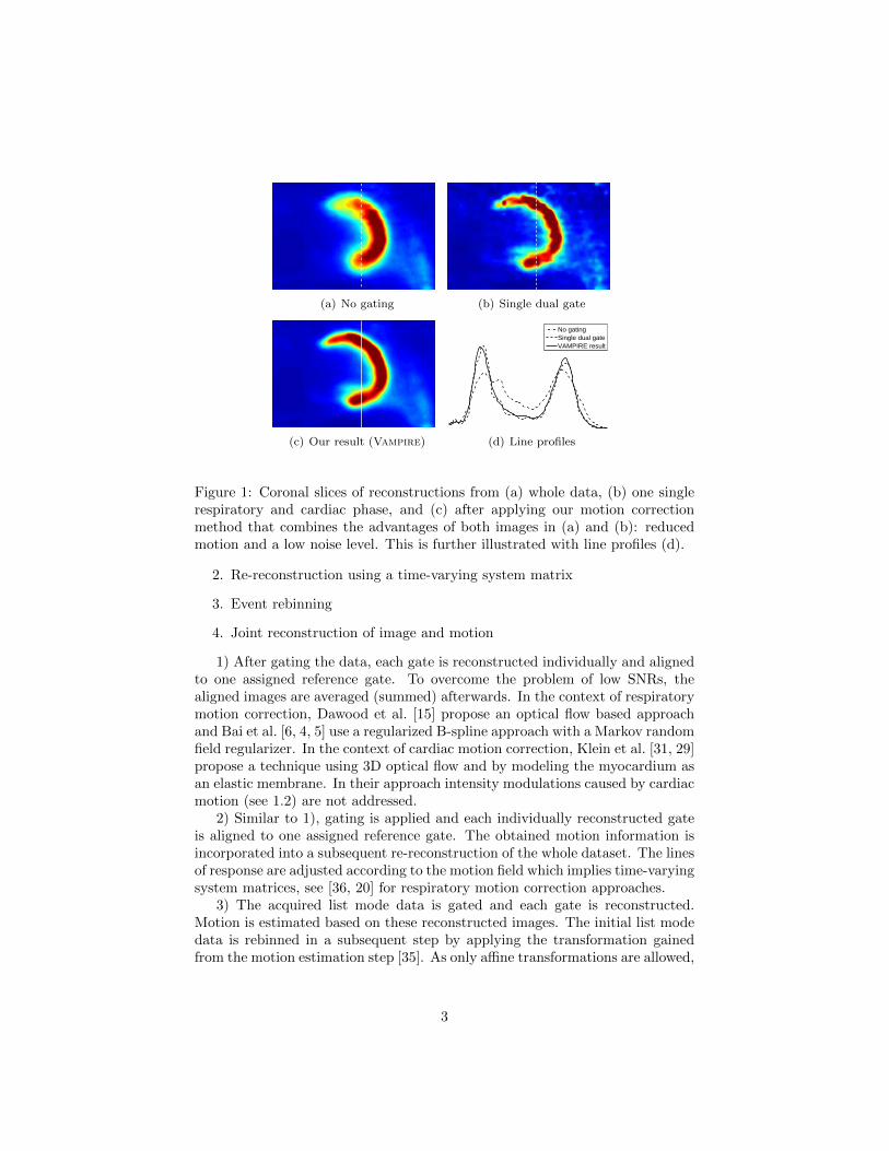
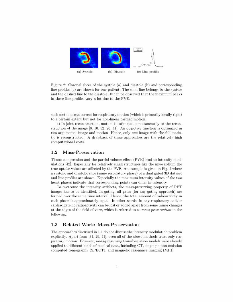


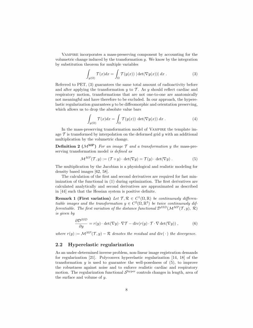
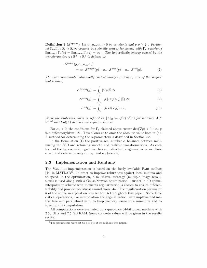

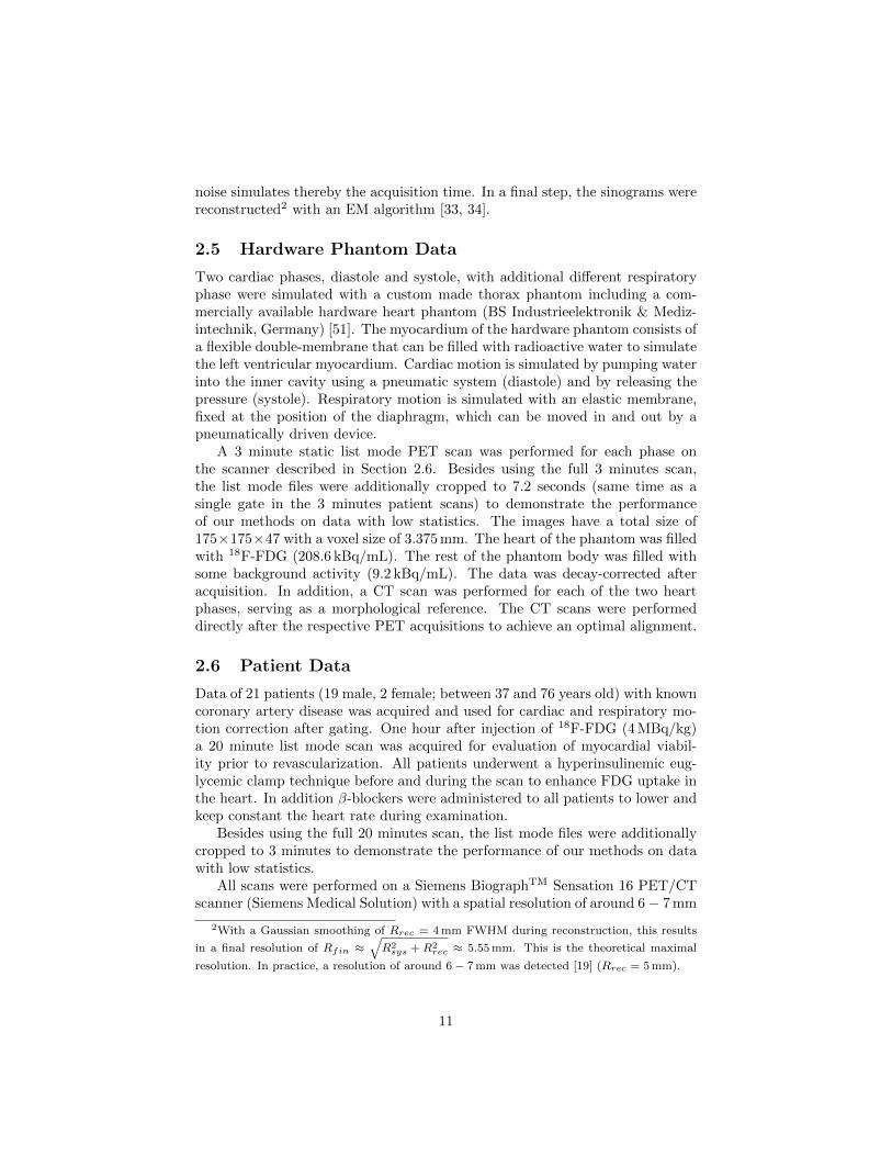
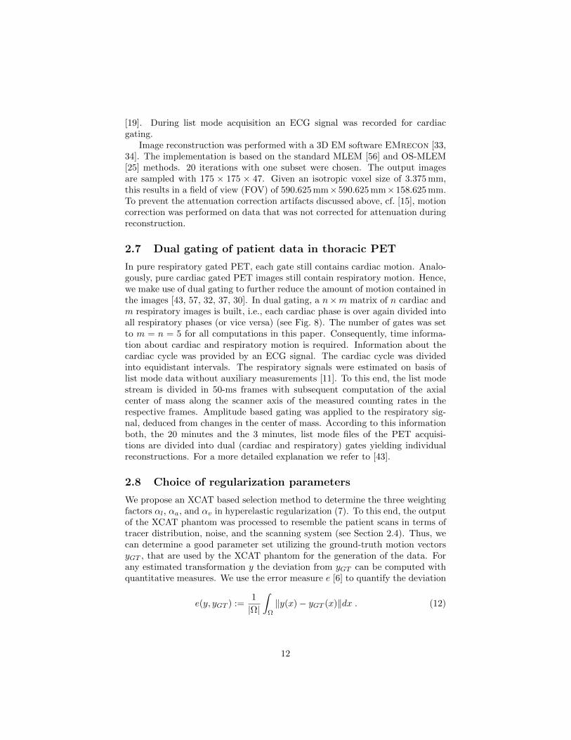

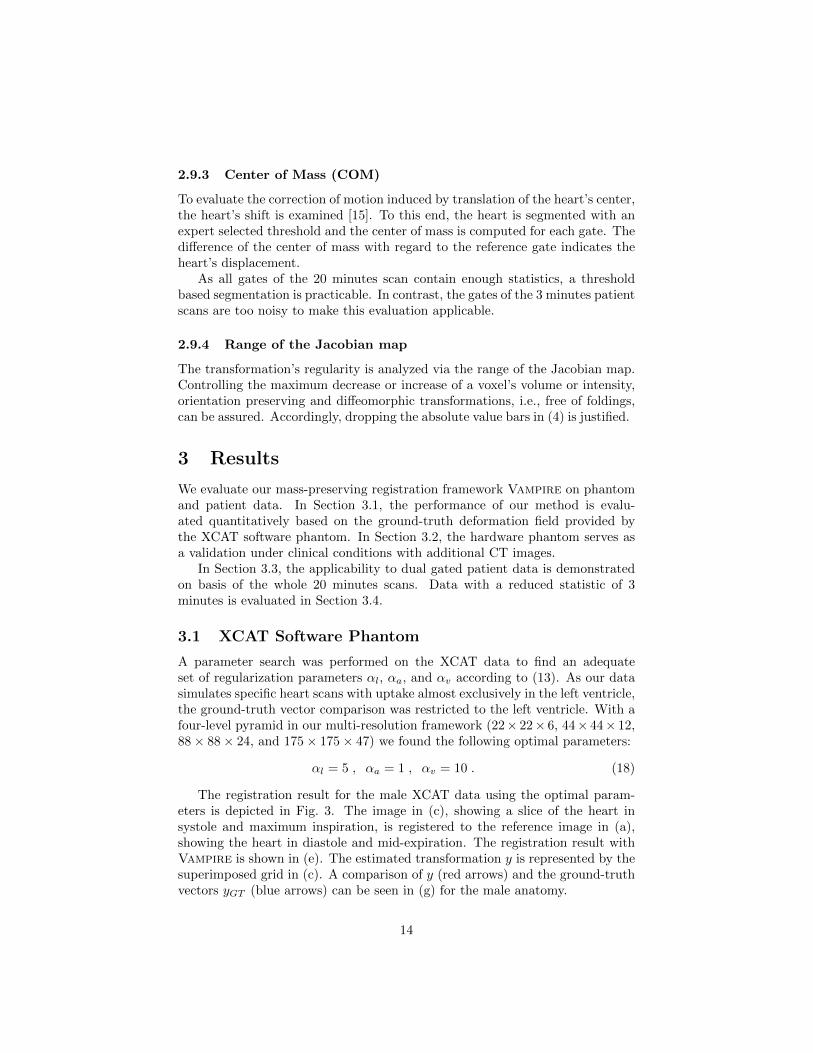


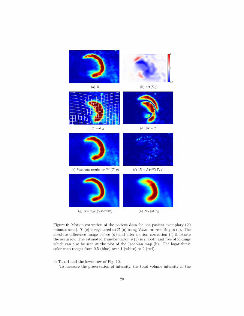


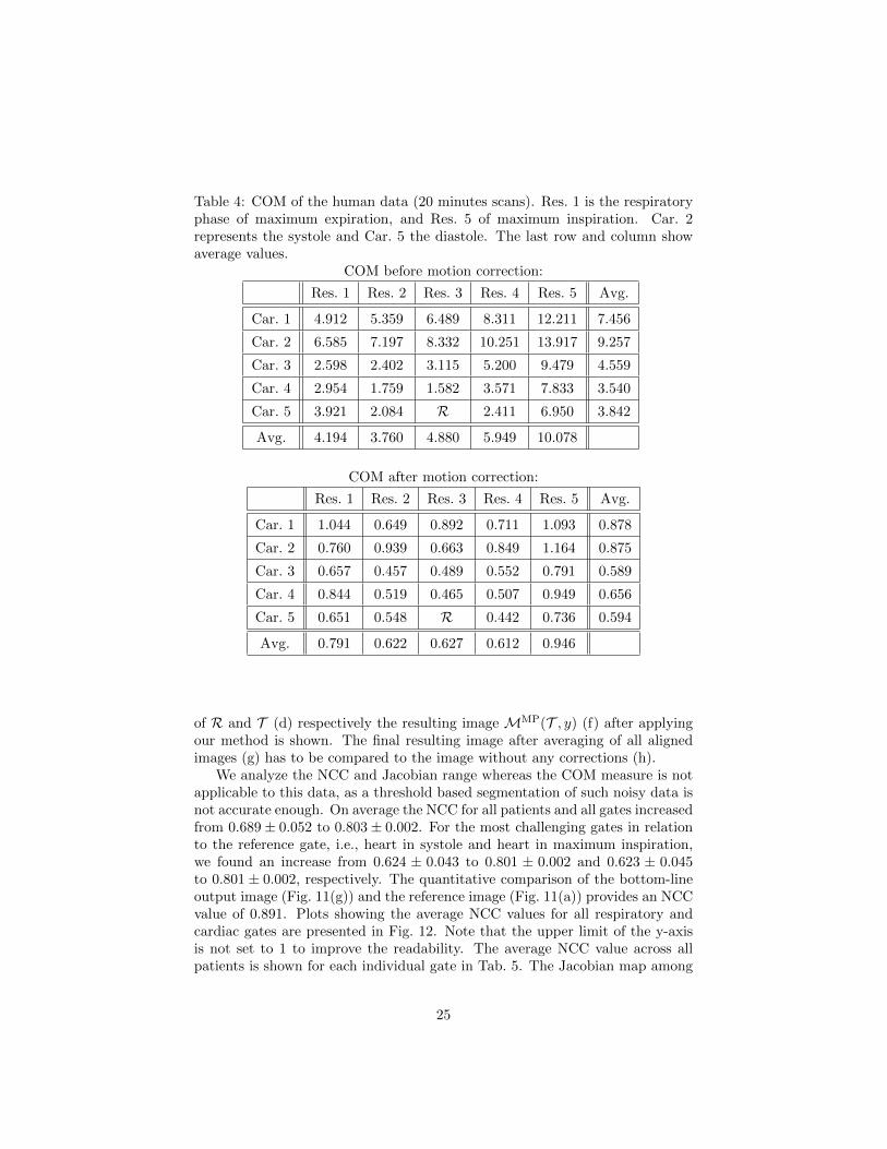


![NIH Public Access estimation in a four-compartment finite element head model …wolters/PaperWolters/2009/LewEtAl... · 2010. 9. 24. · 1992] using the isolated skull approach [Hämäläinen](https://static.documents.pub/doc/80x56/6128e457053ae86df3128c23/nih-public-access-estimation-in-a-four-compartment-finite-element-head-model-wolterspaperwolters2009lewetal.jpg)


![J org Pretz arXiv:2005.12203v1 [physics.class-ph] 25 May 2020 › pdf › 2005.12203.pdf · (t)/ m mass 1 mass 2 mass 3 mass 4 mass 5 mass 6 mass 7 mass 8 mass 9 mass 10 Figure 2.](https://static.documents.pub/doc/80x56/5f1ec529f26656179f60ee75/j-org-pretz-arxiv200512203v1-25-may-2020-a-pdf-a-200512203pdf-t.jpg)
