WestminsterResearchhttp://www.westminster.ac.uk/westminsterresearch
State-dependent TMS reveals representation of affective body
movements in the anterior intraparietal cortex
Mazzoni, N., Jacobs, C., Venuti, P., Silvanto, J. and Cattaneo, L.
Copyright © 2017 the authors.
The article was originally published by the Society for Neuroscience, in the Journal of
Neuroscience, 37 (30), pp. 7231-7239, 2017 and is available at:
https://dx.doi.org/10.1523/JNEUROSCI.0913-17.2017
The WestminsterResearch online digital archive at the University of Westminster aims to make the
research output of the University available to a wider audience. Copyright and Moral Rights remain
with the authors and/or copyright owners.
Whilst further distribution of specific materials from within this archive is forbidden, you may freely
distribute the URL of WestminsterResearch: ((http://westminsterresearch.wmin.ac.uk/).
In case of abuse or copyright appearing without permission e-mail [email protected]
TITLE: State-dependent TMS reveals representation of affective body movements in the anterior
intraparietal cortex
ABBREVIATED TITLE: TMS over aIPS reversed adaptation effect to fearful PLDs
AUTHORS: Noemi Mazzoni [1] [2], Christianne Jacobs [1] [3], Paola Venuti [2], Juha Silvanto [1], Luigi
Cattaneo [4].
AFFILIATIONS:
[1] Department of Psychology, Faculty of Science and Technology, University of Westminster, W1W 6UW
London, United Kingdom
[2] Department of Psychology and Cognitive Science, University of Trento, 38068 Rovereto (TN), Italy
[3] Faculty of Psychology and Educational Sciences, Université Catholique de Louvain, Louvain-la-Neuve,
1348 Belgium
[4] Department of Neuroscience, Biomedicine and Movement, Section of Physiology and Psychology,
University of Verona, 37134 Verona, Italy.
Corresponding author:
Noemi Mazzoni,
ODFLab – Department of Psychology and Cognitive Science, University of Trento,
Via Matteo del Ben 5b, 38068, Rovereto (TN) Italy
Tel: +39 0464 808103
E-mail: [email protected]
Number of pages: 28
Number of figures: 4
Number of tables: 2
Number of multimedia and 3D models: 0
Number of words for Abstract: 249
Number of word for Introduction: 634
Number of words for Discussion: 1476
Conflict of Interest: The authors declare no competing financial interests
Acknowledgements: JS is supported by the ERC (336152). CJ is supported by F.R.S.-F.N.R.S. (“Charge de
recherches”). We are obliged to Antony P. Atkinson (Durham University), Paola Ricciardelli and Rossana
Actis-Grosso (University of Milano-Bicocca) for sharing with us the stimuli. We are grateful to Birkbeck –
University College of London Centre for NeuroImaging (BUCNI) and in particular to Christina Moutsiana,
Benjamin de Haas and Iroise Dumontheil for technical assistance during MRI scans acquisition.
1
ABSTRACT 1
In humans, recognition of others’ actions involves a cortical network that comprises, among other 2
cortical regions, the posterior superior temporal sulcus (pSTS), where biological motion is coded and the 3
anterior intraparietal suclus (aIPS), where movement information is elaborated in terms of meaningful goal-4
directed actions. This action observation system (AOS) is thought to encode neutral voluntary actions, and 5
possibly some aspects of affective motor repertoire, but the role of the AOS’ areas in processing affective 6
kinematic information has never been examined. Here we investigated whether the action observation system 7
plays a role in representing dynamic emotional bodily expressions. In the first experiment, we assessed 8
behavioural adaptation effects of observed affective movements. Participants watched series of happy or 9
fearful whole-body point-light displays (PLDs) as adapters and were then asked to perform an explicit 10
categorization of the emotion expressed in test PLDs. Participants were slower when categorizing any of the 11
two emotions as long as it was congruent with the emotion in the adapter sequence. We interpreted this effect 12
as adaptation to the emotional content of PLDs. In the second experiment, we combined this paradigm with 13
TMS applied over either the right aIPS, pSTS and the right half of the occipital pole (corresponding to 14
Brodmann’s area 17 and serving as control) to examine the neural locus of the adaptation effect. TMS over 15
the aIPS (but not over the other sites) reversed the behavioural cost of adaptation, specifically for fearful 16
contents. This demonstrates that aIPS contains an explicit representation of affective body movements. 17
SIGNIFICANCE STATEMENT: In humans, a network of areas – the action observation system (AOS) - 18
encodes voluntary actions. However, the role of these brain regions in processing affective kinematic 19
information has not been investigated. Here we demonstrate that the aIPS contains a representation of 20
affective body movements. Firstly, in a behavioural experiment, we found an adaptation after-effect for 21
emotional PLDs, indicating the existence of a neural representation selective for affective information in 22
biological motion. To examine the neural locus of this effect, we then combined the adaptation paradigm 23
with TMS. Stimulation of the aIPS (but not over pSTS and control site) reversed the behavioural cost of 24
adaptation, specifically for fearful contents, demonstrating that aIPS contains a representation of affective 25
body movements. 26
27
2
Introduction 28
Perception of movements of other living beings is crucial for survival in most species, to the extent 29
that many vertebrate species have specialized neural systems for action observation. In humans, a 30
widespread network of interconnected brain areas (known as the action observation system - AOS) underlies 31
the comprehension of conspecifics’ body movements and actions. This network includes the posterior 32
superior temporal sulcus (pSTS) (Puce and Perrett, 2003), and two mirror system areas, the putative human 33
anterior intraparietal area (aIPS) and the ventral premotor/caudal inferior frontal gyrus complex (PMv/cIFG) 34
(Cattaneo and Rizzolatti, 2009). Several TMS studies have demonstrated that stimulating the pSTS, the aIPS 35
and the PMv/cIFG regions produces selective impairment in visual recognition of neutral actions (Candidi et 36
al., 2008; Cattaneo et al., 2010; Grossman et al., 2005; van Kemenade et al., 2012; Pobric and Hamilton, 37
2006). But is the AOS also important for the encoding of the emotional aspects of biological motion? 38
The perception of affective stimuli, irrespective of stimulus type, generally enhances the neural 39
response of core affective systems, situated within the limbic system (Adolphs, 2002; Phillips et al., 2003) 40
but emotional body movements are complex and their perception activates also a more widespread network 41
of subcortical and cortical regions, related to analysis of visual body features and more generally to action 42
observation and preparation (de Gelder et al., 2006, 2010, 2015; Tamietto and de Gelder, 2011). It is thus 43
crucial to understand whether the activation within the AOS is a mere side-effect of the type of stimuli (body 44
actions), independent from their content or whether AOS activity is causally linked to emotional recognition. 45
This issue has been explored in the literature in only two TMS studies; these found that perturbation of pSTS 46
(Candidi et al., 2011) and IPL (Engelen et al., 2015) selectively improved the recognition of fearful body 47
images. However, a limitation of both of these studies was that participants observed static images; human 48
bodies are dynamic in nature and the brain substrates used in processing static postures are likely to differ 49
from those engaged in perception of body movements. Furthermore, while conventional TMS paradigms can 50
reveal the causal role of cortical regions in cognitive functions, they do not inform us about the neural 51
representations in those regions. 52
Here we examined whether specific regions of the action observation network contain 53
representations of affective body movements. This was accomplished by the use of state-dependent TMS 54
which enables the selectivity of neural representations in a cortical region to be assessed (Romei et al., 2016; 55
3
Silvanto et al., 2008). This approach has been previously used to examine the selectivity of neural 56
representations in various cognitive functions such as colour and motion perception (Silvanto et al., 2007; 57
Cattaneo and Silvanto, 2008), numerical cognition (Kadosh et al., 2010) and action observation (Cattaneo et 58
al., 2011, 2010; Jacquet and Avenanti 2015; Sato et al. 2011). In order to examine the role of AOS in 59
encoding the emotional aspects of dynamic biological motion, we used point-light displays (PLDs), also 60
referred to as biological motion (BM) stimuli (Johansson, 1973), which allow isolation of motion signals 61
from others visual cues. Kinematic information contained in PLDs is sufficient for detection of emotional 62
content of human movements (Alaerts et al. 2011; Atkinson et al. 2004, 2007, 2012; Chouchourelou et al., 63
2006; Clarke et al., 2005; Dittrich et al. 1996). In Experiment 1, we examined behavioural adaptation effects 64
of observed affective PLDs. We found an adaptation-like bias with incongruent stimuli recognized faster 65
than congruent ones. In Experiment 2, we used the TMS-adaptation paradigm to examine the cortical locus 66
of adaptation effects observed in Experiment 1. TMS over the aIPS – but not over pSTS nor over a visual 67
control area - reversed the behavioural adaptation for fearful stimuli, indicating that this region contains 68
neural representations selective for the fearful characteristics of human movements. 69
70
Material and methods 71
Visual stimuli and validation of emotional valence. A total of 20 PLDs were presented, depicting 10 72
different expressions of happiness and fear, respectively. These stimuli are part of a wider dataset created by 73
Atkinson and collaborators (Atkinson et al., 2004, 2012). The PLDs consisted of 2 second-long digitalized 74
video clips (see Atkinson et al. 2012 for details), displaying a single actor represented as 13 white dot-lights 75
moving on a black background. The dots were positioned over the head and the main joints (one dot over 76
each ankle, knee, hip, elbow, shoulder, and hand) of the actor. Examples of the stimuli can be viewed at 77
http://community.dur.ac.uk/a.p.atkinson/Stimuli.html. We selected happy and fearful stimuli because they 78
are roughly equally arousing emotions, with opposite emotional valence (positive or negative). Prior to the 79
main experiments, we ran a pilot study to validate the PLDs in terms of quantity of movement contained in 80
the PLDs and of type and intensity of portrayed emotion. Sixteen healthy adults took part in this pilot 81
experiment (13 females, mean age = 29.63 (SD = 7.65)). All the participants provided informed consent 82
before taking part in the experiment. They were seated in front of a 24-inch monitor at a distance of about 60 83
4
centimetres. The stimuli were presented foveally. Each PLD was presented once, and for each video 84
participants were asked to recognize the conveyed emotion among 4 options (Fear, Happiness, Neutral and 85
Other) by pressing the corresponding button on the keyboard. The response options (appearing on the screen 86
after each stimulus) were indicated with a label placed over the keys “F, G, H, J” and were randomized 87
across participants. After the emotion recognition task, participants were asked to rate the “Intensity of the 88
emotion” and the “Quantity of movement” on a scale from 1 to 5, using the numeric keys on the top of the 89
keyboard. Stimuli were presented and responses recorded with E-Prime 2.0® (Psychology Software Tools, 90
Inc.). For each individual PLD we calculated the accuracy of emotion categorization, the rated intensity of 91
the emotion and the rated quantity of movement. Data distribution was tested for normality with Shapiro-92
Wilk test. Accuracy, Intensity and Movement were not normally distributed, so they were analysed using a 93
non-parametric test for paired data, the Wilcoxon signed rank test with continuity correction. Significance 94
thresholds were Bonferroni-corrected for 3 multiple comparisons (for each variable, we compared results 95
between the three emotional valences, hence the critical alpha was set as p < .017). There were no significant 96
differences between the happy and fearful movements for Accuracy, Movement, and Intensity, while – 97
predictably - the neutral movements were rated as less intense compared with the two emotions (Table 1). 98
This implies that the stimuli used in Experiment 1 and 2 (i.e. fearful and happy PLDs) do not differ in terms 99
of: i) recognizability between the emotional categories, ii) intensity of the expressed emotion or iii) quantity 100
of movement contained in the stimuli. 101
Insert Table 1 102
103
Experiment 1: behavioural assessment of adaptation to observed emotional body movements. 104
Participants. Twenty-six healthy adults (14 females and 12 males, age mean = 23.58 years (SD = 105
2.95 years)) took part in the behavioural study (Experiment 1). All participants had normal or corrected-to-106
normal vision. Prior to the experiment, all participants provided written informed consent, in accordance 107
with the Declaration of Helsinki. 108
5
Design and procedure. Participants were seated in a comfortable chair in front of a 24-inch computer 109
screen at a distance of around 60 cm. E-Prime version 2.0 (Psychology Software Tools, Inc.) software was 110
used for stimulus presentation and response recordings. The study consisted of 12 adaptation blocks (6 with 111
happy and 6 with fearful adapters), consisting of a 1 minute adapting period followed by 8 test trials. Each 112
trial began with a white central fixation cross over a black background, lasting for 10 seconds. This was 113
followed by an adaptation period in which the same PLD was repeated 30 times (for a total duration of 60 114
sec). Participants were asked to simply watch the stimuli and focus on the emotion expressed by the actor. 115
The order of adaptation blocks was randomized. At the end of adaptation, a screen appeared asking 116
participant to “Get ready for the task”, after which 8 test stimuli (4 fearful and 4 happy PLDs) were 117
presented. Half of the test stimuli were emotionally congruent and half were emotionally incongruent with 118
the adapter, and their order was randomized. The test stimuli and the adapter stimuli belong to the same 119
dataset, i.e. the same stimulus could be used as an adapter in one block or as a test stimulus in another block. 120
However, in single blocks, the adapter stimulus was always different from the test stimuli presented 121
thereafter. In other words, every stimulus could appear randomly as adapter or as a test in different blocks, 122
but not in the same block. The movie clip was presented centrally. Simultaneously with the stimulus 123
presentation, the question “Which emotion?” appeared on the upper part of the screen, and the two response 124
options (“Fear” and “Happiness”) were presented on the lower part of the monitor. For each test stimulus, 125
participants were asked to categorize the expressed emotion as fast as possible by key-press. The response 126
options were indicated with a label placed over the keys “G” and “H”, and the key-emotion correspondence 127
was randomized across participants. Participants were asked to respond using the index and the middle finger 128
of their right hand. The PLD was presented for a maximum of 2 seconds, while the question and the response 129
period lasted until participants responded. Accuracy and response time (RTs) were recorded. 130
Data analyses. The dependent variable was mean response times (RTs). Only correct responses were 131
included in the analyses (the overall error rate was 4.43%). Data distributions failed the normality (Shapiro-132
Wilk’s test) and homoscedasticity of variance (Bartlett’s test) tests. To normalize the distribution, the 133
averaged RTs were log-transformed prior to analyses (logRT). A two-way repeated-measures ANOVA was 134
conducted with emotional content of the test stimuli (“emoTest”: Fear or Happiness) and emotional 135
congruence between test and adapter stimuli (congruent or incongruent) as within-subject factors. Post hoc 136
6
comparisons were performed with two-tailed paired-samples t-tests with correction of the significance 137
threshold for multiple comparisons whenever appropriate. All analyses were performed using R, version 138
3.3.1 (R Development Core Team, 2016). 139
140
Experiment 2: Effects of TMS on perceptual adaptation 141
Participants. Seventeen healthy adults (11 females and 6 males, mean age = 25.63 (SD = 5.17)) 142
participated in the TMS experiment (Experiment 2). Three participants were excluded from the analysis 143
because of difficulties in determining their resting motor threshold. In these participants, the TMS 144
stimulation over M1 did not produce any visible hand twitch, and no motor sensation was perceived. Hence, 145
the final analyses were performed on a total of 14 participants. Participants in the TMS experiment were 146
screened for MRI and TMS contraindication prior to the experiment and received a £ 15 voucher refund for 147
their participation. All participants had normal or corrected-to-normal vision. Prior to the experiment, all 148
participants provided written informed consent. The protocol was approved by the University of 149
Westminster’s ethical committee, in accordance with the Declaration of Helsinki. 150
Neuronavigation and identification of stimulation sites on individual anatomy. We used MRI-guided 151
neuronavigation (BrainInnovation BV, the Netherlands) for accurate positioning of the TMS coil. For each 152
participant, a high resolution T1-weighted MPRAGE scan (176 partitions, 1 x 1 x 1 mm, flip angle = 7°, TI = 153
1,000 ms, TE = 3.57 ms, TR = 8.4 ms) was acquired before the TMS experiment. Structural MRI images 154
were obtained with a 1.5 T whole-body TIM Avanto System (Siemens Healthcare), at the 155
Birkbeck/University College London Centre for NeuroImaging (BUCNI), with a 32-channel head coil. A 3D 156
reconstruction of the gray matter surfaces and the scalp was created for each participant, which were co-157
registered to the participant’s head in order to position the coil over the site of stimulation and to control coil 158
position throughout the experiment. In each participant, three different sites in the right hemisphere were 159
stimulated: the posterior part of the right superior temporal sulcus (pSTS), the anterior part of the right 160
intraparietal sulcus (aIPS) and a posterior occipital control area located next to the midline. The three loci 161
were identified on the basis of macro-anatomical landmarks. Specifically, pSTS was targeted over the 162
transition between its posterior segment and its horizontal segment (see Ochiai et al. 2004 for an overview of 163
7
STS anatomy). We defined the aIPS as the most rostral part of the IPS at the intersection between the 164
postcentral gyrus and the IPS (Caspers et al., 2006). Control TMS was applied to a site corresponding to a 165
secondary visual area not primarily implied in coding for emotional aspect of visual stimuli, located between 166
BA 17 and BA 18 (see Figure 1) 167
TMS. Biphasic TMS pulses were applied with a figure-of-eight coil (D70mm coil) connected to a 168
Magstim Rapid2 stimulator (Magstim Co Ltd, Whitland, UK). At first we searched in each participant the 169
visually assessed resting motor threshold (rMT), defined as the stimulator’s output intensity necessary to 170
obtain a visible twitch in the contralateral intrinsic hand muscles in exactly 50% of trials in a series of at least 171
eight consecutive pulses (Rossini et al, 1994). The intensity of stimulation in the actual experiment was set to 172
120% of the individual’s rMT with a maximum of 65% maximal stimulator output due to coil overheating 173
and limiting discomfort to participants. The coil was attached to a Magstim coil stand and placed tangentially 174
to the scalp. Coil orientation was medial-lateral with the handle pointing laterally and slightly posteriorly 175
(70° from the midline) for the aIPS position, in order to induce a current in the underlying cortical tissue 176
roughly perpendicular to the IPS. A similar orientation was used for the stimulation of pSTS, but with the 177
coil handles pointing upwards. Due to pSTS proximity to the ears, in some participants the coil orientation 178
was changed to minimize discomfort. For the occipital (control) stimulation, the coil was positioned 179
perpendicular to the midline with the handle pointing outward. TMS was delivered in triplets. In every trial 180
participants received three 10 Hz pulses time-locked to the onset of the PLD, starting synchronously with the 181
visual stimulus. 182
183
Insert Figure 1 184
185
Procedure. The TMS paradigm was identical to that used in Experiment 1 described above. Every 186
block consisted of 1 minute of adapting period followed by 8 test trials. A total of 12 adapter stimuli (6 187
happy and 6 fearful PLDs) and 96 test stimuli were presented for each of the three sites of stimulation. The 188
order of adaptation blocks was randomized. During the adaptation period the same PLD was repeated 30 189
times (for 60 sec). Participants were asked to simply watch the adapter stimuli and focus on the emotion 190
expressed by the actor. At the end of adaptation, 8 test stimuli (4 fearful and 4 happy PLDs) were presented. 191
8
Half of the test stimuli were emotionally congruent (i.e. same emotion) and half were emotionally 192
incongruent (i.e. different emotion) with the adapter, and their order was randomized. Participants were 193
asked to categorize the expressed emotion (Fear or Happiness) as fast as possible by key-press, using the 194
index and the middle finger of their right hand. Accuracy and response time (RTs) were recorded. The three 195
stimulation sites (right pSTS, right aIPS and the control site) were stimulated on the same day, with 30 196
minutes of delay between sessions. The order of stimulation sites was counterbalanced between participants. 197
Participants wore earplugs and were seated in a comfortable chair in a quiet room, in front of a 24-inch 198
computer screen at a distance of 60 cm, with their head on a chin-rest. 199
200
Insert Figure 2 201
202
Data analyses: All analyses were performed using R, version 3.3.1 (R Development Core Team, 203
2016). The dependent variable was the mean of response time (RTs). Only correct responses were included 204
in the analyses. Data were tested for normality (Shapiro test) and homoscedasticity of variance (Bartlett test). 205
To normalize the distribution, the averaged RTs were log-transformed prior to analyses (logRT). A three-206
way repeated-measures ANOVA (3x2x2) was performed. The site of TMS stimulation (“stimSite”), the 207
emotional valence of the test stimuli (“emoTest”) and the emotional congruence between test and adapter 208
stimuli (“congruence”) were entered as within-subject factors. Post hoc comparisons were performed with 209
two-tailed paired-samples t-tests. The significance threshold for the p-values was corrected for multiple 210
comparisons when appropriate. As a measure of the effects size, the Generalized Eta squared (η2) is reported 211
when appropriate. In addition, we calculated the Cohen’s d for the significant comparisons using bootstrap 212
resamples method (Gerlanc and Kirby, 2015). The number of bootstrap resamples (R) was set at 2000. 213
Bootstrap Cohen’s d effect size measures and their corresponding 95% confidence intervals (CIs) are also 214
reported when appropriate. 215
216
Results 217
9
Experiment 1: behavioural evidence of perceptual adaptation to the emotional content of PLDs. 218
In Experiment 1, the overall error rate was 4.43%. A summary of the results of Experiment 1 is 219
presented in Table 2 and in Figure 3. The two-way ANOVA showed a significant main effect of 220
“Congruence” (F(1,25) = 7.31, p-value = .012) with incongruent stimuli being recognized faster than congruent 221
ones, while the interaction between “emoTest” and “Congruence” was not significant (F(1, 25) = 0.856, p-value 222
= .364; η2 = 0.014; Cohen’s d = -0.236, C.I. = -0.660; 0.166). 223
224
Insert Table 2. 225
226
Insert Figure 3. 227
228
Experiment 2: state-dependent effects of TMS over aIPS on explicit categorization of fearful PLDs. 229
In Experiment 2 the overall error rate was 3.87%. The three-way ANOVA showed a significant main 230
effect of “Congruence” (F(1,13) = 14.994, p-value = .002), with congruent stimuli being recognized slower than 231
incongruent ones (mean RTs: congruent = 1194 ms; incongruent = 1148 ms), suggesting the presence of an 232
adaptation after-effect for affective PLDs and confirming the results of the behavioural experiment 233
(Experiment 1). More importantly, we found a significant three-way interaction between “stimSite, emoTest 234
and Congruence” (F(2,26) = 3.546, p-value = .043). To better understand this interaction, we performed three 235
2x2 repeated measures ANOVAs in the three stimulation sites separately, with “emoTest” and “Congruence” 236
as within factors. We found a significant main effect of “Congruence” in the control site (F(1,13) = 9.329; p-237
value = .009; η2 = .017) and in pSTS (F(1,13) = 9.393; p-value = .009; η2 = .029), showing that the adaptation 238
after-effect persisted and hence suggesting that TMS stimulation did not have any effect on those two brain 239
areas. On the contrary, ANOVA in aIPS showed a significant interaction between “emoTest and 240
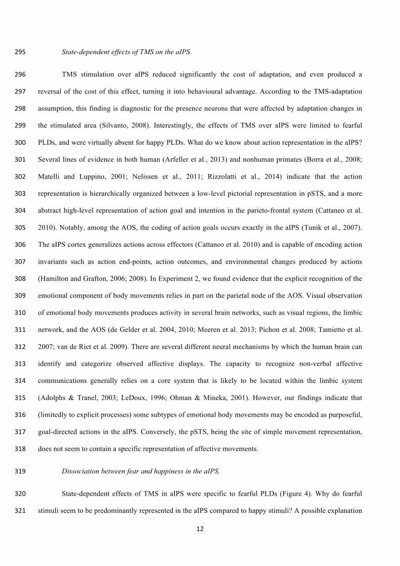
Congruence” (F(1,13) = 8.474; p-value = .012; η2 = .022), but no significant main effects. In particular, the 241
adaptation after-effect was still present for happy test stimuli (p-value = .009; Cohen’s d = -0.311, C.I. = -242
10
1.114, 0.459) with incongruent stimuli recognized faster than congruent ones. Conversely, the adaptation 243
after-effect was completely abolished for fearful test stimuli, to the point that we observed a trend towards an 244
inversion of the adaptation effects, i.e. congruent test stimuli were recognized faster than congruent ones (p-245
value = 0.066; Cohen’s d = 0.267, C.I. = -0.459, 1.075). 246
Insert Figure 4 247
248
DISCUSSION 249
Perceptual adaptation to emotional content of PLDs. In the first experiment, we examined 250
behaviourally whether adaptation to the emotional content of PLD produces perceptual aftereffects. When 251
categorizing an affective PLD, participants’ performance was markedly biased (slower RTs) by their 252
previous exposure to congruent emotions. Adaptation aftereffects for features contained in PLDs have been 253
reported previously. For example, prolonged exposure to human actions conveying gender characteristics 254
generates an aftereffect that biases the perception of gender in subsequently observed actions (Troje et al., 255
2006). Similar adaptation-like aftereffects have been observed for action category (de la Rosa et al., 2014; 256
van Boxtel & Lu, 2013) and for spatial components of the observed bodily trajectories (Jackson and Blake 257
2010; Theusner et al., 2011). Also judgments about the interaction between a human hand and an object have 258
been shown to be susceptible to visual adaptation, with viewing the grasping of a light object biasing 259
subsequent grasped objects to appear heavier (Barraclough et al., 2009). Besides, a number of studies 260
reported adaptation aftereffect to affective facial (Fox and Barton, 2007; Russell and Fehr, 1987; Webster 261
and MacLeod, 2011; Webster et al., 2004) and vocal expressions (Skuk and Schweinberger, 2013; 262
Bestelmeyer et al., 2014). However, the extent to which emotional bodily expression can produce adaptation 263
aftereffects has remained unexplored so far. Our study fills this gap, providing the first evidence that 264
perception of emotional whole body movements can undergo selective perceptual adaptation. The finding is 265
indicative of the existence of a neural representation selective for affective information in biological motion. 266
Absence of state-dependent effects of TMS on the early visual cortex (control condition). 267
11
The aim of Experiment 2 was to examine the neural locus of this adaptation effect for affective dynamic 268
bodily expressions. We found adaptation after-effects similar to those observed in Experiment 1 following 269
control stimulation, consisting in a behavioural disadvantage in recognizing PLDs emotionally congruent 270
with the adapter sequences (Figure 4). Given the assumptions of TMS-adaptation paradigms, we did not 271
expect any effect of TMS on this region, because the adapted features (bodily movements) are not supposed 272
to be coded in the early visual cortex. The earliest visual body representation to be found along the visual 273
pathways is in the lateral occipital complex, way more rostral than the area that we chose as control 274
(Downing et al., 2001). Studies in blindsight patients suggest that the processing of emotional information 275
can efficaciously occur in spite of lesions of the early visual areas, either when conveyed by faces (de Gelder 276
et al., 1999; Morris et al., 2001) or by body postures (De Gelder and Hadjikhani, 2006). Accordingly, in 277
another study, TMS perturbation of V1 impaired the discrimination of neutral – but not emotional - body 278
postures, supporting the hypothesis that the encoding of the emotional content does not depend on V1 279
(Filmer and Monsell, 2013). 280
Absence of state-dependent effects of TMS on the pSTS. 281
In contrast to the early visual cortex, the pSTS is tuned to biological motion. However, to our 282
surprise, no state-dependent effects of TMS were found. We interpreted this finding in the light of the 283
functional specialization of the pSTS. The integrity of STS is fundamental to biological motion identification 284
(Vaina et al., 1990; Grossman et al., 2005; Saygin, 2007), it encodes low-level pictorial aspects of BM 285
(Cattaneo et al., 2010), and it represents bodily movements separately for different body parts (upper limb, 286
face, whole body, gaze) (Hein and Knight, 2008), probably in a viewpoint-invariant manner (Grossman et 287
al., 2010). In one TMS study, stimulation of pSTS improved the visual match of body forms specifically for 288
fearful body postures (Candidi et al, 2011). However this type of task relies on pictorial analysis likely 289
encoded in pSTS, while we asked to recognize the emotional meaning of dynamic PLDs, potentially related 290
to higher level of action representation implemented in aIPS (Cattaneo et al., 2010; Fogassi et al., 2005; 291
Shmuelof and Zohary, 2005; Hamilton and Grafton, 2006). Similarly, another study (Tseng et al., 2010) 292
showed that the specific effects of static fearful facial displays as distracters in a visual search task could be 293
disrupted by anodal transcranial direct current stimulation (tDCS) over the right pSTS. 294
12
State-dependent effects of TMS on the aIPS. 295
TMS stimulation over aIPS reduced significantly the cost of adaptation, and even produced a 296
reversal of the cost of this effect, turning it into behavioural advantage. According to the TMS-adaptation 297
assumption, this finding is diagnostic for the presence neurons that were affected by adaptation changes in 298
the stimulated area (Silvanto, 2008). Interestingly, the effects of TMS over aIPS were limited to fearful 299
PLDs, and were virtually absent for happy PLDs. What do we know about action representation in the aIPS? 300
Several lines of evidence in both human (Arfeller et al., 2013) and nonhuman primates (Borra et al., 2008; 301
Matelli and Luppino, 2001; Nelissen et al., 2011; Rizzolatti et al., 2014) indicate that the action 302
representation is hierarchically organized between a low-level pictorial representation in pSTS, and a more 303
abstract high-level representation of action goal and intention in the parieto-frontal system (Cattaneo et al. 304
2010). Notably, among the AOS, the coding of action goals occurs exactly in the aIPS (Tunik et al., 2007). 305
The aIPS cortex generalizes actions across effectors (Cattaneo et al. 2010) and is capable of encoding action 306
invariants such as action end-points, action outcomes, and environmental changes produced by actions 307
(Hamilton and Grafton, 2006; 2008). In Experiment 2, we found evidence that the explicit recognition of the 308
emotional component of body movements relies in part on the parietal node of the AOS. Visual observation 309
of emotional body movements produces activity in several brain networks, such as visual regions, the limbic 310
network, and the AOS (de Gelder et al. 2004, 2010; Meeren et al. 2013; Pichon et al. 2008; Tamietto et al. 311
2007; van de Riet et al. 2009). There are several different neural mechanisms by which the human brain can 312
identify and categorize observed affective displays. The capacity to recognize non-verbal affective 313
communications generally relies on a core system that is likely to be located within the limbic system 314
(Adolphs & Tranel, 2003; LeDoux, 1996; Ohman & Mineka, 2001). However, our findings indicate that 315
(limitedly to explicit processes) some subtypes of emotional body movements may be encoded as purposeful, 316
goal-directed actions in the aIPS. Conversely, the pSTS, being the site of simple movement representation, 317
does not seem to contain a specific representation of affective movements. 318
Dissociation between fear and happiness in the aIPS. 319
State-dependent effects of TMS in aIPS were specific to fearful PLDs (Figure 4). Why do fearful 320
stimuli seem to be predominantly represented in the aIPS compared to happy stimuli? A possible explanation 321
13
is that the affective state of fear itself is represented in the aIPS. Alternatively, it is possible the motor pattern 322
expressing fear has characteristics that are best encoded by the aIPS, which preferentially processes goal-323
directed, purposeful movements (Cattaneo et al., 2010). The fearful bodily movements represented in our 324
stimuli were in most cases directed towards a position in space as they depicted self-protective or avoidance 325
body movements directed away from specific threatening agents (See example videos at 326
http://community.dur.ac.uk/a.p.atkinson/Stimuli.html). On the contrary, happy stimuli (e.g. exulting, 327
clapping hands, joyful hopping) were not directed towards or away from specific sectors in space. Therefore, 328
the fear-happiness dissociation could be explained by a higher goal-directed or space oriented in fearful 329
movements, compared to happy ones. From an evolutionary point of view, the emotional movements are 330
communicative in nature, and our brain’s prompt reactions to them is essential for the survival ( Darwin, 331
1872; Ekman, 1957; Grèzes et al., 2007). In this sense, each emotional subtype has an own identity, and its 332
affective state is not dissociable from its stereotyped communicative motor behaviour. The effective 333
communication of fearful content is more likely relied on goal-directed and spatially-oriented actions than 334
happiness. We therefore favour the hypothesis that fearful movements have a more “praxic” and “goal-335
directed” quality compared to happiness. In line with that, several studies has reported that the motor system 336
is specifically tuned to fearful body movements as shown by changes in corticospinal excitability in response 337
to fearful body postures (Borgomaneri et al., 2012; Borgomaneri et al., 2015), fearful facial expressions 338
(Borgomaneri et al., 2014), and negative natural complex scenes (Schutter et al., 2008). However, the role of 339
activity in the corticospinal system in action comprehension remains unclear. 340
341
Conclusions 342
We conclude that, while performing explicit categorizations (i.e. high-level cognitive task), the 343
human brain considers fearful emotional body movements as goal-directed actions. This conclusion is 344
supported by the specific recruitment of the cortical network that is specialized in processing actions. The 345
AOS therefore contains representations of affective movements, as long as these are interpreted as finalistic, 346
goal-directed, meaningful actions. On the contrary, the pSTS is known to encode biological motion 347
according to its characteristic kinematic, distinguishing it from non-human motion and is apparently not 348
encoding specifically neither fearful nor happy bodily actions. 349
15
References 351
Adolphs R (2002) Neural systems for recognizing emotion. Curr Opin Neurobiol 12:169–177. 352
Adolphs R., Tranel D, Damasio AR (2003) Dissociable neural systems for recognizing emotions. Brain and 353
Cognition, 52:61–69. 354
Alaerts K, Nackaerts E, Meyns P, Swinnen SP, Wenderoth N (2011) Action and emotion recognition from 355
point light displays: An investigation of gender differences. PLoS One 6. 356
Arfeller C, Schwarzbach J, Ubaldi S, Ferrari P, Barchiesi G, Cattaneo L (2013) Whole-brain haemodynamic 357
after-effects of 1-Hz magnetic stimulation of the posterior superior temporal cortex during action 358
observation. Brain Topogr 26:278–291. 359
Atkinson AP, Dittrich WH, Gemmell AJ, Young AW (2004) Emotion perception from dynamic and static 360
body expressions in point-light and full-light displays. Perception 33:717–746. 361
Atkinson AP, Tunstall ML, Dittrich WH (2007) Evidence for distinct contributions of form and motion 362
information to the recognition of emotions from body gestures. Cognition 104:59–72. 363
Atkinson AP, Vuong QC, Smithson HE (2012) Modulation of the face- and body-selective visual regions by 364
the motion and emotion of point-light face and body stimuli. Neuroimage 59:1700–1712 365
Barraclough NE, Keith RH, Xiao D, Oram MW, Perrett DI (2009) Visual Adaptation to Goal-directed Hand 366
Actions. J Cogn Neurosci 21:1805–1819 367
Bestelmeyer PEG, Maurage P, Rouger J, Latinus M, Belin P (2014) Adaptation to vocal expressions reveals 368
multistep perception of auditory emotion. J Neurosci 34:8098–8105 369
Borgomaneri S, Gazzola V, Avenanti A (2012) Motor mapping of implied actions during perception of 370
emotional body language. Brain Stimul 5:70–76 371
Borgomaneri S, Gazzola V, Avenanti A (2014) Temporal dynamics of motor cortex excitability during 372
perception of natural emotional scenes. Soc Cogn Affect Neurosci 9:1451–1457 373
Borgomaneri S, Gazzola V, Avenanti A (2015a) Transcranial magnetic stimulation reveals two functionally 374
16
distinct stages of motor cortex involvement during perception of emotional body language. Brain Struct 375
Funct 220:2765–2781. 376
Borgomaneri S, Vitale F, Gazzola V, Avenanti A (2015b) Seeing fearful body language rapidly freezes the 377
observer’s motor cortex. Cortex 65:232–245. 378
Borra E, Belmalih A, Calzavara R, Gerbella M, Murata A, Rozzi S, Luppino G (2008) Cortical connections 379
of the macaque anterior intraparietal (AIP) area. Cereb Cortex 18:1094–1111 380
Candidi M, Stienen BMC, Aglioti SM, de Gelder B (2011) Event-Related Repetitive Transcranial Magnetic 381
Stimulation of Posterior Superior Temporal Sulcus Improves the Detection of Threatening Postural 382
Changes in Human Bodies. J Neurosci 31:17547–17554. 383
Candidi M, Urgesi C, Ionta S, Aglioti SM (2008) Virtual lesion of ventral premotor cortex impairs visual 384
perception of biomechanically possible but not impossible actions. Soc Neurosci 3:388–400. 385
Caspers S, Geyer S, Schleicher A, Mohlberg H, Amunts K, Zilles K (2006) The human inferior parietal 386
cortex: Cytoarchitectonic parcellation and interindividual variability. Neuroimage 33:430–448. 387
Cattaneo L, Barchiesi G, Tabarelli D, Arfeller C, Sato M, Glenberg AM (2011) One’s motor performance 388
predictably modulates the understanding of others’ actions through adaptation of premotor visuo-motor 389
neurons. Soc Cogn Affect Neurosci 6. 390
Cattaneo L, Rizzolatti G (2009) The mirror neuron system. Arch Neurol 66:557–560 391
Cattaneo L, Sandrini M, Schwarzbach J (2010) State-dependent TMS reveals a hierarchical representation of 392
observed acts in the temporal, parietal, and premotor cortices. Cereb Cortex 20:2252–2258. 393
Cattaneo Z, Silvanto J (2008) Investigating visual motion perception using the transcranial magnetic 394
stimulation-adaptation paradigm. Neuroreport 19:1423–1427 395
Chouchourelou A, Matsuka T, Harber K, Shiffrar M (2006) The visual analysis of emotional actions. Soc 396
Neurosci 1:63–74. 397
Clarke TJ, Bradshaw MF, Field DT, Hampson SE, Rose D (2005) The perception of emotion from body 398
17
movement in point-light displays of interpersonal dialogue. Perception 34:1171–1180. 399
de Gelder B (2006) Towards the neurobiology of emotional body language. Nat Rev Neurosci 7:242–249. 400
de Gelder B, de Borst AW, Watson R (2015) The perception of emotion in body expressions. Wiley 401
Interdiscip Rev Cogn Sci 6:149–158. 402
de Gelder B, Hadjikhani N (2006) Non-conscious recognition of emotional body language. Neuroreport 403
17:583–586. 404
de Gelder B, Snyder J, Greve D, Gerard G, Hadjikhani N (2004) Fear fosters flight: a mechanism for fear 405
contagion when perceiving emotion expressed by a whole body. Proc Natl Acad Sci U S A 101:16701–406
16706. 407
de Gelder B, Van den Stock J, Meeren HKM, Sinke CBA, Kret ME, Tamietto M (2010) Standing up for the 408
body. Recent progress in uncovering the networks involved in the perception of bodies and bodily 409
expressions. Neurosci Biobehav Rev 34:513–527. 410
de Gelder B, Vroomen J, Pourtois G (2004b) Multisensory perception of emotion, its time course and its 411
neural basis. Handb multisensory Process:581–596. 412
de Gelder B, Vroomen J, Pourtois G, Weiskrantz L (1999) Non-conscious recognition of affect in the 413
absence of striate cortex. Neuroreport 10:3759–3763. 414
de la Rosa S, Streuber S, Giese M, Bülthoff HH, Curio C (2014) Putting Actions in Context: Visual Action 415
Adaptation Aftereffects Are Modulated by Social Contexts Canal-Bruland R, ed. PLoS One 9:e86502 416
Darwin, C. R. 1872. The expression of the emotions in man and animals. London: John Murray. 417
Dittrich WH, Troscianko T, Lea SE, Morgan D (1996) Perception of emotion from dynamic point-light 418
displays represented in dance. Perception 25:727–738 419
Downing PE, Jiang YH, Shuman M, Kanwisher N* (2001) A cortical area selective for visual processing of 420
the human body. Science (80- ) 293:2470–2473. 421
Ekman P (1957) A methodological discussion of nonverbal behavior. J Psychol 43:141–149. 422
18
Engelen T, de Graaf TA, Sack AT, de Gelder B (2015) A causal role for inferior parietal lobule in emotion 423
body perception. Cortex 73:195–202. 424
Filmer HL, Monsell S (2013) TMS to V1 spares discrimination of emotive relative to neutral body postures. 425
Neuropsychologia 51:2485–2491. 426
Fogassi L, Ferrari PF, Gesierich B, Rozzi S, Chersi F, Rizzolatti G (2005) Parietal lobe: from action 427
organization to intention understanding. Science 308:662–667 428
Fox CJ, Barton JJS (2007) What is adapted in face adaptation? The neural representations of expression in 429
the human visual system. Brain Res 1127:80–89. 430
Grefkes C, Weiss PH, Zilles K, Fink GR (2002) Crossmodal Processing of Object Features in Human 431
Anterior Intraparietal Cortex: An fMRI Study Implies Equivalencies between Humans and Monkeys. 432
Neuron 35:173–184 433
Grossman ED, Battelli L, Pascual-Leone A (2005) Repetitive TMS over posterior STS disrupts perception of 434
biological motion. Vision Res 45:2847–2853. 435
Grossman ED, Jardine NL, Pyles JA (2010) fMR-Adaptation Reveals Invariant Coding of Biological Motion 436
on the Human STS. Front Hum Neurosci 4:15 437
Hamilton AF d. C, Grafton ST (2006) Goal Representation in Human Anterior Intraparietal Sulcus. J 438
Neurosci 26:1133–1137 439
Hamilton, AF d. C, Grafton, ST (2008) Action Outcomes Are Represented in Human Inferior Frontoparietal 440
Cortex. Cerebral Cortex, 18(5):1160–1168. 441
Hein G, Knight RT (2008) Superior temporal sulcus--It’s my area: or is it? J Cogn Neurosci 20:2125–2136. 442
Jackson S, Blake R (2010) Neural Integration of Information Specifying Human Structure from Form, 443
Motion, and Depth. J Neurosci 30. 444
Jacquet PO, Avenanti A (2015) Perturbing the action observation network during perception and 445
categorization of actions’ goals and grips: State-dependency and virtual lesion TMS effects. Cereb 446
19
Cortex 25:598–608. 447
Johansson G (1973) Visual perception of biological motion and a model for its analysis. Percept Psychophys 448
14:201–211 449
Kadosh RC, Muggleton N, Silvanto J, Walsh V (2010) Double Dissociation of Format-Dependent and 450
Number-Specific Neurons in Human Parietal Cortex. Cereb Cortex 20:2166–2171 451
LeDoux, J. E. (1996). The emotional brain. New York: Simon & Shuster 452
Matelli M, Luppino G (2001) Parietofrontal Circuits for Action and Space Perception in the Macaque 453
Monkey. Neuroimage 14:S27–S32. 454
Meeren HKM, de Gelder B, Ahlfors SP, Hämäläinen MS, Hadjikhani N (2013) Different Cortical Dynamics 455
in Face and Body Perception: An MEG study. PLoS One 8. 456
Morris JS, DeGelder B, Weiskrantz L, Dolan RJ (2001) Differential extrageniculostriate and amygdala 457
responses to presentation of emotional faces in a cortically blind field. Brain 124:1241–1252 458
Nelissen K, Borra E, Gerbella M, Rozzi S, Luppino G, Vanduffel W, Rizzolatti G, Orban GA (2011) Action 459
Observation Circuits in the Macaque Monkey Cortex. J Neurosci 31:3743–3756. 460
Ochiai T, Grimault S, Scavarda D, Roch G, Hori T, Rivière D, Mangin JF, Régis J (2004) Sulcal pattern and 461
morphology of the superior temporal sulcus. Neuroimage 22:706–719. 462
Öhman A, Mineka S (2001) Fears, phobias, and preparedness: Toward an evolved module of fear and fear 463
learning. Psychological Review, 108(3):483–522. 464
Peelen M V, Downing PE (2005) Selectivity for the human body in the fusiform gyrus. J Neurophysiol 465
93:603–608. 466
Peelen M V, Downing PE (2007) The neural basis of visual body perception. Nat Rev Neurosci 8:636–648. 467
Phillips ML, Drevets WC, Rauch SL, Lane R (2003) Neurobiology of emotion perception I: The neural basis 468
of normal emotion perception. Biol Psychiatry 54:504–514. 469
Pichon S, de Gelder B, Grezes J (2008) Emotional modulation of visual and motor areas by dynamic body 470
20
expressions of anger. Soc Neurosci 3:199–212. 471
Pobric G, Hamilton AF de C (2006) Action understanding requires the left inferior frontal cortex. Curr Biol 472
16:524–529. 473
Puce A, Perrett D (2003) Electrophysiology and brain imaging of biological motion. Philos Trans R Soc B-474
Biological Sci 358:435–445. 475
R Development Core Team (2016). R: A language and environment for statistical computing. R Foundation 476
for Statistical Computing, Vienna, Austria. ISBN 3-900051-07-0, URL http://www.R-project.org. 477
Rizzolatti G, Cattaneo L, Fabbri-Destro M, Rozzi S (2014) Cortical mechanisms underlying the organization 478
of goal-directed actions and mirror neuron-based action understanding. Physiol Rev 94:655–706. 479
Romei V et al. (2016) Empowering Reentrant Projections from V5 to V1 Boosts Sensitivity to Motion. Curr 480
Biol 26:2155–2160. 481
Rossini PM, Barker AT, Berardelli A, Caramia MD, Caruso G, Cracco RQ, Dimitrijević MR, Hallett M, 482
Katayama Y, Lücking CH, Maertens de Noordhout AL, Marsden CD, Murray NMF, Rothwell JC, 483
Swash M, Tomberg C (1994) Non-invasive electrical and magnetic stimulation of the brain, spinal cord 484
and roots: basic principles and procedures for routine clinical application. Report of an IFCN 485
committee. Electroencephalogr Clin Neurophysiol 91:79–92. 486
Russell JA, Fehr B (1987) Relativity in the perception of emotion in facial expressions. J Exp Psychol Gen 487
116:223–237. 488
Sato M, Grabski K, Glenberg AM, Brisebois A, Basirat A, Ménard L, Cattaneo L (2011) Articulatory bias in 489
speech categorization: Evidence from use-induced motor plasticity. Cortex 47. 490
Saygin AP (2007) Superior temporal and premotor brain areas necessary for biological motion perception. 491
Brain 130:2452–2461. 492
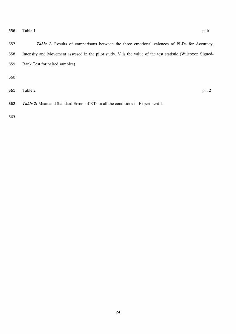
Schutter DJLG, Hofman D, Van Honk J (2008) Fearful faces selectively increase corticospinal motor tract 493
excitability: A transcranial magnetic stimulation study. Psychophysiology 45:345–348 494
21
Shmuelof L, Zohary E (2006) A Mirror Representation of Others’ Actions in the Human Anterior Parietal 495
Cortex. Journal of Neuroscience, 26(38):9736–9742. 496
Shmuelof L, Zohary E (2005) Dissociation between ventral and dorsal fMRI activation during object and 497
action recognition. Neuron, 47(3):457–70. 498
Silvanto J (2008) State-dependency of transcranial magnetic stimulation. Brain Topogr 21:1–10. 499
Silvanto J, Muggleton N, Walsh V (2008) State-dependency in brain stimulation studies of perception and 500
cognition. Trends Cogn Sci 12:447–454 501
Silvanto J, Muggleton NG, Cowey A, Walsh V (2007) Neural activation state determines behavioral 502
susceptibility to modified theta burst transcranial magnetic stimulation. Eur J Neurosci 26:523–528 503
Skuk VG, Schweinberger SR (2013) Adaptation aftereffects in vocal emotion perception elicited by 504
expressive faces and voices. PLoS One 8:1–13. 505
Tamietto M, Adenzato M, Geminiani G, de Gelder B (2007) Fast recognition of social emotions takes the 506
whole brain: Interhemispheric cooperation in the absence of cerebral asymmetry. Neuropsychologia 507
45:836–843. 508
Tamietto M, de Gelder B (2011) Sentinels in the visual system. Front Behav Neurosci 5:6. 509
Theusner S, de Lussanet MHE, Lappe M (2011) Adaptation to biological motion leads to a motion and a 510
form aftereffect. Atten Percept Psychophys 73:1843–1855. 511
Troje NF, Sadr J, Geyer H, Nakayama K (2006) Adaptation aftereffects in the perception of gender from 512
biological motion. J Vis 6:7. 513
Tunik E, Rice NJ, Hamilton A, Grafton ST (2007) Beyond grasping: representation of action in human 514
anterior intraparietal sulcus. Neuroimage 36 Suppl 2:T77-86 515
Vaina LM, Lemay M, Bienfang DC, Choi AY, Nakayama K (1990) Intact "biological motion" 516
and "structure from motion" perception in a patient with impaired motion mechanisms: a 517
case study. Vis Neurosci 5:353–369 518
22
van Boxtel JJA, Lu H (2013) Impaired global, and compensatory local, biological motion processing in 519
people with high levels of autistic traits. Front Psychol 4:209. 520
van de Riet W a C, Grezes J, de Gelder B (2009) Specific and common brain regions involved in the 521
perception of faces and bodies and the representation of their emotional expressions. Soc Neurosci 522
4:101–120. 523
van Kemenade BM, Muggleton N, Walsh V, Saygin AP (2012) Effects of TMS over Premotor and Superior 524
Temporal Cortices on Biological Motion Perception. J Cogn Neurosci 24:896–904. 525
Webster M a, MacLeod DI a (2011) Visual adaptation and face perception. Philos Trans R Soc London B 526
366:1702–1725. 527
Webster MA, Kaping D, Mizokami Y, Duhamel P (2004) Adaptation to natural facial categories. Nature 528
428:557–561. 529
530
531
23
Legend of Figures 532
Figure 1 p. 10 533
Figure 1. Representation of stimulation sites and respective anatomical landmarks. Right panel: 534
individual renderings of the gray-white matter border in each of the 14 participants. Left panel: the same 535
brains as in the right panel are shown with the main anatomical landmarks used for localization of TMS 536
targets. Blue: central sulcus; green: postcentral sulcus; yellow: intraparietal sulcus; purple: Silvian fissure; 537
red: superior temporal sulcus. The 3 stimulation sites (aIPS, pSTS and control) are represented with white 538
spots. 539
540
Figure 2 p. 11 541
Figure 2: Timeline of TMS Experiments. 542
543
Figure 3 p. 13 544
Figure 3. Visualization of results in Experiment 1. The performance of each participant is represented 545
with a black bar. The grey columns represent the mean of RTs in congruent and incongruent conditions. 546
Main analysis revealed an adaptation after-effect for affective PLDs, with congruent stimuli being 547
recognized significantly slower than incongruent ones. 548
549
Figure 4 p. 14 550
Figure 4: Visualization of Results of Experiment 2. Mean RTs are shown, classified according to emotion 551
in the test PLD (happiness or fear); congruence with the adapter sequence (congruent or incongruent); and to 552
the site of TMS (aIPS, pSTS or occipital control). The vertical bars represent the standard errors. 553
554
Legend of Tables 555
24
Table 1 p. 6 556
Table 1. Results of comparisons between the three emotional valences of PLDs for Accuracy, 557
Intensity and Movement assessed in the pilot study. V is the value of the test statistic (Wilcoxon Signed-558
Rank Test for paired samples). 559
560
Table 2 p. 12 561
Table 2: Mean and Standard Errors of RTs in all the conditions in Experiment 1. 562
563
26
Figure 2
Figure 3
28
Table 1 564
Accuracy Movement Intensity
V p-value V p-value V p-value
Fearful vs Happy 30 0.39 12.5 0.139 26 0.919
Fearful vs Neutral 21 0.034 31 0.759 55 0.002
Happy vs Neutral 28.5 0.154 45 0.083 55 0.002
565
Table 2 566
emoTest Congruence Mean RT (ms) SE (ms)
Fear Congruent 1317 50.32
Fear Incongruent 1219 46.75
Happiness Congruent 1267 43.92
Happiness Incongruent 1252 36.84
567