111
MR: Finger and Thumb Injuries Laura W. Bancroft, M.D. Professor of Radiology University of Central Florida Florida State University
MR: Finger and Thumb Injuries
Laura W. Bancroft, M.D.Professor of Radiology
University of Central Florida
Florida State University
Outline
• Normal anatomy of the fingers and thumb
• MR imaging protocols
• MRI findings of sports injuries of the fingers and thumb

NORMAL ANATOMY

Clavero JA et al. Extensor Mechanism of the Fingers: MR Imaging–Anatomic Correlation. RadioGraphics 2003; 23:593-611.
Zone specific Anatomy (Verdan)-distal to proximal
I - DIP jointII - middle phalanxIII - PIP jointIV - proximal phalanxV - MCP jointVI - dorsum of handVII -wrist extensor compartmentVIII - extrinsic extensor muscles

Clavero JA et al. Extensor Mechanism of the Fingers: MR Imaging–Anatomic Correlation. RadioGraphics 2003; 23:593-611.
terminal tendon
triangular ligament med/lat conjoined tendons

Clavero JA et al. Extensor Mechanism of the Fingers: MR Imaging–Anatomic Correlation. RadioGraphics 2003; 23:593-611.
lateral slip
med/lat conjoined tendons
central slip
medial slip

Clavero JA et al. Extensor Mechanism of the Fingers: MR Imaging–Anatomic Correlation. RadioGraphics 2003; 23:593-611.
interosseous muscle
retinacular ligament
oblique fibers
transverse fibers
sagittal band

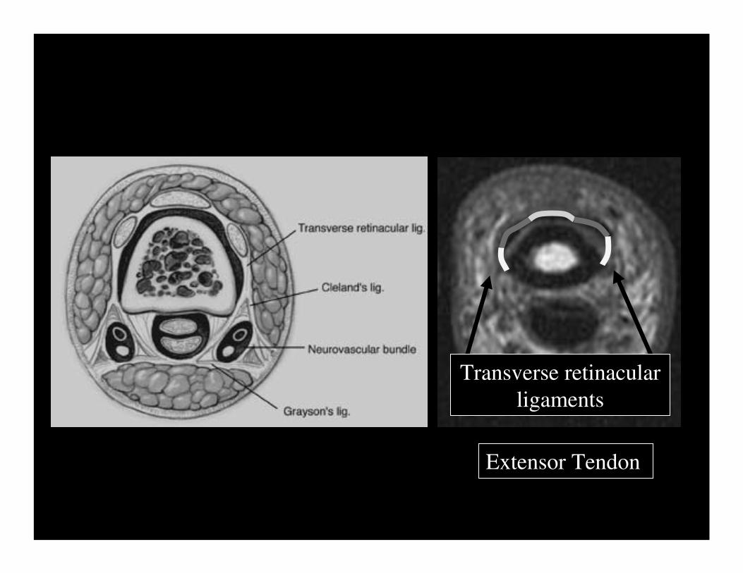
Extensor Tendon
Central slip

Extensor Tendon
Conjoined tendons
Extensor Tendon
Transverse retinacularligaments

palmar lateral
FDS
FDS FDS

palmar lateralFDS FDS
FDPFDP
FDS
FDP
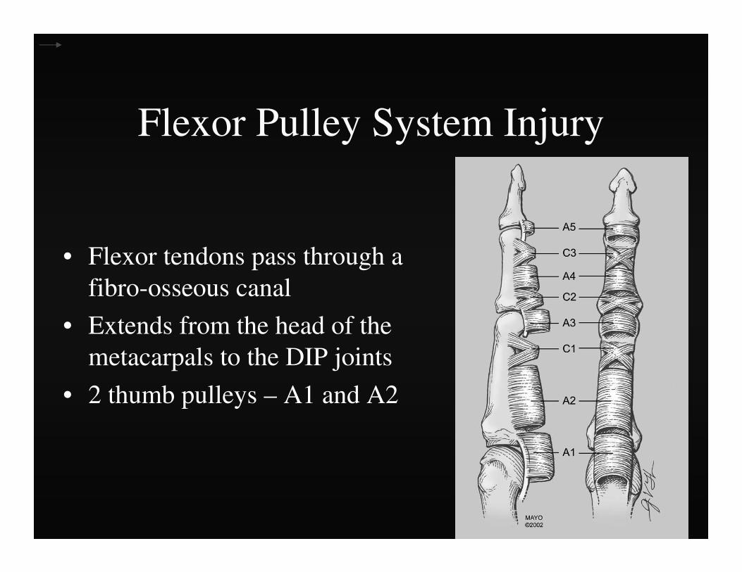
Flexor Pulley System Injury
• Flexor tendons pass through a fibro-osseous canal
• Extends from the head of the metacarpals to the DIP joints
• 2 thumb pulleys – A1 and A2

Flexor Pulley System Injury
• A2 and A4 – Largest
– Prevent bowstringing

Flexor Pulley System Injury
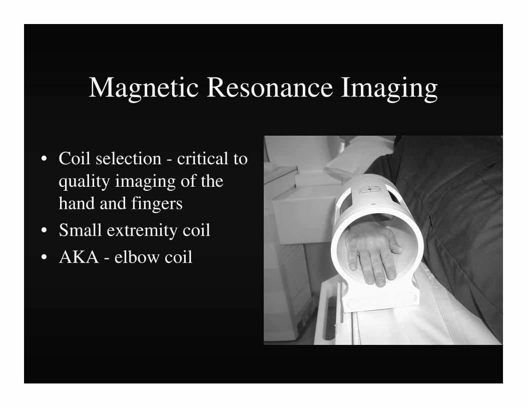
Magnetic Resonance Imaging
• Coil selection - critical to quality imaging of the hand and fingers
• Small extremity coil
• AKA - elbow coil
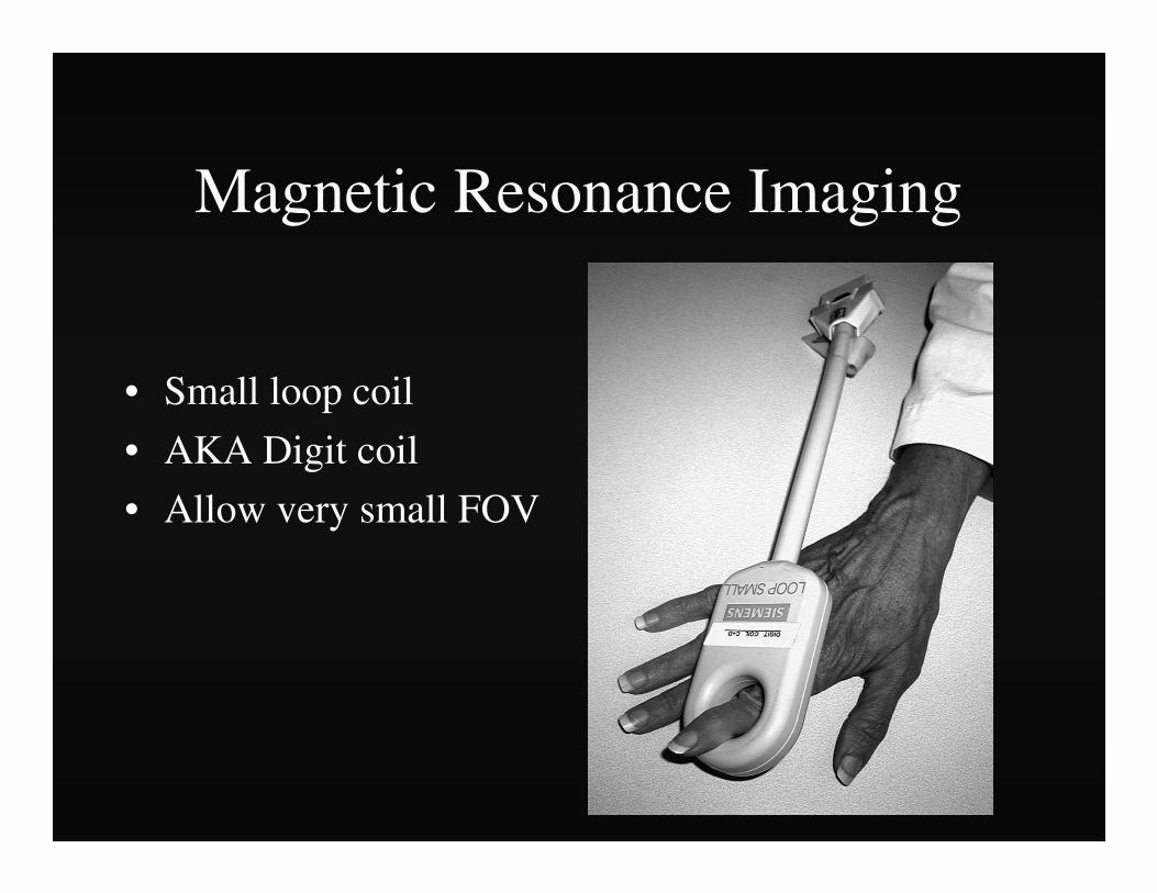
Magnetic Resonance Imaging
• Small loop coil
• AKA Digit coil
• Allow very small FOV

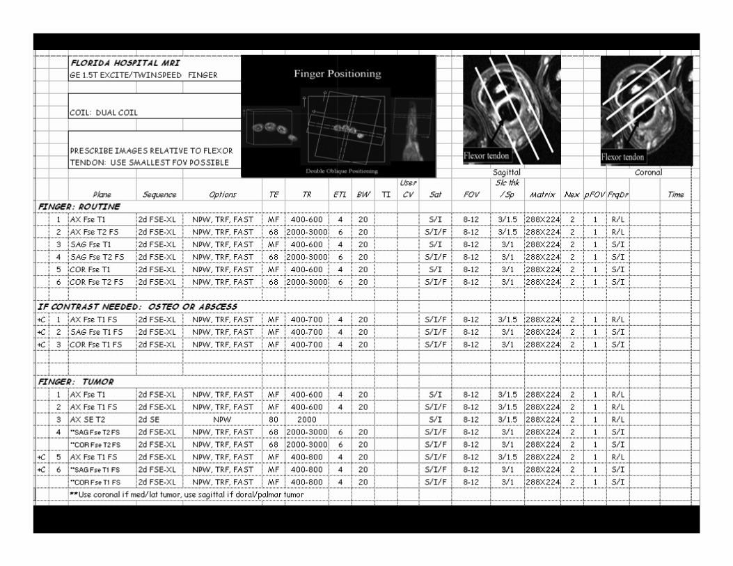
Finger Positioning
Double Oblique Positioning
S
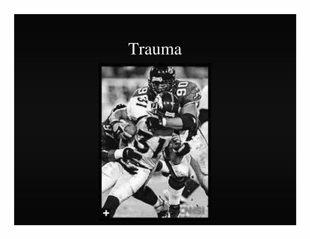
Trauma
Finger and Thumb Injuries
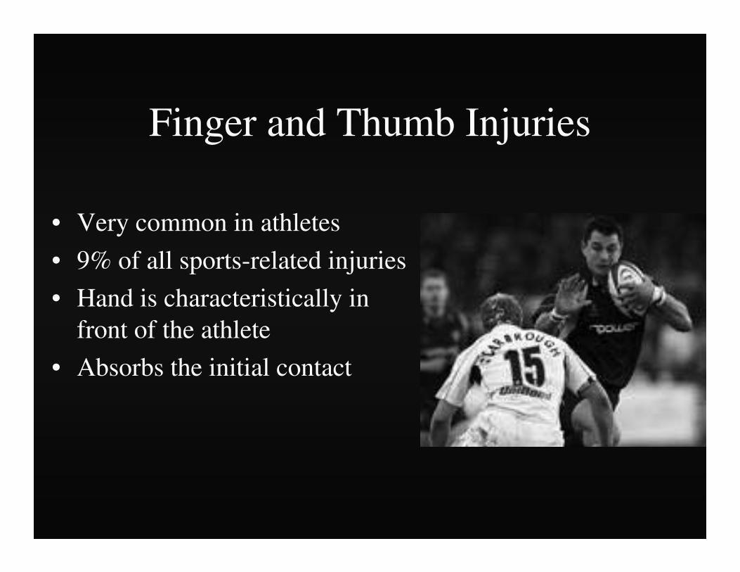
• Very common in athletes
• 9% of all sports-related injuries
• Hand is characteristically in front of the athlete
• Absorbs the initial contact

Finger and Thumb Injuries
• Hands are used in a majority of sports
• Many competitive team sports – Fingers and thumb - most often
injured
Fingers and Thumb Injuries
• Common in sports with a high risk of falling– Skiing
– Biking
– Gymnastics
– In-line skating

Fingers and Thumb Injuries
• In-line skating –– >50% of injuries
involve hand/wrist
• Football –– Hand and wrist injuries
account for 15-20%

Finger and Thumb Injuries
• “Minor” injuries– Finger sprain
• “Major” injuries– Fracture or dislocation
• Hard to prevent

Imaging
• Radiographs
• Computed Tomography
• Magnetic Resonance Imaging

Hyperextension InjuryCapsular and Volar Plate Disruption

• Volar Plate
– Thick fibrocartilaginousstructure
– Prevents hyperextension
– Palmar aspect of PIP joint
– Distal – firm attachment
Hyperextension InjuryCapsular and Volar Plate Disruption
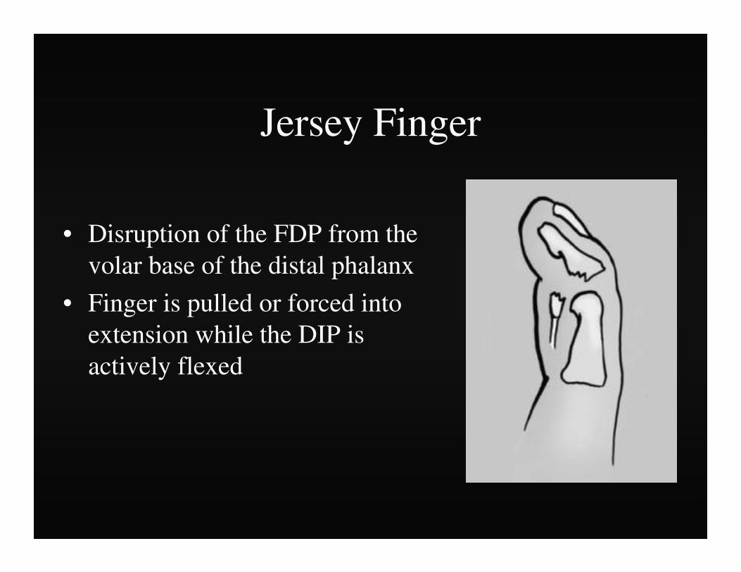
Jersey Finger
• Disruption of the FDP from the volar base of the distal phalanx
• Finger is pulled or forced into extension while the DIP is actively flexed

Jersey Finger• Attempting to grab
someone by the jersey while making a tackle
– Football
– Rugby
• Ring finger - 75% of cases
• Localized pain and swelling
• Inability to flex DIP joint
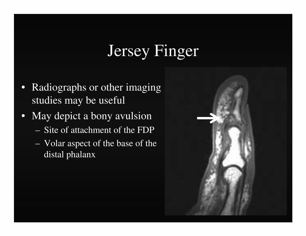
Jersey Finger
• Radiographs or other imaging studies may be useful
• May depict a bony avulsion– Site of attachment of the FDP
– Volar aspect of the base of the distal phalanx
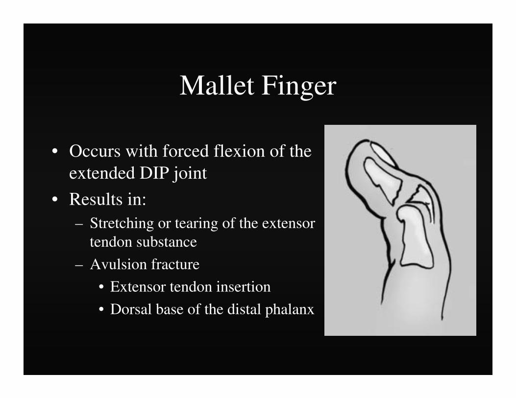
Mallet Finger
• Occurs with forced flexion of the extended DIP joint
• Results in:– Stretching or tearing of the extensor
tendon substance
– Avulsion fracture
• Extensor tendon insertion
• Dorsal base of the distal phalanx

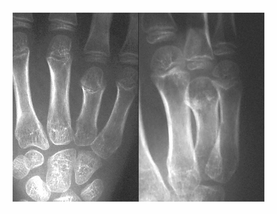
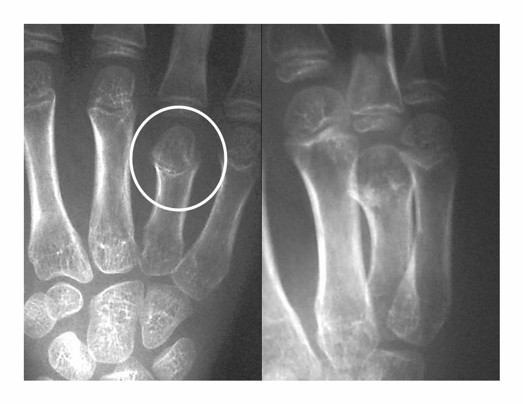
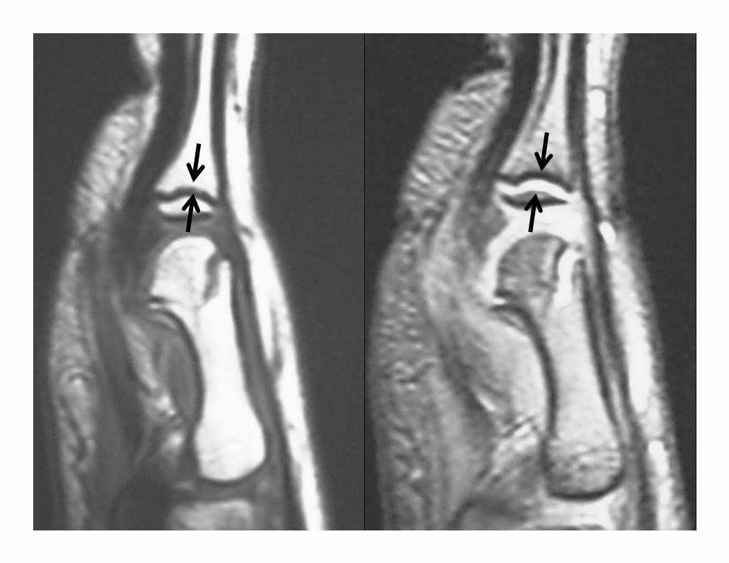
Mallet Finger
• Extensor tendon tear or bony avulsion
• Bony avulsion of dorsal base of the distal phalanx

Mallet Finger
• Classic mechanism of injury – Tip of the extended finger
struck by a ball• Softball, baseball, or basketball
• Often referred to as a “Jammed” Finger

Applications:Physeal Injury
• MR imaging accurately depicts the physealanatomy
• Cartilage sensitive sequences are especially useful in mapping physeal bars


Flexor Tendon Zones
• I – between FDS and FDP attachments
• II – FDS attachment to palmar fold
• III – A1 pulley to retinaculum
• IV – carpal tunnel
• V – forearm proximal to retinaculum
I II III

Flexor Tenosynovitis
• MR imaging useful
• Increased signal within the tendon sheath – fluid sensitive sequences
• Enhancement – Post-gadolinium images

Flexor Tenosynovitis
• Cortisone injections –injected directly into the region of concern
• 90% improve with non-operative treatment

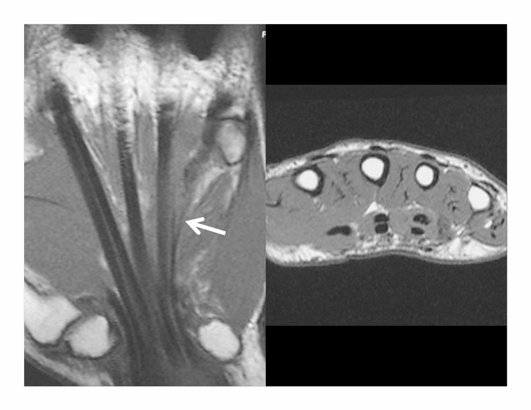
Tendon Evaluation
• Normal tendons demonstrate low signal intensity on all pulse sequence
• MR accurately depicts tendon morphology and the gap for severed tendons
• MR is also useful for pulley injuries.
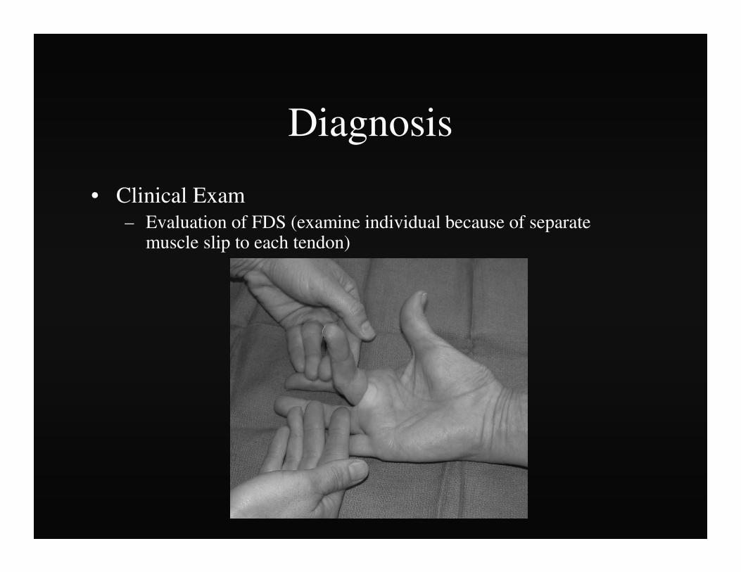
Diagnosis
• Clinical Exam– Evaluation of FDS (examine individual because of separate
muscle slip to each tendon)

Diagnosis
• Clinical Exam– Evaluation of FDP ( Examine together since tendons
share common muscle belly, FDP to index separate)

Diagnosis
• Clinical Exam– “Squeeze” test- squeeze volar mid-forearm
and assess flexion of digits
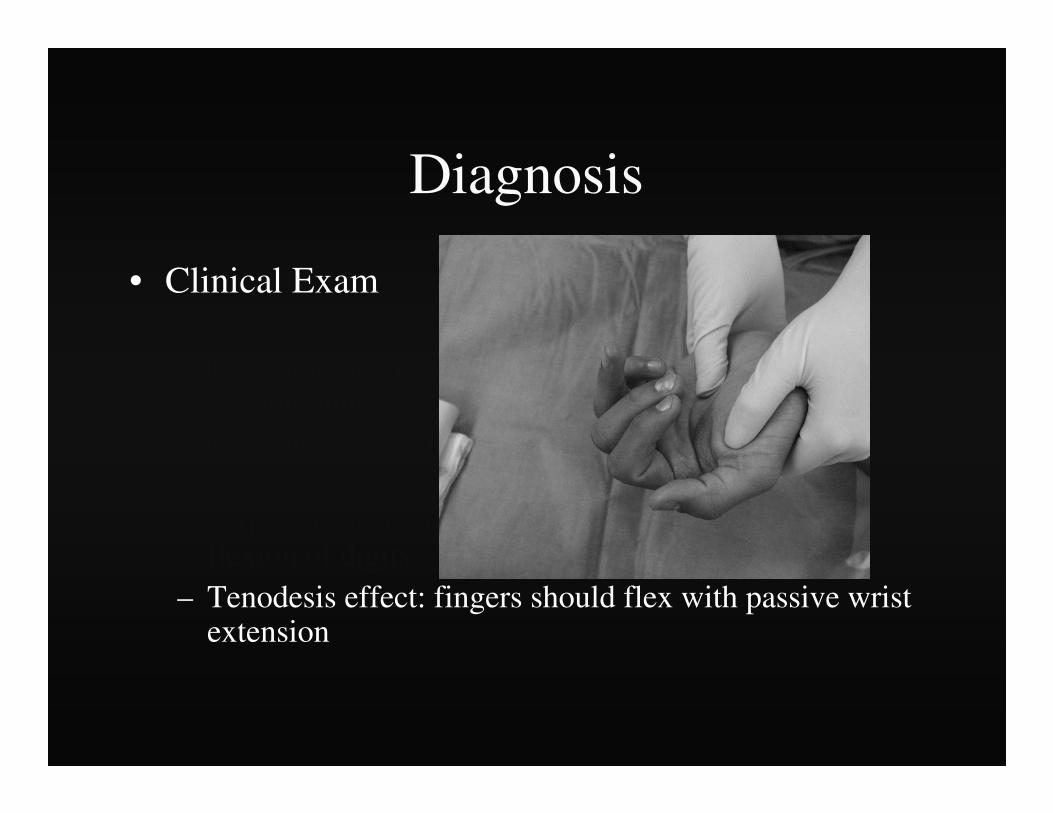
Diagnosis
• Clinical Exam– Observation- “Cascade effect” of digits– Evaluation of FDS (examine individual because of
separate muscle slip to each tendon)– Evaluation of FDP ( Examine together since tendons
share common muscle belly, FDP to index separate)– “Squeeze” test- squeeze volar mid-forearm and assess
flexion of digits– Tenodesis effect: fingers should flex with passive wrist
extension
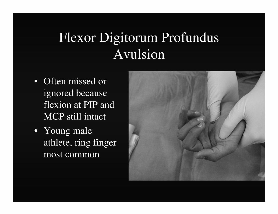
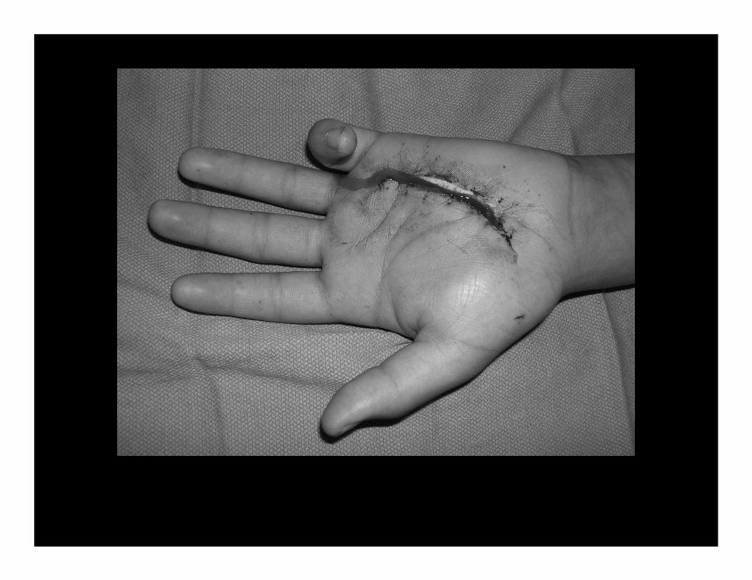
Flexor Digitorum ProfundusAvulsion
• Often missed or ignored because flexion at PIP and MCP still intact
• Young male athlete, ring finger most common

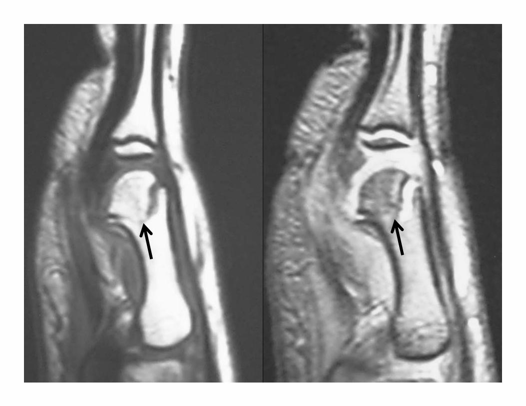
Flexor Tendon Tears
• Commonly result from sports-related injuries
• May occur anywhere along the course of the tendons
• Localized pain and swelling
• Inability to flex the IP joints
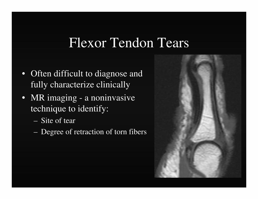
Flexor Tendon Tears
• Often difficult to diagnose and fully characterize clinically
• MR imaging - a noninvasive technique to identify:– Site of tear
– Degree of retraction of torn fibers
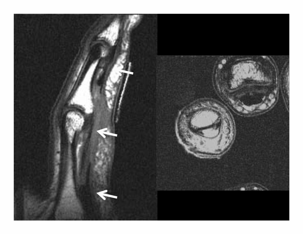

Flexor digitorum profundus and superficialis rupture


Flexor Digitorum ProfundusAvulsion
• Leddy and Packer classification:– Type I: Tendon retracts into palm- repair within 7-10
days
Courtesy of Dr. Peter Murray


Flexor Digitorum ProfundusAvulsion
• Leddy and Packer classification:– Type II: Small bony fragment avulsed-
usually trapped proximally at A3 pulley-repair in first 6 wks possible III- large bony fragment avulsed- usually trapped at A4 pulley- ORIF
From Strickland J, Green’s Hand Surgery

Courtesy of Dr. Peter Murray

Courtesy of Dr. Peter Murray

Courtesy of Dr. Peter Murray

Flexor Digitorum ProfundusAvulsion
• Leddy and Packer classification:– Type III- large bony fragment avulsed- usually
trapped at A4 pulley- ORIF
From Strickland J, Green’s Hand Surgery
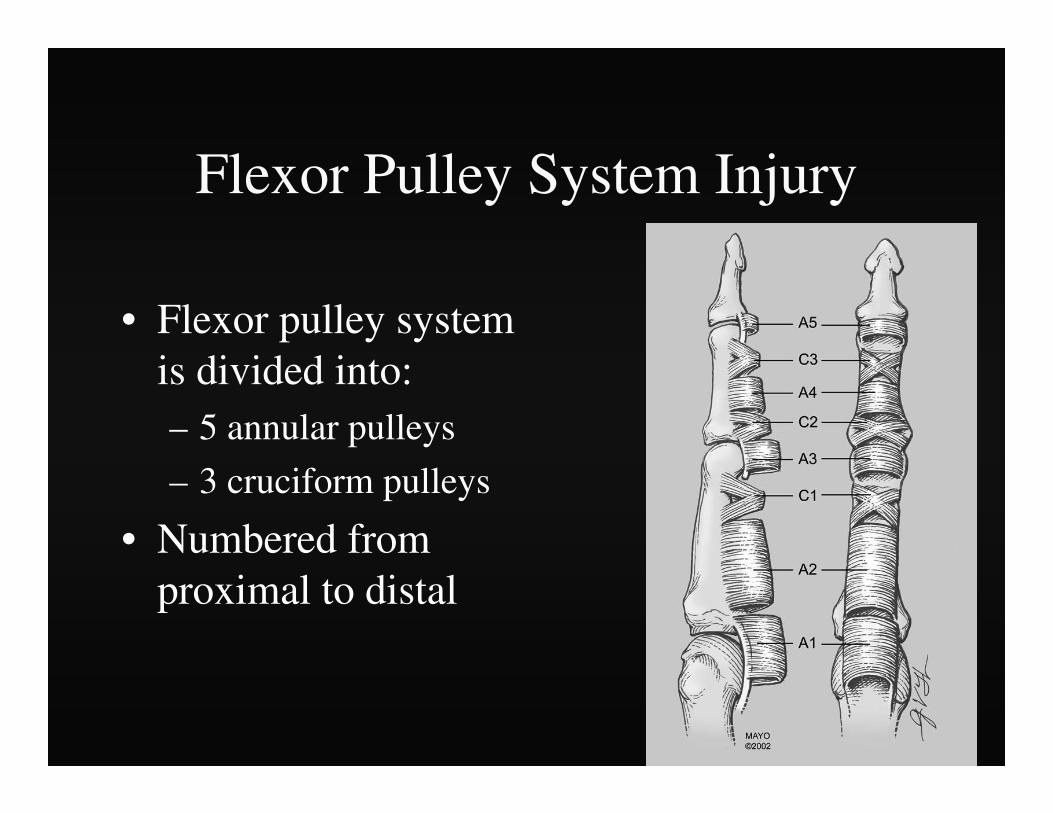
Flexor Pulley System Injury
• Flexor pulley system is divided into:– 5 annular pulleys
– 3 cruciform pulleys
• Numbered from proximal to distal

Flexor Pulley System Injury
• A2 pulley - most important to flexor tendon function– Injury typically begins with the A2 pulley
– Followed sequentially by the A3 and A4 pulley
– Rarely - A1 pulley
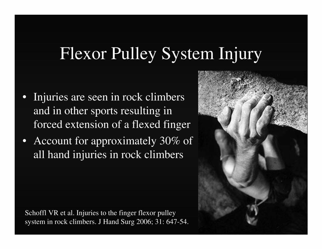
Flexor Pulley System Injury
• Injuries are seen in rock climbers and in other sports resulting in forced extension of a flexed finger
• Account for approximately 30% of all hand injuries in rock climbers
Schoffl VR et al. Injuries to the finger flexor pulley system in rock climbers. J Hand Surg 2006; 31: 647-54.

Flexor Pulley System Injury
• Crimp Position– DIP joints - extended
– PIP joints – flexed
– MCP joints – extended
– Carpus – slightly extended
Flexor Pulley System Injury
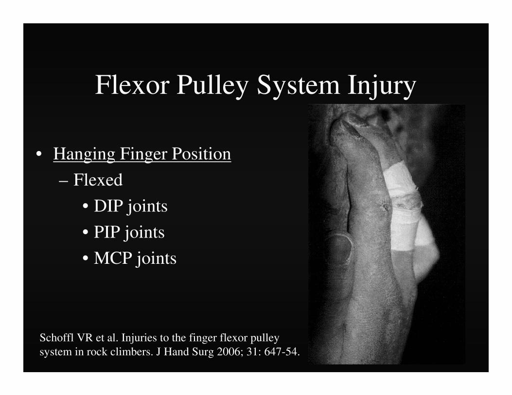
• Hanging Finger Position
– Flexed
• DIP joints
• PIP joints
• MCP joints
Schoffl VR et al. Injuries to the finger flexor pulley system in rock climbers. J Hand Surg 2006; 31: 647-54.

• I – pulley strain
• II - A4 rupture or partial rupture A2/A3
• III – A2/A3 rupture
• IV
• Multiple ruptures
• Single rupture with lumbrical ms or collateral ligament injury
Pulley Injury - Grades
Schoffl VR et al. Injuries to the finger flexor pulley system in rock climbers. J Hand Surg2006; 31: 647-54.

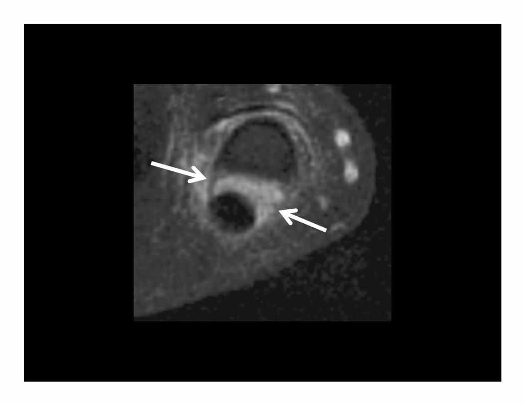
MRI (3T)
• A2 PULLEY RUPTURES– Sensitivity: 87.5 %
– Specificity: 100 %
– Positive predictive value (PPV): 100 %
– Negative predictive value (NPV): 95.2 %


Reconstruct pulley
Courtesy of Dr. Peter Murray
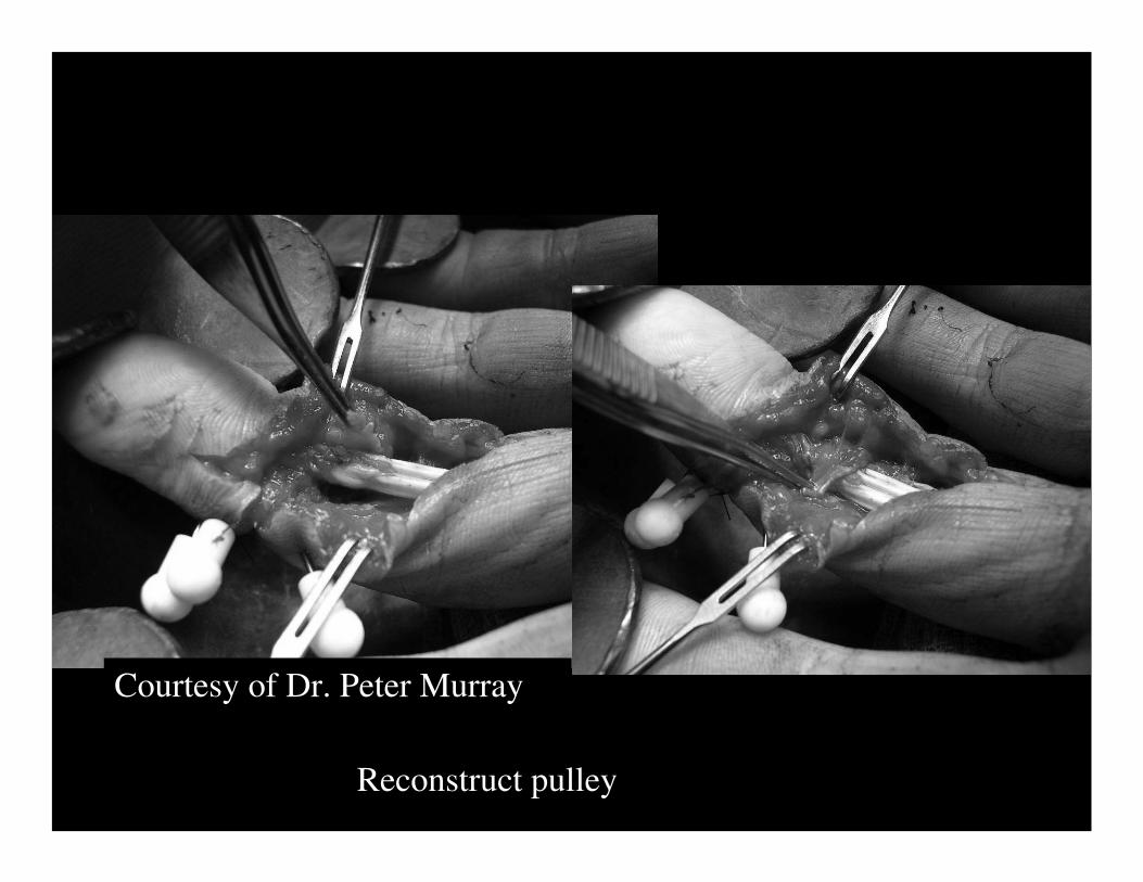
Reconstruct pulley
Courtesy of Dr. Peter Murray

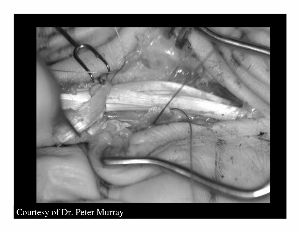
Reconstruct flexor tendon
Courtesy of Dr. Peter Murray

Reconstruct flexor tendon
Courtesy of Dr. Peter Murray
Courtesy of Dr. Peter Murray

Courtesy of Dr. Peter Murray

THUMB
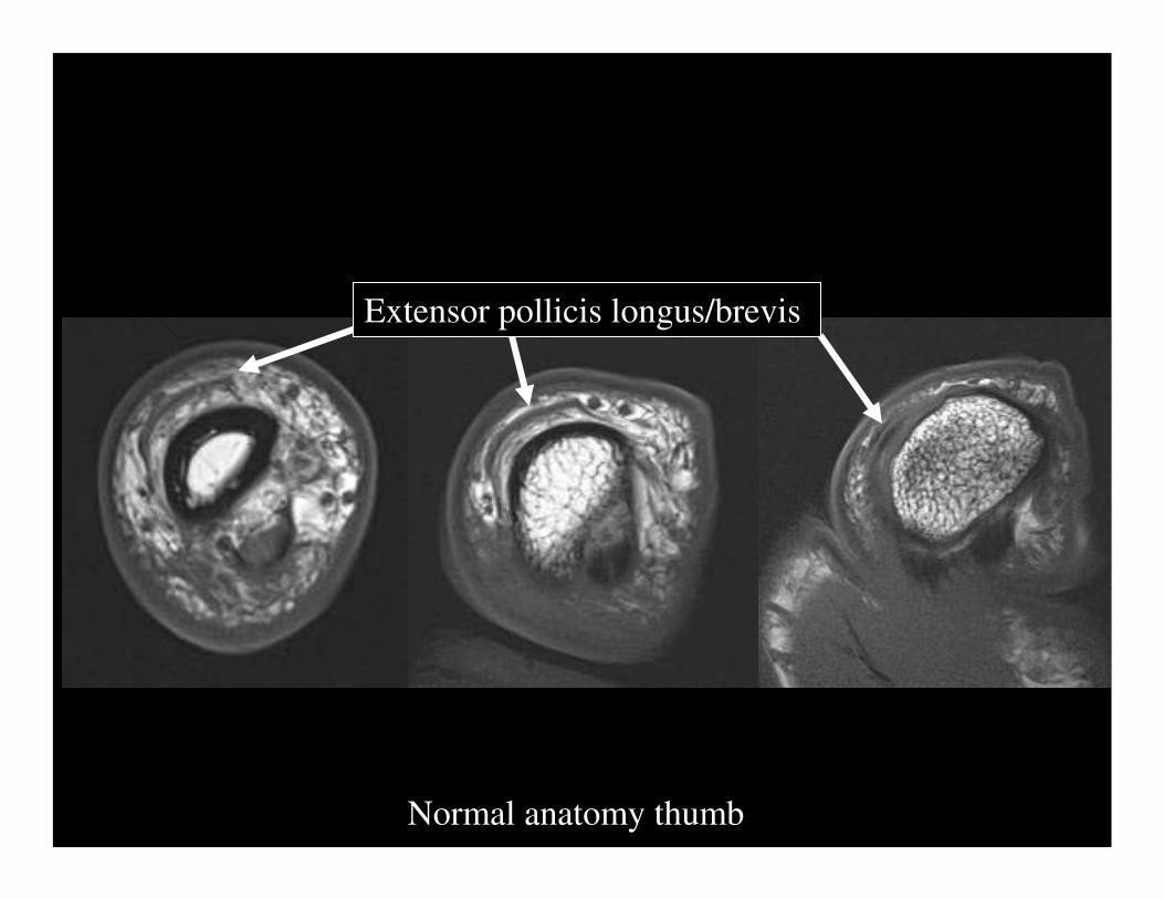
Normal anatomy thumb
Extensor pollicis longus/brevis

Normal anatomy thumb
flexor pollicis longus

Normal anatomy thumb
pulleys

Normal anatomy thumb
flexor pollicis longus

Normal anatomy thumb
Ulnar collateral ligament
Adductor aponeurosis

Normal anatomy thumb
radial collateral ligament

IML=Intermetacarpal lig.
POL=Posterior oblique lig.
DRL=Dorsoradial lig.
ECRL=Ext. carpi radialis longus
APL=Abductor pollicis longus
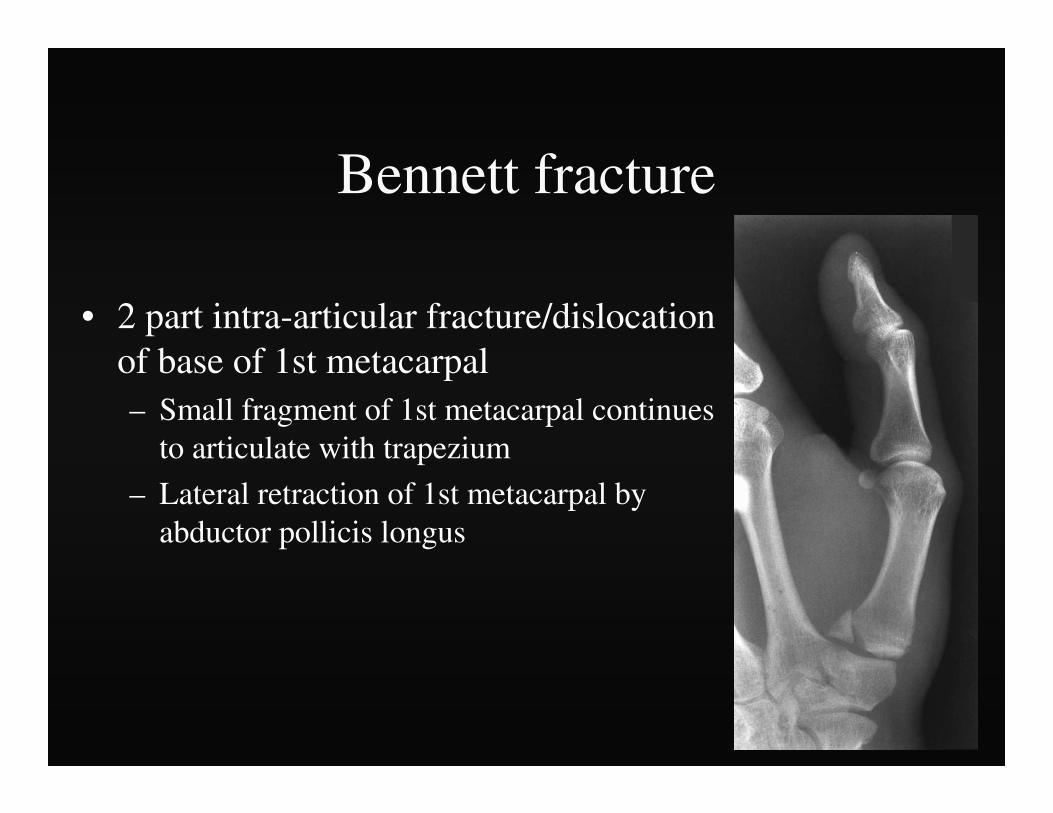
Bennett fracture
• 2 part intra-articular fracture/dislocation of base of 1st metacarpal– Small fragment of 1st metacarpal continues
to articulate with trapezium
– Lateral retraction of 1st metacarpal by abductor pollicis longus


Rolando Fracture
• Originally described -Y-shaped 3-fragment fracture – Extended to the articular surface
• Today the eponym is widely used for any comminuted intra-articular fracture at the base of the thumb

Gamekeeper’s Thumb
• Disruption of the ulnar collateral ligament of the 1st MCP joint
• Result of an acute radial or valgus stress on the thumb
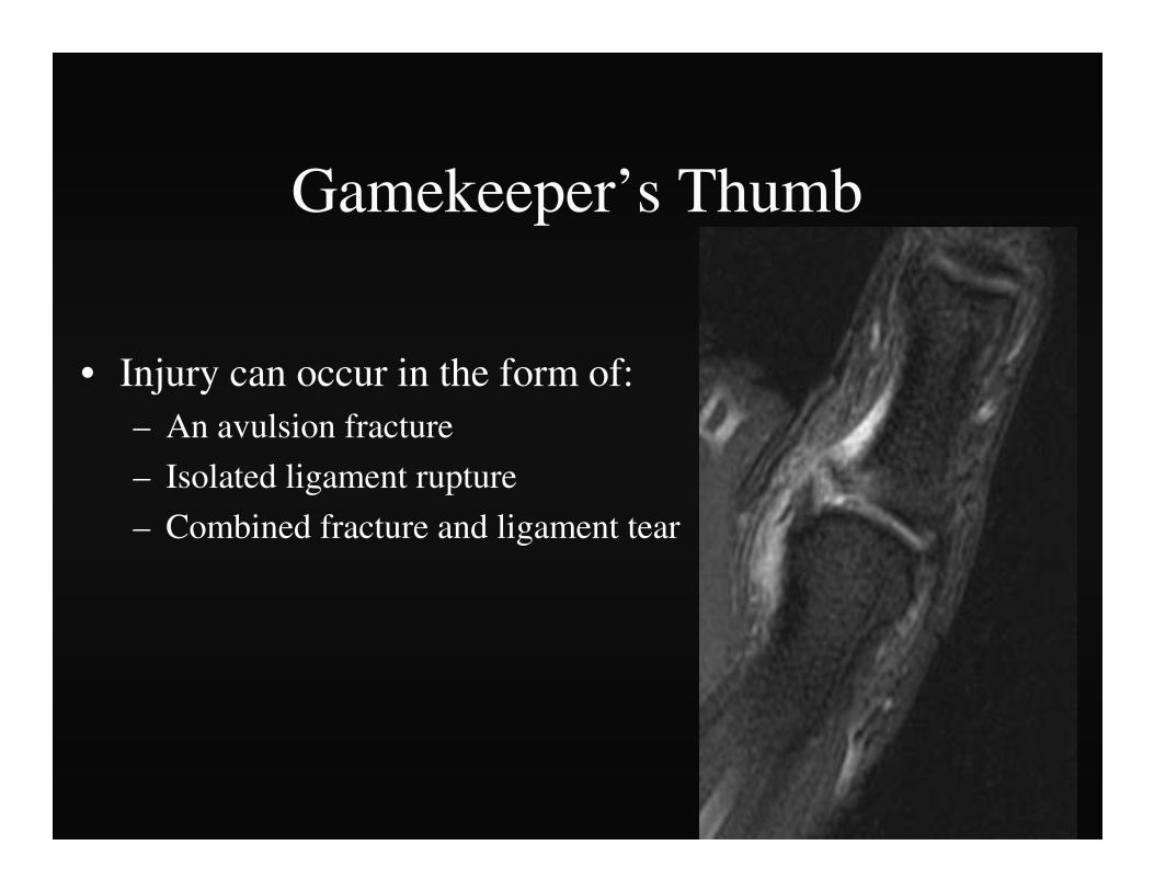
Gamekeeper’s Thumb
• Injury can occur in the form of:– An avulsion fracture
– Isolated ligament rupture
– Combined fracture and ligament tear

Gamekeeper’s Thumb
• Commonly referred to as “Skier’s Thumb”
• Most commonly seen in snow skier’s
• Fall holding a ski pole causing forced abduction and extension of the thumb

Gamekeeper’s Thumb
• Radiographs may be useful
• May depict a small avulsion fracture – Ulnar aspect of the base of
the 1st proximal phalanx
– Attachment of the UCL


Gamekeeper’s Thumb
• Stress radiographs – Neutral radiographs – Radiographs with abduction
and extension
• Greater than 30 degrees difference – Abnormal - UCL disruption

Gamekeeper’s Thumb
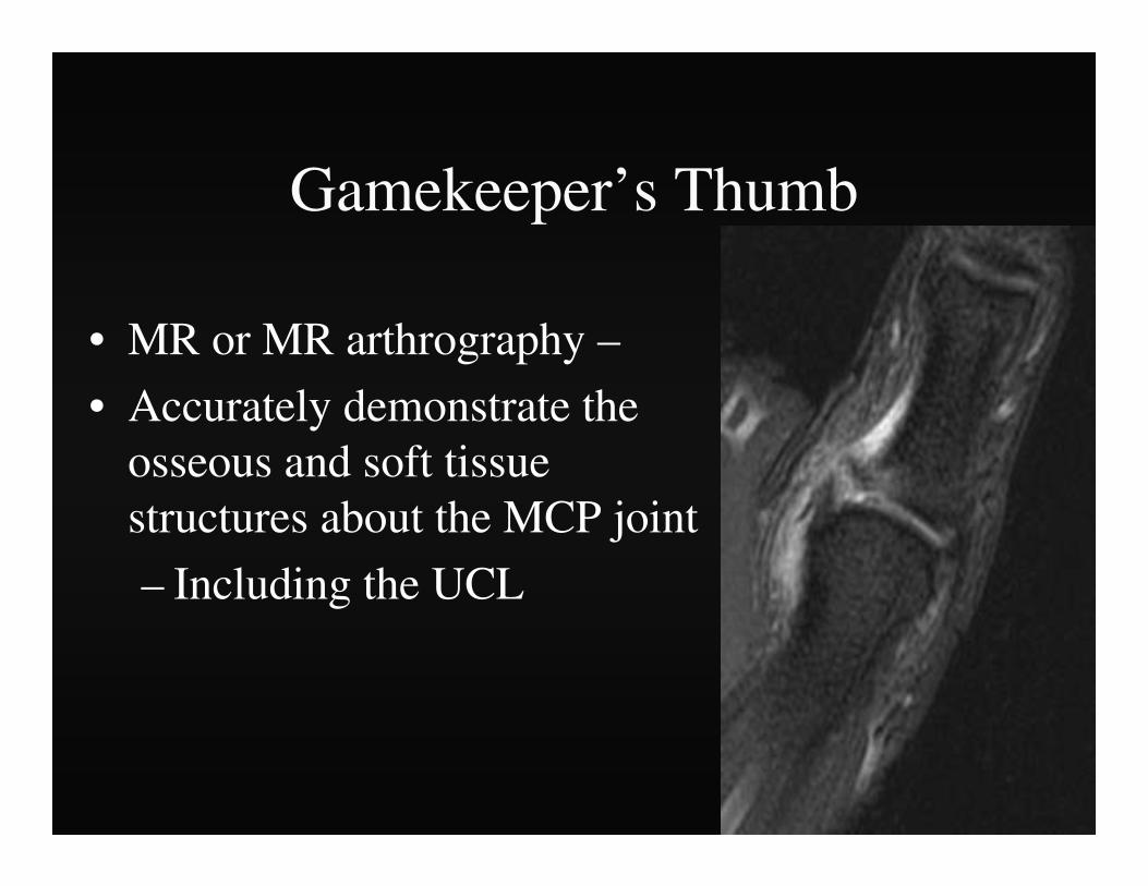
• MR or MR arthrography –
• Accurately demonstrate the osseous and soft tissue structures about the MCP joint
– Including the UCL


Gamekeeper’s thumb
• If the fracture fragment is nondisplaced -splinting of the thumb may lead to healing and restoration of joint stability
• In most patients surgical repair is preferable

Stener Lesion
• Torn UCL displaces superficial to the adductor aponeurosis
• Prevents spontaneous ligament healing
• 29% of UCL injuries

Stener Lesion
• MR imaging can depict
– UCL
– Adductor Aponeurosis
• Operative intervention – Normal anatomic apposition
– Healing of the displaced UCL

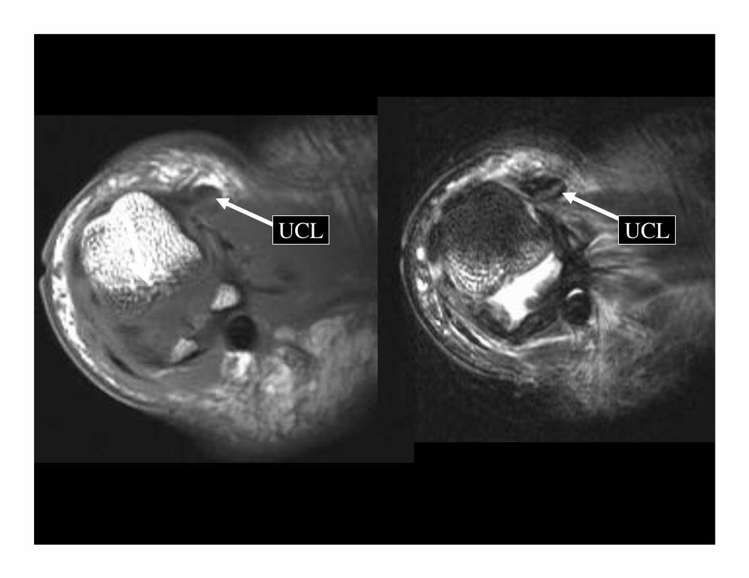
UCL UCL
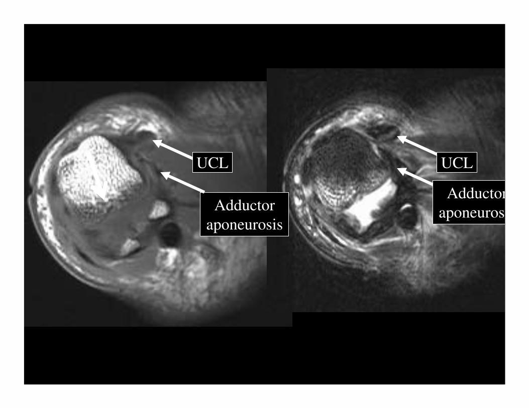
UCL UCL
Adductor aponeurosis
Adductoraponeurosi

Stener Lesion
• If a Stener lesion is present
• Only operative intervention – Normal anatomic apposition
– Healing of the displaced UCL
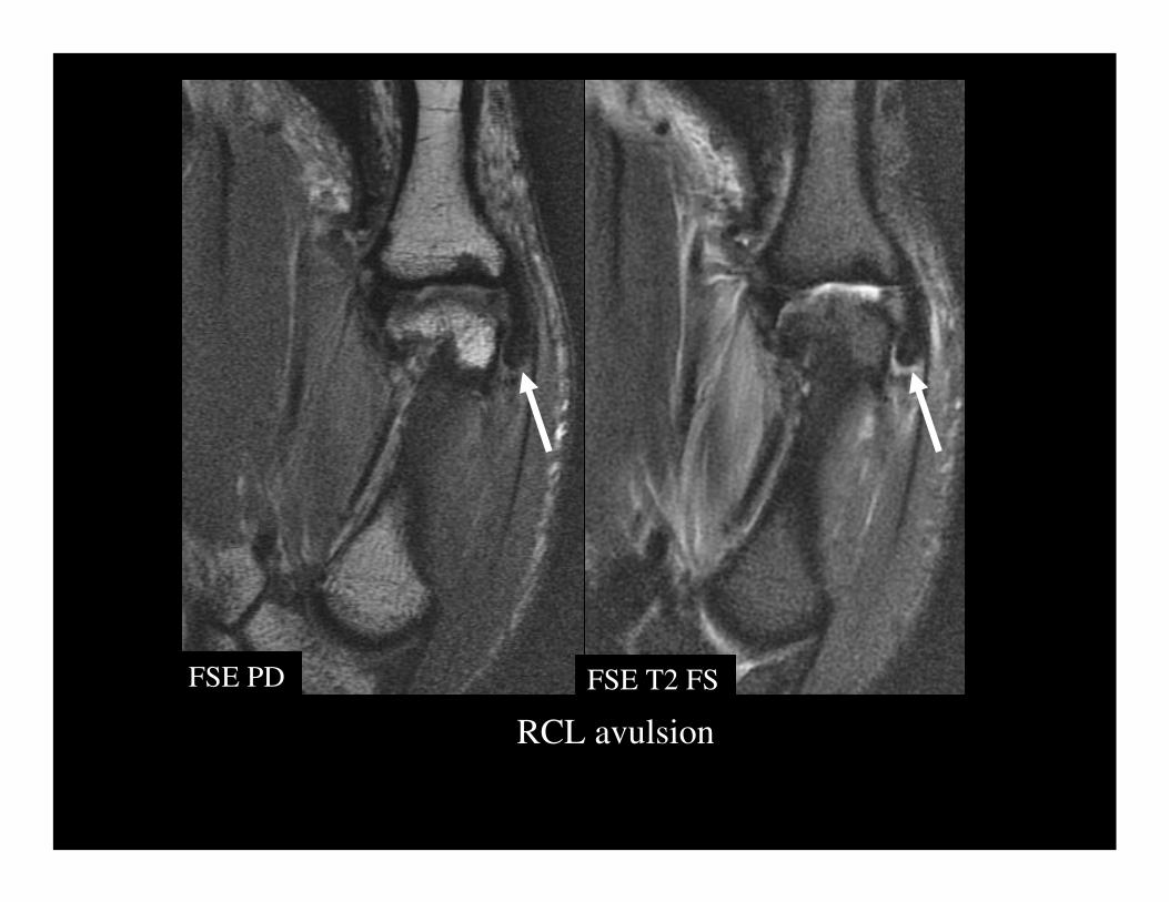
RCL avulsion
FSE PD FSE T2 FS

RCL avulsion
FSE T2 FSFSE PD

RCL avulsion
FSE T2 FSFSE PD

Conclusion
• Knowledge of the mechanism of injury and various injury patterns of osseous and soft tissue injury may direct the appropriate imaging studies for the fingers and thumb
• Imaging features of infection
• Review of the most common benign and malignant tumors of the fingers and thumb


