International Journal of Radiology and Radiation Oncology Citation: Meesa IR, Mammen L (2016) MR Imaging of Pregnant Women with Abdominal Pain and Suspected Appendicitis: Diagnostic Accuracy and Outcomes. Int J Radiol Radiat Oncol 2: 004-007. DOI: http://dx.doi.org/10.17352/ijrro.000010 004 Abstract Acute appendicitis is the most common cause of acute surgical abdomen during pregnancy. Our study was conducted to review our experience and diagnostic accuracy with MRI during pregnancy and clinical outcomes over a two year period. All pregnant women who underwent an MRI examination of the abdomen between January 2008 and January 2010 at Spectrum Health hospitals were included in the study. We retrospectively reviewed the medical records and MRI findings in 46 pregnant women. 46 pregnant women underwent a total of 53 MRI scans and of these, 23/46 (50%) presented with RLQ pain and other signs suspicious for appendicitis. 10/23 (43%) had MRI findings positive for acute appendicitis: 5/10 (50%) had uncomplicated acute appendicitis by MRI criteria, all were confirmed at surgery. 3/5 had a ruptured appendix with abscess, 1/5 had a perforated appendix with abscess and 1/5 had MRI findings suspicious for appendicitis but was discharged without surgery or further follow up. The sensitivity for diagnosing appendicitis by MRI in our 10 patients with positive findings was 89% (8 of 9 cases, with one case lost to follow up). The specificity was 100%, since all of the patients who had a normal appendix and/or no secondary signs of appendicitis on MRI were managed medically and were discharged without readmission for surgery. The negative predictive value (NPV) was 93% (13/14). Our study shows that MRI of the abdomen without contrast is an excellent alternative to CT and ultrasound for diagnosing and excluding acute appendicitis during pregnancy. Our study was conducted to review our experience at Spectrum Health to evaluate our diagnostic accuracy with MRI during pregnancy and clinical outcomes over a two year period. Materials and Methods Patients IRB approval was obtained. All pregnant women who underwent an MRI examination of the abdomen between January 2008 and January 2010 at Spectrum Health hospitals were included in the study. We retrospectively reviewed the medical records and MRI findings in 46 pregnant women (age range, 17 - 39; mean age 28). Eleven patients were in the first trimester, 20 patients were in the second trimester, and 15 patients were in the third trimester. Out of this group, women presenting with right lower quadrant pain and clinical concern for acute appendicitis were identified. e MRI used, as well as the image interpretation performed at the time of the procedure, are described below. MR imaging MRI was performed with a 1.5 - T General Electric magnet with a torso array coil except in instances the patient was too large when the body coil was utilized. e MRI sequences included axial and coronal T2 single shot fast spin Echo (SSFSE), axial and coronal steady state Objective Acute appendicitis is the most common cause of acute surgical abdomen during pregnancy, occurring in approximately 1 in 1500 deliveries [1]. Acute appendicitis in pregnant women has been associated with premature labor and maternal and/or fetal death, particularly when perforation with peritonitis occurs [2]. Alteration of the position of the intra-abdominal contents by the pregnant uterus, and the overlapping signs and symptoms of normal pregnancy and acute appendicitis, are the two most important factors leading to difficulties in identification and diagnosis [3]. Computed tomography (CT) [4] and ultrasound (US) [5], have been traditionally used for diagnosing and excluding appendicitis. e major disadvantage of using CT in pregnancy is the radiation exposure to the fetus and the mother. e major disadvantages of US in pregnancy are that it is highly operator dependent and its diagnostic performance decreases with advancing gestational age, obesity, and bowel gas [6]. MRI is the recommended as the primary imaging modality for right lower quadrant (RLQ) pain during pregnancy if US is inconclusive [7]. Recent literature indicates that MRI can be used to accurately diagnose or exclude appendicitis. MRI also enables the diagnosis of alternative causes of RLQ pain (eg, pelvis abscess, tuboovarian abscess, ovarian torsion, adnexal mass, bowel obstruction) [8]. Furthermore, evaluation of the placenta and fetal anatomy can be obtained. Research Article MR Imaging of Pregnant Women with Abdominal Pain and Suspected Appendicitis: Diagnostic Accuracy and Outcomes Indu Rekha Meesa 1 * and Leena Mammen 2 1 Summit Radiology, 5001 US Highway 30 W. Ste D, Fort Wayne, IN 46818, USA 2 Advanced Radiology Services, PC, MSU College of Human Medicine, Spectrum Health Hospitals, Grand Rapids MI, USA Dates: Received: 05 November, 2015; Accepted: 09 January, 2016; Published: 18 January, 2016 *Corresponding author: Indu Rekha Meesa, Summit Radiology, 5001 US Highway 30 W. Ste D, Fort Wayne, IN 46818, USA, E-mail: www.peertechz.com
Transcript
International Journal of Radiology and Radiation Oncology
Citation: Meesa IR, Mammen L (2016) MR Imaging of Pregnant Women with Abdominal Pain and Suspected Appendicitis: Diagnostic Accuracy and Outcomes. Int J Radiol Radiat Oncol 2: 004-007. DOI: http://dx.doi.org/10.17352/ijrro.000010
004
Abstract
Acute appendicitis is the most common cause of acute surgical abdomen during pregnancy. Our study was conducted to review our experience and diagnostic accuracy with MRI during pregnancy and clinical outcomes over a two year period. All pregnant women who underwent an MRI examination of the abdomen between January 2008 and January 2010 at Spectrum Health hospitals were included in the study. We retrospectively reviewed the medical records and MRI findings in 46 pregnant women. 46 pregnant women underwent a total of 53 MRI scans and of these, 23/46 (50%) presented with RLQ pain and other signs suspicious for appendicitis. 10/23 (43%) had MRI findings positive for acute appendicitis: 5/10 (50%) had uncomplicated acute appendicitis by MRI criteria, all were confirmed at surgery. 3/5 had a ruptured appendix with abscess, 1/5 had a perforated appendix with abscess and 1/5 had MRI findings suspicious for appendicitis but was discharged without surgery or further follow up. The sensitivity for diagnosing appendicitis by MRI in our 10 patients with positive findings was 89% (8 of 9 cases, with one case lost to follow up). The specificity was 100%, since all of the patients who had a normal appendix and/or no secondary signs of appendicitis on MRI were managed medically and were discharged without readmission for surgery. The negative predictive value (NPV) was 93% (13/14). Our study shows that MRI of the abdomen without contrast is an excellent alternative to CT and ultrasound for diagnosing and excluding acute appendicitis during pregnancy.
Our study was conducted to review our experience at Spectrum Health to evaluate our diagnostic accuracy with MRI during pregnancy and clinical outcomes over a two year period.
Materials and MethodsPatients
IRB approval was obtained. All pregnant women who underwent an MRI examination of the abdomen between January 2008 and January
2010 at Spectrum Health hospitals were included in the study. We retrospectively reviewed the medical records and MRI findings in 46 pregnant women (age range, 17 - 39; mean age 28). Eleven patients were in the first trimester, 20 patients were in the second trimester, and 15 patients were in the third trimester. Out of this group, women presenting with right lower quadrant pain and clinical concern for acute appendicitis were identified. The MRI used, as well as the image interpretation performed at the time of the procedure, are described below.
MR imagingMRI was performed with a 1.5 - T General Electric magnet with a
torso array coil except in instances the patient was too large when the body coil was utilized. The MRI sequences included axial and coronal T2 single shot fast spin Echo (SSFSE), axial and coronal steady state
ObjectiveAcute appendicitis is the most common cause of acute surgical
abdomen during pregnancy, occurring in approximately 1 in 1500 deliveries [1]. Acute appendicitis in pregnant women has been associated with premature labor and maternal and/or fetal death, particularly when perforation with peritonitis occurs [2]. Alteration of the position of the intra-abdominal contents by the pregnant uterus, and the overlapping signs and symptoms of normal pregnancy and acute appendicitis, are the two most important factors leading to difficulties in identification and diagnosis [3].
Computed tomography (CT) [4] and ultrasound (US) [5], have been traditionally used for diagnosing and excluding appendicitis. The major disadvantage of using CT in pregnancy is the radiation exposure to the fetus and the mother. The major disadvantages of US in pregnancy are that it is highly operator dependent and its diagnostic performance decreases with advancing gestational age, obesity, and bowel gas [6].
MRI is the recommended as the primary imaging modality for right lower quadrant (RLQ) pain during pregnancy if US is inconclusive [7]. Recent literature indicates that MRI can be used to accurately diagnose or exclude appendicitis. MRI also enables the diagnosis of alternative causes of RLQ pain (eg, pelvis abscess, tuboovarian abscess, ovarian torsion, adnexal mass, bowel obstruction) [8]. Furthermore, evaluation of the placenta and fetal anatomy can be obtained.
Research Article
MR Imaging of Pregnant Women with Abdominal Pain and Suspected Appendicitis: Diagnostic Accuracy and Outcomes
Indu Rekha Meesa1* and Leena Mammen2
1Summit Radiology, 5001 US Highway 30 W. Ste D, Fort Wayne, IN 46818, USA2Advanced Radiology Services, PC, MSU College of Human Medicine, Spectrum Health Hospitals, Grand Rapids MI, USA
*Corresponding author: Indu Rekha Meesa, Summit Radiology, 5001 US Highway 30 W. Ste D, Fort Wayne, IN 46818, USA, E-mail:
www.peertechz.com
Citation: Meesa IR, Mammen L (2016) MR Imaging of Pregnant Women with Abdominal Pain and Suspected Appendicitis: Diagnostic Accuracy and Outcomes. Int J Radiol Radiat Oncol 2: 004-007. DOI: http://dx.doi.org/10.17352/ijrro.000010
Meesa and Mammen. (2016)
005
- 2/3 treated with surgical debridement
- 1/3 treated with percutaneous drainage by interventional radiology
• 1/5 had a perforated appendix with abscess (confirmed on f/u MRI), treated with surgical debridement
• 1/5 had findings suspicious for appendicitis but was discharged without surgery or further follow up
• 13/23 (57%) patients had a normal appendix and/or no secondary signs of appendicitis on MRI; all were managed medically and discharged in an improved condition
• 23/46 (50%) cases were performed for conditions other than RLQ pain, in which a variety of abnormalities were found: placenta previa (2), placenta accreta (1), thinning of lower uterine segment (1), uterine fibroids (1), cystic ovarian mass(1), ovarian dermoid (2), paraovarian cyst (1), cornual pregnancy (1), sub-septate uterus (1), left adrenal mass (1), polycystic kidneys (1), kidney stones (1), and thickened terminal ileum (1). Renal agenesis was the only fetal abnormality diagnosed.
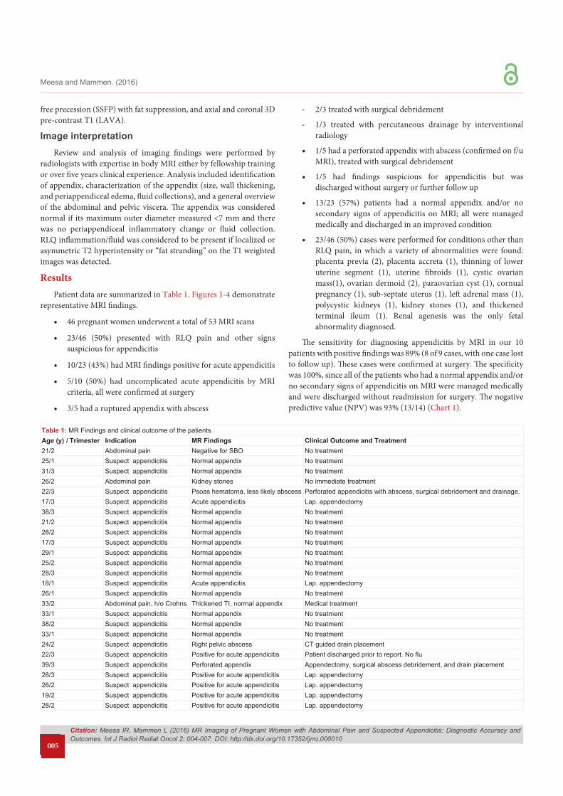
The sensitivity for diagnosing appendicitis by MRI in our 10 patients with positive findings was 89% (8 of 9 cases, with one case lost to follow up). These cases were confirmed at surgery. The specificity was 100%, since all of the patients who had a normal appendix and/or no secondary signs of appendicitis on MRI were managed medically and were discharged without readmission for surgery. The negative predictive value (NPV) was 93% (13/14) (Chart 1).
free precession (SSFP) with fat suppression, and axial and coronal 3D pre-contrast T1 (LAVA).
Image interpretationReview and analysis of imaging findings were performed by
radiologists with expertise in body MRI either by fellowship training or over five years clinical experience. Analysis included identification of appendix, characterization of the appendix (size, wall thickening, and periappendiceal edema, fluid collections), and a general overview of the abdominal and pelvic viscera. The appendix was considered normal if its maximum outer diameter measured <7 mm and there was no periappendiceal inflammatory change or fluid collection. RLQ inflammation/fluid was considered to be present if localized or asymmetric T2 hyperintensity or “fat stranding” on the T1 weighted images was detected.
Results Patient data are summarized in Table 1. Figures 1-4 demonstrate
representative MRI findings.
• 46 pregnant women underwent a total of 53 MRI scans
• 23/46 (50%) presented with RLQ pain and other signs suspicious for appendicitis
• 10/23 (43%) had MRI findings positive for acute appendicitis
• 5/10 (50%) had uncomplicated acute appendicitis by MRI criteria, all were confirmed at surgery
• 3/5 had a ruptured appendix with abscess
Table 1: MR Findings and clinical outcome of the patients.Age (y) I Trimester Indication MR Findings Clinical Outcome and Treatment21/2 Abdominal pain Negative for SBO No treatment25/1 Suspect appendicitis Normal appendix No treatment31/3 Suspect appendicitis Normal appendix No treatment26/2 Abdominal pain Kidney stones No immediate treatment22/3 Suspect appendicitis Psoas hematoma, less likely abscess Perforated appendicitis with abscess, surgical debridement and drainage.17/3 Suspect appendicitis Acute appendicitis Lap. appendectomy38/3 Suspect appendicitis Normal appendix No treatment21/2 Suspect appendicitis Normal appendix No treatment28/2 Suspect appendicitis Normal appendix No treatment17/3 Suspect appendicitis Normal appendix No treatment29/1 Suspect appendicitis Normal appendix No treatment25/2 Suspect appendicitis Normal appendix No treatment28/3 Suspect appendicitis Normal appendix No treatment18/1 Suspect appendicitis Acute appendicitis Lap. appendectomy26/1 Suspect appendicitis Normal appendix No treatment33/2 Abdominal pain, h/o Crohns Thickened TI, normal appendix Medical treatment33/1 Suspect appendicitis Normal appendix No treatment38/2 Suspect appendicitis Normal appendix No treatment33/1 Suspect appendicitis Normal appendix No treatment24/2 Suspect appendicitis Right pelvic abscess CT guided drain placement22/3 Suspect appendicitis Positive for acute appendicitis Patient discharged prior to report. No flu39/3 Suspect appendicitis Perforated appendix Appendectomy, surgical abscess debridement, and drain placement28/3 Suspect appendicitis Positive for acute appendicitis Lap. appendectomy26/2 Suspect appendicitis Positive for acute appendicitis Lap. appendectomy19/2 Suspect appendicitis Positive for acute appendicitis Lap. appendectomy28/2 Suspect appendicitis Positive for acute appendicitis Lap. appendectomy
Citation: Meesa IR, Mammen L (2016) MR Imaging of Pregnant Women with Abdominal Pain and Suspected Appendicitis: Diagnostic Accuracy and Outcomes. Int J Radiol Radiat Oncol 2: 004-007. DOI: http://dx.doi.org/10.17352/ijrro.000010
Meesa and Mammen. (2016)
006
DiscussionAcute appendicitis is the most common surgical emergency in
pregnancy. Diagnosis is difficult because this condition presents with signs and symptoms that are not unusual with pregnancy (e.g., nausea, vomiting, and leukocytosis). The enlarged uterus, especially in the 2nd and 3rd trimester, can elevate the anterior peritoneum and displace the appendix superiorly, making US and clinical diagnosis more difficult [2,9,10].
Traditionally, US and CT are the proven imaging modalities for diagnosing and excluding acute appendicitis in non-pregnant women with reported sensitivities and specificities greater than 85% and 95%, respectively. The American College of Radiology Appropriateness Criteria rates US of the RLQ with graded compression highest in this
Figure 1: 2nd trimester with RLQ pain. MRI demonstrated a normal appendix (arrow).
Figure 2: 3rd trimester with acute retrocecal appendicitis (arrow).
Figure 3: 3rd trimester with appendicitis and appendicoliths (arrows).
Figure 4b: Coronal image of RLQ abscess (arrows).
Figure 4c: CT guided drain placement in RLQ abscess (arrow).
Figure 4a: 2nd trimester with a RLQ abscess (arrows).
Citation: Meesa IR, Mammen L (2016) MR Imaging of Pregnant Women with Abdominal Pain and Suspected Appendicitis: Diagnostic Accuracy and Outcomes. Int J Radiol Radiat Oncol 2: 004-007. DOI: http://dx.doi.org/10.17352/ijrro.000010
Meesa and Mammen. (2016)
007
clinical scenario. If US is non-diagnostic, MRI of the abdomen and pelvis without contrast is rated above CT as the next imaging step.7
With recent developments of ultrafast sequences and coil technology which allow imaging with less motion and faster scan times, MRI is increasingly being used in the obstetric setting for both maternal and fetal diseases [8]. Excellent soft-tissue contrast resolution and multi-planar imaging capability lessens the need for contrast administration. There is no clinical or experimental evidence of teratogenic or other adverse biologic effects of MRI during pregnancy [11,12].
In this series of 23 pregnant women suspected of having appendicitis, 10 patients had positive MRI findings in the RLQ. MRI accurately detected 8 cases of acute appendicitis with one case diagnosed on the second scan and one case lost to follow up. MRI was successfully used to follow up all three patients with ruptured appendicitis with abscess after drain placement. In these patients, the appendix was not directly visualized on the MRI, but inflammatory changes, including fluid collections in the RLQ, were seen.
In the remaining 13 pregnant women with RLQ pain, the appendix was identified as being normal and/or there was absence of periappendiceal inflammatory change. None of these patients underwent laparotomy after a normal MRI reading.
Diagnostic imaging offers the clinician more confidence in clinical dilemmas such as appendicitis during pregnancy as there is a greater impact if the diagnosis is missed or if the patient is over treated since a fetus is also involved. MRI reduces possible psychological pressures in pregnant women who undergo MRI vs. CT because they do not have to worry about the effect of potentially hazardous radiation exposure to the fetus (6). The single case of late diagnosis of appendicitis in our study demonstrates how difficult this diagnosis can be by clinical exam, even in the setting of perforation.
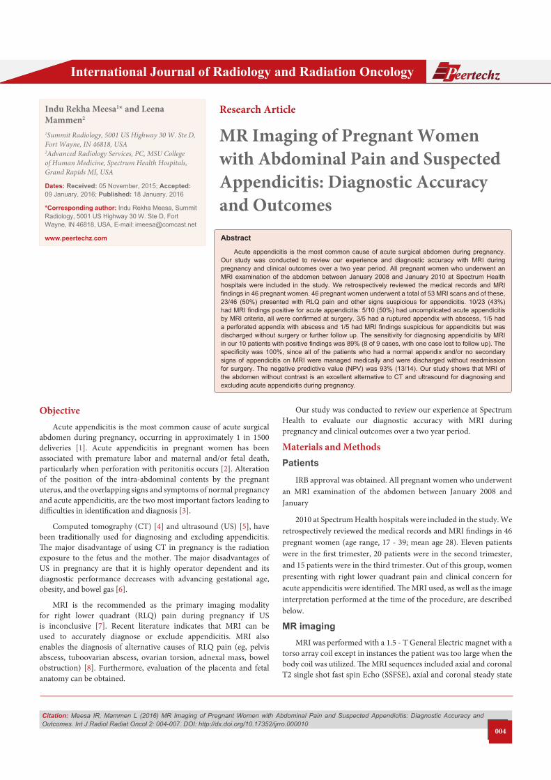
Figure 4d: Post abscess drain MRI with resolution of RLQ fluid collection (arrow).
Chart 1Sensitivity 89%Specificity 100%NPV 93%
In our study, the sensitivity and specificity for diagnosing appendicitis by MRI was 89% and 100%, respectively. The negative predictive value of MRI was 93%. Interestingly, positive laparotomy rates for appendicitis during pregnancy are reported to be approximately 50%, which is similar to the incidence of appendicitis (43%) by MRI in this patient group.
ConclusionOur study shows that MRI of the abdomen without contrast is
an excellent alternative to CT and ultrasound for diagnosing and excluding acute appendicitis during pregnancy. When ultrasound does not reveal the appendix, MRI offers another option to confirm a normal appendix without radiation risk. Our experience is concordant with the initial experiences published on this imaging technique and has the potential to significantly decrease negative laparotomy rates. Alternative etiologies for the patient’s pain were also discovered which provided valuable information. This study also reminds us that imaging results must be correlated closely with the clinical exam, particularly if there is clinical discordance.
Interestingly, the results from our study also show a novel application of MRI to follow up abdominal/pelvic abscesses in pregnant patients with ruptured appendicitis.
References1. Babaknia A, Parsa H, Woodruff JD (1977) Appendicitis during pregnancy.
3. Mourad J, Elliott JP, Erickson L, Lisboa L (2000) Appendicitis in pregnancy: new information that contradicts long-held clinical beliefs. Am J Obstet Gynecol 182: 1027-1029.
4. Ames Castro M, Shipp TD, Castro EE, Ouzounian J, Rao P (2001) The use of helical computed tomography in pregnancy for the diagnosis of acute appendicitis. Am J Obstet Gynecol 184: 954-957.
5. Rioux M (1992) Sonographic detection of the normal and abnormal appendix. AJR 158: 773-778.
6. Lodewijk P Cobben, Groot I, Haans L, Johan G Blickman, et al. (2004) Puylaert. MRI for Clinically Suspected Appendicitis During Pregnancy. American Journal of Roentgenology 183: 671-675.
7. Rosen MP, Ding A, Blake MA, Baker ME, Cash BD, et al. (2007) American College of Radiology Appropriateness Criteria: Right lower quadrant pain, fever, leukocytosis in the pregnant woman. Revised 2007.
8. Oto A, Ernst RD, Shah R, Koroglu M, Chaljub G, et al. (2005) Right-Lower-Quadrant Pain and Suspected Appendicitis in Pregnant Women: Evaluation with MR Imaging-Initial Experience. Radiology 234: 445-451.
9. Anderson B, Nielsen TF (1999) Appendicitis in pregnancy: diagnosis, management and complications. Acta Obstet Gynecol Scand 78: 758-762.
10. Maslovitz S, Gutman G, Lessing JB, Kupdermine MJ, Gamzu R (2003) The significance of clinical signs and blood indices for the diagnosis of appendicitis during pregnancy. Gynecology Obstet Invest 56: 188-191.
11. Nagayama M, Watanabe Y, Okumura A, Amoh Y, Nakashita S, et al. (2002) Fast MR imaging in obstetrics. RadioGraphics 22: 563-582.
12. Jung SE, Byun JY, Lee JM, Rha SE, Kim H, et al. (2001) MR imaging of maternal diseases in pregnancy. AJR Am J Roentgenol 177: 1293-1300.