1 Mr.Epithelium’s Anatomy and Physiology Test SSSS 2020 KEY One 8.5 x 11 notes sheet is allowed, along with 1 non-programmable calculator dedicated to computation. Good luck! Name(s): ______________________________ , ______________________________ Points: 180 Instructions: - You have 40 minutes to complete this test packet. o There are 9 stations, with 5 minutes per station allotted. - One 8.5 x 11 notes sheet is allowed, along with 1 non-programmable calculator dedicated to computation per person. - Point values are denoted using brackets [ ] in front of each question. - Tiebreakers are the total score of the following stations, in order: 6, 8, 1 - Good luck! Have fun, feedback is greatly appreciated (PM Mr.Epithelium on Scioly).
Transcript
1
Mr.Epithelium’s Anatomy and Physiology Test SSSS 2020
KEY
One 8.5 x 11 notes sheet is allowed, along with 1 non-programmable calculator dedicated to computation.
- You have 40 minutes to complete this test packet.
o There are 9 stations, with 5 minutes per station allotted.
- One 8.5 x 11 notes sheet is allowed, along with 1 non-programmable calculator
dedicated to computation per person.
- Point values are denoted using brackets [ ] in front of each question.
- Tiebreakers are the total score of the following stations, in order: 6, 8, 1
- Good luck! Have fun, feedback is greatly appreciated (PM Mr.Epithelium on
Scioly).
2
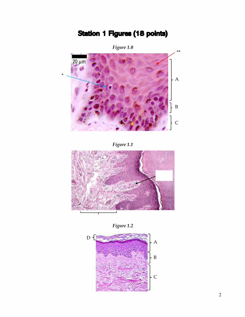
Station 1 Figures (18 points)
Figure 1.0
Figure 1.1
Figure 1.2
A
B
C
A
B
C
D
*
**
3
Station 1 (18 points) Questions 1-7 refer to Figure 1.0.
1. [1] What type of cell is indicated by the blue arrow (*)? Melanocyte 2. [1] What type of cell is indicated by the red arrow (**)? Keratinocyte 3. [1] What does label A indicate? Stratum spinosum 4. [1] What does label B indicate? Stratum basale 5. [1] What does label C indicate? Papillary layer (of dermis) 6. [1] What type of connection occurs between layers B and C? Hemidesmosomes 7. [1] Using the measurement given, estimate the width of the cell labeled with the
blue arrow. 5-7 µm Questions 8-9 refer to Figure 1.1.
8. [1] What skin receptor is shown in the picture? Meissner/tactile corpuscle a. [1] What type of touch does this receptor sense? Light touch (accept
similar) b. [1] What type of skin are these receptors most concentrated in? Thick
9. [1] What type of tissue makes up the layer indicated by the bracket? Areolar tissue
Questions 10-13 refer to Figure 1.2.
10. [1] What type of epithelium makes up label A? Keratinized stratified squamous epithelium
11. [1] What type of tissue makes up label C? Dense irregular connective tissue 12. [1] What does label D indicate? Stratum corneum
a. [1] An acidic secretion sometimes covers label D. What is this called? Acid mantle
b. [1] What is the function of this secretion? (up to 1 point) *(+1) Create acidic environment *(+1) Slows microbial (bacterial) growth
13. [1] What are the finger-like projections in label B called? Dermal papillae a. [1] Name one function of these projections. (up to 1 point) *(+1) Increases
surface area for exchange of nutrients, gases, and wastes *(+1) Creates strong connection between the papillary dermis and epidermis.
4
Station 2 Figures (20 points)
Figure 2.0
(a) (b) (c) Word Bank Anagen Club Spinosum Basale Granulosum Telogen Catagen Lanugo Terminal Corneum Lucidum Vellus
5
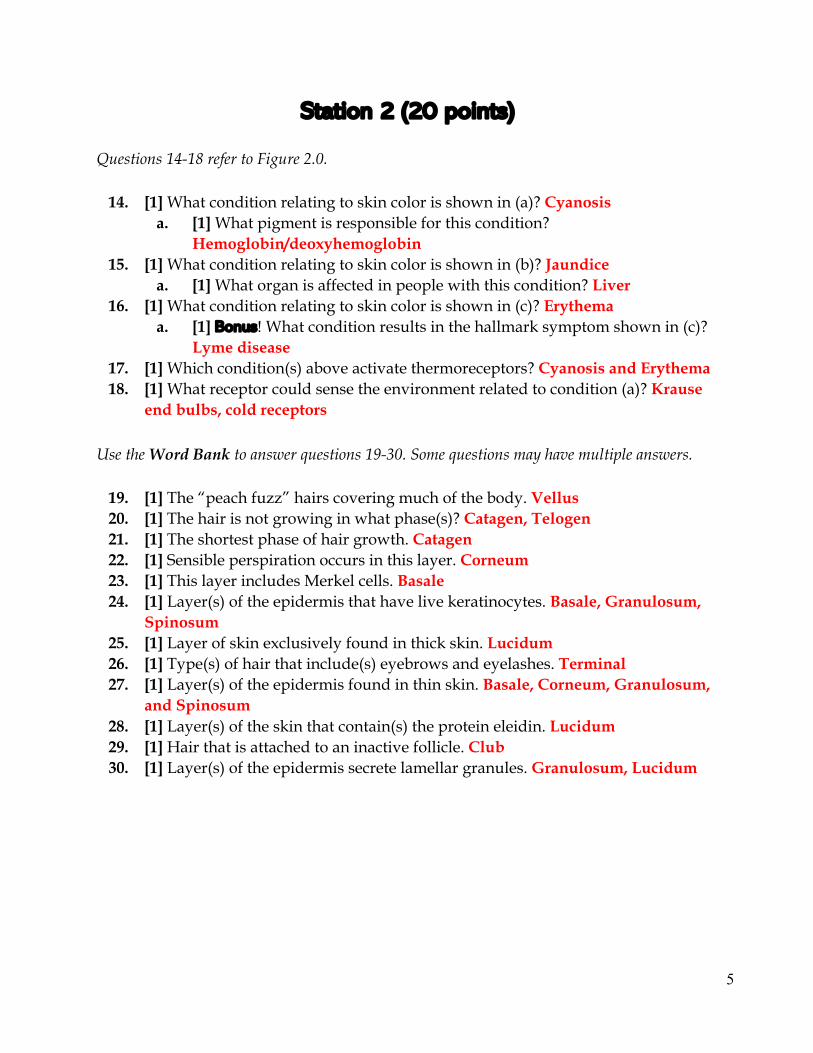
Station 2 (20 points) Questions 14-18 refer to Figure 2.0.
14. [1] What condition relating to skin color is shown in (a)? Cyanosis a. [1] What pigment is responsible for this condition?
Hemoglobin/deoxyhemoglobin 15. [1] What condition relating to skin color is shown in (b)? Jaundice
a. [1] What organ is affected in people with this condition? Liver 16. [1] What condition relating to skin color is shown in (c)? Erythema
a. [1] Bonus! What condition results in the hallmark symptom shown in (c)? Lyme disease
17. [1] Which condition(s) above activate thermoreceptors? Cyanosis and Erythema 18. [1] What receptor could sense the environment related to condition (a)? Krause
end bulbs, cold receptors
Use the Word Bank to answer questions 19-30. Some questions may have multiple answers.
19. [1] The “peach fuzz” hairs covering much of the body. Vellus 20. [1] The hair is not growing in what phase(s)? Catagen, Telogen 21. [1] The shortest phase of hair growth. Catagen 22. [1] Sensible perspiration occurs in this layer. Corneum 23. [1] This layer includes Merkel cells. Basale 24. [1] Layer(s) of the epidermis that have live keratinocytes. Basale, Granulosum,
Spinosum 25. [1] Layer of skin exclusively found in thick skin. Lucidum 26. [1] Type(s) of hair that include(s) eyebrows and eyelashes. Terminal 27. [1] Layer(s) of the epidermis found in thin skin. Basale, Corneum, Granulosum,
and Spinosum 28. [1] Layer(s) of the skin that contain(s) the protein eleidin. Lucidum 29. [1] Hair that is attached to an inactive follicle. Club 30. [1] Layer(s) of the epidermis secrete lamellar granules. Granulosum, Lucidum
6
Station 3 Figures (22 points)
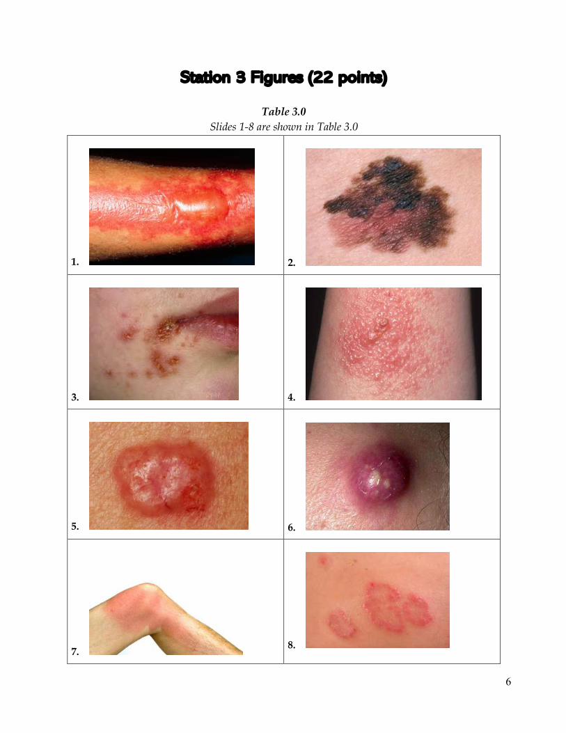
Table 3.0 Slides 1-8 are shown in Table 3.0
1.
2.
3.
4.
5.
6.
7.
8.
7
Station 3 (22 points) Questions 31-40 refer to Table 3.0. Note: Disease, disorder, and injuries are used synonymously in this section
31. [1] What disease is shown in slide 1? 2nd degree burn a. [1] What layers of the skin are affected by this disease? Epidermis and
dermis 32. [1] Does slide 2 show malignant melanoma? Yes
a. [2] Use one letter of the ABCD’s of melanoma to back up your answer. +2 for any ONE of the following: A- the melanoma is asymmetrical; B- the border of the melanoma is irregular (some parts are patchy, faded, etc); C- the color varies throughout (red, black, lighter shades, etc); D- the diameter of the melanoma is greater than 6 mm.
33. [1] What disease is shown in slide 3? Impetigo a. [1] Who is most at risk for this disease? Children
34. [1] What disease is shown in slide 4? Poison ivy allergy (Poison ivy rash) a. [1] What is the primary cause of this disease? Be specific! Contact with the
oily resin urushiol (Note: on any object or from the plant itself) 35. [1] What disease is shown in slide 5? Basal cell carcinoma
a. [1] Name a prevention method for this disease. Use sunscreen (SPF > 30), avoid tanning beds, wear protective clothing, other acceptable answers
36. [1] What disease is shown in slide 6? Carbuncle a. [1] What pathogen can be a cause of this disease? Staphylococcus aureus /
Streptococcus pyogenes 37. [1] What disease is shown in slide 7? 1st degree burn
a. [2] What layers of the dermis are affected by this disease, if any at all? None
38. [1] What disease is shown in slide 8? Eczema a. [1] What specific type of this disease is shown in the slide? Nummular
eczema 39. [2] Which slide(s) can be caused by bacteria? Slides 3 and 6 40. [2] Which slide(s) can be caused by overexposure to sunlight? Slides 2, 5, and 7
8
Station 4 Figures (20 points)
Figure 4.2
A
B
C
Figure 4.0
A
B
C
Figure 4.1
A
B
C
9
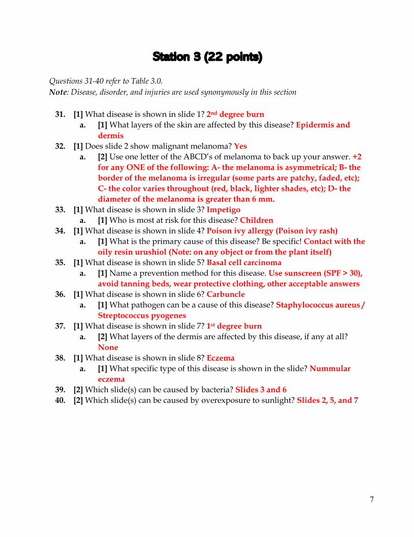
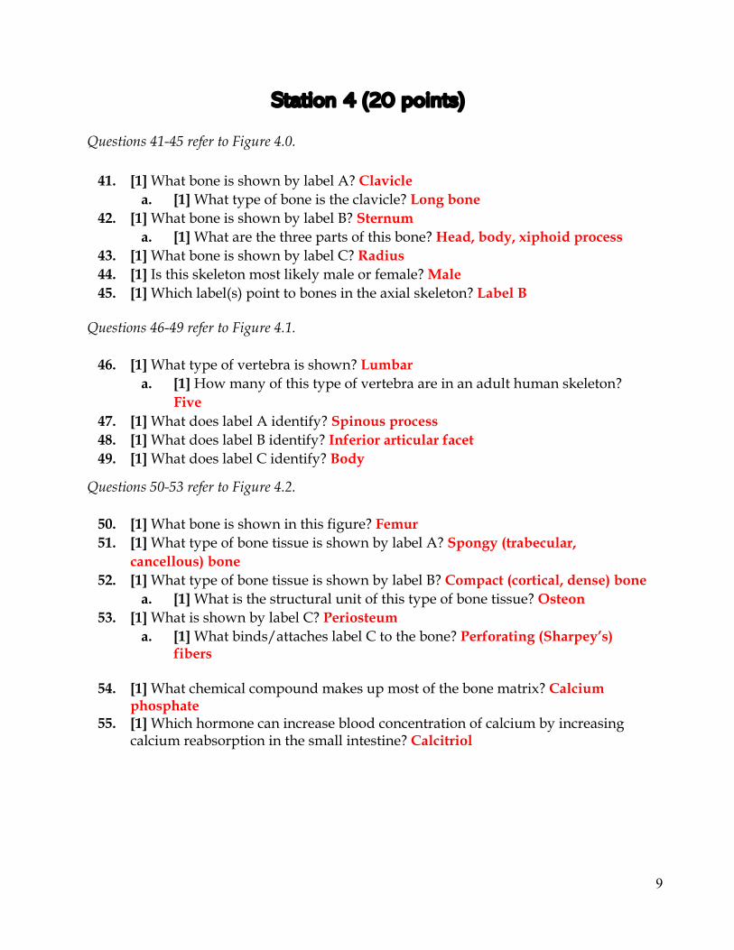
Station 4 (20 points) Questions 41-45 refer to Figure 4.0.
41. [1] What bone is shown by label A? Clavicle a. [1] What type of bone is the clavicle? Long bone
42. [1] What bone is shown by label B? Sternum a. [1] What are the three parts of this bone? Head, body, xiphoid process
43. [1] What bone is shown by label C? Radius 44. [1] Is this skeleton most likely male or female? Male 45. [1] Which label(s) point to bones in the axial skeleton? Label B
Questions 46-49 refer to Figure 4.1.
46. [1] What type of vertebra is shown? Lumbar a. [1] How many of this type of vertebra are in an adult human skeleton?
Five 47. [1] What does label A identify? Spinous process 48. [1] What does label B identify? Inferior articular facet 49. [1] What does label C identify? Body
Questions 50-53 refer to Figure 4.2.
50. [1] What bone is shown in this figure? Femur 51. [1] What type of bone tissue is shown by label A? Spongy (trabecular,
cancellous) bone 52. [1] What type of bone tissue is shown by label B? Compact (cortical, dense) bone
a. [1] What is the structural unit of this type of bone tissue? Osteon 53. [1] What is shown by label C? Periosteum
a. [1] What binds/attaches label C to the bone? Perforating (Sharpey’s) fibers
54. [1] What chemical compound makes up most of the bone matrix? Calcium
phosphate 55. [1] Which hormone can increase blood concentration of calcium by increasing
calcium reabsorption in the small intestine? Calcitriol
10
Station 5 Figures (22 points)
Figure 5.0
Figure 5.1
A
B
C D E F
1 2 3
G H
I
A
11
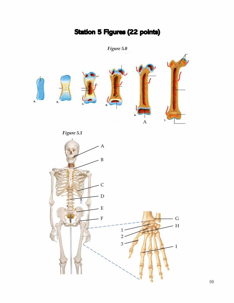
Station 5 (22 points) Questions 56-60 refer to Figure 5.0. Stages a-f are shown.
56. [1] What type of ossification is shown? Endochondral ossification 57. [1] In which stage does the primary ossification center first appear? Stage c. 58. [1] In which stage does the secondary ossification center first appear? Stage d. 59. [1] What does label A indicate? Epiphyseal plate 60. [1] The medullary cavity is shown in Stage f. What bone cell is mainly
responsible for the formation of the medullary cavity? Osteoclasts a. [1] What ion do these cells release to aide in their main function?
Hydrogen ions (H+)
Questions 61-70 refer to Figure 5.1. Question number
Label Structural
classification Functional
classification Bones involved
61. [2] A [0.5] Fibrous [0.5] Synarthrosis [1] Maxilla
62. [2] B [0.5] Synovial [0.5] Diarthrosis
(gliding) [1] Sternum,
clavicle
63. [2] D [0.5] Cartilaginous [0.5]
Amphiarthrosis
[1] Be specific! 1st, 2nd lumbar
vertebrae (L1, L2)
64. [2] E [0.5] Synovial [0.5] Diarthrosis
(gliding) [1] Ilium, sacrum
65. [2] F [0.5] Synovial [0.5] Diarthrosis (ball and socket)
[1] Ilium, ischium, pubis, femur
66. [1] What type of cartilage is shown in Label C? Hyaline cartilage 67. [1] What bone is Label 1 pointing to? Lunate
a. [1] What bone is Label 2 pointing to? Capitate 68. [1] What type of synovial joint is Label G? Pivot 69. [1] What type of bone is Label 3 pointing to? Sesamoid bone 70. [1] What Label(s) (A-I) are pointing to areas of the appendicular skeleton
exclusively, if any? Labels F, G, H, I
12
Station 6 Figures (18 points)
Figure 6.0
Figure 6.1
*
13
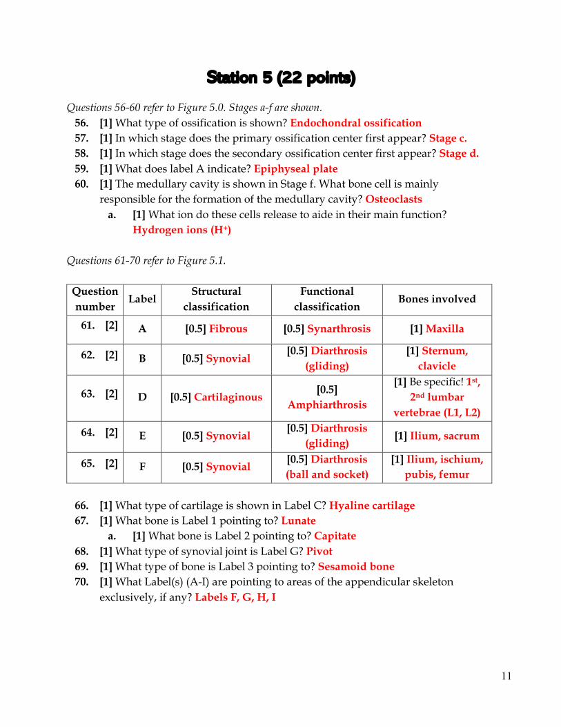
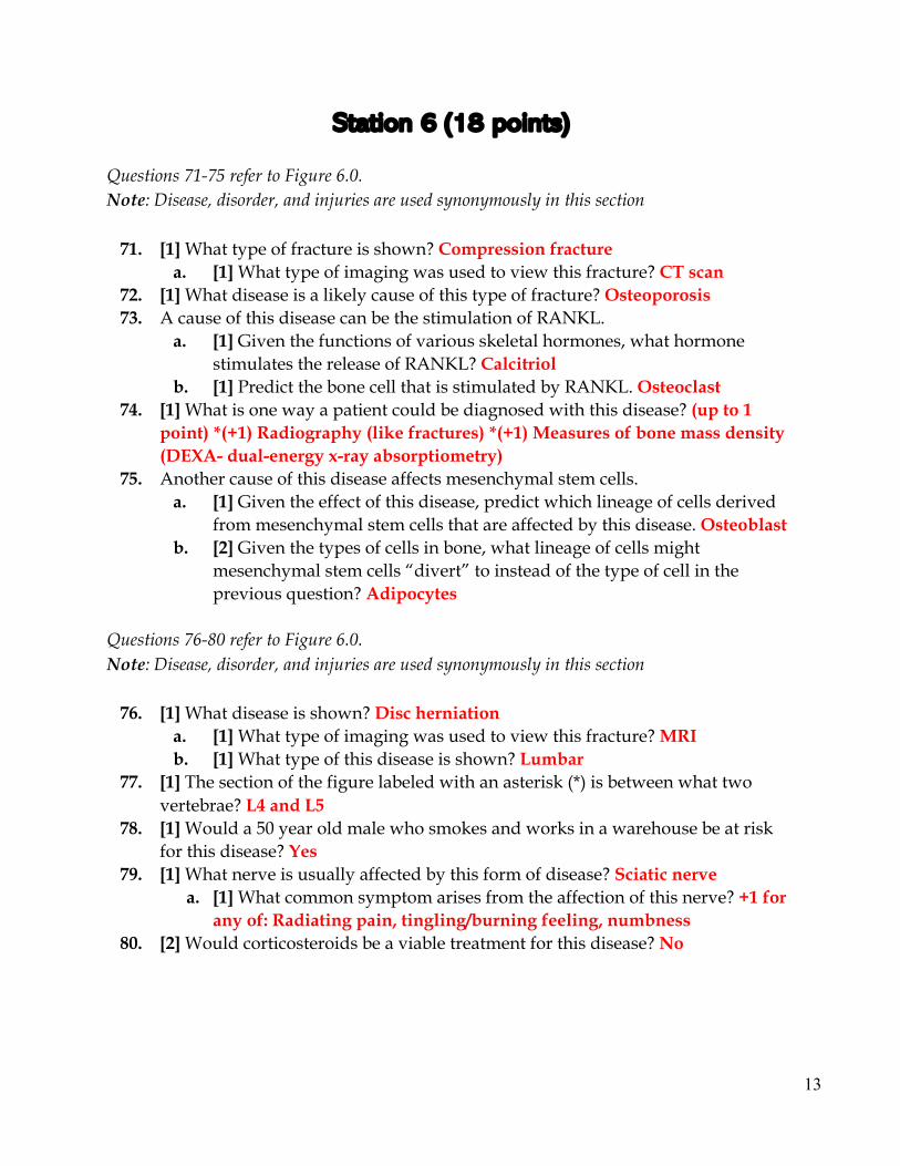
Station 6 (18 points) Questions 71-75 refer to Figure 6.0. Note: Disease, disorder, and injuries are used synonymously in this section
71. [1] What type of fracture is shown? Compression fracture a. [1] What type of imaging was used to view this fracture? CT scan
72. [1] What disease is a likely cause of this type of fracture? Osteoporosis 73. A cause of this disease can be the stimulation of RANKL.
a. [1] Given the functions of various skeletal hormones, what hormone stimulates the release of RANKL? Calcitriol
b. [1] Predict the bone cell that is stimulated by RANKL. Osteoclast 74. [1] What is one way a patient could be diagnosed with this disease? (up to 1
point) *(+1) Radiography (like fractures) *(+1) Measures of bone mass density (DEXA- dual-energy x-ray absorptiometry)
75. Another cause of this disease affects mesenchymal stem cells. a. [1] Given the effect of this disease, predict which lineage of cells derived
from mesenchymal stem cells that are affected by this disease. Osteoblast b. [2] Given the types of cells in bone, what lineage of cells might
mesenchymal stem cells “divert” to instead of the type of cell in the previous question? Adipocytes
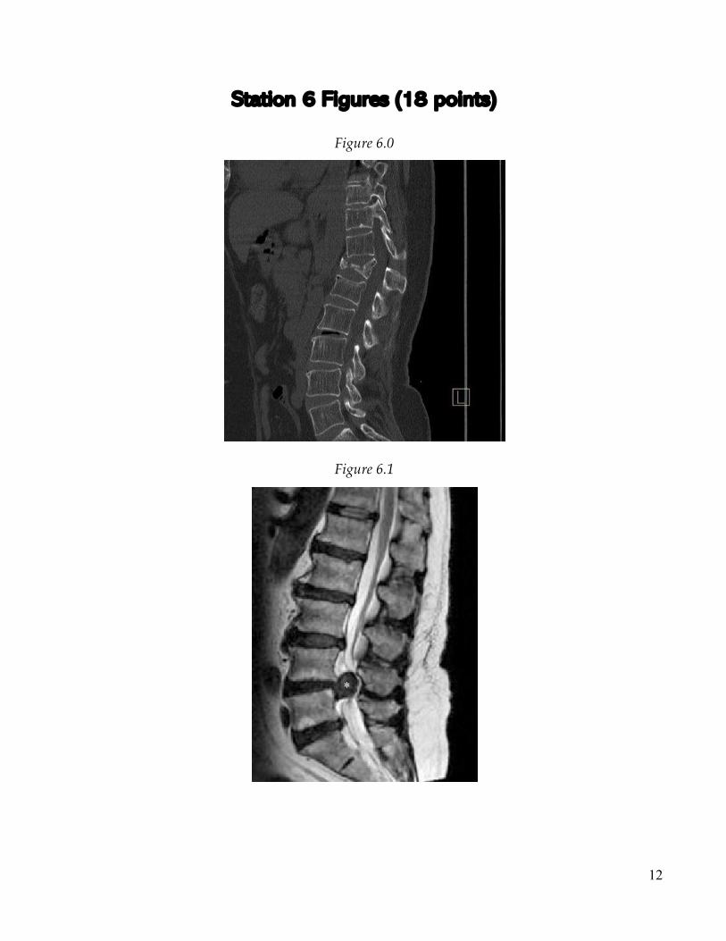
Questions 76-80 refer to Figure 6.0. Note: Disease, disorder, and injuries are used synonymously in this section
76. [1] What disease is shown? Disc herniation a. [1] What type of imaging was used to view this fracture? MRI b. [1] What type of this disease is shown? Lumbar
77. [1] The section of the figure labeled with an asterisk (*) is between what two vertebrae? L4 and L5
78. [1] Would a 50 year old male who smokes and works in a warehouse be at risk for this disease? Yes
79. [1] What nerve is usually affected by this form of disease? Sciatic nerve a. [1] What common symptom arises from the affection of this nerve? +1 for
any of: Radiating pain, tingling/burning feeling, numbness 80. [2] Would corticosteroids be a viable treatment for this disease? No
14
Station 7 Figures (20 points)
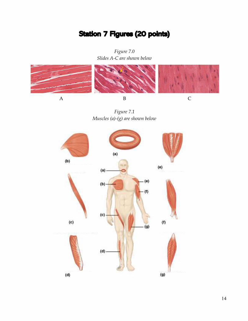
Figure 7.0
Slides A-C are shown below
Figure 7.1
Muscles (a)-(g) are shown below
A
B
C
15
Station 7 (20 points) Questions 81-90 refer to Figure 7.0.
81. [1] What type of muscle tissue is shown in Slide A? Skeletal 82. [1] What type of muscle tissue is shown in Slide B? Cardiac 83. [1] What type of muscle tissue is shown in Slide C? Smooth 84. [1] What structure is the arrow in Slide B pointing to? Nucleus 85. [1] In cardiac muscle, what protein does calcium bind to initiate contraction?
Troponin 86. [1] In smooth muscle, what protein does calcium bind to initiate contraction?
Calmodulin 87. [1] Which Slide(s) have terminal cisternae? A 88. [1] Which Slide(s) can be controlled by pacemaker cells? B, C 89. [1] Which Slide(s) is/are striated? A, B 90. [1] Which Slide(s) can undergo tetanus? A, C
Questions 91-100 refer to Figure 7.1.
91. [1] What muscle is shown in (a)? Orbicularis oris 92. [1] What muscle fiber organization is shown in (a)? Circular 93. [1] What muscle is shown in (b)? Pectoralis major 94. [1] What muscle fiber organization is shown in (b)? Convergent 95. [1] What muscle is shown in (c)? Sartorius 96. [1] What muscle fiber organization is shown in (c)? Parallel 97. [1] What muscle is shown in (e)? Deltoid 98. [1] What muscle is shown in (f)? Biceps brachii 99. [1] What muscle is shown in (g)? Rectus femoris 100. [1] How many muscles in Figure 7.1 have an origin within the axial skeleton? 2
16
Station 8 Figures (21 points)
Figure 8.0
Figure 8.1
Three different scenarios (A-C) are shown below.
A
B
C
1 2
17
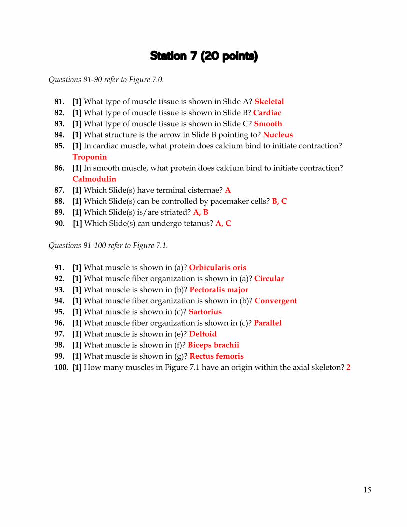
Station 8 (21 points) Questions 101-105 refer to Figure 8.0.
101. [1] What does Label B indicate? Z line (disc) a. [2] There are two proteins that are anchored at Label B. What are they? +1
each: Actin and titin 102. [1] What does Label D indicate? M line
a. [1] There is one protein that is anchored at Label D. What is it? Myosin 103. [1] What does Label 1 indicated? A band
a. [1] Does Label 1 shorten during contraction? No b. [1] Does Label 1 contain thin filaments? Yes
104. [1] What does Label 2 indicate? I band a. [1] Does Label 2 shorten during contraction? Yes b. [1] Does Label 2 contain thin filaments? Yes
105. [1] Is the H zone visible in this Figure? Yes Questions 106-110 refer to Figure 8.1.
106. [1] What type of contraction is shown in scenario A? Isometric a. [1] What is the relationship between the value of tension produced by the
muscle and the load? They are equal 107. [1] What type of contraction is shown in scenario B? Concentric
a. [1] What is the relationship between the value of tension produced by the muscle and the load? Tension produced is greater than load
108. [1] What type of contraction is shown in scenario C? Eccentric a. [1] What is the relationship between the value of tension produced by the
muscle and the load? Load is greater than tension produced 109. [1] What muscle is shown in this Figure? Biceps brachii
a. [1] What is the origin of this muscle shown in the Figure (name a bone)? Scapula
110. [1] What scenarios are examples of an isotonic contraction? Scenarios B and C
18
Station 9 Figures (19 points)
Figure 9.0
Figure 9.1
Figure 9.2
19
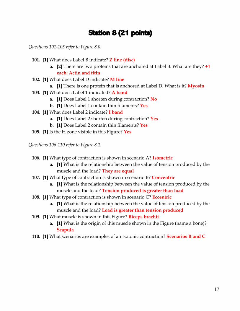
Station 9 (19 points) Questions 111-115 refer to Figure 9.0.
111. [1] Is the injury shown a strain or sprain? Sprain a. [1] Sprains and strains can be difficult to tell apart due to symptoms.
Name one symptom that strains and sprains have in common. +1 for ANY of: *Pain around affected joint *Swelling around affected joint *Range of motion is limited
b. [1] What structure do these injuries affect? Ligaments 112. [1] A special kind of fracture can arise when a bone is pulled rather sharply
during the injury. What is it? Avulsion 113. [1] Where in the body was this Figure taken? Knee, legs, etc. 114. [2] What grade of this injury occurs when the affected structure has been
completely torn? Grade 3 115. A common treatment option for mild forms of this injury is R.I.C.E.
a. [2] What does R.I.C.E. stand for? +0.5 each for: R- rest, I- ice, C- compression, E- elevation
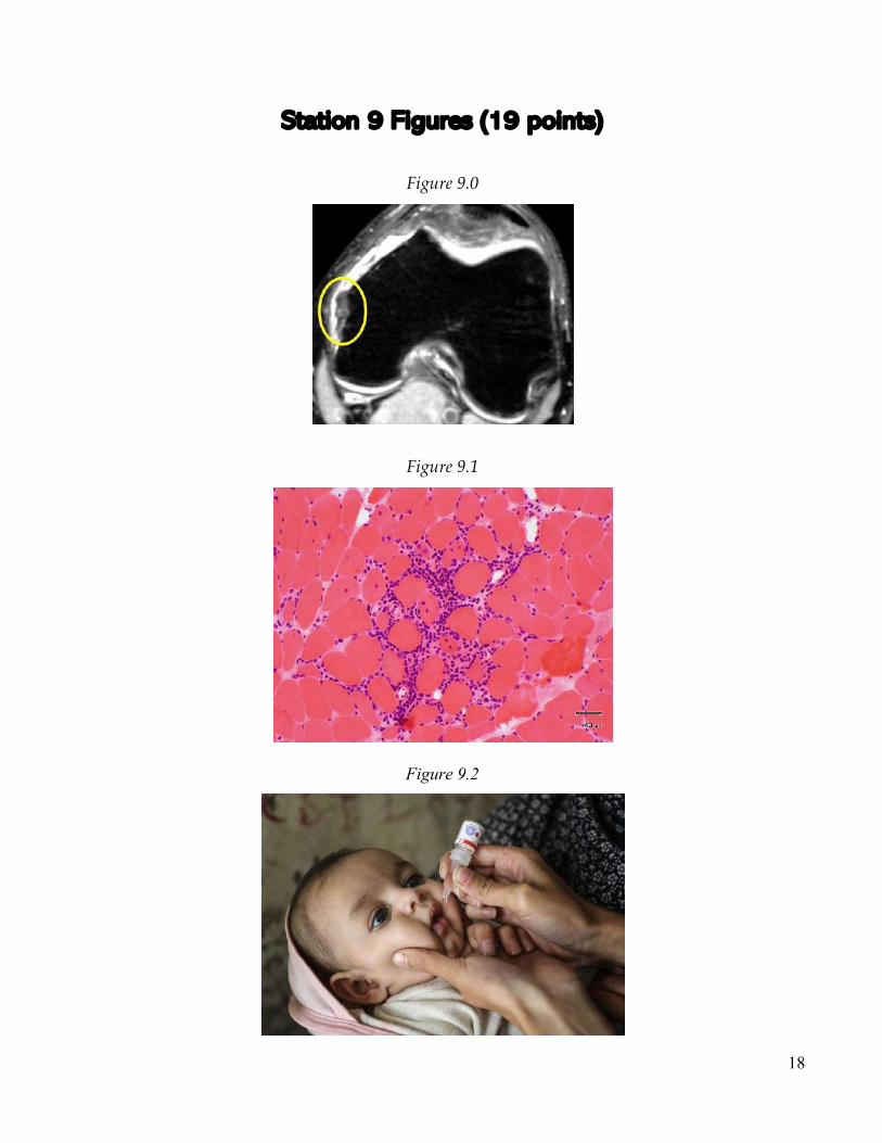
Questions 116-117 refer to Figure 9.1.
116. [1] What disease is shown in this Figure? Myositis a. [2] What specific form of this disease is shown? Inclusion body myositis b. [1] What is a common symptom of this form? Progressive muscle
weakness 117. [1] What type of diseases can be a cause of this disease? Examples include
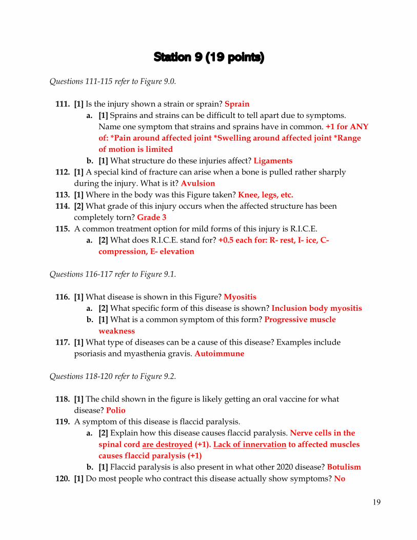
psoriasis and myasthenia gravis. Autoimmune Questions 118-120 refer to Figure 9.2.
118. [1] The child shown in the figure is likely getting an oral vaccine for what disease? Polio
119. A symptom of this disease is flaccid paralysis. a. [2] Explain how this disease causes flaccid paralysis. Nerve cells in the
spinal cord are destroyed (+1). Lack of innervation to affected muscles causes flaccid paralysis (+1)
b. [1] Flaccid paralysis is also present in what other 2020 disease? Botulism 120. [1] Do most people who contract this disease actually show symptoms? No


![OOO OOOOnnnnnnn]]]]]]]llllSSSSSSS6666666«««««««ssss ...](https://static.documents.pub/doc/80x56/6159386e91a31b7a2427aef2/ooo-oooonnnnnnnllllsssssss6666666ssss.jpg)


