Page 1
1
Ms # M0:02138 Revised: July 25, 2000
Regulatable Expression of p21-Activated Kinase-1 Promotes Anchorage-Independent Growth
and Abnormal Organization of Mitotic Spindles in Human Epithelial Breast Cancer Cells*
Ratna K. Vadlamudi, Liana Adam, Rui-An Wang, Mahitosh Mandal, Diep Nguyen, Aysegul
Sahin, Jonathan Chernoff#, Mien-Chie Hung, and Rakesh Kumar # #
From The University of Texas M. D. Anderson Cancer Center, Houston, Texas 77030; and #The
Fox Chase Cancer Center, Philadelphia, Pennsylvania 19100.
# #To whom correspondence and requests for reprints should be addressed at Department of
Molecular and Cellular Oncology, The University of Texas M. D. Anderson Cancer Center-
36, 1515 Holcomble Blvd., Houston, TX 77030. E-mail: [email protected]
*This study was supported by the NIH grants CA 80066 and CA65746, and the Breast and
Ovarian Cancer Research Programs of the University of Texas M. D. Anderson Cancer
Center (to R.K.), and the Cancer Center Core grant CA16672.
Running Title: Pak1 regulation of anchorage-independent growth
Key Words: Pak1, Signaling, Anchorage-independent growth, Breast Cancer.
Copyright 2000 by The American Society for Biochemistry and Molecular Biology, Inc.
JBC Papers in Press. Published on August 16, 2000 as Manuscript M002138200 by guest on M
arch 22, 2018http://w
ww
.jbc.org/D
ownloaded from
Page 2
2
The abbreviations used are HER, human epidermal growth factor receptor; Pak1, P21-
activated kinase, HRG, heregulin beta-1; JNK, Jun-N-terminal kinase; ERK, extracellular
signal related protein kinases.
ABSTRACT
Stimulation of growth factor signaling has been implicated in the development of
invasive phenotypes and the activation of p21-activated kinase (Pak1) in human breast
cancer cells (Adam et al. J. Biol. Chem. 273, 28238-28246, 1998; Adam et al. J. Biol.
Chem. 275, 12041-12050, 2000). To study the role of Pak1 in the regulation of motility
and growth of breast epithelial cells, we developed human epithelial MCF-7 clones that
over expressed the kinase-active T423E Pak1 mutant under an inducible tetracycline
promoter or that stably expressed the kinase-active H83, 86L Pak1 mutant, which is
deficient in small GTPase binding site. The expression of both T423E and H83, 86L Pak1
mutants in breast epithelial cells was accompanied by increased cell motility without any
apparent effect on the growth rate of cells. The T423E Pak1 mutant was primarily localized
to filopodia and the H83, 86L Pak1 mutant was primarily localized to ruffles. Cells
expressing T423E Pak1 exhibited a regulatable stimulation of mitogen-activated protein
kinase and Jun-N-terminal kinase activities. The expression of kinase-active Pak1 mutants
significantly stimulated anchorage-independent growth of cells in soft agar in a preferential
mitogen-acivated protein kinase sensitive manner. In addition, regulatable expression of
kinase active Pak1 resulted in an abnormal organization of mitotic spindles characterized by
appearance of multiple spindle orientations. We also provide evidence to suggest a close
correlation between the status of Pak1 kinase activity and baseline invasiveness of human
by guest on March 22, 2018
http://ww
w.jbc.org/
Dow
nloaded from
Page 3
3
breast cancer cells and breast tumor grades. This study is the first demonstration of Pak1
regulation of anchorage-independent growth, potential Pak1 regulation of invasiveness and
abnormal organization of mitotic spindles of human epithelial breast cancer cells.
by guest on March 22, 2018
http://ww
w.jbc.org/
Dow
nloaded from
Page 4
4
INTRODUCTION
Breast cancer is one of the most common malignancies in the United States affecting one in nine women.
Overexpression of the human epidermal growth factor receptor-2 (HER 2, also known as c-erbB2 or c-neu) is
associated with increased progression and metastasis, an aggressive clinical course, and decreased disease-free
survival in human breast cancer patients’ (1). Accumulating evidence suggests that in addition to HER 2 over
expression, the heregulin (HRG, a combinatorial ligand for HER 3, HER 4) pathway be involved in the progression of
breast cancer cells to a more invasive phenotype (2-5). Exposure of cells to growth factors induces cytoskeleton
reorganization, lamellipodia formation, and membrane ruffling; such changes contribute to increased cell migration
and invasion (3, 6). Members of Rho family of the small GTPases Rac1, Cdc42 and RhoA have been implicated in the
regulation of cytoskeletal rearrangements; RhoA is involved in the maintenance of actin stress-fibers and focal
adhesion points; Rac1 in the formation of lamellipodia and membrane ruffles (7); and Cdc42 in the formation of
peripheral actin microspikes and filopodia (8). The small GTPases also regulate gene expression: Cdc42 and Rac1
activate Jun N-terminal kinase JNK and p38 mitogen-activated protein kinase (p38MAPK) pathways (9). Thus
GTPases Rac1, Cdc42 and Rho are implicated in cellular transformation (10, 11). Cdc42 and Rac1 family members also
activate extracellular signal-regulated protein kinases (ERKs) and ternary-complex factor (12). The signaling pathway
by which the small GTPases regulate their diverse cellular functions is an evolving area of investigation.
In mammalian cells, p21-activated kinases (Paks) are identified as one target of the small GTPases Cdc42 and
Rac1, and binding of GTP-bound GTPases to Pak1 stimulates its kinase activity via autophosphorylation (13).
Expression of the activated T423E Pak1 mutant triggers the formation of lamellipodia, dissolution of stress fibers, and
dissolution of focal adhesion complexes in fibroblast cells (14, 15). Expression of another kinase-active Pak1 mutant
with a mutation in GTPase binding-sites, H83, 86L, supports the formation of actin ruffles (16).
The Pak1 contains five potential SH3 domains (16, 17). In addition to kinase activity, SH3 domains of Pak1
have been implicated in cytoskeletal changes and Pak1 localization probably via their interaction with adapter
molecules such as Nck, guanine nucleotide factor PIX and paxillin (18-20). Pak1 kinase activity is essential for the
formation of polarized lamellipodia at the leading edge (21) and for actin-myosin-mediated cytoskeletal changes
involving MLC phosphorylation (22). Pak1 also activates a number of signaling pathways, including p38MAPK,
ERKs, JNK and NF-κB (12, 23). Expression of the kinase-dead K299R Pak1 mutant blocks the transformation of Rat1
by guest on March 22, 2018
http://ww
w.jbc.org/
Dow
nloaded from
Page 5
5
fibroblast by Ras (24) and cooperative ERK activation by Ras, Rho, and Rac1 GTPases, suggesting that Pak1 plays a
role in the cell transformation and ERK activation (25).
Treatment of noninvasive human breast epithelial MCF-7 cells with HRG induces
Pak1 kinase activity and motility (4). To further understand the role of Pak1 in the
regulation of motility and growth of breast epithelial cells, we established stable breast
cancer cell lines expressing T423E and H83, 86L Pak1 mutants. The expression of kinase-
active Pak1 mutants was accompanied by a significant stimulation of motility, invasiveness,
and anchorage-independent growth of epithelial cells. The Pak1-linked enhanced ability of
cells to grow in soft agar was preferentially sensitive to a specific MAPK inhibitor
compared to p38MAPK inhibitor, implying that Pak1 regulates anchorage-independent growth
of epithelial cells via a MAPK kinase pathway.
MATERIALS AND METHODS
Cell Cultures and Reagents. MCF-7 human breast cancer cells (4) were maintained in
DMEM-F12 (1:1) supplemented with 10% fetal calf serum. Antibodies were purchased
from the following companies. Pak1 from the Santa Cruz; Vinculin from Sigma; anti-HA
from Boehringer Mannheim; Phos-MEK1, phos-P42/44MAPK, phos-p38MAPK from the
New England Biolabs. Inhibitors SB203580 and PD980599 were purchased from the
Calbiochem.
Constructs and Production of Stable Cell Lines. MCF-7 cells were sequentially transfected
with pTak (Teton vector, Clontech) plus pNeomycin plasmid (pNeo) and pTet-Splice-HA-
Pak1 (T423E) (21) and hygromycin plasmid (pHyg, Clontech) using calcium phospahate
method. Forty-eight hours post transfection, cells were selected in media containing 1000
by guest on March 22, 2018
http://ww
w.jbc.org/
Dow
nloaded from
Page 6
6
µg/ml G418 (to retain the tetracycline-VP16 transactivator), 200 µg/ml hygromycin (to
select for the T423E Pak1 regulated expression vector). Stable cells expressing H83, 86L
Pak1 were transfected and selected with hygromycin. Expression of Pak1 mutants was
verified by immunoblotting using anti-HA mAb.
Cell Migration and Immunoflurescence Assays. The cell migration assays were performed
using the Boyden chambers using a confocal microscope (4). T423E cells were induced for
24 hours with doxycycline (1 µg/ml) in DMEM containing 10% serum. Cells grown in the
absence of doxycycline were used as control. After a short treatment with trypsin, the cells
were washed, resuspended in DMEM/F12 plus 0.1% bovine serum albumin in the presence
or absence of doxycycline, and loaded on the upper well of a Boyden chamber at a
concentration of 20,000 cells/well. The lower side of the separating filter was coated with
chemoattractant (a thick layer of 1:2 diluted Matrigel (Life Technologies, Inc) in serum-
free DMEM/F-12. The number of cells that successfully migrated through the filter and
invaded the 2-mm Matrigel layer in order to spread, as well as those that remained on the
upper side of the filter, were counted by confocal microscopy after staining with propidium
iodide (Sigma). The percentage of migrating cells compared with the total number of cells
was recorded and represents the means ± S.E. of triplicate wells from three separate
experiments. For co-staining of filamentous actin and HA-tagged Pak1, 0.1 µM Alexa 546-
conjugated phalloidin was included during incubation with the FITC-goat anti-mouse
secondary antibody. Slides were mounted, and analyzed by a Zeiss-LSM inverted
microscope or by confocal microscopy.
by guest on March 22, 2018
http://ww
w.jbc.org/
Dow
nloaded from
Page 7
7
Cell Proliferation and Soft-Agar Assays. Cell proliferation assays were performed using MTT
dye method as described (4). Colony growth assays were performed as described previously
(26). Briefly 1 ml of solution of 0.6% DIFCO Agar in DMEM supplemented with 10 %
FBS with insulin was layered onto 60 × 15 mm tissue culture plates. MCF-7 cell (10,000
cells) were mixed with 1 ml of 0.36% Bactoagar solution in DMEM prepared in a similar
manner and layered on top of the 0.6% Bactoagar layer. Plates were incubated at 37°C in
5% CO2 for 21 days. For cells expressing T423 Pak1, doxycycline (1µg/ml) was added
where indicated. As a positive control HRG 10 ng/ml was also included in the medium.
When indicated, some cultures were treated with MEK1/2 inhibitor PD980599 (10 µM),
p38MAPK inhibitor SB203580 (10 µM).
Biochemical Assays. We have prepared the cell extracts, and performed
immunoprecipitation, immunoblotting, kinase reactions, and DNA-binding gel-shift assays
following the methods published by us in the earlier (3-6).
Immunohistochemistry. Nine cases of 10% formalin fixed, paraffin embedded human breast
carcinoma samples was processed for PAP immunohistochemical staining to reveal the
Pak-1 expression. Briefly, the sections were deparaffinized with xylene and rehydrated
through graded ethanol. The sections were then incubated with rabbit-anti-Pak-1 (1:50
dilution, Santa Cruz Biotech) for 2 h, goat-anti-rabbit IgG (1:100 dilution, Sigma) for 1 h
and rabbit PAP (1:200 dilution, Sigma) for 1 h at room temperature. The sections were
washed three times with PBS after each incubation. Finally the staining was visualized with
DAB-H2O2 and counter-stained with hematoxylin. For specificity control, the sections were
stained with the same concentration of normal rabbit IgG in place of the primary antibody.
by guest on March 22, 2018
http://ww
w.jbc.org/
Dow
nloaded from
Page 8
8
RESULTS AND DISCUSSION
Characterization of Inducible Expression of Kinase-active T423E Pak1 in Epithelial
Cancer Cells - To study the influence of Pak1 kinase on the biology of epithelial breast
cancer cells, we established stable cell lines with inducible expression of the activated form
of Pak1 under the control of the tetracycline regulated promoter. We used the T423E Pak1
mutant, which behaves as activated Pak1 kinase but retains its ability to bind Cdc42 and
Rac1 (16, 21). We also used another stable clone expressing the H83, 86L Pak1 mutant,
which lacks the ability to bind Cdc42/Rac1 but behaves as an activated Pak1 kinase (16, 21).
These Pak1 mutants allowed us to differentiate the effects that were caused by sequestering
of Cdc42/Rac1, Pak1 kinase activity, or both.
To induce the expression of T423E Pak1, MCF-7 cells stable transfected with Tet-
T423E plasmid were treated with doxycycline for various time intervals or with different
doses of doxycycline (Dox), and T423E Pak1 expression was analyzed by western blotting
analysis using an anti-HA tag monoclonal antibody. Doxycycline induced expression of
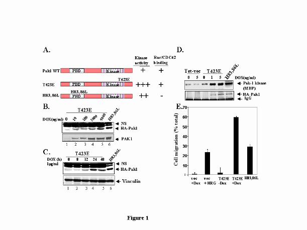
T423E Pak1 protein in a dose- and time-dependent manner (Figs. 1B and C). The identity of
induced band as T423E Pak1 was also confirmed by immunblotting witn an anti-Pak1
antibody. The levels of induced expression of T423E Pak1 were three to five times those of
endogenous Pak1. To determine if the expressed protein was functional and retained its
kinase activity, HA-tagged T423E Pak1 was immunoprecipiated from cells grown in serum-
free conditions with an anti-HA mAb and exogenously expressed Pak1 kinase activity was
measured by in vitro kinase assay. Myelin basic protein was used as a substrate and signal
was quantitated by phosphoimager. As shown in Fig.1D, Pak1 kinase activity was also
induced by doxycycline in a dose-dependent manner. For subsequent studies, we used
by guest on March 22, 2018
http://ww
w.jbc.org/
Dow
nloaded from
Page 9
9
1µg/ml dose of doxycycline, which could effectively induce about 3-fold induction of
T423E Pak1 protein and its kinase activity as compared to endogenous Pak1 levels. The
level of expression of H83, 86L Pak1 in stable clones was 5- to 6-fold that of vector
transfected control cells. The kinase activity of H83, 86L Pak1, however was lower than
that of T423E Pak1 cells taking into account the 3-fold lower expression of T423E Pak1
(Fig. 1D, compare lane 4 and 6).
We next analyzed the influence of Pak1 expression on the migration of MCF-7 cells using Boyden chamber
assay. Vector-tranfected cells showed low motility (Fig. 1E). In contrast, induction of T423E Pak1 with doxycycline
resulted in a significant increase in cell motility. HRG treatment was used as a positive control. H83, 86L Pak1 mutant
also induced motility of epithelial cells to a level similar to that induced with HRG treatment. Recently it was shown
that kinase activity affects directional cell movement (21). Although, both active mutants (T423E Pak1 and H83, 86L
Pak1) stimulated cell migration, the higher activity of T423E suggests that kinase activity and its localization and
interaction with other proteins also play a role in increasing the migratory potential of MCF-7 cells.
Localization of T423E Pak1 and H83, 86L Pak1. The subcellular localization of active
Pak1 mutants was visualized by immunofluorescence staining for tagged-HA and actin.
MCF-7 cells expressing T423E and H83, 86L Pak1 exhibited distinct actin localization.
Cells expressing T423E Pak1 showed the presence of filopodia and HA-Pak1 was
predominantly localized at the cell periphery that corresponds to the filopodia structures
(Fig. 2A). Cells expressing T423E Pak1 showed very little stress fibers compared with
vector-transfected cells (Fig. 2A) and exhibited scattered phenotype. Thirty to forty percent
of cells expressing T423E Pak1 generated a leading edge reminiscent of motile phenotype.
In contrast, cells expressing H83, 86L Pak1 showed lower levels of filopodia or stress-
fibers, but exhibited extensive membrane ruffling to which HA-Pak1 staining was localized.
Formation and induction of actin-containing structures, including filopodia, ruffles and
by guest on March 22, 2018
http://ww
w.jbc.org/
Dow
nloaded from
Page 10
10
leading edge and dissolution of stress fibers by active kinase mutants which in-turn loosen
their contacts with the substratum may provide an advantage for the cells and may have
contributed to the increased migration observed in Boyden chamber assays (Fig. 2A).
Increased Anchorage-independent Growth of MCF-7 Cells Expressing Kinase-active
Pak1 Mutants- Kinase-deficient Pak1 can suppress transformation that is mediated by Ras,
Rho, and Rac1 (24, 25) in Rat1 cells but not in NIH3T3 cells, suggesting that the
transformation-blocking function of Pak1 depends on the cellular context. To examine the
potential influence of regulatable expression of kinase-active Pak1 on the growth
characteristics of breast epithelial cancer cells, we measured the growth rate and ability of
cells to grow in an anchorage-independent manner. Expression of kinase-active Pak1
mutants had very little or no significant effect on the growth-rate of MCF-7 cells on plastic
(data not shown). Kinase-active Pak1 mutants did however significantly enhanced the ability
of MCF-7 cells to form colonies on soft agar (Fig. 2B). There was no difference in the
number or size of colonies between vector transfected control cells and uninduced T423E
expressing cells. Doxycycline mediated increase in the level of expression of T423E Pak1
was accompanied by a significant reproducible enhancement of the ability of cells to form
colonies in soft agar comparable to that of the cells treated with HRG as a positive control.
Cells expressing H83, 86L Pak1 also exhibited a profound increase in ability to grow in an
anchorage-independent manner (Fig. 2B). This finding contradicts an earlier observation
(24) showing the inability of H83, 86L Pak1 in Rat1 cells to form colonies in soft agar. It
is possible that these different results reflect the use of fibroblast versus epithelial cells in
these two studies.
by guest on March 22, 2018
http://ww
w.jbc.org/
Dow
nloaded from
Page 11
11
Regulation of MAPK Signaling Pathways by T423E Pak1-To study the biochemical
basis of the increased ability of MCF-7 cells to form colonies in soft agar, we analyzed the
signaling pathways activated in cells expressing T423E Pak1. We used doxycycline to
induce the expression of T423E Pak1 and analyzed the activation status of the signaling
components by blotting with phospho-specific antibodies. Both T423E and H83, 86L Pak1
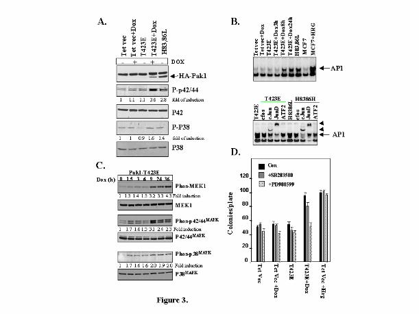
mutants demonstrated increased p42/44MAPK activity (2 to 4 fold) over vector transfected
cells. T423E Pak1 mutant demonstrated a modest increase in the level of p38MAPK (1.6-2
fold, Fig. 3A). Both Pak1 mutants exhibited increased AP1-DNA binding activity, which
was determined by enzymatic mobility shift assay (EMSA) assay (Fig. 3B, upper Panel).
Results of supershift experiments using specific antibodies which recognize specific
components of AP -1 complex suggested that the increased AP1 DNA binding activity was
caused primarily by c-jun and JunD transcription factors (Fig 3B, lower panel). The
exhibition of similar patterns of super shifts of AP1 DNA complex by both pak1 mutants
suggested the possibility of regulation of c-jun and Jun D transcription factors by Pak1
pathway. To further characterize the regulation of the p42/44MAPK pathway by Pak1, we used
doxycycline to induce T423E Pak1 expression in MCF-7 as a function of time. Consistent
with the earlier results, p42/44MAPK and its upstream kinase MEK1/2 were activated in a
time dependent manner by the induction of Pak 1 (Fig. 3C). When vector transfected cells
were exposed to doxycycline; the p42/44MAPK pathway was not induced (data not shown).
To address the possibility that activation of p42/44MAPK contributed to the
anchorage-independent growth by active Pak1 mutants, the soft agar experiment was
repeated in the presence or absence of PD98059, a specific MAPK inhibitor, or
by guest on March 22, 2018
http://ww
w.jbc.org/
Dow
nloaded from
Page 12
12
SB203580, a specific p38MAPK inhibitor. As illustrated in Fig. 3D, inclusion of a nontoxic
dose of PD98059 blocked the T423E Pak1-mediated increase in anchorage-independent
growth; inclusion of SB203580 had a small inhibitory effect compared to PD98059.
Nonspecific inhibitory effects of PD98059 did not cause the observed blockade effect of
PD98059 on anchorage-independent growth, because it failed to suppress the anchorage-
independent growth supported by HRG treatment (Fig. 3D). These results suggest that Pak1
play a role in activating p42/44MAPK. Although it is well accepted that Pak1 activates JNK
and p38MAPK pathways, there are contradictory reports on whether Pak1 activates p42/44MAPK
(23, 27, and 12). The use of different cell systems or the use of transient and inducible
systems could have caused the variation between the earlier reports and the results
presented in this study. These results suggest that p42/44MAPK may have a possible
preferential role of supporting the anchorage-independent growth of cells expressing
kinase-active T423E Pak1.
Active Pak1 Expression leads to an Abnormal Organization of Mitotic Spindles. Earlier
studies with Pak homologues in Saccharomyces cervisiae (Ste20) showed Pak homologues
have a role in cytokinesis and in mitosis (28, 29). To determine whether extended
expression of the mammalian Pak1 homologue affected cell-cycle progression, we induced
expression of kinase active Pak1 by treating MCF-7 cells expressing T423E for 72 h with
doxycycline. Cell-cycle progression was analyzed by FACS analysis, and there were no
significant differences between the cell-cycle progression of these treated cells and that of
the control vector-transfected cells (data not shown). We next examined whether prolonged
activation of Pak1 could influence the organization of mitotic spindles and thus, possibly
by guest on March 22, 2018
http://ww
w.jbc.org/
Dow
nloaded from
Page 13
13
contribute to anchorage independent growth via modifying the status of DNA ploidy and
genomic instability. To explore this possibility, MCF-7 cells expressing Tet vector or
T423E-Pak1 were treated with doxycycline for 72 h to induce the expression of active
Pak1. Cells were costained with monoclonal antibody against beta-tubulin (green) to mark
spindles and polyclonal antibody against the BTAK protein (red) to localize the centromere.
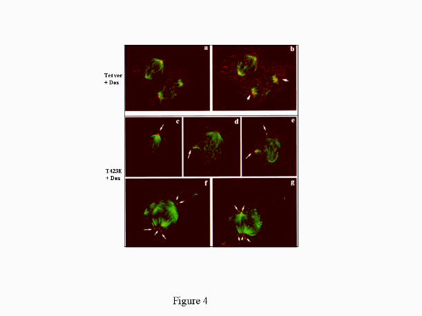
The cells were analyzed by confocal microscopy. As shown in Fig. 4, cells expressing
activated Pak1 exhibited multiple spindles with several orientations and centromere spots
(arrows); vector transfected cells did not show multiple spindles. For quantitation, we
scored 40 mitotic cells per field in eight different field’s (X20) and counted cells
exhibiting multiple spindles by analyzing each of them at a higher magnification. Induction
of kinase-active T423E Pak1 for 72 hours resulted in appearance of multiple spindles in
11+ 1 % of mitotic cells compared to <2% in vector transfected cells or T423E cells
without doxycycline. The multiple spindles in T423E-Pak1 expressing MCF-7 cells could
have been caused by defects in cytokinesis, but we could not differentiate this possibility in
the present study. Additional studies are needed to delineate the potential role of Pak1 in
mitosis, including the effect of abnormal Pak activation on cellular events responsible for
segregation of mitotic spindles.
Pak Expression and Breast Cancer- Since HRG activates Pak1 activity and breast
cancer progression (3-6), we hypothesized that Pak1 expression and activity may be closely
associated with the invasive phenotypes of breast cancer cells. To explore this possibility,
we analyzed the level of Pak1 expression and activity in a small numbers of grade II and
grade III breast tumor biopsies. As shown in Fig. 5A, there was a significant increase in the
by guest on March 22, 2018
http://ww
w.jbc.org/
Dow
nloaded from
Page 14
14
expression and kinase activity of Pak1 in grade III tumors, as compared to grade II tumors
(4). Interestingly, increased expression another band at 55 kDa was also observed in grade
III tumors. Since Pak1 antiserum is also known to react with Pak2 isoform, we believe that
Pak2 may be also elevated in grade III samples.
We also examined the level of endogenous Pak1 activity in a panel of invasive and non-invasive breast
cancer cell lines grown in complete medium supplemented with 10% fetal calf serum. Invasive breast cell lines (MDA-
MB435, MDA-MB231) exhibited a significant elevation in the level of Pak activity as compared to non-invasive
breast cancer cell lines (MDA-453, BT-474, and MCF-7) (Fig. 5B). Although Pak1 expression was observed in MCF-7
cells, the level of Pak1 kinase activity in MCF-7 cells were significantly lower as compared to its levels higher in
invasive cells, suggesting that Pak activity rather that amount Pak1 may be responsible for the phenotype. Similarly,
grade III tumors (4 out of 5) exhibited an elevation in the level of Pak kinase activity compared to grade II tumors.
There was no direct correlation between the expression level of Pak1 protein and its activity. It is possible that the
Pak1 kinase activity in tumor cells may be stimulated by mutation or signaling pathways or autocrine/paracrine
growth factors. Taken together, these observations support the notion that Pak1 activity rather than the expression
of Pak1 protein may be closely related with the invasiveness of breast cancer.
We next analyzed the expression of Pak1 in a panel of tumor biopsies by
immunohistochemical staining. As shown in Figs. 5C-H, there was low levels of Pak1
immunoreactivity in low-grade tumors. In contrast, poorly differentiated ductal carcinomas
of the breast (documented as grade III) demonstrated a significant intense Pak1 staining
(Figs. 5C, D, and E). Panel F is grade 2-breast carcinoma showing low level staining for
Pak1. A larger study is needed to further establish the validity these new results.
In summary, our findings demonstrated the ability of Pak1 activity to stimulate the
growth of breast epithelial cancer cells in an anchorage-independent manner, and to
promote an abnormal organization of mitotic spindles. We also provide new evidence to
by guest on March 22, 2018
http://ww
w.jbc.org/
Dow
nloaded from
Page 15
15
suggest a close relationship between the levels of Pak1 expression and activity Pak1 and
invasive phenotypes of human breast cancer cell lines and tumor grades.
ACKNOWLEDGEMENT. We thank Subrato Sen for providing us with the
centromere marker BTAC antibody.
REFERENCES
1. Slamon, D. J., and Clark, G. M. (1988) Science 240, 1795-1798.
2. Tang, C. K., Perez, C., Grunt, T., Waibel, C., Cho, C., and Lupu, R. (1996) Cancer Res.,
56, 3350-3358.
3. Adam, L., Vadlamudi, R., Kondapaka, S.B., Chernoff, J., Mendelsohn, J., and Kumar, R.
(1998) J. Biol. Chem. 273, 28238-28246.
4. Vadlamudi, R., Adam, L., Tseng, B., Costa, L., and Kumar, R. (1999) Cancer Res. 59,
2843-2846.
5. Vadlamudi, R., Adam, L., Talukder, A., Mendelsohn, J., and Kumar, R. (1999) Oncogene
18, 7253-7264.
6. Adam, L., Vadlamudi, R., Mandal, M., Chernoff, J., and Kumar, R. (2000) J. Biol. Chem.,
275, 12041-12050.
7. Ridley, A. J, Paterson, H. F. Johnston C. L. Diekmann, D., and Hall A. (1992) Cell, 70,
401-410.
8. Kozma., R., Ahmed, S., Best, A., and Lim, L. (1996) Mol. Cell. Biol. 16, 5069-5080.
9. Coso, O., Chiariello, M., Yu, J.C., Teramoto, H., Crespo, P., Xu, N., Miki, T., and
Gutkind, J.S. (1995) Cell 81, 1137-1146.
by guest on March 22, 2018
http://ww
w.jbc.org/
Dow
nloaded from
Page 16
16
10. Qiu. A, Chen, J., McCormick, F., and Symons, M. (1992) Proc. Natl. Acad. Sci. USA. 92,
11781-11781.
11. Qiu. A, Chen J., Kir, D., McCormick, F., and Symons, M. (1995) Nature 374, 457-459.
12. Frost, J.A., Steen, H., Shapiro, P., Lewis, T., Ahn, N., Shaw, P.E., and Cobb, M.H. (1997)
EMBO J. 16, 426-6438.
13. Manser, E., Leung, T., Salhuddin, H., Zhao, Z.S., and Lim, L. (1994) Nature, 367, 40-46.
14. Zhao, Z., Manser, E., Chen, X., Chong, C. Leung T., and Lim, L (1998) Mol Cell. Biol. 18,
2153-2163.
15. Manser, E., Loo, T. H., Koh C. G., Zhao, Z. S., Chen, X. Q., Tan, L., Tan, I., Leung, T.,
and Lim, L. (1998) Molecular Cell 1, 183-192.
16. Sells, M.A., Knaus, U.G., Bagrodia, S., Ambrose, D. M., Bakoch, G. M., and Chernoff, J.
(1997) Curr. Biol. 7, 202-210.
17. Pawson, T., and Scott, J. D. (1998) Science 278, 2075-2080.
18. Frost, J.A., Khokhlatchev, A., Stippec, S., White, M. A., and Cobb, M.H. (1998) J. Biol.
Chem. 273, 28191-28198.
19. Dharmawardhane, S., Sanders, L.C., Martin, S. S., Daniels, R. H., and Bokoch, G. M.
(1997) J. Cell Biol. 138, 1265-1278.
20. Turner, C. E., Brown, M.C., Perrotta, J. A., Riedy, M.C., Nikolopoulos, S.N., McDonald,
A.R., Bagrodia, S., Thomas, S., Leventhal, P.S. (1999) J. Cell. Biol. 145, 851-863.
21. Sells, M. A., Boyd, J. T., and Chernoff, J. (1999) J. Cell Biol. 145, 837-849.
22. Sanders L. C., Mastsumura, F., Bokoch, G. M., and de Lanerolle, P. (1999) Science 283,
2083-2085.
by guest on March 22, 2018
http://ww
w.jbc.org/
Dow
nloaded from
Page 17
17
23. Bagrodia, S., Derijard, B., Davis, R. J., and Cerione, R.A. (1995) J. Biol. Chem. 270,
27995-27998.
24. Tang, Y., Chen, Z., Ambrose, D., Liu, J., Gibbs, J.B., Chernoff, J., and Field, J. (1997)
Mol. Cell. Biol. 17, 4454-4464.
25. Tang, Y., Yu, J., and Field, J. (1999) Mol. Cell. Biol. 19, 1881-1891.
26. Cox, A. D., and Der, C. J. (1994) Methods Enzy. 238, 277-294.
27. Frost, J.A., S. Xu, M.R. Hutchison, S. Marcus, and M.H. Cobb. (1996) Mol .Cell. Biol. 16,
3707-3713.
28. Cvrckova, F., De Vigilio, C., Manser, E., Pringle, J.R., and Nasmyth, K. (1995) Genes
and Dev. 9, 1817-1830.
29. Wu, C., Lytvyn, V., Thomas, D. V., and Leberer, E. (1997) J. Biol. Chem. 272, 30623-
30626.
by guest on March 22, 2018
http://ww
w.jbc.org/
Dow
nloaded from
Page 18
18
FIGURE LEGENDS
Fig. 1. Characterization of Kinase-active Pak1 Expressing Cell Lines. (A) Schematic
representation of Pak1 mutants (T423E and H83, 86L) used in the study, PBD: p21 GTPase
binding domain. (B) T423E expressing MCF-7 cells were treated with increasing
concentrations of Dox and induction of T423E Pak1 was analyzed by immunoblotting with
HA mAb. Lysates from stable clones expressing HA-H83, 86L Pak1 was loaded in the last
lane. A duplicate set of blot was immunoblotted with a Pak1 antibody. (C) Kinetics of HA-
Pak1 expression was analyzed by treating cells at various time points with 1µg/ml of Dox
and analyzed by Western using a mouse-anti-HA mAb. The blot was reprobed with an anti-
vinculin Ab, as a loading control. (D) Equal amount of protein from serum-starved cells was
immunoprecipitated with an anti-HA antibody and kinase activity of exogenously expressed
Pak1 mutants was analyzed by in-vitro kinase assay using myelin basic protein as a
substrate. (E) Migration of T423E and H83, 86L expressing clones were analyzed using a
modified Boyden chamber as described in “Materials and Methods”. Results shown are
representative of three separate experiments.
Fig. 2. (A) Localization of Kinase-active Pak1 mutants. Vector transfected or active Pak1
expressing cells were co-stained with Alexa 546-Phalloidin to visualize F-actin containing
structures and with an anti-HA mAb to visualize the localization of active HA-tagged Pak1
mutants. Yellow color indicates colocalization between the F-actin and Ha-tagged Pak1
protein (arrows). T423E cells were treated with or without doxcycycline for 24 h to induce
the expression of Pak1 mutant. T423E Pak1 cells exhibited numerous F-actin containing
filopodia (T423E+Dox), and H83, 86L Pak1 expressing cells exhibited extensive
by guest on March 22, 2018
http://ww
w.jbc.org/
Dow
nloaded from
Page 19
19
membrane ruffling; as compared to control Tet vector transfected cells. Arrows indicate
co-localization of HA-tagged Pak1 with F-actin. Similar results were obtained in three
separate experiments. (B) Effect of kinase-active Pak1 expression on anchorage
independent growth of MCF-7 cells. Anchorage independent growth potential of the active
Pak1 expressing clones was measured by their ability to form colonies on soft agar. HRG
treatment was used as a positive control. Similar results were seen on four independent
experiments. Error bars represent standard error of mean. (C) Representative photographs
of soft agar colonies. Results shown are representative of four independent experiments.
Fig. 3. Signaling pathways activated byT423E and H83, 86L Pak1 mutants. MCF-7 cells
expressing either vector or Pak1 mutants were serum starved for 24 hours and induced to
express T423E Pak1 by treating cells with doxycycline (1 µg/ml) for 24 h. (A) Activation
of signaling molecules was measured by western blotting using Phospho-specific
antibodies. (B) AP1 binding activity was measured as a measure of activation of JNK
pathway by EMSA assay (upper panel) and Supershift (arrowheads) assays with various
antibodies (bottom panel); (C) Kinetics of activation of MEK1 and 42/44MAPK kinases by
induction of T423E. Results shown are representative of three separate experiments with
similar findings. Fold induction over control was calculated using the Sigma Scan program.
(D) MEK1 inhibitor PD98059 significantly reduced active Pak1 mediated anchorage
independent growth. Soft agar colony assays were performed in the presence of p42/44MAPK
pathway inhibitor (PD98059) and p38MAPK kinase pathway inhibitor (SB203580). Treatment
with HRG was used as a positive control. Results shown are representative of three
independent experiments.
by guest on March 22, 2018
http://ww
w.jbc.org/
Dow
nloaded from
Page 20
20
Fig. 4. Prolonged Expression of Pak1 Leads to Appearance of Multiple Spindles. MCF-7
cells expressing Tet vector (Panels a and b) or Tet-T423E construct (Panels c-g) were
treated with doxycycline (Dox) for 72 h. Cells were co-stained with anti-beta1-tubulin mAb
(green) and anti-BTAK Ab (red) and analyzed by confocal microscopy. Two mock-
transfected cells are shown: one in metaphase (upper cell) and one in anaphase (lower cell)
sectioned at one 400 nm interval (Panels a and b). Note the typical localization of BTAK at
the level of each spindle pole and a quasi-planar distribution of the symmetric spindles. By
contrast, cells expressing the active T4 23E Pak1 showed three (Panels c-e) or multiple
(Panels f and g) spindles, that form different angles between them (one antero-superior, one
latero-posterior and one supero-posterior (Panel d). Multiple spindles with different
orientations are shown in another example of the effect of T423E Pak-1 induction (Panels f
and g).
Fig. 5. Levels of Pak1 expression and activity in human breast cancer cell lines and tumors.
(A) Pak1 levels and activity in breast tumors. Breast tumor lysates (ref. 4) were analyzed by
Western blotting for Pak1 expression (upper panel) and subsequently, reprobed with a
vinculin Ab as a loading control (middle panel). Tumor lysates were immunoprecipitated
with a Pak1 mAb, and assayed for in vitro Pak1 kinase activity (bottom panel). Quantitation
of Pak1 kinase activity is shown as fold change. (B) Status of endogenous Pak1 expression
was analyzed by immunoblotting in a panel of breast cancer cells grown in complete
medium supplemented with 10% fetal calf serum (upper panel). Pak1 was
immunoprecipitated and kinase activity was determined by in vitro kinase assay using myelin
basic protein as a substrate (bottom panel). Quantitation of Pak1 kinase activity is shown as
by guest on March 22, 2018
http://ww
w.jbc.org/
Dow
nloaded from
Page 21
21
fold change. (C-H) Immunohistochemical demonstration of Pak1 in breast tissue samples.
Panels C, D, and E are invasive poorly differentiated ductal carcinomas of the breast
(documented as grade III). All show very strong positively for Pak1. Panel F is grade 2-
breast carcinoma showing low level staining for PAK1. Panel E is ductual epithelial
hyperplasia with low level of Pak1 staining. Panel H is negative control of section adjacent
to stained with normal rabbit IgG in place of the primary antibody. The sections were
stained with PAP method and counter stained with hematoxylin.
by guest on March 22, 2018
http://ww
w.jbc.org/
Dow
nloaded from
Page 22
by guest on March 22, 2018
http://ww
w.jbc.org/
Dow
nloaded from
Page 23
by guest on March 22, 2018
http://ww
w.jbc.org/
Dow
nloaded from
Page 24
by guest on March 22, 2018
http://ww
w.jbc.org/
Dow
nloaded from
Page 25
by guest on March 22, 2018
http://ww
w.jbc.org/
Dow
nloaded from
Page 26
by guest on March 22, 2018
http://ww
w.jbc.org/
Dow
nloaded from
Page 27
Sahin, Jonathan Chernoff, Mien-Chie Hung and Rakesh KumarRatna K Vadlamudi, Liana Adam, Rui-An Wang, Mahitosh Mandal, Diep Nguyen, Aysegul
cancer cellsgrowth and abnormal organization of mitotic spindles in human epithelial breast
Regulatable expression of p21-activated kinase-1 promotes anchorage-independent
published online August 16, 2000J. Biol. Chem.
10.1074/jbc.M002138200Access the most updated version of this article at doi:
Alerts:
When a correction for this article is posted•
When this article is cited•
to choose from all of JBC's e-mail alertsClick here
by guest on March 22, 2018
http://ww
w.jbc.org/
Dow
nloaded from