102
Mamdouh Mahfouz MD [email protected] MSK Imaging Shoulder joint I Normal anatomy and rotator cuff lesions ssregypt.com
Mamdouh Mahfouz MD [email protected]
MSK Imaging Shoulder joint I
Normal anatomy and rotator cuff lesions
ssregypt.com

Suspected inflammation or tumors
Examination Technique
Patient preparation:
Fasting 4- 6h
Patient position: supine
Procedure
Surface coil [one side ]
Indications
Shoulder joint complaint
Pain
Trauma
Swelling
Osteoarthritis
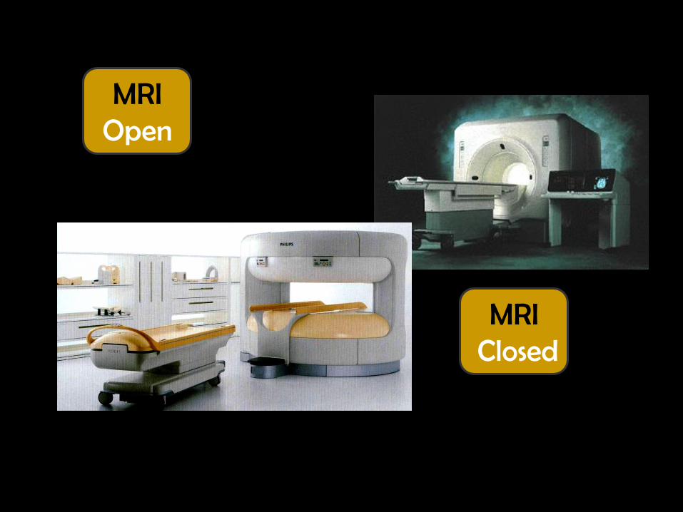
MRI Open
MRI Closed
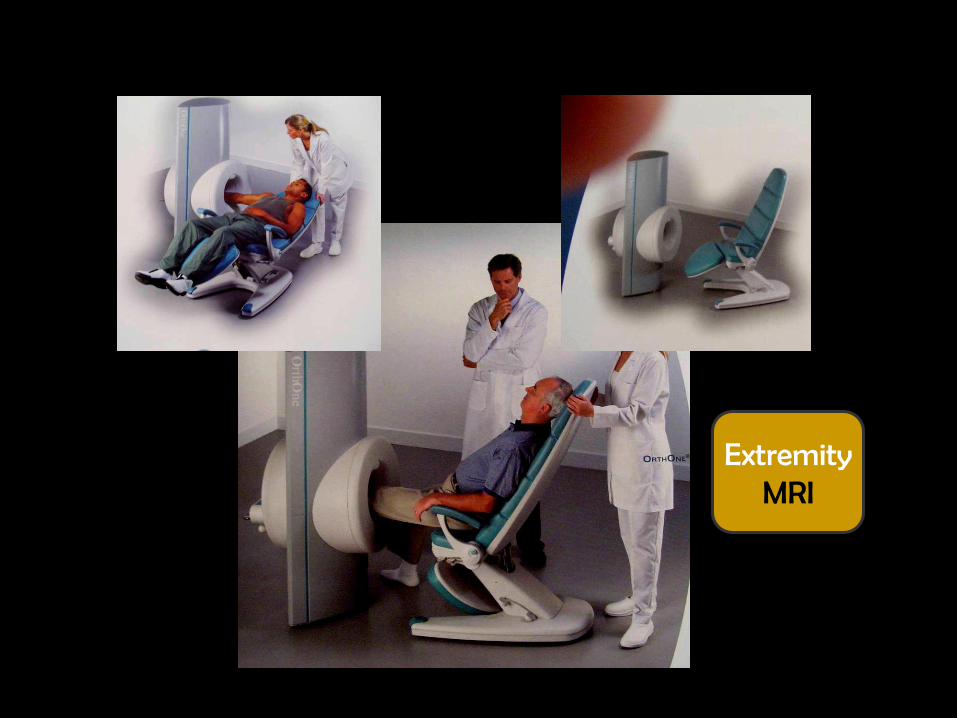
Extremity MRI
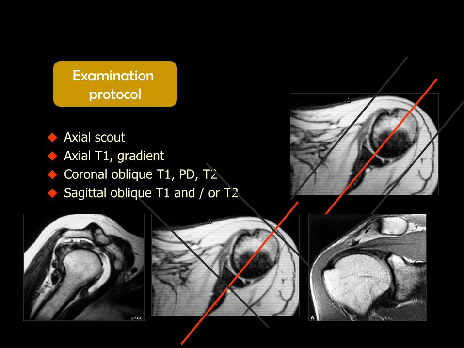
Axial scout
Axial T1, gradient
Coronal oblique T1, PD, T2
Sagittal oblique T1 and / or T2
Examination protocol

How to know the pulse sequence ?!
All these pulse sequences belong to T2
Gradient T1 T2 STIR
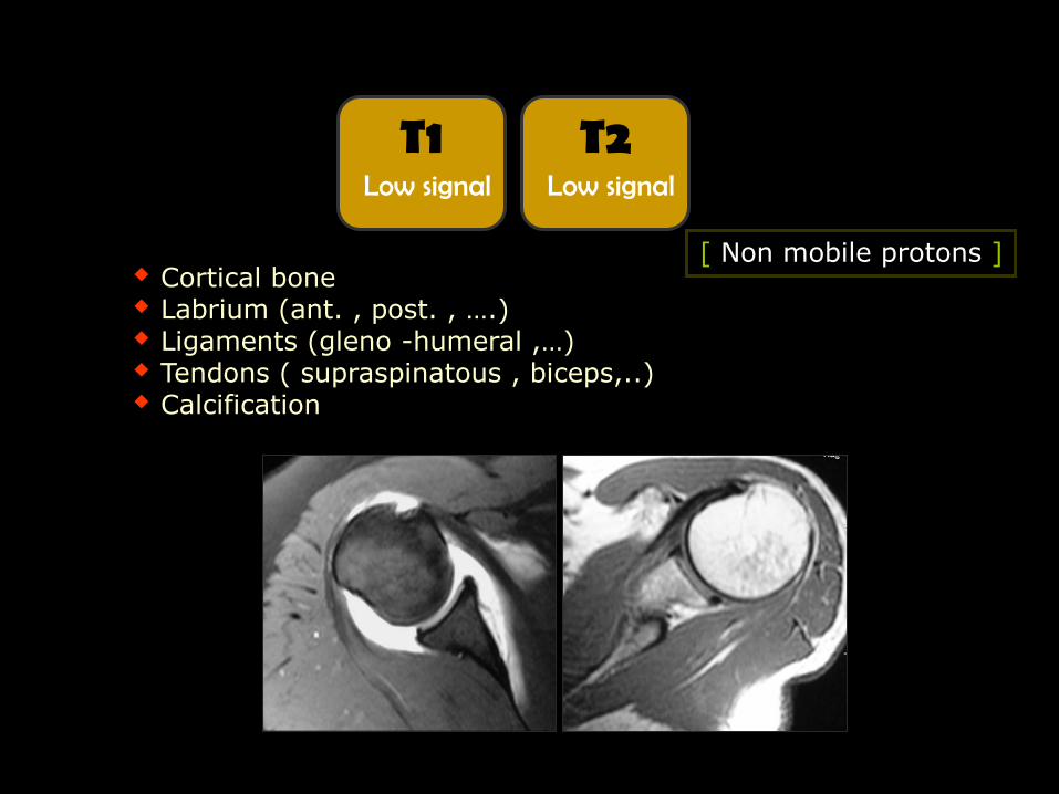
T1 Low signal
T2 Low signal
[ Non mobile protons ] Cortical bone Labrium (ant. , post. , ….) Ligaments (gleno -humeral ,…) Tendons ( supraspinatous , biceps,..) Calcification

Fat [ Subcutaneous
fat, dermoid cyst ,…]
Fluid [Effusion , cyst ,
articular cartilage ,… ]
T1 High signal
T2 Low signal
T1 Low signal
T2 High signal

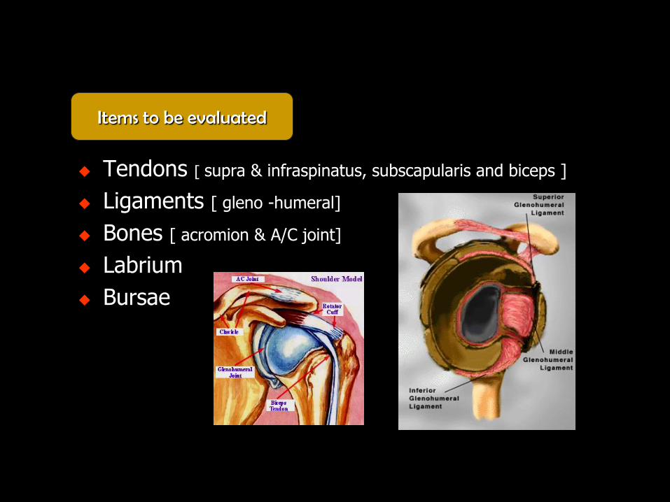
Tendons [ supra & infraspinatus, subscapularis and biceps ]
Ligaments [ gleno -humeral]
Bones [ acromion & A/C joint]
Labrium
Bursae
Items to be evaluated

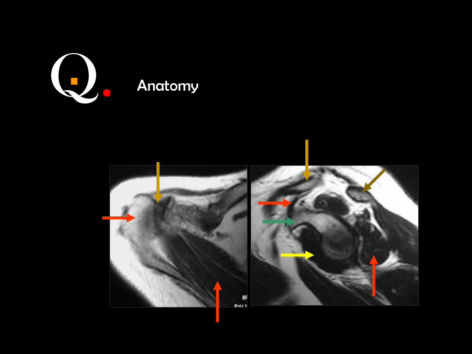
Anatomy
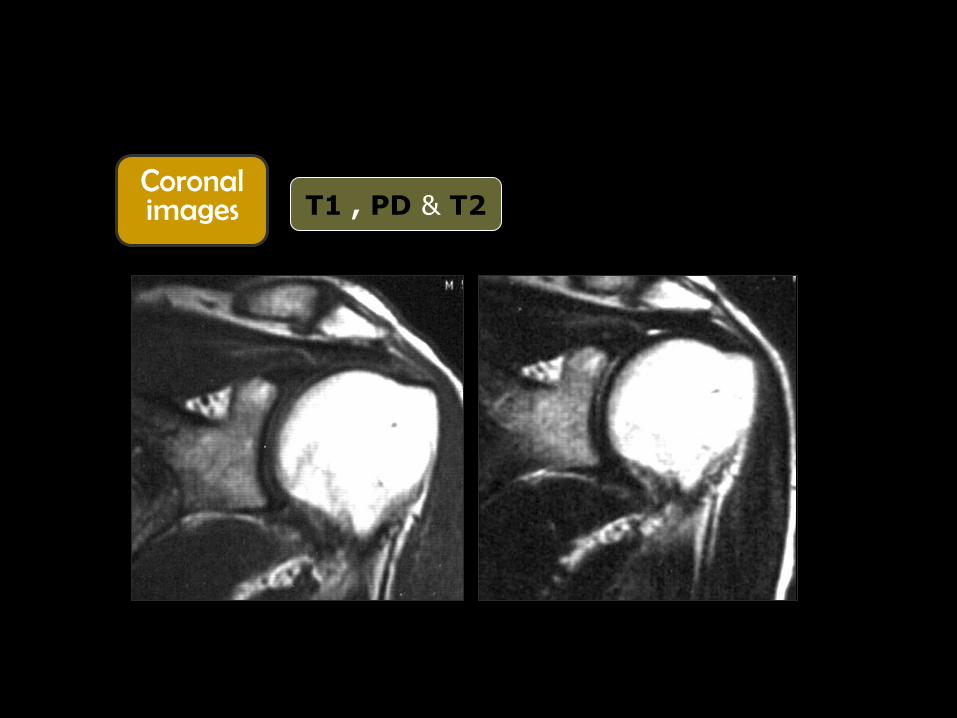
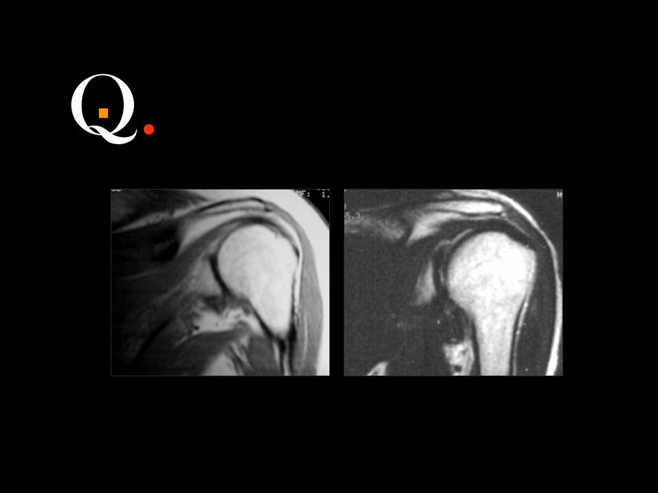
Coronal images
T1 , PD & T2

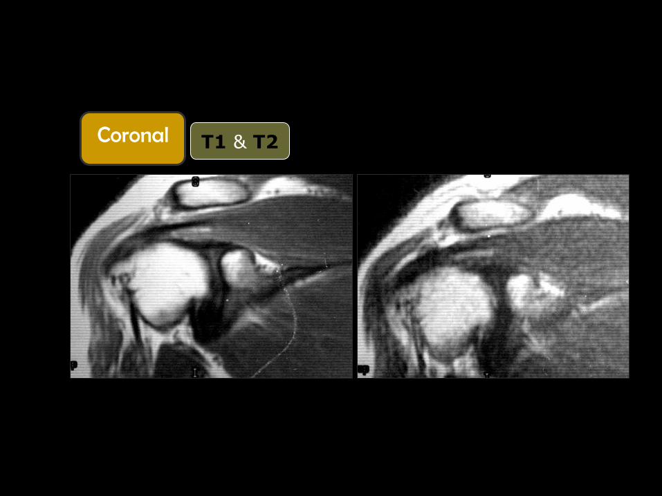
Coronal
T1 & T2

Coronal
T1 & T2

Coronal

Q. .
Coronal
T1 & T2
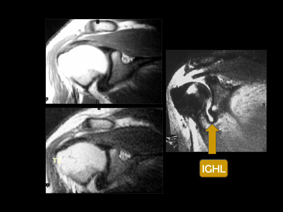
T1
T2
IGHL
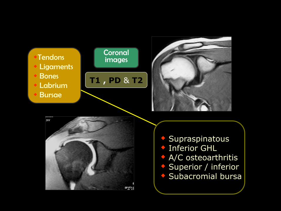
Tendons Ligaments Bones Labrium Bursae
Supraspinatous Inferior GHL A/C osteoarthritis Superior / inferior Subacromial bursa
Coronal images
T1 , PD & T2
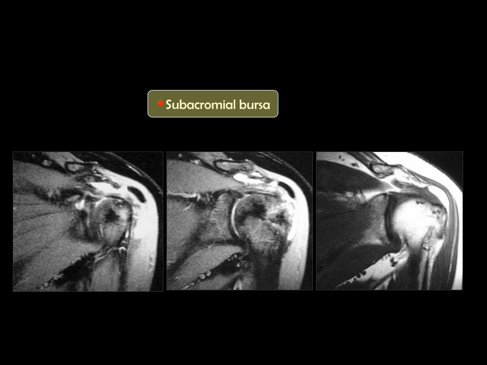
Subacromial bursa
Q. .
Q. .
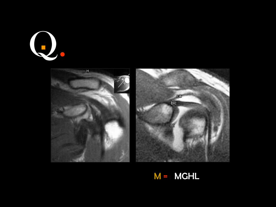
M = MGHL
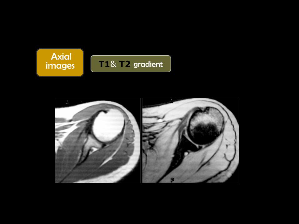
Axial images
T1& T2 gradient

Q. .

Middle
GH ligament

Q. .

Hill-Sach’s
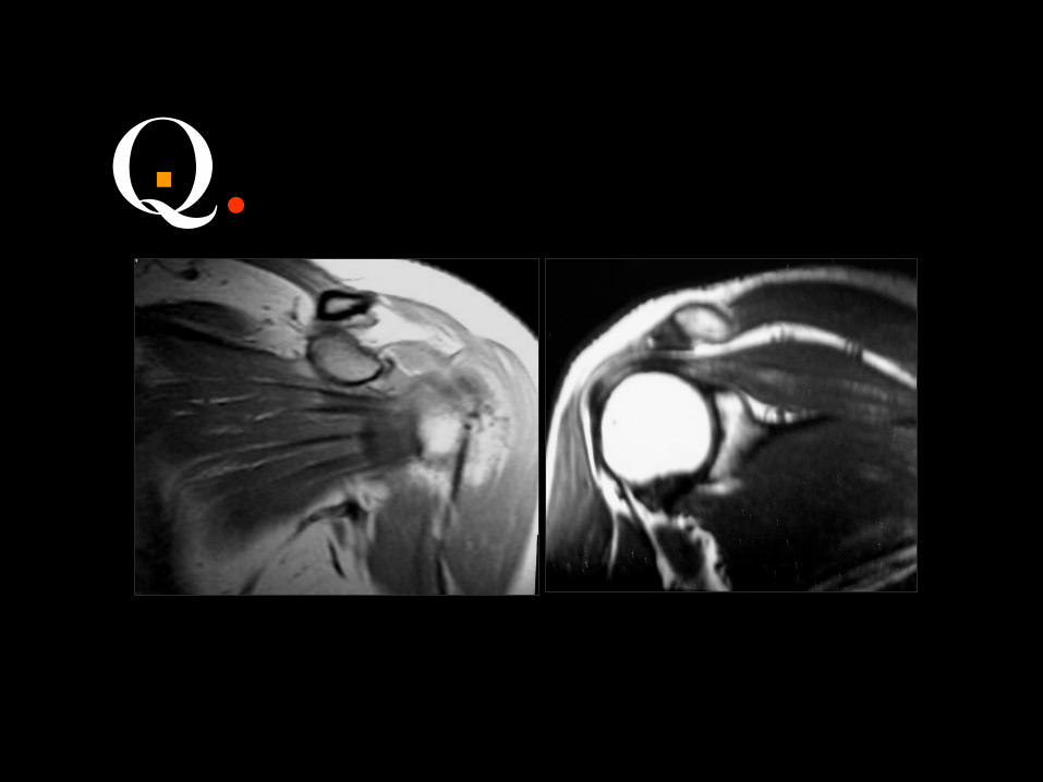
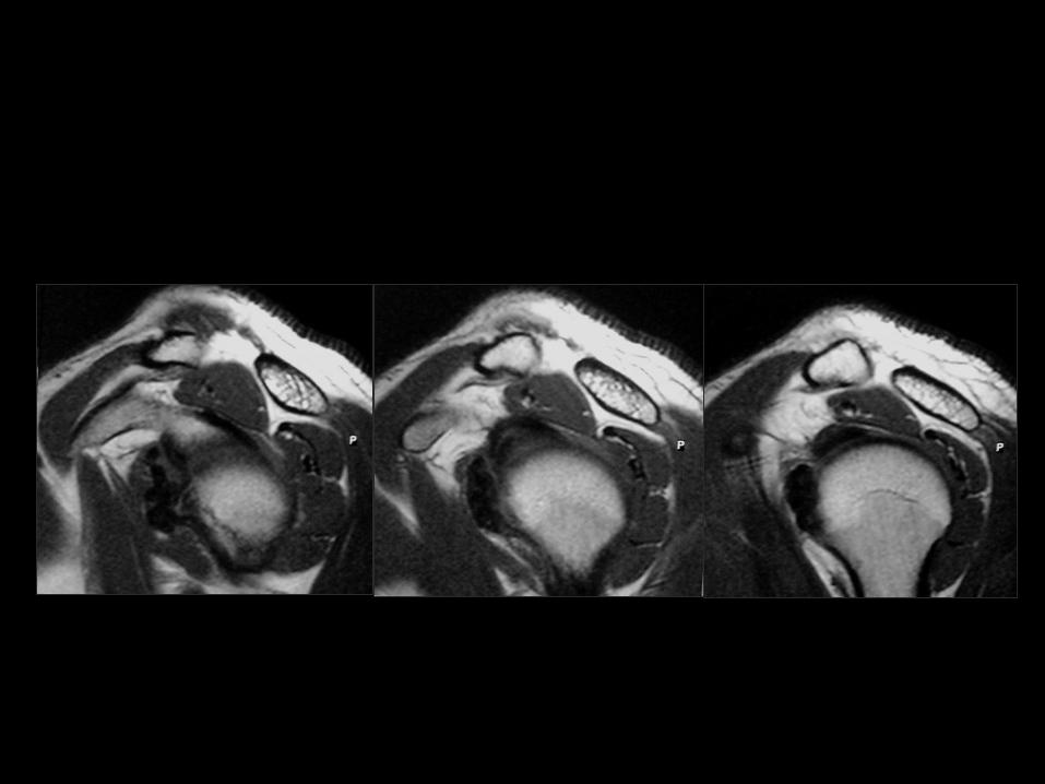
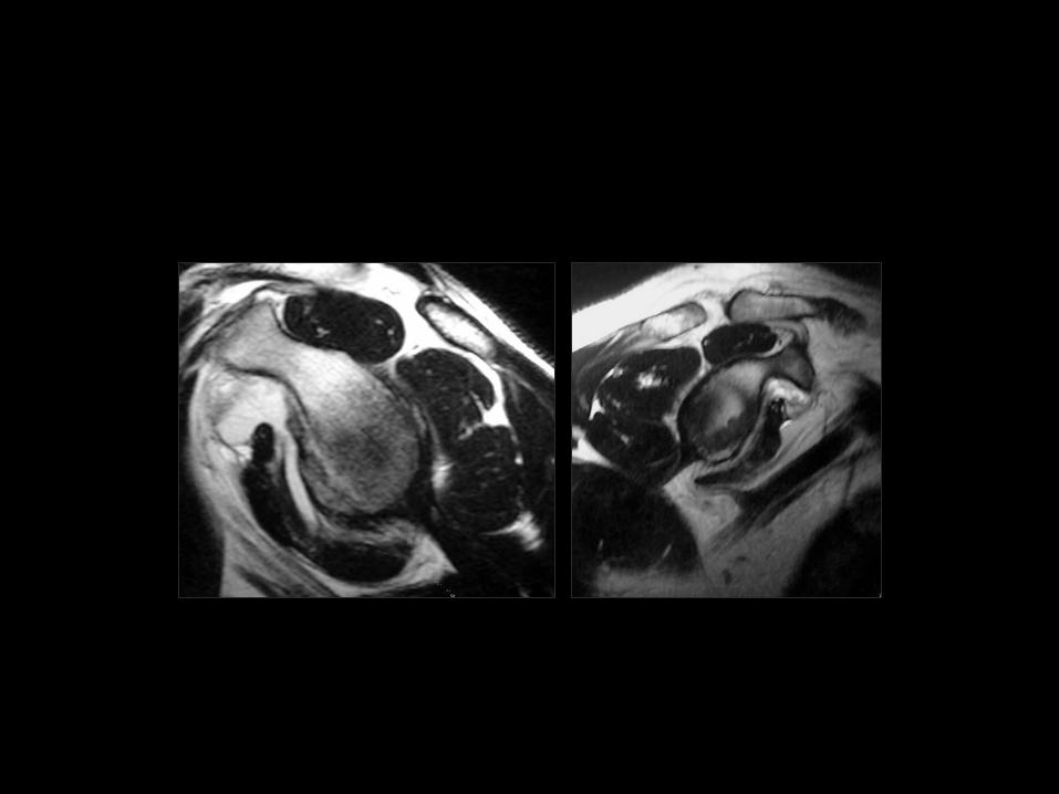
Axial images
T1& T2 gradient
Tendons Ligaments Bones Labrium Bursae
Subscapularis, Infraspinatous Biceps (in the groove) Middle GHL Humeral head [ Hill-Sack’s ] Anterior / posterior labrium
Subscapularis / subcoracoid
Q. .
Q. .

Q. .
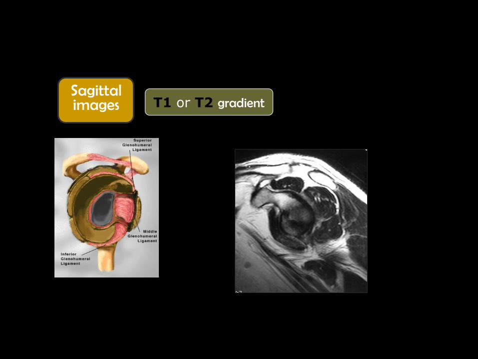
Sagittal images
T1 or T2 gradient
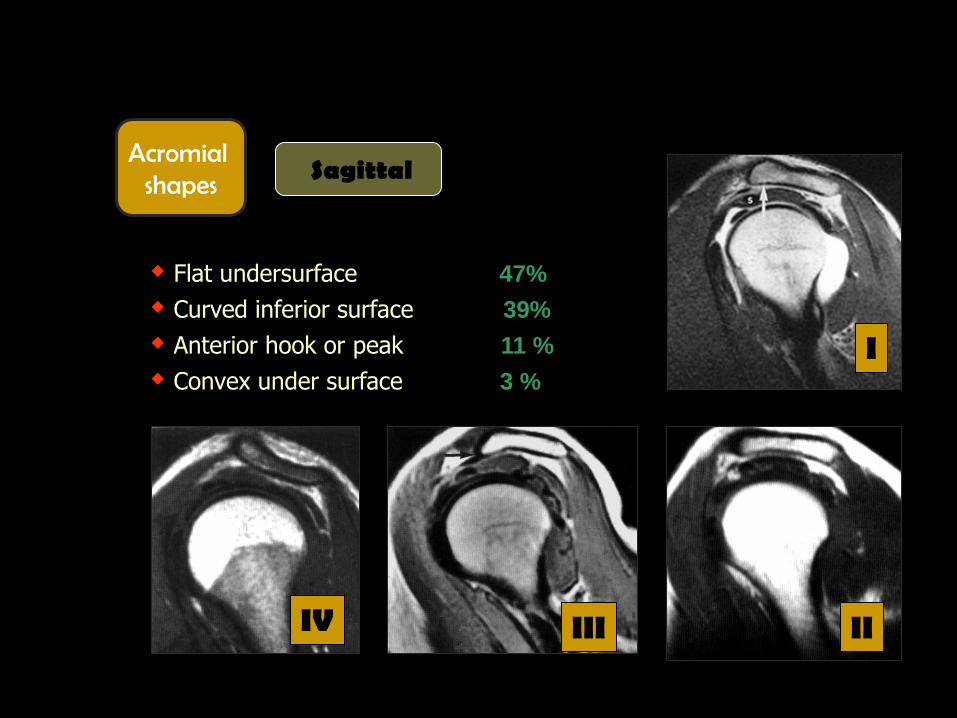
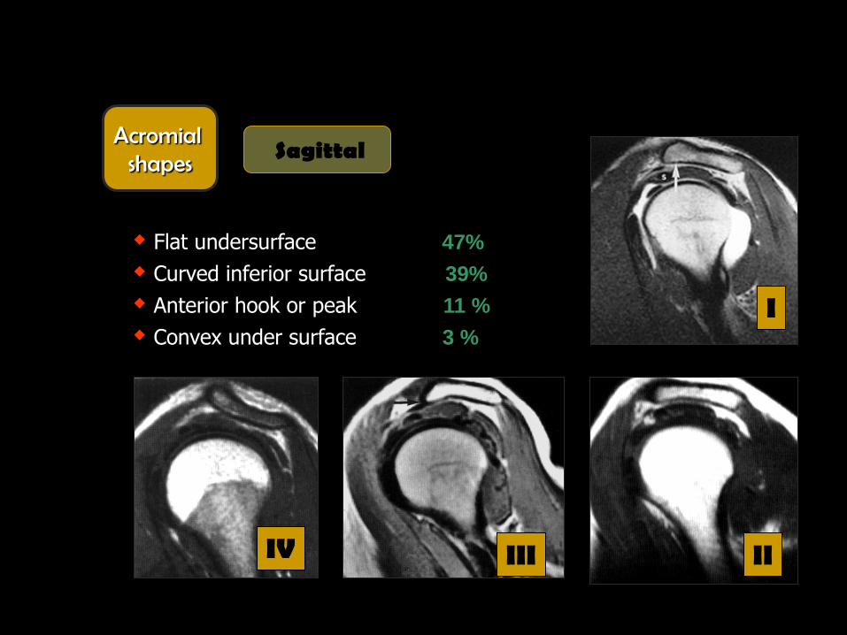
Flat undersurface 47%
Curved inferior surface 39%
Anterior hook or peak 11 %
Convex under surface 3 %
I
IV III II
Acromial shapes
Sagittal
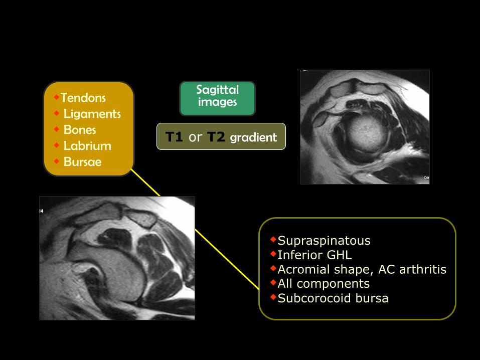
Sagittal images
T1 or T2 gradient
Tendons Ligaments Bones Labrium Bursae
Supraspinatous Inferior GHL Acromial shape, AC arthritis All components
Subcorocoid bursa
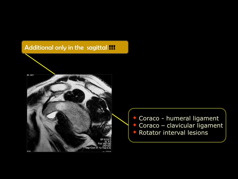
Additional only in the sagittal !!!
Coraco - humeral ligament Coraco – clavicular ligament Rotator interval lesions
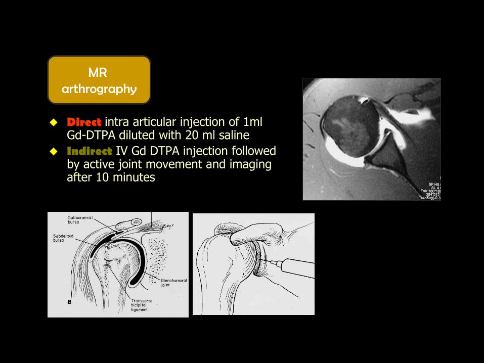
Direct intra articular injection of 1ml Gd-DTPA diluted with 20 ml saline
Indirect IV Gd DTPA injection followed by active joint movement and imaging after 10 minutes
MR arthrography

MR arthrography
Tendons [ supra & infraspinatus, subscapularis and biceps ]
Ligaments [ gleno -humeral]
Bones [ acromion & A/C joint]
Labrium
Bursae
Items to be evaluated

Supraspinatous
Infraspinatous
Teres minor
Rotator cuff Tendons
Subscapularis
Origin Insertion Posterior aspects of scapula
Anterior aspects of scapula
Biceps tendon

Supraspinatous Tendon
T1 Low signal
T2 Low signal

Supraspinatous Tendon
Supraspinatous
Two muscle slips
Intermediate signal in T1 & PD not in T2 WIs
Magic angle tendon at 55 to the static magnetic field
Prominent muscle slip
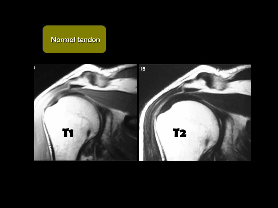
T1 T2
Normal tendon
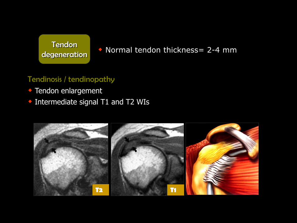
Tendinosis / tendinopathy
Tendon enlargement
Intermediate signal T1 and T2 WIs
T1 T2
Tendon degeneration
Normal tendon thickness= 2-4 mm

Intermediate signal in T1 and in T2 WIs within the tendon
Tendon degeneration

Tendon degeneration
Type III acromion
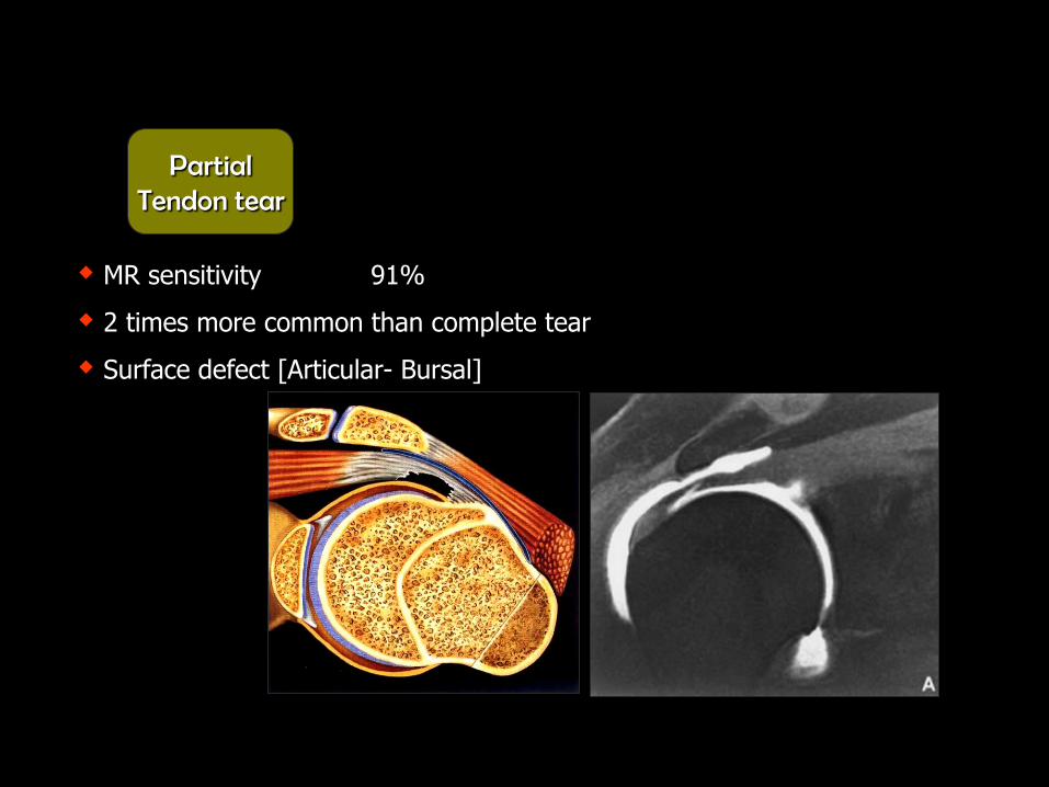
MR sensitivity 91%
2 times more common than complete tear
Surface defect [Articular- Bursal]
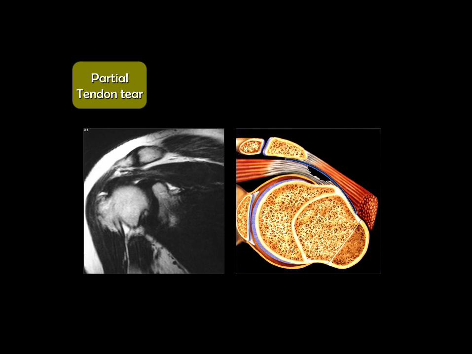
Partial Tendon tear
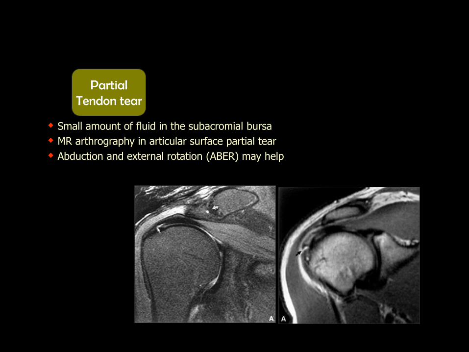
Small amount of fluid in the subacromial bursa
MR arthrography in articular surface partial tear
Abduction and external rotation (ABER) may help
Partial Tendon tear
Partial Tendon tear

Partial Tendon tear

Tendenopathy complicated by partial tear
T1 T2 PD
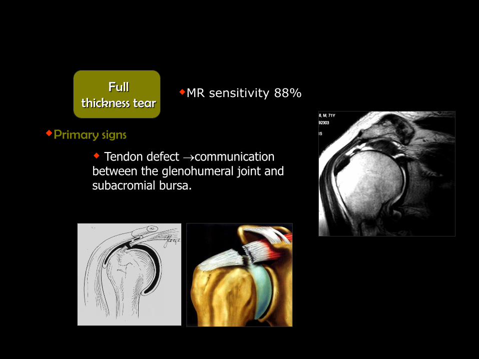
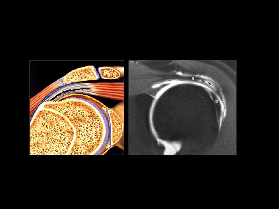
Primary signs
Tendon defect communication between the glenohumeral joint and subacromial bursa.
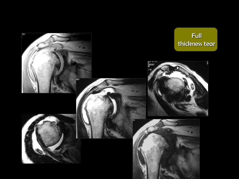
Full thickness tear
MR sensitivity 88%

Full thickness tear
Length of the gap
State of the muscle

Full thickness tear

Tendon defect
Complete absence humeral head in direct contact with the acromion
Full thickness tear
Full thickness tear
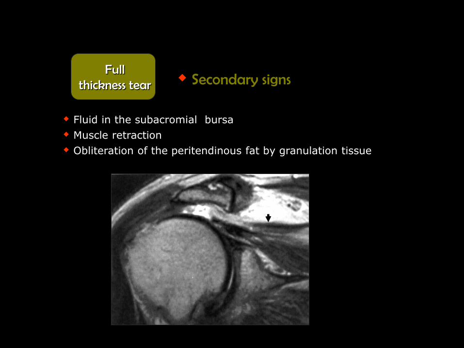
Fluid in the subacromial bursa
Muscle retraction
Obliteration of the peritendinous fat by granulation tissue
Secondary signs Full
thickness tear

Q. .
Q. .

Sher et al. 1995
34% of asymptomatic individuals have rotator cuff tears
54% of asymptomatic individuals above 60 years have tears

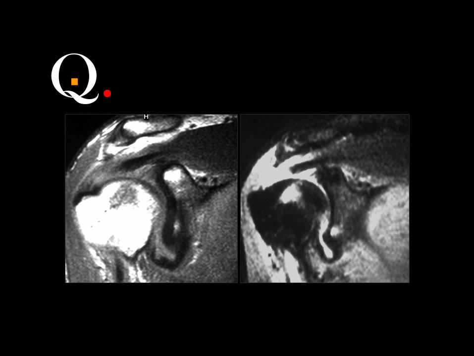
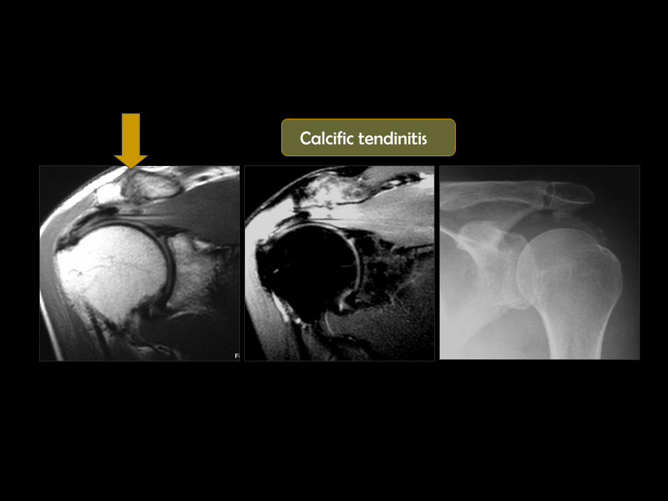
Deposition of calcium in the supraspinatous tendon tendon thickness
Low signal in T1 and T2 WIs
Usually asymptomatic
Pain in 30- 45% of cases
Common at the critical zone
Calcific tendinitis
Calcific tendinitis
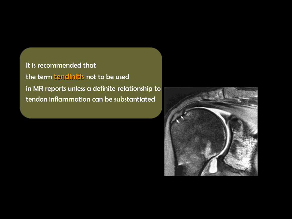
It is recommended that
the term tendinitis not to be used
in MR reports unless a definite relationship to
tendon inflammation can be substantiated

Infraspinatous Tendon
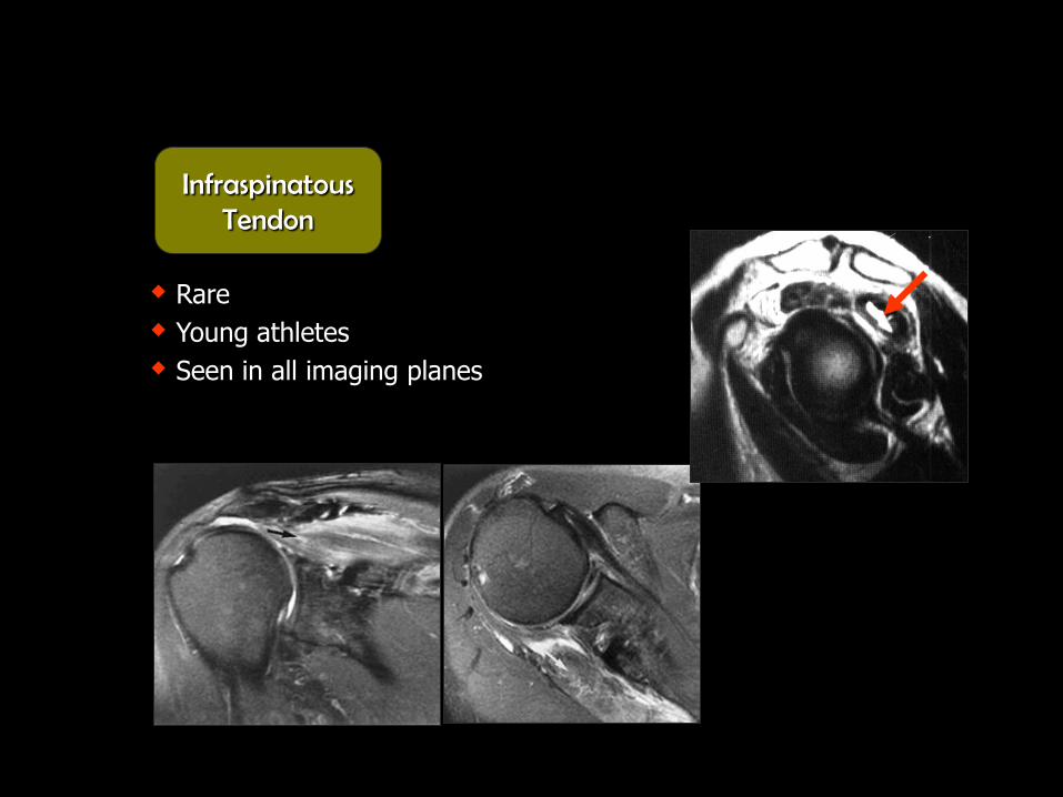
Rare
Young athletes
Seen in all imaging planes
Infraspinatous Tendon
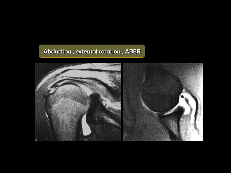
Abduction , external rotation , ABER
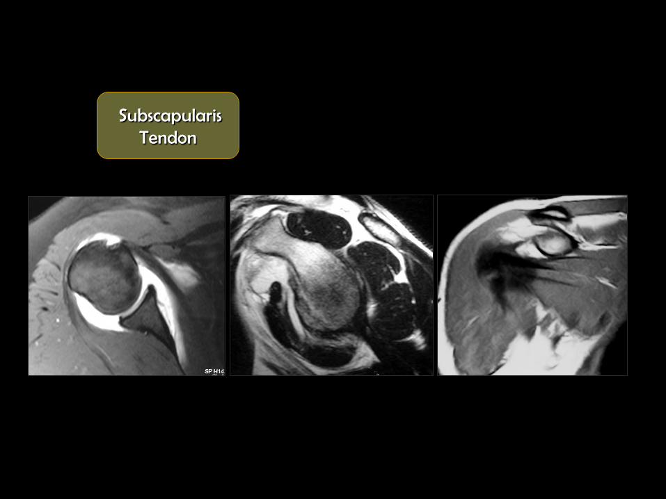
Subscapularis Tendon

Uncommon
old patient with recurrent dislocation
Axial images
Subscapularis Tendon

Supra and infraspinatous tears
Biceps tendon abnormalities
Subscapularis Tendon
Common associations

Q. .

Q. .

Progressive painful compression of the supraspinatous tendon
Usually affects the critical zone
95% rotator cuff tears result from chronic impingement
Impingement
syndrome

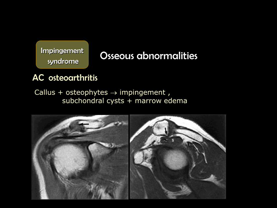
Osseous abnormalities
Acromion shape , 4 types
AC osteoarthritis
A C
Impingement
syndrome
Flat undersurface 47%
Curved inferior surface 39%
Anterior hook or peak 11 %
Convex under surface 3 %
I
IV III II
Acromial shapes
Sagittal
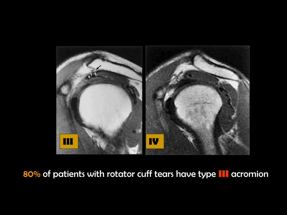
80% of patients with rotator cuff tears have type III acromion
III IV
Osseous abnormalities
AC osteoarthritis
Impingement
syndrome
Callus + osteophytes impingement , subchondral cysts + marrow edema
Q. .


Soft tissue abnormalities
Supraspinatous tendon
- Tendinosis
- Calcific tendinitis
Subacromial brusitis
Biceps tendon abnormalities
Impingement
syndrome
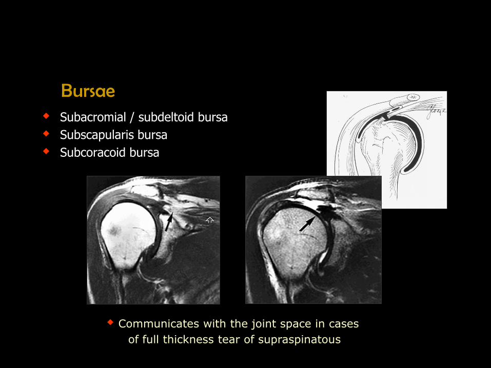
Bursae Subacromial / subdeltoid bursa
Subscapularis bursa
Subcoracoid bursa
Communicates with the joint space in cases
of full thickness tear of supraspinatous
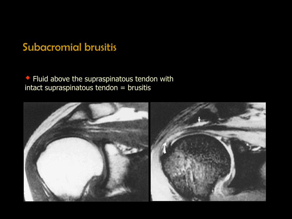
Subacromial brusitis
Fluid above the supraspinatous tendon with intact supraspinatous tendon = brusitis

Between the subscapularis and the MGHL may communicate with the joint space
Between the coracoid process and subscapularis muscle communicates with the joint space
Subscapularis bursa
Subcoracoid bursa

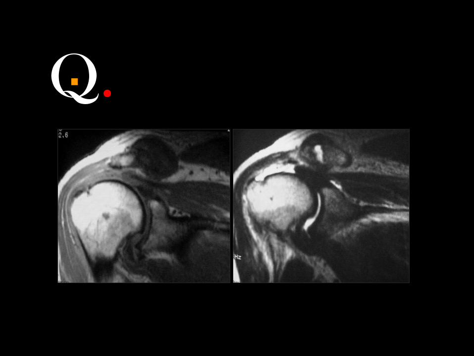
Q. . AC osteoarthritis with supraspinatous tendenopathy

Q. . Subscapularis tear with joint effusion extending around the biceps tendon

Q. . Supraspinatous partial ,articular surface, tear with normal infraspinatus tendon
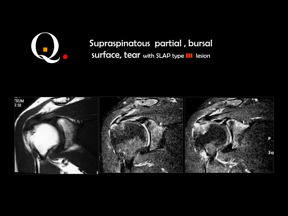
Q. . Supraspinatous partial , bursal surface, tear with SLAP type III lesion

Q. . Anatomy and technique
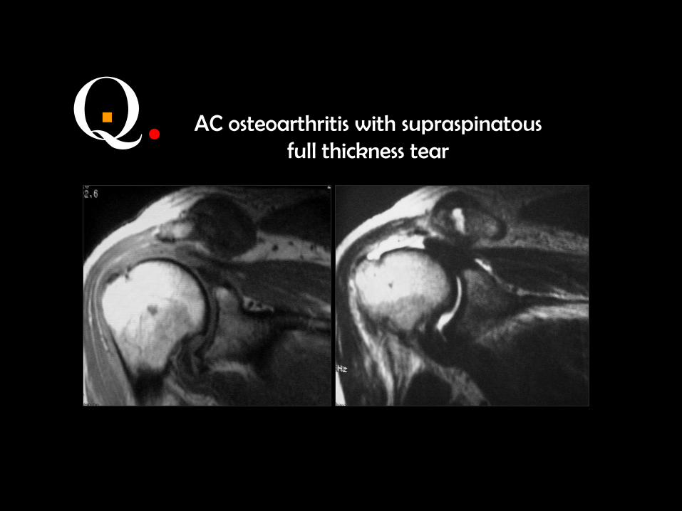
Q. . AC osteoarthritis with supraspinatous full thickness tear
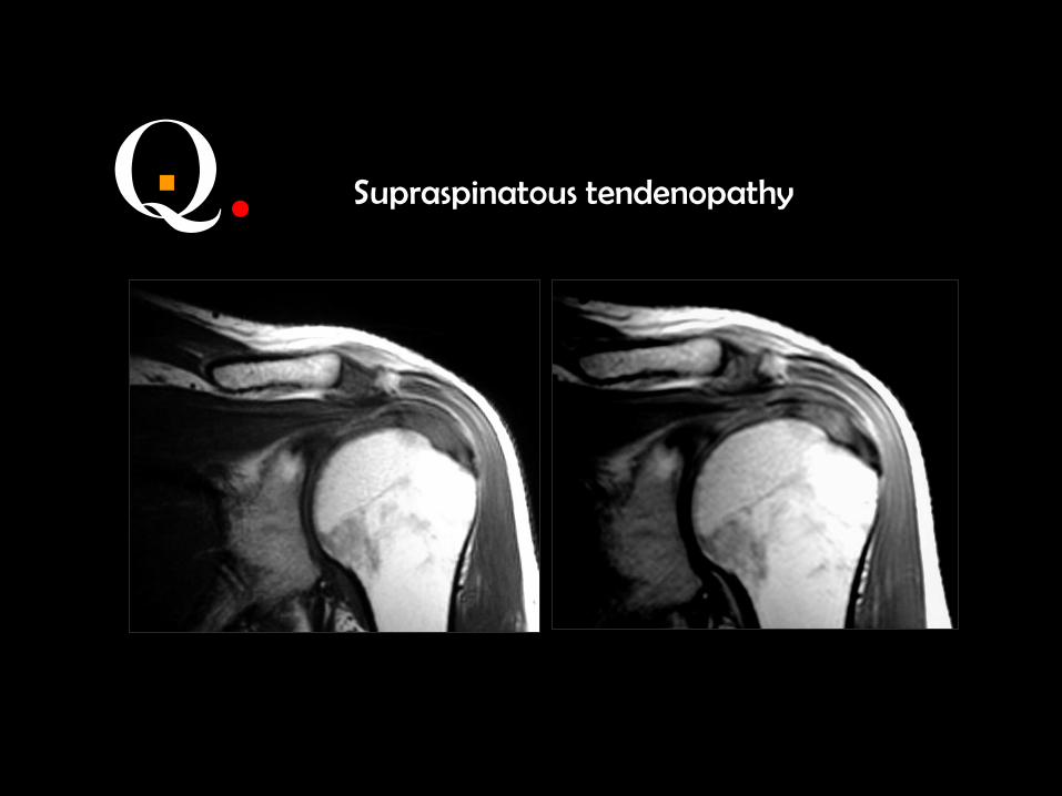
Q. . Supraspinatous tendenopathy
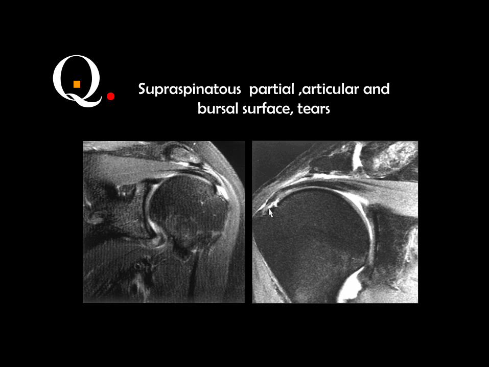
Q. . Supraspinatous partial ,articular and bursal surface, tears
Q. . Anatomy

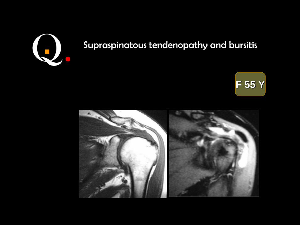
Q. . AC osteoarthritis with supraspinatous tendenopathy and bursitis
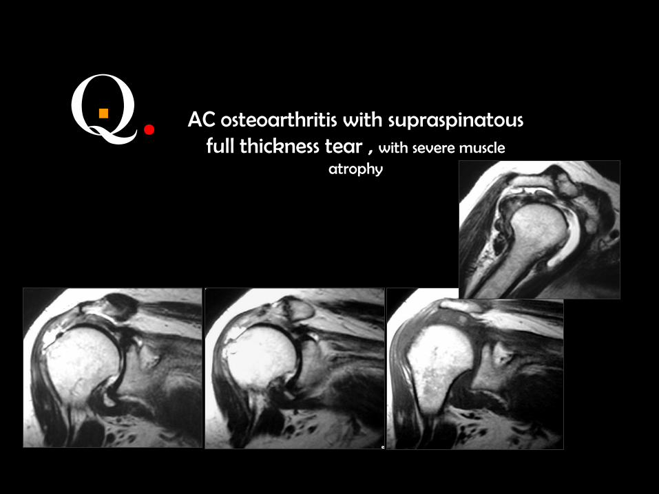
Q. . AC osteoarthritis with supraspinatous full thickness tear , with severe muscle
atrophy

Q. . Supraspinatous partial thickness tear bursal surface
M 43 Y
Q. .
F 55 Y
Supraspinatous tendenopathy and bursitis

Thank you
نستغفرك و نتوب اليك ×نشهد ان ال اله اال انت ×سبحانك اللهم و بحمدك
Mamdouh mahfouz MD [email protected]
ssr-eg.net

Q. . AC osteoarthritis with supraspinatous tendenopathy

Q. . AC osteoarthritis with supraspinatous tendenopathy

Q. . AC osteoarthritis with supraspinatous tendenopathy
Q. . AC osteoarthritis with supraspinatous tendenopathy

Q. . AC osteoarthritis with supraspinatous tendenopathy

Q. . AC osteoarthritis with supraspinatous tendenopathy


![MSK CT PROTOCOL[2] - jefferson.edu · AC joint. SHOULDER Coronal Imaging Plane Coronal Imaging Plane •Prescribe coronal plane off of axial images parallel to supraspinatus muscle](https://static.documents.pub/doc/80x56/5d645f8588c9930e728b6075/msk-ct-protocol2-ac-joint-shoulder-coronal-imaging-plane-coronal-imaging.jpg)


