Page 1
REVIEW
Multi-level regulation of cellular recognition of viral dsRNA
Alys Peisley • Sun Hur
Received: 7 June 2012 / Revised: 20 August 2012 / Accepted: 23 August 2012
� Springer Basel AG 2012
Abstract Effective antiviral immunity depends on accu-
rate recognition of viral RNAs by the innate immune
system. Double-stranded RNA (dsRNA) often accumulates
in virally infected cells and was initially considered a
unique viral signature that was sufficient to initiate antiviral
response through dsRNA receptors and dsRNA-dependent
effectors such as Toll-like receptor 3, retinoic acid inducible
gene-1, protein kinase RNA-activated and oligoadenylate
synthetase. However, dsRNA is also present in many cel-
lular RNAs, raising a question of how these receptors and
effectors discriminate between viral and cellular dsRNAs.
Accumulating evidence suggests that innate immune sen-
sors detect not only dsRNA structure but also other and
often multiple features of RNA such as length, sequence,
cellular location, post-transcriptional processing and mod-
ification, which are divergent between viral and cellular
RNAs. This review summarizes recent findings on the
substrate specificities of a few selected dsRNA-dependent
effectors and receptors, which have revealed more complex
mechanisms involved in cellular discrimination between
self and non-self RNA.
Keywords Antiviral innate immunity �Double-stranded RNA � PKR � OAS � ADAR � TLR3 �RIG-I � MDA5
Abbreviations
ADAR Adenosine deaminase acting on RNA
CARD Caspase activation and recruitment domain
DsRNA Double-stranded RNA
SsRNA Single-stranded RNA
DsRBD DsRNA binding domain
eIF2 Eukaryotic initiation factor 2
IFN Interferon
ISG Interferon-stimulated gene
LGP2 Laboratory of genetics and physiology-2
MAVS Mitochondrial antiviral signaling protein
MDA5 Melanoma differentiation-associated gene 5
MiRN Micro RNA
OAS Oligoadenylate synthetase
polyI:C Polyriboinosinic:polyribocytidylic acid
PKR Protein kinase RNA-activated
RIG-I Retinoic acid inducible gene-1
RNase-L Ribonuclease L
RLR RIG-I-like receptor
SAXS Small-angle X-ray scattering
SiRNA Small interfering RNA
TIR Toll/interleukin-1 receptor
TLR Toll-like receptor
TRIF TIR-containing adaptor inducing interferon-bUTR Untranslated region
Introduction
A successful host defense against viral infection depends
on both accurate recognition of viral invasion by germ-line
encoded pattern recognition receptors (PRRs) and proper
functioning of innate immune effectors to suppress viral
replication (Fig. 1) [1, 2]. Recognition of invariant virus-
A. Peisley � S. Hur
Department of Biological Chemistry and Molecular
Pharmacology, Harvard Medical School, Boston, USA
A. Peisley � S. Hur (&)
Program in Cellular and Molecular Medicine, Children’s
Hospital Boston, Center for Life Science Boston, 3 Blackfan
Circle, Boston, MA 02115, USA
e-mail: [email protected]
Cell. Mol. Life Sci.
DOI 10.1007/s00018-012-1149-4 Cellular and Molecular Life Sciences
123
Page 2
associated molecular patterns by PRRs activates signaling
pathways to generate antiviral cytokines including, but not
limited to, the type I interferons (e.g., IFNa/b). These
cytokines in turn stimulate the expression of a series of
interferon-stimulated genes (ISGs), such as antiviral
effector proteins, to establish antiviral states in infected and
neighboring cells (Fig. 1) [1, 3], and activate appropriate
adaptive immune response [4]. In vertebrates, several of
these receptors and effectors regulate their signaling
activity and effector functions, respectively, in a manner
dependent on viral RNA binding (Fig. 1) [1, 2]. Such viral
RNA-specific PRRs include the Toll-like receptors (TLRs)
3 and 7–8 and the retinoic acid inducible gene-1 (RIG-I)-
like receptors (RLRs), RIG-I and melanoma differentia-
tion-associated gene 5 (MDA5) [2]. Viral RNA-dependent
antiviral effectors include protein kinase RNA-activated
(PKR), oligoadenylate synthetase (OAS) and adenosine
deaminase acting on RNA (ADAR) [5]. With the exception
of TLR7 and 8, which recognize viral single-stranded
RNAs (ssRNAs), these cellular receptors and effectors
were shown to recognize double-stranded RNA (dsRNA).
As dsRNA structure had been thought to be a unique fea-
ture of viral RNAs, it had been widely accepted that
dsRNA binding alone is sufficient to activate their
respective antiviral functions.
The dsRNA duplex adopts an A-form helix that is dis-
tinct from the typical B-form helix of dsDNA. The major
groove of dsRNA is narrower and deeper than that of
dsDNA (4 vs. 11–12 A width), whereas the minor groove
of dsRNA is wider and shallower than dsDNA (10–11 vs.
6 A width)[6]. This distinct configuration of the phosphate
backbone of dsRNA along with the unique 20 hydroxyl
groups exposed in the minor groove can be specifically
recognized by conserved protein motifs such as dsRNA
binding domain (dsRBD) motifs in PKR and ADAR [7, 8].
In particular, the narrow major groove, which contains
sequence-specific information and is a common site of
interaction between protein and dsDNA [9], does not allow
insertion of protein to interact with dsRNA bases.
Accordingly, protein–dsRNA interaction is largely medi-
ated by the minor groove, which contains degenerate
sequence information, and the phosphate backbone, thus is
generally RNA sequence-independent [7].
Recent studies on the human transcriptome revealed that
dsRNAs, originally thought not to be expressed in the cell,
are generated in the form of secondary structures in pre and
mature micro or small interfering RNAs (miRNAs or
siRNAs) [10, 11], and in the form of long duplexes formed
by inverted repeat sequence elements [12, 13] or sense–
antisense hybrids [14, 15]. These observations raise a
question about the viral selection mechanism of receptors
and effectors previously thought to discriminate between
viral and cellular origin solely on the basis of presence of
dsRNA structure. Recent data suggests that viral RNA
receptors and effectors can recognize other features of
RNA, in addition to duplex structure such as 50 or 30
functional groups, post-transcriptional modification,
length, tertiary structure, and in some cases, sequence [18–
23]. This sensitivity to multiple other features of RNA is
likely important for robust and accurate discrimination
between cellular and viral dsRNAs. It also provides an
explanation for how some cellular or viral dsRNAs act as
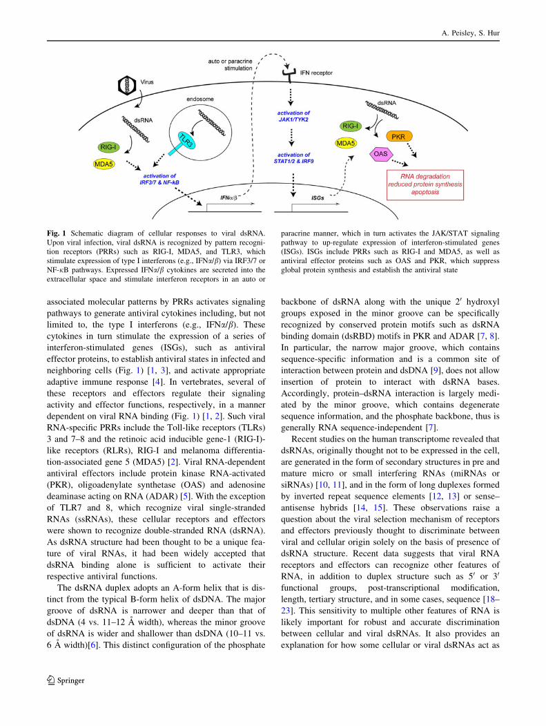
Fig. 1 Schematic diagram of cellular responses to viral dsRNA.
Upon viral infection, viral dsRNA is recognized by pattern recogni-
tion receptors (PRRs) such as RIG-I, MDA5, and TLR3, which
stimulate expression of type I interferons (e.g., IFNa/b) via IRF3/7 or
NF-jB pathways. Expressed IFNa/b cytokines are secreted into the
extracellular space and stimulate interferon receptors in an auto or
paracrine manner, which in turn activates the JAK/STAT signaling
pathway to up-regulate expression of interferon-stimulated genes
(ISGs). ISGs include PRRs such as RIG-I and MDA5, as well as
antiviral effector proteins such as OAS and PKR, which suppress
global protein synthesis and establish the antiviral state
A. Peisley, S. Hur
123
Page 3
antagonists rather than agonists for dsRNA receptors and
effectors [16, 17]. It remains to be addressed, however,
how these proteins interact with dsRNA in a manner that
allows for simultaneous recognition of multiple, seemingly
disparate features of RNA to bring about self versus non-
self discrimination. In this review, we will discuss multi-
layered aspects of RNA specificity and the structural and
biochemical mechanisms of dsRNA-dependent effectors
(PKR, ADAR, and OAS) and receptors (TLR3, RIG-I, and
MDA5). More detailed reviews on the biological functions
of each of these proteins can be found elsewhere.
Protein kinase RNA-activated (PKR)
PKR is a cytoplasmic Ser/Thr protein kinase that is up-
regulated by type I interferons and plays an important
role in the establishment of an antiviral and antiprolif-
erative cell state in response to viral infection [24]. PKR
consists of two N-terminal tandem dsRBDs and a C-ter-
minal catalytic kinase domain [11]. In the absence of
dsRNA, the kinase domain is in the autorepressed,
monomeric state [25]. Binding of dsRNA leads to a
conformational change in PKR, which is believed to
release the catalytic domain from the autoinhibitory
dsRBDs. Binding to dsRNA also brings multiple PKR
molecules into close proximity, which enables trans-
phosphorylation of PKR at several Ser or Thr residues
throughout the protein [26–28]. Phosphorylated PKR then
dissociates from dsRNA [29], and functions as a consti-
tutively active kinase that in turn phosphorylates serine
51 of eukaryotic translational initiation factor (eIF2a) and
suppresses global protein synthesis by blocking transla-
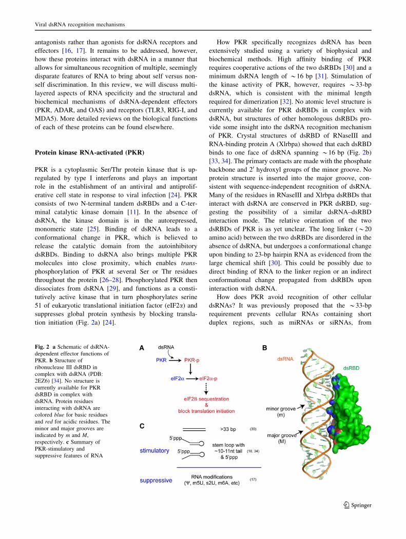
tion initiation (Fig. 2a) [24].
How PKR specifically recognizes dsRNA has been
extensively studied using a variety of biophysical and
biochemical methods. High affinity binding of PKR
requires cooperative actions of the two dsRBDs [30] and a
minimum dsRNA length of *16 bp [31]. Stimulation of
the kinase activity of PKR, however, requires *33-bp
dsRNA, which is consistent with the minimal length
required for dimerization [32]. No atomic level structure is
currently available for PKR dsRBDs in complex with
dsRNA, but structures of other homologous dsRBDs pro-
vide some insight into the dsRNA recognition mechanism
of PKR. Crystal structures of dsRBD of RNaseIII and
RNA-binding protein A (Xlrbpa) showed that each dsRBD
binds to one face of dsRNA spanning *16 bp (Fig. 2b)
[33, 34]. The primary contacts are made with the phosphate
backbone and 20 hydroxyl groups of the minor groove. No
protein structure is inserted into the major groove, con-
sistent with sequence-independent recognition of dsRNA.
Many of the residues in RNaseIII and Xlrbpa dsRBDs that
interact with dsRNA are conserved in PKR dsRBD, sug-
gesting the possibility of a similar dsRNA–dsRBD
interaction mode. The relative orientation of the two
dsRBDs of PKR is as yet unclear. The long linker (*20
amino acid) between the two dsRBDs are disordered in the
absence of dsRNA, but undergoes a conformational change
upon binding to 23-bp hairpin RNA as evidenced from the
large chemical shift [30]. This could be possibly due to
direct binding of RNA to the linker region or an indirect
conformational change propagated from dsRBDs upon
interaction with dsRNA.
How does PKR avoid recognition of other cellular
dsRNAs? It was previously proposed that the *33-bp
requirement prevents cellular RNAs containing short
duplex regions, such as miRNAs or siRNAs, from
Fig. 2 a Schematic of dsRNA-
dependent effector functions of
PKR. b Structure of
ribonuclease III dsRBD in
complex with dsRNA (PDB:
2EZ6) [34]. No structure is
currently available for PKR
dsRBD in complex with
dsRNA. Protein residues
interacting with dsRNA are
colored blue for basic residues
and red for acidic residues. The
minor and major grooves are
indicated by m and M,
respectively. c Summary of
PKR-stimulatory and
suppressive features of RNA
Viral dsRNA recognition mechanisms
123
Page 4
activating PKR. However, recent studies revealed more
complex RNA selectivity of PKR beyond recognition of a
simple dsRNA structure (Fig. 2c). The 50 untranslated
region (UTR) of IFN-c mRNA folds into a complex tertiary
structure that forms a coaxially stacked 33-bp stem, which
then activates PKR [35]. In addition, an RNA library
selection experiment revealed that a short (*16-bp) stem-
loop of ssRNA can activate PKR as well as a perfect
duplex RNA longer than 33 bp [36]. Unlike long dsRNA,
these short stem-loops require a *10-nt single-stranded
tail at either the 50 or 30 end and a triphosphate group at the
50 end to activate PKR [20]. In addition, some post-tran-
scriptional modifications, such as pseudouridine and
5-methyluridine, which are known to preserve the RNA
secondary structure, abolish or diminish the PKR-stimula-
tory activity of ssRNA containing short stem-loop [19]. As
most cellular RNAs undergo extensive post-transcriptional
modifications and 50 processing in the nucleus, which
removes the 50 triphosphate group present in all nascent
transcripts, the sensitivity of PKR to the 50 triphosphate
group and modified nucleotides provides an explanation
for how PKR avoids inappropriate activation by cellular
ssRNAs with secondary structures. However, it remains to
be understood how PKR utilizes dsRBDs to recognize
ssRNA tails and the 50 triphosphate group, how it dis-
criminates among ssRNAs on the basis of nucleotide
modification and whether it undergoes similar dimerization
upon binding to hairpin-containing ssRNA as with dsRNA.
Adenosine deaminase acting on RNA (ADAR)
ADAR is an RNA-modifying enzyme that converts aden-
osine to inosine (I) within dsRNA by hydrolytic
deamination (Fig. 3a) [37]. This A-to-I conversion is one
of many processes commonly referred to as RNA editing.
ADAR recognizes dsRNA structure using one to three
dsRBDs depending on the organism and the isotype [38].
Mammals express three isotypes of ADAR isotypes 1 and 2
display editing activity and are ubiquitously expressed,
whereas isotype 3 lacks a demonstrable editing activity and
its expression is limited to the central nervous system [39].
Adenosine editing by ADAR1 and 2 can occur in either a
site-specific or non-specific manner depending on the tar-
get dsRNA structure (Fig. 3b) [40–42]. In a perfect duplex
RNAs, A-to-I editing is non-specific, and can occur for up
to *50 % of adenosines [41, 43]. On the other hand, for an
imperfect duplex RNA with bulges or mismatches, A-to-I
editing occurs in a more restricted manner that is often
sequence-dependent [44, 45]. The best-studied examples of
those site-specific targets include the Q/R and R/G sites
within mRNAs encoding glutamate receptors (GluR) and
serotonin receptor (5-HT2cR) [46, 47]. Since inosine pairs
with cytidine, ADAR-mediated editing results in an ino-
sine–uridine mismatch, which decreases the stability of the
duplex [48]. Inosine is also read as guanosine by the cel-
lular ribosome, spliceosome, and viral RNA-dependent
RNA polymerase, and thus A-to-I editing can alter protein-
coding potential, mRNA splicing pattern, and can be
propagated into viral genomes (Fig. 3a) [37, 49].
The effect of RNA editing by ADAR on virus and
antiviral immunity appears complex and depends greatly
on specific cell types and viruses [50]. A splice variant of
ADAR1, p150, is up-regulated upon interferon stimulation
and was shown to suppress replication of measles virus and
virus-induced cytotoxicity [51]. Although the precise
mechanism for the antiviral function of p150 is not clear,
extensive mutations of A-to-G and U-to-C were observed
Fig. 3 a Schematic of dsRNA-
dependent effector functions of
ADAR. b Summary of RNA
specificity of ADAR. Examples
of site-specific editing targets
include the Q/R and R/G sites of
GluR-B pre-mRNA. c Structure
of ADAR2 dsRBDs in complex
with a RNA stem-loop
containing the R/G editing site
of the GluR-2 pre-mRNA (PDB:
2L3J [73]). Protein residues
interacting with dsRNA are
shown in a stick representation
and the flexible linker
connecting between the two
dsRBDs is represented by a
dotted line. The minor and
major grooves are indicated by
m and M, respectively
A. Peisley, S. Hur
123
Page 5
in the measles virus genome [52]. Similar antiviral effects
of p150 were observed with influenza A, Newcastle dis-
ease, and Sendai viruses [51]. However, not all viruses are
negatively affected by ADAR1 as positive effects have also
been observed [53–55]. For example, site-selective editing
of hepatitis D virus mRNA by ADAR is essential for
proper synthesis of a viral protein, HDAg-L [56, 57]. In the
case of vesicular stomatitis virus, ADAR helps viral rep-
lication in a manner independent of its editing activity, but
rather by antagonizing PKR [53, 54]. In addition, recent
studies showed that inosine containing dsRNAs can inhibit
activation of IRF3 and therefore the downstream interferon
signaling pathway, possibly by functioning as competitive
inhibitors of RIG-I and MDA5 [17]. While it is clear that
ADAR is involved in determining the fate of host–virus
interaction, how exactly ADAR exerts pro- and anti-viral
effects and how these seemingly opposing functions are
coordinated remain to be investigated in the future studies.
ADAR-mediated RNA editing is not limited to viral
RNAs, but also occurs relatively frequently for cellular
RNAs as evidenced by recent transcriptome analyses [37,
58, 59]. The examples include *70-nt-long pre-miRNAs
[58, 60] and inverted Alu elements, which fold into a near-
perfect duplex of *250–300 bp [12, 61]. The precise
biological consequences of these RNA-editing events are
still incompletely understood, but are likely to be multi-
faceted through their effects on RNA stability, function,
and subcellular localization [37, 49, 62–65]. It is also
tempting to speculate that editing could prevent aberrant
activation of the dsRNA-dependent innate immune system
by disrupting the long duplex structures present among the
cellular RNAs. Interestingly, deletion of ADAR1 in
hematopoietic stem cells has been shown to increase the
level of type I and II interferons [66], which could be
possibly due to the inability of the ADAR knock-out cells
to disrupt cellular RNA duplex structures. In whole
organisms, ADAR deficiency was shown to cause devel-
opmental abnormality in vertebrates [67–70] and
behavioral defects in invertebrates [71, 72], consistent with
an essential and versatile role of ADAR in cellular RNA
metabolism.
How does ADAR recognize a specific site on an
imperfect duplex RNA while promiscuously modifying a
perfect dsRNA? A recent NMR structure of the two
dsRBDs of ADAR2 in complex with one of the target sites
(R/G site) within GluR-2 mRNA provided an important
insight into the sequence-specific dsRNA recognition by
dsRBD [73]. In this structure, the overall interaction
between dsRNA and ADAR dsRBD was similar to that
seen with other homologous dsRBDs [33, 34, 74, 75], i.e.,
each dsRBD binds to one face of dsRNA, forming an
interaction with two successive minor grooves of dsRNA
(Fig. 3c). The structure also revealed, however, several
unexpected contacts between protein residues and edges of
bases in the minor groove at or near the site of mismatch
(Fig. 3c). Although these interactions appear to depend on
the presence of a mismatch and thus are unlikely to occur
in a perfect duplex, the structure provides an intriguing
example of the potential of the dsRNA minor groove in
sequence-dependent interaction with proteins and demon-
strates the versatility of dsRBDs in identifying structural
irregularities embedded within a dsRNA.
Oligoadenylate synthetase (OAS)
OAS belongs to a family of template-independent RNA
polymerases, which includes the eukaryotic polyadenosine
polymerase (PAP) and the class I CCA-adding enzyme
(CCA) from Archaea [76]. Mammals express four types
of OAS among which three isoforms, OAS1, OAS2, and
OAS3, are likely to have evolved from gene duplication.
These three members of OAS display an enzymatic
activity of linking two ATP molecules (donor and
acceptor) via a 20 and 50 phosphodiester bond to synthe-
size 20,50-linked oligoadenylates [pxA(20p50A)n; x = 1–3;
n [2] [77–79]. This 20,50-linked oligoadenylate then
functions as a cofactor to activate a latent ribonuclease,
RNase-L. RNase-L degrades both viral and cellular ssR-
NAs, such as ribosomal RNAs and mRNAs, with little
sequence specificity (typically after UU or UA sites),
which results in inhibition of global protein synthesis
(Fig. 4a) [80–82]. In a normal, resting state, the level of
20,50-oligoadenylate is tightly regulated by the enzymes
50-phosphatase and 20-phosphodiesterase, which inacti-
vates and degrades 20,50-oligoadenylates, respectively [83,
84]. During viral infection, however, the level of OAS is
transiently up-regulated by interferon, which results in
transient activation of RNase-L and suppression of viral
replication [79, 82, 85, 86].
Unlike PKR or ADAR, OAS does not harbor dsRBDs.
The crystal structure of OAS revealed a single globular
domain, composed of the N-terminal and C-terminal lobes
[87]. Although the structure was obtained without either a
donor or an acceptor ATP molecule bound, comparison of
the active site of OAS with that of PAP or CCA led to the
proposal that the donor ATP binds at the interface between
the two lobes (Fig. 4b) [87, 88]. It is as yet unclear how the
acceptor ATP and dsRNA bind, and how dsRNA binding
stimulates the catalytic activity of OAS. Based on the
location of the positively charged groove on the OAS
surface, it was proposed that dsRNA binds across the N and
C terminal domains on the opposite side of the ATP
binding surface (Fig. 4b) [87]. Interestingly, OAS can bind
to multiple types of nucleic acids, including non-activating
ssRNA with little or no secondary structure [89]. Based on
Viral dsRNA recognition mechanisms
123
Page 6
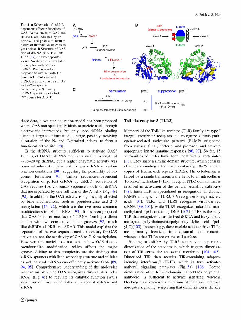
these data, a two-step activation model has been proposed
where OAS non-specifically binds to nucleic acids through
electrostatic interactions, but only upon dsRNA binding
can it undergo a conformational change, possibly involving
a rotation of the N- and C-terminal halves, to form a
functional active site [79].
Is the dsRNA structure sufficient to activate OAS?
Binding of OAS to dsRNA requires a minimum length of
*18–20 bp dsRNA, but a higher enzymatic activity was
observed when stimulated with longer dsRNA in certain
reaction conditions [90], suggesting the possibility of oli-
gomer formation [91]. Unlike sequence-independent
recognition of perfect dsRNA by dsRBD, activation of
OAS requires two consensus sequence motifs on dsRNA
that are separated by one full turn of the A-helix (Fig. 4c)
[92]. In addition, the OAS activity is significantly affected
by base modifications, such as pseudouridine and 20-Omethylation [23, 92], which are the two most common
modifications in cellular RNAs [93]. It has been proposed
that OAS binds to one face of dsRNA forming a direct
contact with two consecutive minor grooves [92], much
like dsRBDs of PKR and ADAR. This model explains the
separation of the two sequence motifs necessary for OAS
activation, and the sensitivity of OAS to 20-O methylation.
However, this model does not explain how OAS detects
pseudouridine modification, which affects the major
groove. Adding to this complexity are the findings that
ssRNA aptamers with little secondary structure and cellular
as well as viral mRNAs can efficiently activate OAS [89,
94, 95]. Comprehensive understanding of the molecular
mechanism by which OAS recognizes diverse, dissimilar
RNAs (Fig. 4c) to regulate its catalytic function awaits
structures of OAS in complex with agonist dsRNA and
ssRNA.
Toll-like receptor 3 (TLR3)
Members of the Toll-like receptor (TLR) family are type I
integral membrane receptors that recognize various path-
ogen-associated molecular patterns (PAMP) originated
from viruses, fungi, bacteria, and protozoa, and activate
appropriate innate immune responses [96, 97]. So far, 15
subfamilies of TLRs have been identified in vertebrates
[98]. They share a similar domain structure, which consists
of a ligand-binding ectodomain containing 19–25 tandem
copies of leucine-rich repeats (LRRs). The ectodomain is
linked by a single transmembrane helix to an intracellular
Toll-like/interleukin-1 (IL-1) receptor (TIR) domain that is
involved in activation of the cellular signaling pathways
[98]. Each TLR is specialized in recognition of distinct
PAMPs among which TLR3, 7–9 recognize foreign nucleic
acids [97]. TLR7 and TLR8 recognize virus-derived
ssRNA [99–101], while TLR9 recognizes microbial non-
methylated CpG-containing DNA [102]. TLR3 is the only
TLR that recognizes virus-derived dsRNA and its synthetic
analogue, polyriboinosinic:polyribocytidylic acid (pol-
yI:C)[103]. Interestingly, these nucleic acid-sensitive TLRs
are primarily localized in endosomal compartments,
whereas other TLRs are on the cell surface.
Binding of dsRNA by TLR3 occurs via cooperative
dimerization of the ectodomain, which triggers dimeriza-
tion of TIR across the endosomal membrane [104, 105].
Dimerized TIR then recruits TIR-containing adapter-
inducing interferon-b (TRIF), which in turn activates
antiviral signaling pathways (Fig. 5a) [106]. Forced
dimerization of TLR3 ectodomain via a-TLR3 polyclonal
antibodies is sufficient to activate signaling, whereas
blocking dimerization via mutations of the dimer interface
abrogates signaling, suggesting that dimerization is the key
Fig. 4 a Schematic of dsRNA-
dependent effector functions of
OAS. Active states of OAS and
RNase-L are indicated by an
asterisk. The precise molecular
nature of their active states is as
yet unclear. b Structure of OAS
free of dsRNA or ATP (PDB:
1PX5 [87]) in two opposite
views. No structure is available
in complex with ATP or
dsRNA. Protein residues
proposed to interact with the
donor ATP molecule and
dsRNA are shown as red sticksand yellow spheres,
respectively. c Summary
of RNA specificity of OAS.
‘W’ stands for A or U
A. Peisley, S. Hur
123
Page 7
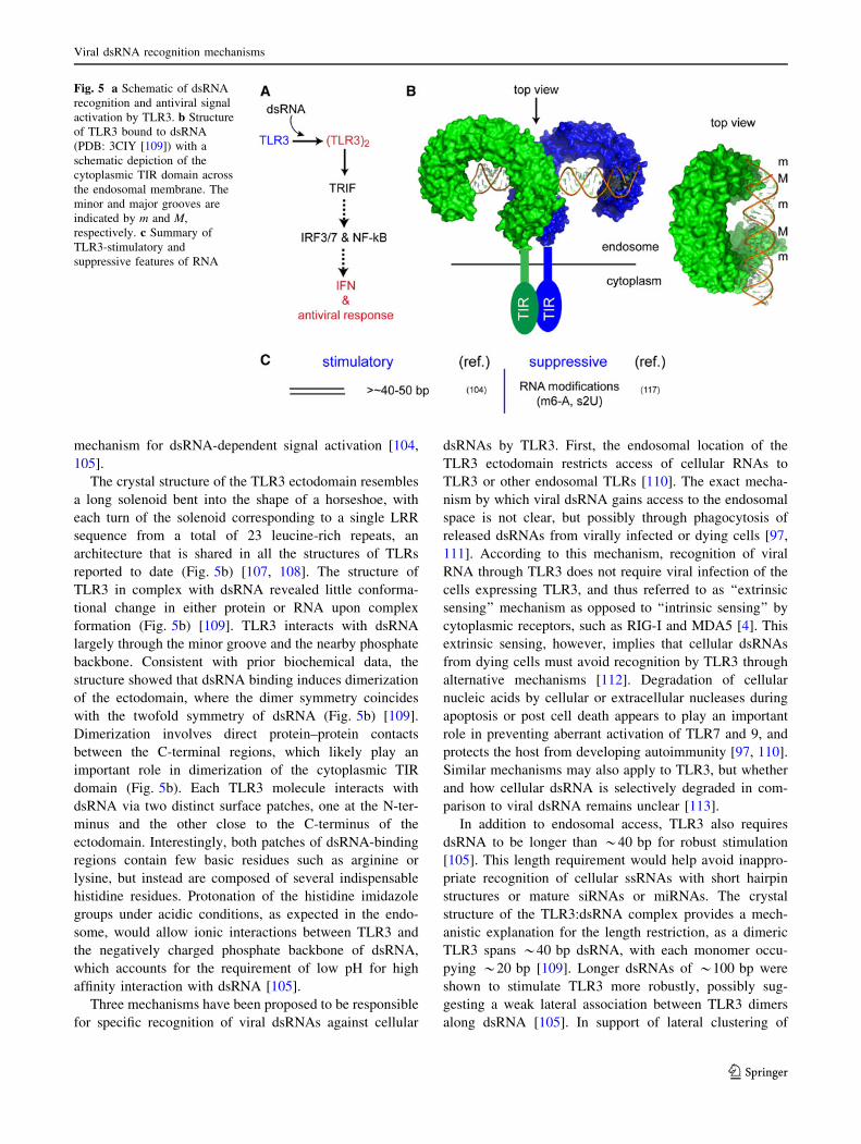
mechanism for dsRNA-dependent signal activation [104,
105].
The crystal structure of the TLR3 ectodomain resembles
a long solenoid bent into the shape of a horseshoe, with
each turn of the solenoid corresponding to a single LRR
sequence from a total of 23 leucine-rich repeats, an
architecture that is shared in all the structures of TLRs
reported to date (Fig. 5b) [107, 108]. The structure of
TLR3 in complex with dsRNA revealed little conforma-
tional change in either protein or RNA upon complex
formation (Fig. 5b) [109]. TLR3 interacts with dsRNA
largely through the minor groove and the nearby phosphate
backbone. Consistent with prior biochemical data, the
structure showed that dsRNA binding induces dimerization
of the ectodomain, where the dimer symmetry coincides
with the twofold symmetry of dsRNA (Fig. 5b) [109].
Dimerization involves direct protein–protein contacts
between the C-terminal regions, which likely play an
important role in dimerization of the cytoplasmic TIR
domain (Fig. 5b). Each TLR3 molecule interacts with
dsRNA via two distinct surface patches, one at the N-ter-
minus and the other close to the C-terminus of the
ectodomain. Interestingly, both patches of dsRNA-binding
regions contain few basic residues such as arginine or
lysine, but instead are composed of several indispensable
histidine residues. Protonation of the histidine imidazole
groups under acidic conditions, as expected in the endo-
some, would allow ionic interactions between TLR3 and
the negatively charged phosphate backbone of dsRNA,
which accounts for the requirement of low pH for high
affinity interaction with dsRNA [105].
Three mechanisms have been proposed to be responsible
for specific recognition of viral dsRNAs against cellular
dsRNAs by TLR3. First, the endosomal location of the
TLR3 ectodomain restricts access of cellular RNAs to
TLR3 or other endosomal TLRs [110]. The exact mecha-
nism by which viral dsRNA gains access to the endosomal
space is not clear, but possibly through phagocytosis of
released dsRNAs from virally infected or dying cells [97,
111]. According to this mechanism, recognition of viral
RNA through TLR3 does not require viral infection of the
cells expressing TLR3, and thus referred to as ‘‘extrinsic
sensing’’ mechanism as opposed to ‘‘intrinsic sensing’’ by
cytoplasmic receptors, such as RIG-I and MDA5 [4]. This
extrinsic sensing, however, implies that cellular dsRNAs
from dying cells must avoid recognition by TLR3 through
alternative mechanisms [112]. Degradation of cellular
nucleic acids by cellular or extracellular nucleases during
apoptosis or post cell death appears to play an important
role in preventing aberrant activation of TLR7 and 9, and
protects the host from developing autoimmunity [97, 110].
Similar mechanisms may also apply to TLR3, but whether
and how cellular dsRNA is selectively degraded in com-
parison to viral dsRNA remains unclear [113].
In addition to endosomal access, TLR3 also requires
dsRNA to be longer than *40 bp for robust stimulation
[105]. This length requirement would help avoid inappro-
priate recognition of cellular ssRNAs with short hairpin
structures or mature siRNAs or miRNAs. The crystal
structure of the TLR3:dsRNA complex provides a mech-
anistic explanation for the length restriction, as a dimeric
TLR3 spans *40 bp dsRNA, with each monomer occu-
pying *20 bp [109]. Longer dsRNAs of *100 bp were
shown to stimulate TLR3 more robustly, possibly sug-
gesting a weak lateral association between TLR3 dimers
along dsRNA [105]. In support of lateral clustering of
Fig. 5 a Schematic of dsRNA
recognition and antiviral signal
activation by TLR3. b Structure
of TLR3 bound to dsRNA
(PDB: 3CIY [109]) with a
schematic depiction of the
cytoplasmic TIR domain across
the endosomal membrane. The
minor and major grooves are
indicated by m and M,
respectively. c Summary of
TLR3-stimulatory and
suppressive features of RNA
Viral dsRNA recognition mechanisms
123
Page 8
TLR3, neutralizing Fab fragments, which bind to the TLR3
ectodomain in a manner that could disrupt its lateral
clustering, were shown to inhibit the signaling activity of
TLR3 without disrupting its dsRNA binding or dimeriza-
tion activity [114]. In an apparent contradiction to the
importance of dimerization or oligomerization, recent
studies showed that exogenously introduced 21-bp siRNA
can also stimulate TLR3 [115, 116], suggesting that low-
affinity interaction with short dsRNA can be compensated
for by high dose of RNA. It is possible that TLR3 can still
dimerize on 21-bp dsRNA, albeit inefficiently, in the same
manner as on 40-bp dsRNA or through an alternative
binding mode [117]. These observations suggest that
dsRNA length is not an absolute criterion used by TLR3
for self and non-self discrimination, but rather a relative
condition that is dependent on and can be scaled by the
abundance of RNA and receptors in the cell.
Finally, dsRNA recognition by TLR3 is suppressed by
the presence of modified nucleotides in RNA [118].
Modified nucleotides such as N6-methyladenosine and
2-thiouridine ablate the interferon signaling activity of
TLR3, whereas pseudouridine and 5-methyluridine have
more minor effects on TLR3. Interestingly, in vitro tran-
scribed or mitochondrial RNAs, but not cytoplasmic RNAs
from mammalian cell extracts, can activate the innate
immune response in dendritic cells [112, 118]. Considering
that mitochondrial RNAs contain a low level of modified
nucleotides in comparison to cytoplasmic cellular RNAs
[93], these observations suggest that nucleotide modifica-
tion provides an additional physicochemical specificity for
TLR3 to efficiently discriminate between self and non-self
dsRNAs (Fig. 5c).
Retinoic acid-inducible gene-I (RIG-I)
RIG-I-like receptors, which include RIG-I, MDA5, and
LGP2, represent another antiviral PRR pathway parallel to
that of TLRs 3 and 7–9. While nucleic acid-specific TLRs
are functional in the endosome, RIG-I-like receptors are
located in the cytoplasm and directly sense viral RNAs in
the infected cell (‘‘intrinsic sensing’’) [119, 120]. RIG-I and
MDA5 share a common domain architecture consisting of
two tandem caspase activation recruitment domains
(CARDs), which interact with the downstream signaling
adaptor, mitochondrial antiviral-signaling protein
(MAVS); a central DExD/H motif helicase domain
responsible for RNA-dependent ATP hydrolysis; and a
C-terminal domain (CTD) that binds to dsRNA [120–123].
LGP2 also has a similar domain architecture to RIG-I and
MDA5, but lacks the CARD domain [120]. Accordingly,
LGP2 does not possess an immune signaling activity by
itself, but is thought to up- and down-regulate the signaling
activities of MDA5 and RIG-I, respectively [124, 125].
Exactly how RIG-I and MDA5 relay antiviral signals to
MAVS is currently poorly understood, but several recent
studies collectively propose the following series of events
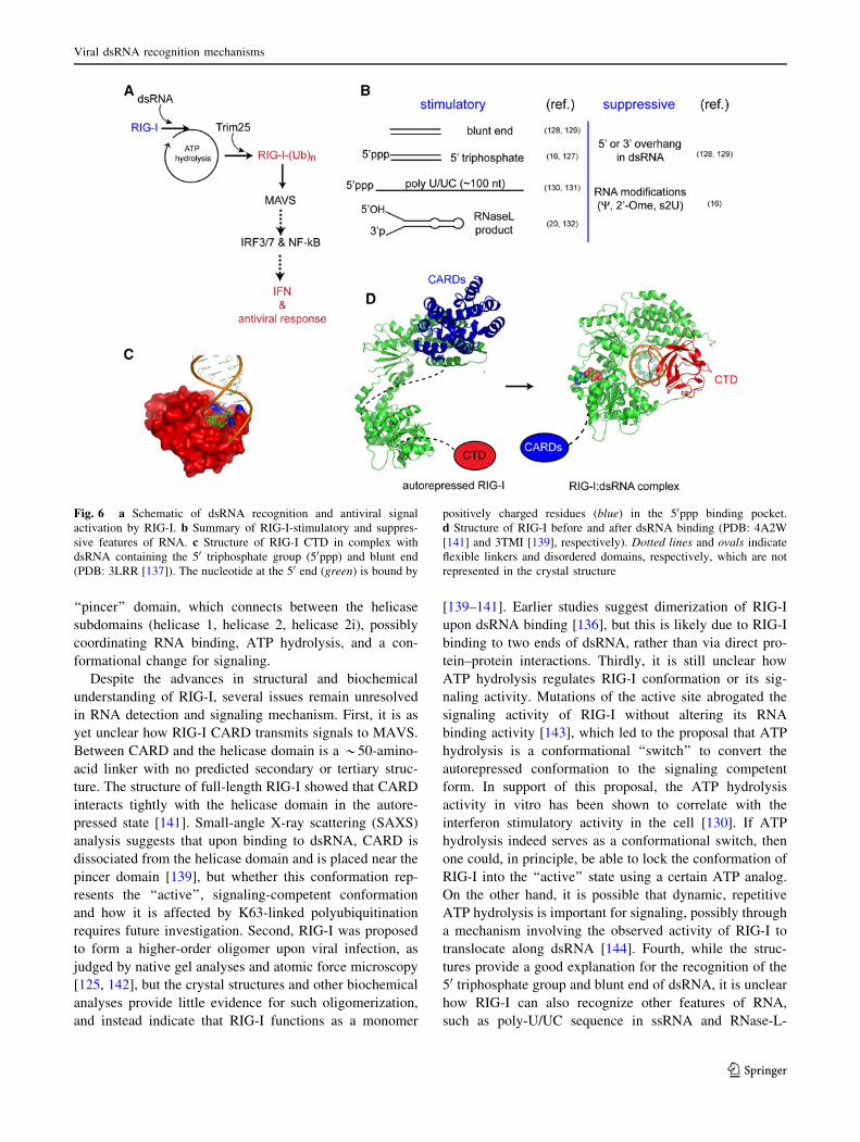
during RIG-I signaling. Upon viral RNA binding, RIG-I
hydrolyzes ATP and the second CARD domain becomes
covalently conjugated with K63-linked polyubiquitin by
Trim25 [126]. The ubiquitinated RIG-I CARD domain then
self-oligomerizes, interacts with CARD of MAVS, and
triggers formation of filamentous oligomers of MAVS
CARD on the mitochondrial surface [127, 128]. This
oligomeric form of MAVS CARD then recruits down-
stream signaling molecules such as TRAF2 and 3, which in
turn activate IRF3/7 or NF-jB signaling pathways in the
interferon antiviral response (Fig. 6a) [128].
The RNA selectivity of RIG-I appears to be complex
and has been much debated over the last several years
(Fig. 6b). It was first identified as a receptor stimulated by
a dsRNA mimic, polyI:C, and thus thought to recognize
simple dsRNA structure [119]. Later studies revealed,
however, that the 50 triphosphate group and blunt end of
RNA are important for viral recognition of short
(*20–25 bp) dsRNA by RIG-I [18, 129–131]. RIG-I was
also reported to recognize long ([100 nt) ssRNA with a 50
triphosphate group, such as the polyU/UC region of the
HCV genomic RNA, in a sequence- and length-dependent
manner [132, 133]. In addition, RNA cleavage products
produced by RNase L, which contain the 50 hydroxyl and 30
monophosphate group, can also activate RIG-I [22, 134].
As with PKR, OAS, and TLR3, modified nucleotides
(pseudouridine, 2-thio-uridine and 20-O-methyl-uridine)
suppress RIG-I stimulation by RNA [133].
More detailed biochemical and biophysical studies
revealed that the CTD, which displays little similarity to
any previously characterized RNA binding proteins, is
responsible for recognition of the 50 triphosphate group and
blunt end of dsRNA [135, 136]. Structures of the CTD
bound to dsRNA with the 50 triphosphate group and blunt
end revealed that a conserved, essential phenylalanine in
the CTD forms a face-to-face contact with the blunt-end
bases [137, 138]. The 50 triphosphate group forms elec-
trostatic interactions with a cluster of lysine residues
(Fig. 6c). The combination of pi-stacking and electrostatic
interactions provides an explanation for the observed
preference of RIG-I for dsRNA ends. Recently, three
groups have independently determined crystal structures of
isolated helicase domain or helicase-CTD of RIG-I in
complex with blunt-ended dsRNA [139–141]. The inter-
action between the CTD and dsRNA is similarly preserved
in the helicase-CTD–dsRNA structure, but helicase wraps
around dsRNA, forming additional contacts with the RNA
phosphate backbone (Fig. 6d). The most striking feature of
these structures was a long, previously unrecognized
A. Peisley, S. Hur
123
Page 9
‘‘pincer’’ domain, which connects between the helicase
subdomains (helicase 1, helicase 2, helicase 2i), possibly
coordinating RNA binding, ATP hydrolysis, and a con-
formational change for signaling.
Despite the advances in structural and biochemical
understanding of RIG-I, several issues remain unresolved
in RNA detection and signaling mechanism. First, it is as
yet unclear how RIG-I CARD transmits signals to MAVS.
Between CARD and the helicase domain is a *50-amino-
acid linker with no predicted secondary or tertiary struc-
ture. The structure of full-length RIG-I showed that CARD
interacts tightly with the helicase domain in the autore-
pressed state [141]. Small-angle X-ray scattering (SAXS)
analysis suggests that upon binding to dsRNA, CARD is
dissociated from the helicase domain and is placed near the
pincer domain [139], but whether this conformation rep-
resents the ‘‘active’’, signaling-competent conformation
and how it is affected by K63-linked polyubiquitination
requires future investigation. Second, RIG-I was proposed
to form a higher-order oligomer upon viral infection, as
judged by native gel analyses and atomic force microscopy
[125, 142], but the crystal structures and other biochemical
analyses provide little evidence for such oligomerization,
and instead indicate that RIG-I functions as a monomer
[139–141]. Earlier studies suggest dimerization of RIG-I
upon dsRNA binding [136], but this is likely due to RIG-I
binding to two ends of dsRNA, rather than via direct pro-
tein–protein interactions. Thirdly, it is still unclear how
ATP hydrolysis regulates RIG-I conformation or its sig-
naling activity. Mutations of the active site abrogated the
signaling activity of RIG-I without altering its RNA
binding activity [143], which led to the proposal that ATP
hydrolysis is a conformational ‘‘switch’’ to convert the
autorepressed conformation to the signaling competent
form. In support of this proposal, the ATP hydrolysis
activity in vitro has been shown to correlate with the
interferon stimulatory activity in the cell [130]. If ATP
hydrolysis indeed serves as a conformational switch, then
one could, in principle, be able to lock the conformation of
RIG-I into the ‘‘active’’ state using a certain ATP analog.
On the other hand, it is possible that dynamic, repetitive
ATP hydrolysis is important for signaling, possibly through
a mechanism involving the observed activity of RIG-I to
translocate along dsRNA [144]. Fourth, while the struc-
tures provide a good explanation for the recognition of the
50 triphosphate group and blunt end of dsRNA, it is unclear
how RIG-I can also recognize other features of RNA,
such as poly-U/UC sequence in ssRNA and RNase-L-
Fig. 6 a Schematic of dsRNA recognition and antiviral signal
activation by RIG-I. b Summary of RIG-I-stimulatory and suppres-
sive features of RNA. c Structure of RIG-I CTD in complex with
dsRNA containing the 50 triphosphate group (50ppp) and blunt end
(PDB: 3LRR [137]). The nucleotide at the 50 end (green) is bound by
positively charged residues (blue) in the 50ppp binding pocket.
d Structure of RIG-I before and after dsRNA binding (PDB: 4A2W
[141] and 3TMI [139], respectively). Dotted lines and ovals indicate
flexible linkers and disordered domains, respectively, which are not
represented in the crystal structure
Viral dsRNA recognition mechanisms
123
Page 10
degradation products. As with other dsRNA receptors
discussed above, understanding the molecular mechanisms
for the diverse RNA selectivity of RIG-I would require
additional structural and biochemical analyses in the
future.
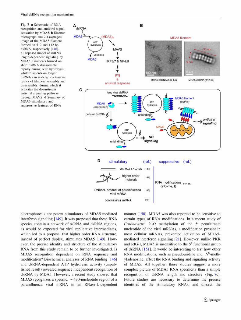
Melanoma differentiation-associated gene 5 (MDA5)
Our understanding of MDA5 lags significantly behind that
of RIG-I, despite the conserved domain architecture and
the shared signaling adaptor, MAVS (Fig. 7a). The
molecular features of RNA recognized by MDA5 have
long remained enigmatic, but a pioneering work by Kato
et al. [145] revealed that dsRNA length is the major
determinant that allows MDA5 to distinguish between
cellular and viral dsRNAs. While RIG-I prefers short
dsRNAs, MDA5-mediated signaling positively correlates
with the length of dsRNA in the range of *1–7 kb [145].
The length discrimination at this scale distinguishes MDA5
from that of other dsRNA sensors such as PKR, OAS, and
TLR3.
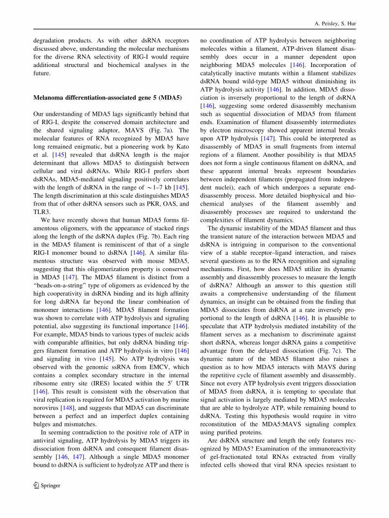
We have recently shown that human MDA5 forms fil-
amentous oligomers, with the appearance of stacked rings
along the length of the dsRNA duplex (Fig. 7b). Each ring
in the MDA5 filament is reminiscent of that of a single
RIG-I monomer bound to dsRNA [146]. A similar fila-
mentous structure was observed with mouse MDA5,
suggesting that this oligomerization property is conserved
in MDA5 [147]. The MDA5 filament is distinct from a
‘‘beads-on-a-string’’ type of oligomers as evidenced by the
high cooperativity in dsRNA binding and its high affinity
for long dsRNA far beyond the linear combination of
monomer interactions [146]. MDA5 filament formation
was shown to correlate with ATP hydrolysis and signaling
potential, also suggesting its functional importance [146].
For example, MDA5 binds to various types of nucleic acids
with comparable affinities, but only dsRNA binding trig-
gers filament formation and ATP hydrolysis in vitro [146]
and signaling in vivo [145]. No ATP hydrolysis was
observed with the genomic ssRNA from EMCV, which
contains a complex secondary structure in the internal
ribosome entry site (IRES) located within the 50 UTR
[146]. This result is consistent with the observation that
viral replication is required for MDA5 activation by murine
norovirus [148], and suggests that MDA5 can discriminate
between a perfect and an imperfect duplex containing
bulges and mismatches.
In seeming contradiction to the positive role of ATP in
antiviral signaling, ATP hydrolysis by MDA5 triggers its
dissociation from dsRNA and consequent filament disas-
sembly [146, 147]. Although a single MDA5 monomer
bound to dsRNA is sufficient to hydrolyze ATP and there is
no coordination of ATP hydrolysis between neighboring
molecules within a filament, ATP-driven filament disas-
sembly does occur in a manner dependent upon
neighboring MDA5 molecules [146]. Incorporation of
catalytically inactive mutants within a filament stabilizes
dsRNA bound wild-type MDA5 without diminishing its
ATP hydrolysis activity [146]. In addition, MDA5 disso-
ciation is inversely proportional to the length of dsRNA
[146], suggesting some ordered disassembly mechanism
such as sequential dissociation of MDA5 from filament
ends. Examination of filament disassembly intermediates
by electron microscopy showed apparent internal breaks
upon ATP hydrolysis [147]. This could be interpreted as
disassembly of MDA5 in small fragments from internal
regions of a filament. Another possibility is that MDA5
does not form a single continuous filament on dsRNA, and
these apparent internal breaks represent boundaries
between independent filaments (propagated from indepen-
dent nuclei), each of which undergoes a separate end-
disassembly process. More detailed biophysical and bio-
chemical analyses of the filament assembly and
disassembly processes are required to understand the
complexities of filament dynamics.
The dynamic instability of the MDA5 filament and thus
the transient nature of the interaction between MDA5 and
dsRNA is intriguing in comparison to the conventional
view of a stable receptor–ligand interaction, and raises
several questions as to the RNA recognition and signaling
mechanisms. First, how does MDA5 utilize its dynamic
assembly and disassembly processes to measure the length
of dsRNA? Although an answer to this question still
awaits a comprehensive understanding of the filament
dynamics, an insight can be obtained from the finding that
MDA5 dissociates from dsRNA at a rate inversely pro-
portional to the length of dsRNA [146]. It is plausible to
speculate that ATP hydrolysis mediated instability of the
filament serves as a mechanism to discriminate against
short dsRNA, whereas longer dsRNA gains a competitive
advantage from the delayed dissociation (Fig. 7c). The
dynamic nature of the MDA5 filament also raises a
question as to how MDA5 interacts with MAVS during
the repetitive cycle of filament assembly and disassembly.
Since not every ATP hydrolysis event triggers dissociation
of MDA5 from dsRNA, it is tempting to speculate that
signal activation is largely mediated by MDA5 molecules
that are able to hydrolyze ATP, while remaining bound to
dsRNA. Testing this hypothesis would require in vitro
reconstitution of the MDA5:MAVS signaling complex
using purified proteins.
Are dsRNA structure and length the only features rec-
ognized by MDA5? Examination of the immunoreactivity
of gel-fractionated total RNAs extracted from virally
infected cells showed that viral RNA species resistant to
A. Peisley, S. Hur
123
Page 11
electrophoresis are potent stimulators of MDA5-mediated
interferon signaling [149]. It was proposed that these RNA
species contain a network of ssRNA and dsRNA regions,
as would be expected for viral replicative intermediates,
which led to a proposal that higher order RNA structure,
instead of perfect duplex, stimulates MDA5 [149]. How-
ever, the precise identity and structure of the stimulatory
RNA from this study remain to be further investigated. Is
MDA5 recognition dependent on RNA sequence and
modification? Biochemical analyses of RNA binding [146]
and dsRNA-dependent ATP hydrolysis activity (unpub-
lished result) revealed sequence independent recognition of
dsRNA by MDA5. However, a recent study showed that
MDA5 recognizes a specific, *430-nucleotide region of a
parainfluenza viral mRNA in an RNase-L-dependent
manner [150]. MDA5 was also reported to be sensitive to
certain types of RNA modifications. In a recent study of
Coronavirus, 20-O methylation of the 50 penultimate
nucleotide of the viral mRNAs, a modification present in
most cellular mRNAs, prevented activation of MDA5-
mediated interferon signaling [21]. However, unlike PKR
and RIG-I, MDA5 is insensitive to the 50 functional group
of dsRNA [151]. It would be interesting to test how other
RNA modifications, such as pseudouridine and N6-meth-
yladenosine, affect the RNA binding and signaling activity
of MDA5. All together, these studies suggest a more
complex picture of MDA5 RNA specificity than a simple
recognition of dsRNA length and structure (Fig. 7c).
Future studies are necessary to determine the precise
identities of the stimulatory RNAs, and dissect the
Fig. 7 a Schematic of RNA
recognition and antiviral signal
activation by MDA5. b Electron
micrograph and 2D-averaged
image of the MDA5 filament
formed on 512 and 112 bp
dsRNA, respectively [146].
c Proposed model of dsRNA
length-dependent signaling by
MDA5. Filaments formed on
short dsRNA disassemble
rapidly during ATP hydrolysis,
while filaments on longer
dsRNA can undergo continuous
cycles of filament assembly and
disassembly, during which it
activates the downstream
antiviral signaling pathway
through MAVS. d Summary of
MDA5-stimulatory and
suppressive features of RNA
Viral dsRNA recognition mechanisms
123
Page 12
importance of sequence, secondary structure, and position
and type of nucleotide modification for MDA5 activation.
DHX9 and DDX1
Since the discovery of RLRs, several other helicases have
been identified that are involved in viral dsRNA sensing in
the cytoplasm. These helicases include DHX9 (a.k.a. RNA
helicase A) and DDX1, which were proposed to recognize
dsRNA (and also dsDNA for DHX9) in a manner inde-
pendent of RIG-I and MDA5 [152–154]. DHX9 was shown
to utilize two distinct domains to bind to dsRNA and DNA,
and bifurcate the downstream signal via MAVS and
Myd88, respectively [152, 153]. DDX1 was shown to bind
to dsRNA mimic, polyI:C, and form a complex with two
other helicases, DDX21 and DHX36, which interact with
TRIF and activate the antiviral response [154]. Interest-
ingly, both DHX9 and DDX1 were previously implicated
in diverse cellular functions other than viral nucleic acid
detection. For example, DHX9 was proposed to be
involved in gene regulation of cellular RNAs through
remodeling of ribonucleoprotein particles during transla-
tion [155] and DDX1 was shown to up-regulate NF-jB-
mediated transcriptional activity [156]. Exactly how they
coordinate these multiple divergent biological functions
and whether they recognize any additional features of RNA
beyond a simple duplex structure remain to be investigated.
Conclusions
The distinct backbone arrangement of dsRNA, in com-
parison to dsDNA or ssRNA, has supported a conventional
model that dsRNA structure, much like lipopolysaccha-
rides and other bacterial-specific chemical structures, is
recognized by innate immune receptors as a unique
molecular signature of viruses. However, recent studies of
the human transcriptome have revealed a prevalence and
diversity of non-coding RNAs, many of which contain a
range of secondary structures, varying from an imperfect
duplex of *21 bp in miRNAs to a near-perfect duplex of
*250–300 bp in inverted repeat elements. In parallel, the
innate immune sensors initially thought to recognize the
dsRNA structure are now known to detect and regulate its
antiviral activity in a manner dependent on other, and often
multiple, features of RNA, such as nucleotide modification,
50 functional groups, bulges, mismatches, and sequences.
While the list of these additional immune-stimulatory and
suppressive RNA features is expanding, our understanding
of the versatile selectivity of the receptors and effectors at
the level of molecular structure, thermodynamics and
kinetics remains rudimentary. Future structural and
biochemical studies would help us understand the under-
lying molecular principle, and perhaps identify
combinatorial rules that could be used in therapeutic RNAs
for either immune suppression or activation [157, 158].
References
1. Sadler AJ, Williams BRG (2008) Interferon-inducible antiviral
effectors. Nat Rev Immunol 8:559–568
2. Pichlmair A, Reis e Sousa C (2007) Innate recognition of
viruses. Immunity 27: 370–383
3. Borden EC, Williams BR (2011) Interferon-stimulated genes
and their protein products: what and how? J Interferon Cytokine
Res 31:1–4
4. Iwasaki A, Medzhitov R (2010) Regulation of adaptive immu-
nity by the innate immune system. Science 327:291–295
5. Samuel CE (2001) Antiviral actions of interferons. Clin
Microbiol Rev 14:778–809
6. Saenger W (1984) Principles of nucleic acid structure. Springer,
New York, p 113
7. Doyle M, Jantsch MF (2003) New and old roles of the double-
stranded RNA-binding domain. J Struct Biol 140:147–153
8. Saunders LR, Barber GN (2003) The dsRNA binding protein
family: critical roles, diverse cellular functions. FASEB
961–983
9. Pabo CO, Sauer RT (1984) Protein-DNA recognition. Ann Rev
Biochem 53:293–321
10. Watanabe T et al (2008) Endogenous siRNAs from naturally
formed dsRNAs regulate transcripts in mouse oocytes. Nature
453:539–543
11. Chiang HR et al (2010) Mammalian microRNAs: experimental
evaluation of novel and previously annotated genes. Genes Dev
24:992–1009
12. Athanasiadis A, Rich A, Maas S (2004) Widespread A-to-I RNA
editing of Alu-containing mRNAs in the human transcriptome.
PLoS Biol 2(12):e391
13. Wang Y, Leung FCC (2009) A study on genomic distribution and
sequence features of human long inverted repeats reveals species-
specific intronic inverted repeats. FEBS J 276:1986–1998
14. Yelin R et al (2003) Widespread occurrence of antisense tran-
scription in the human genome. Nat Biotechol 21:379–386
15. Rosok O, Sioud M (2004) Systematic identification of sense-
antisense transcripts in mammalian cells. Nat Biotechnol
22:104–108
16. McKenna SA et al (2007) Viral dsRNA inhibitors prevent self-
association and autophosphorylation of PKR. J Mol Biol
372:103–113
17. Vitali P, Scadden ADJ (2010) Double-stranded RNAs contain-
ing multiple IU pairs are sufficient to suppress interferon
induction and apoptosis. Nat Struct Mol Biol 17(9):1043–1050
18. Hornung V et al (2006) 50-Triphosphate RNA is the ligand for
RIG-I. Science 314:994–997
19. Nallagatla SR, Bevilacqua PC (2008) Nucleoside modifications
modulate activation of the protein kinase PKR in an RNA
structure-specific manner. RNA 14:1201–1203
20. Nallagatla SR et al (2007) 50-Triphosphate-dependent activation
of PKR by RNAs with short stem-loops. Science 318:
1455–1458
21. Zust R et al (2011) Ribose 20-O-methylation provides a
molecular signature for the distinction of self and non-self
mRNA dependent on the RNA sensor Mda5. Nat Immunol
12:137–143
A. Peisley, S. Hur
123
Page 13
22. Malathi K et al (2010) RNase L releases a small RNA from
HCV RNA that refolds into a potent PAMP. RNA
16:2108–2119
23. Anderson BR et al (2011) Nucleoside modifications in RNA
limit activation of 20-50-oligoadenylate synthetase and increase
resistance to cleavage by RNase L. Nucleic Acids Res
39:9329–9338
24. Garcia MA et al (2006) Impact of protein kinase PKR in cell
biology: from antiviral to antiproliferative action. Microbiol Mol
Biol Rev 70:1032–1060
25. Lemaire PA, Cole JL (2008) Mechanism of PKR activation by
dsRNA. J Mol Biol 381:351–360
26. Pfeller CK et al (2011) Protein kinase PKR and RNA adenosine
deaminase ADAR1: new roles for old players as modulators of
the interferon response. Curr Opin Immunol 23:573–582
27. McKenna SA et al (2007) Biophysical and biochemical inves-
tigations of dsRNA-activated kinase PKR. Methods Enzym
430:373–396
28. Cole JL (2010) Analysis of PKR activation using analytical
ultracentrifugation. Macromol Biosci 10:703–713
29. Lemaire PA, Lary J, Cole JL (2005) Mechanism of PKR acti-
vation: dimerization and kinase activation in the absence of
double-stranded RNA. J Mol Biol 345:81–90
30. Kim I, Liu CW, Puglisi JD (2006) Specific recognition of HIV
TAR RNA by the dsRNA binding domains (dsRBD1–dsRBD2)
of PKR. J Mol Biol 358:430–442
31. Bevilacqua PC, Cech TR (1996) Minor-groove recognition of
double-stranded RNA by the double-stranded RNA-binding
domain from the RNA-activated protein kinase PKR. Bio-
chemistry 35:9983–9994
32. Manche L et al (1992) Interactions between double-stranded
RNA regulators and the protein kinase DAI. Mol Cell Biol
12:5238–5248
33. Ryter JM, Schultz SC (1998) Molecular basis of double-stranded
RNA-protein interactions: structure of a dsRNA-binding domain
complexed with dsRNA. EMBO J 17:7505–7513
34. Gan J et al (2006) Structural insight into the mechanism of
double-stranded RNA processing by ribonuclease III. Cell
124:355–366
35. Cohen-Chalamish S et al (2009) Dynamic refolding of IFN-
gamma mRNA enables it to function as PKR activator and
translation template. Nat Chem Biol 5:896–903
36. Zheng X, Bevilacqua PC (2004) Activation of the protein kinase
PKR by short double-stranded RNAs with single-stranded tails.
RNA 10:1934–1945
37. Hundley HA, Bass BL (2010) ADAR editing in double-stranded
UTRs and other noncoding RNA sequences. Trends Biochem
Sci 35:377–383
38. Keegan LP et al (2004) Adenosine deaminases acting on RNA
(ADARs): RNA-editing enzymes. Genome Biol 5:209
39. Chen CX et al (2000) A third member of the RNA-specific
adenosine deaminase gene family, ADAR3, contains both single-
and double-stranded RNA binding domains. RNA 6:755–767
40. Wong SK, Sato S, Lazinski DW (2001) Substrate recognition by
ADAR1 and ADAR2. RNA 7:846–858
41. Lehmann KA, Bass BL (2000) Double-stranded RNA adenosine
deaminases ADAR1 and ADAR2 have overlapping specificities.
Biochemistry 39:12875–12884
42. Wahlstedt H, Ohman M (2011) Site-selective versus promiscu-
ous A-to-I editing. Wiley Interdiscip Rev RNA 2:761–771
43. Polson AG, Bass BL (1994) Preferential selection of adenosines
for modification by double-stranded RNA adenosine deaminase.
EMBO J 13:5701–5711
44. Lehmann KA, Bass BL (1999) The importance of internal loops
within RNA substrates of ADAR1. J Mol Biol 291:1–13
45. Dawson TR, Sansam CL, Emeson RB (2004) Structure and
sequence determinants required for the RNA editing of ADAR2
substrates. J Biol Chem 279:4941–4951
46. Higuchi M et al (1993) RNA editing of AMPA receptor subunit
GluR-B: a base-paired intron-exon structure determines position
and efficiency. Cell 75:1361–1370
47. Burns CM et al (1997) Regulation of serotonin-2C receptor
G-protein coupling by RNA editing. Nature 387:303–308
48. Bass BL, Weintraub H (1988) An unwinding activity that
covalently modifies its double-stranded RNA substrate. Cell
55:1089–1098
49. Nishikura K (2010) Functions and regulation of RNA editing by
ADAR deaminases. Ann Rev Biochem 79:321–349
50. Samuel CE (2011) Adenosine deaminases acting on RNA
(ADARs) are both antiviral and proviral. Virology 411:180–193
51. Ward SV et al (2011) RNA editing enzyme adenosine deami-
nase is a restriction factor for controlling measles virus
replication that also is required for embryogenesis. Proc Natl
Acad Sci USA 108:331–336
52. Cattaneo R et al (1988) Biased hypermutation and other genetic
changes in defective measles viruses in human brain infections.
Cell 55:255–265
53. Nie Y, Hammond GL, Yang JH (2007) Double-stranded RNA
deaminase ADAR1 increases host susceptibility to virus infec-
tion. J Virol 81:917–923
54. Li Z, Wolff KC, Samuel CE (2010) RNA adenosine deaminase
ADAR1 deficiency leads to increased activation of protein
kinase PKR and reduced vesicular stomatitis virus growth fol-
lowing interferon treatment. Virology 396:316–322
55. Casey JL (2006) RNA editing in hepatitis delta virus. Curr Top
Microbiol Immunol 307:67–89
56. Wong SK, Lazinski DW (2002) Replicating hepatitis delta virus
RNA is edited in the nucleus by the small form of ADAR1. Proc
Natl Acad Sci USA 99:15118–15123
57. Sato S, Cornillez-Ty C, Lazinski DW (2004) By inhibiting
replication, the large hepatitis delta antigen can indirectly reg-
ulate amber/W editing and its own expression. J Virol
78:8120–8134
58. Peng Z et al (2012) Comprehensive analysis of RNA-seq data
reveals extensive RNA editing in a human transcriptome. Nat
Biotechol 30:253–260
59. Paz-Yaacov N et al (2010) Adenosine-to-inosine RNA editing
shapes transcriptome diversity in primates. Proc Natl Acad Sci
USA 107:12174–12179
60. Alon S et al (2012) Systematic identification of edited mi-
croRNAs in the human brain. Genome Res [Epub ahead]
61. Carmi S, Borukhov I, Levanon EY (2011) Identification of
widespread ultra-edited human RNAs. PLoS Genet 7:e1002317
62. Heale BS, Keegan LP, O’Connell MA (2010) The effect of RNA
editing and ADARs on miRNA biogenesis and function. Adv
Exp Med Biol 700:76–84
63. Jepson JEC, Reenan RA (2008) RNA editing in regulating gene
expression in the brain. Biochim Biophys Acta 1779:459–470
64. Chen LL, Carmichael GG (2012) Nuclear editing of mRNA 30-UTR. Curr Top Microbiol Immunol 353:111–121
65. Wang Q, Carmichael GG (2004) Effects of length and location
on the cellular response to double-stranded RNA. Microbiol Mol
Biol Rev 68(3):432–452
66. Hartner JC et al (2008) ADAR1 is essential for the maintenance
of hematopoiesis and suppression of interferon signaling. Nat
Immunol 10:109–115
67. Wang Q et al (2000) Requirement of the RNA editing deami-
nase ADAR1 gene for embryonic erythropoiesis. Science
290:1765–1768
68. Hartner JC et al (2004) J Biol Chem 279:4894–4902
Viral dsRNA recognition mechanisms
123
Page 14
69. Wang Q et al (2004) Stress-induced apoptosis associated with
null mutation of ADAR1 RNA editing deaminase. J Biol Chem
279:4952–4961
70. Higuchi M et al (2000) Point mutation in an AMPA receptor
gene rescues lethality in mice deficient in the RNA-editing
enzyme ADAR2. Nature 406:78–81
71. Palladino MJ et al (2000) A-to-I pre-mRNA editing in Dro-sophila is primarily involved in adult nervous system function
and integrity. Cell 102:437
72. Tonkin LA et al (2002) RNA editing by ADARs is important for
normal behavior in Caenorhabditis elegans. EMBO J
21:6025–6035
73. Stefl R et al (2010) The solution structure of the ADAR2
dsRBM-RNA complex reveals a sequence-specific readout of
the minor groove. Cell 143:225–237
74. Ramos A et al (2000) RNA recognition by a Staufen double-
stranded RNA-binding domain. EMBO J 19:997–1009
75. Wu H et al (2004) Structural basis for recognition of the AGNN
tetraloop RNA fold by the double-stranded RNA binding domain
of Rnt1p RNase III. Proc Natl Acad Sci USA 101:8307–8312
76. Torralba S, Sojat J, Hartmann R (2008) 20-50 oligoadenylate
synthetase shares active site architecture with the archaeal CCA-
adding enzyme. Cell Mol Life Sci 65:2613–2620
77. Hovanessian AG et al (1987) Identification of 69-kd and 100-kd
forms of 2–5A synthetase in interferon-treated human cells by
specific monoclonal antibodies. EMBO J 6:1273–1280
78. Rebouillat D, Marie I, Hovanessian AG (1998) Molecular
cloning and characterization of two related and interferon-
induced 56-kDa and 30-kDa proteins highly similar to 2–5 oli-
goadenylate synthetase. Eur J Biochem 257:319–330
79. Kristiansen H et al (2011) The oligoadenylate synthetase family:
an ancient protein family with multiple antiviral activities.
J Interferon Cytokine Res 31:41–47
80. Wreschner DH et al (1981) Interferon action—sequence speci-
ficity of the ppp(A20p)nA-dependent ribonuclease. Nature
278:414–417
81. Floyd-Smith G, Slattery E, Lengyel P (1981) Interferon action:
RNA cleavage pattern of a (20-50)oligoadenylate-dependent
endonuclease. Science 212:1030–1032
82. Chakrabarti A, Jha BK, Silverman RH (2011) New insights into
the role of RNase-L in innate immunity. J Interferon Cytokine
Res 31:49–57
83. Kubota K et al (2004) Identification of 20-phosphodiesterase,
which plays a role in the 2–5A system regulated by interferon.
J Biol Chem 279:37832–37841
84. Knight M et al (1980) Radioimmune, radiobinding and HPLC
analysis of 2–5A and related oligonucleotides from intact cells.
Nature 288:189–192
85. Rebouillat D, Hovanessian AG (1999) The human 2,5-oligoade-
nylate synthetase family: interferon-induced proteins with unique
enzymatic properties. J Interferon Cytokine Res 19:295–308
86. Schoggins JW et al (2011) A diverse range of gene products are
effectors of the type I interferon antiviral response. Nature
472:481–485
87. Hartmann R et al (2003) Crystal structure of the 20-specific and
double-stranded RNA-activated interferon-induced antiviral
protein 20-50-oligoadenylate synthetase. Mol Cell 12:1173–1185
88. Sarkar SN et al (1999) The nature of the catalytic domain of 20-50-oligoadenylate synthetases. J Biol Chem 274:25535–25542
89. Hartmann R et al (1998) Activation of 20-50 oligoadenylate
synthetase by single-stranded and double-stranded RNA apta-
mers. J Biol Chem 273:3236–3246
90. Desai SY, Sen GC (1997) Effects of varying lengths of double-
stranded RNA on binding and activation of 20-50-oligoadenylate
synthetase. J Interferon Cytokine Res 17:531–536
91. Ghosh A et al (1997) Enzymatic activity of 20-50-oligoadenylate
synthetase is impaired by specific mutations that affect oligo-
merization of the protein. J Biol Chem 272:33220–33226
92. Kodym R, Kodym E, Story MD (2009) 20-50-Oligoadenylate
synthetase is activated by a specific RNA sequence motif.
Biochem Biophy Res Comm 388:317–322
93. Grosjean H, Benne R (1998) Modification and editing of RNA.
American Society for Microbiology, Washington
94. Nilsen TW et al (1982) Heterogeneous nuclear RNA promotes
synthesis of (20,50)oligoadenylate and is cleaved by the (20,50)oligoadenylate-activated endoribonuclease. Mol Cell Biol
2:154–160
95. Molinaro RJ et al (2006) Selection and cloning of poly(rC)-
binding protein 2 and Raf kinase inhibitor protein RNA acti-
vators of 20,50-oligoadenylate synthetase from prostate cancer
cells. Nucleic Acids Res 34:6684–6695
96. Kumagai Y, Takeuchi O, Akira S (2008) Pathogen recognition
by innate receptors. J Infect Chemother 14:86–92
97. Blasius AL, Beutler B (2010) Intracellular Toll-like receptors.
Immunity 32:305–315
98. Temperley ND et al (2008) Evolution of the chicken Toll-like
receptor gene family: a story of gene gain and gene loss. BMC
Genomics 9:62
99. Diebold SS et al (2004) Innate antiviral responses by means of
TLR7-mediated recognition of single-stranded RNA. Science
303:1529–1531
100. Heil F et al (2004) Species-specific recognition of single-
stranded RNA via Toll-like receptor 7 and 8. Science
303:1526–1529
101. Lund JM et al (2004) Recognition of single-stranded RNA
viruses by Toll-like receptor 7. Proc Natl Acad Sci USA
101:5598–5603
102. Hemmi H et al (2000) A Toll-like receptor recognizes bacterial
DNA. Nature 408:740–745
103. Alexopoulou L et al (2001) Recognition of double-stranded
RNA and activation of NF-jB by Toll-like receptor 3. Nature
413:732–738
104. Wang Y et al (2010) Dimerization of Toll-like receptor 3
(TLR3) is required for ligand binding. J Biol Chem
285:36836–36841
105. Leonard JN et al (2008) The TLR3 signaling complex forms by
cooperative receptor dimerization. Proc Natl Acad Sci USA
105(1):258–263
106. Oshiumi H et al (2003) TICAM-1, an adaptor molecule that
participates in Toll-like receptor 3-mediated interferon-bold beta
induction. Nat Immunol 4:161–167
107. Bell JK et al (2005) The molecular structure of the Toll-like
receptor 3 ligand-binding domain. Proc Natl Acad Sci USA
102:10976–10980
108. Jim MS, Lee J-O (2008) Structures of the Toll-like receptor
family and its ligand complexes. Immunity 29:182–191
109. Liu L et al (2008) Structural basis of Toll-like receptor 3 sig-
naling with double-stranded RNA. Science 320:379–381
110. Sioud M (2006) Innate sensing of self and non-self RNAs by
Toll-like receptors. Trends Mol Med 12:167–176
111. Schroder M, Bowie AG (2005) TLR3 in antiviral immunity: key
player or bystander? Trends Immunol 26:462–468
112. Kariko K et al (2004) mRNA is an endogenous ligand for Toll-
like receptor 3. J Biol Chem 279:12542–12550
113. Cavassani KA et al (2008) TLR3 is an endogenous sensor of
tissue necrosis during acute inflammatory events. J Exp Med
205:2609–2621
114. Duffy K.E et al (2012) Lateral clustering of TLR3:dsRNA sig-
naling units revealed by TLR3ecd:3Fabs quaternary structure.
J Mol Biol [Epub ahead]
A. Peisley, S. Hur
123
Page 15
115. Kleinman ME et al (2008) Sequence- and target-independent
angiogenesis suppression by siRNA via TLR3. Nature
452:591–597
116. Kariko K et al (2004) Small interfering RNAs mediate sequence
independent gene suppression and induce immune activation by
signaling through Toll-like receptor 3. J Immunol 172:6545–
6549
117. Pirher N et al (2008) A second binding site for double-stranded
RNA in TLR3 and consequences for interferon activation. Nat
Struc Mol Biol 15:761–763
118. Kariko K et al (2005) Suppression of RNA recognition by Toll-
like receptors: the impact of nucleoside modification and the
evolutionary origin of RNA. Immunity 23:165–175
119. Yoneyama M et al (2004) The RNA helicase RIG-I has an
essential function in double-stranded RNA-induced innate
antiviral responses. Nat Immunol 5(7):730–737
120. Yoneyama M et al (2005) Shared and unique functions of the
DExD/H-Box helicases RIG-I, MDA5, and LGP2 in antiviral
innate immunity. J Immunol 175:2851–2858
121. Seth RB et al (2005) Identification and characterization of
MAVS, a mitochondrial antiviral signaling protein that activates
NF-jB and IRF3. Cell 122:699–782
122. Xu LG et al (2005) VISA is an adapter protein required for
virus-triggered IFN-b signaling. Mol Cell 19:727–740
123. Kawai T et al (2005) IPS-1, an adaptor triggering RIG-I- and
Mda5-mediated type I interferon induction. Nat Immunol
6(10):981–988
124. Satoh T et al (2010) LGP2 is a positive regulator of RIG-I and
MDA5-mediated antiviral responses. Proc Natl Acad Sci USA
107:1512–1517
125. Saito T, Hirai R, Loo YM, Owen D, Johnson CL, Sinha SC,
Akira S, Fujita T, Gale M Jr (2007) Regulation of innate anti-
viral defenses through a shared repressor domain in RIG-I and
LGP2. Proc Natl Acad Sci USA 104(2):582–587
126. Gack MU et al (2007) TRIM25 RING-finger E3 ubiquitin ligase
is essential for RIG-I-mediated antiviral activity. Nature
446:916–920
127. Jiang X et al (2012) Ubiquitin-induced oligomerization of the
RNA sensors RIG-I and MDA5 activates antiviral innate
immune response. Immunity 36:959–973
128. Hou F et al (2011) MAVS forms functional prion-like aggre-
gates to activate and propagate antiviral innate immune
response. Cell 146:1–14
129. Pichlmair A et al (2006) RIG-I-mediated antiviral responses to
single-stranded RNA bearing 50-phosphates. Science
314:997–1001
130. Schlee M et al (2009) Recognition of 50triphosphate by RIG-I
helicase requires short blunt double-stranded RNA as contained
in panhandle of negative-strand virus. Immunity 31(1):25–34
131. Marques JT et al (2006) A structural basis for discriminating
between self and nonself double-stranded RNAs in mammalian
cells. Nat Biotechnol 24:559–565
132. Saito T et al (2008) Innate immunity induced by composition-
dependent RIG-I recognition of hepatitis C virus RNA. Nature
454:523–527
133. Uzri D, Gehrke L (2009) Nucleotide sequences and modifica-
tions that determine RIG-I/RNA binding and signaling
activities. J Virol 83:4174–4184
134. Malathi K et al (2007) Small self-RNA generated by RNase L
amplifies antiviral innate immunity. Nature 448:816–819
135. Takahasi K et al (2008) Nonself RNA-sensing mechanism of
RIG-I helicase and activation of antiviral immune responses.
Mol Cell 29(4):428–440
136. Cui S et al (2008) The C-terminal regulatory domain is the RNA
50-triphosphate sensor of RIG-I. Mol Cell 29(2):169–179
137. Lu C et al (2010) The structural basis of 50 triphosphate double-
stranded RNA recognition by RIG-I C-terminal domain.
Structure 18:1032–1043
138. Wang Y et al (2010) Structural and functional insights into 50-ppp RNA pattern recognition by the innate immune receptor
RIG-I. Nat Struct Mol Biol 17(7):781–787
139. Jiang F et al (2011) Structural basis of RNA recognition and
activation by innate immune receptor RIG-I. Nature
479(7373):423–427
140. Luo D et al (2011) Structural insights into RNA recognition by
RIG-I. Cell 147:409–422
141. Kowalinski E et al (2011) Structural basis for the activation of
innate immune pattern-recognition receptor RIG-I by viral
RNA. Cell 147:423–435
142. Binder M et al (2011) Molecular mechanism of signal percep-
tion and integration by the innate immune sensor retinoic acid
inducible gene-I. J Biol Chem 286:27278–27287
143. Bamming D, Horvath CM (2009) Regulation of signal trans-
duction by enzymatically inactive antiviral RNA helicase
proteins MDA5, RIG-I, and LGP2. J Biol Chem 284(15):
9700–9712
144. Myong S et al (2009) Cytosolic viral sensor RIG-I Is a 50-tri-phosphate-dependent translocase on double-stranded RNA.
Science 323:1070–1074
145. Kato H et al (2008) Length-dependent recognition of double-
stranded ribonucleic acids by retinoic acid-inducible gene-I and
melanoma differentiation-associated gene 5. J Exp Med
205(7):1601–1610
146. Peisley A et al (2011) Cooperative assembly and dynamic dis-
assembly of MDA5 filaments for viral dsRNA recognition. Proc
Natl Acad Sci USA 108(52):21010–21015
147. Berke IC, Modis Y (2012) MDA5 cooperatively forms dimers
and ATP-sensitive filaments upon binding double-stranded
RNA. EMBO J 7:1714–1726
148. McCartney SA et al (2008) MDA-5 recognition of a murine
norovirus. PLoS Pathog 4(7):e1000108
149. Pichlmair A et al (2009) Activation of MDA5 requires higher-
order RNA structures generated during virus infection. J Virol
83(20):10761–10769
150. Luthra P et al (2011) Activation of IFN-b expression by a viral
mRNA through RNase L and MDA5. Proc Natl Acad Sci USA
108:2118–2123
151. Kato H et al (2006) Differential roles of MDA5 and RIG-I he-
licases in the recognition of RNA viruses. Nature 441:101–105
152. Kim T et al (2010) Aspartate-glutamate-alanine-histidine box
motif (DEAH)/RNA helicase A helicases sense microbial DNA
in human plasmacytoid dendritic cells. Proc Natl Acad Sci USA
107:15181–15186
153. Zhang Z et al (2011) DHX9 pairs with IPS-1 to sense double-
stranded RNA in myeloid dendritic cells. J Immunol
187:4501–4508
154. Zhang Z et al (2011) DDX1, DDX21, and DHX36 helicases
form a complex with the adaptor molecule TRIF to sense
dsRNA in dendritic cell. Immunity 34:866–878
155. Jin J et al (2011) Evidence that Lin28 stimulates translation by
recruiting RNA helicase A to polysomes. Nucleic Acids Res
39:3724–3734
156. Ishaq M et al (2009) The DEAD-box RNA helicase DDX1
interacts with RelA and enhances nuclear factor kappaB-medi-
ated transcription. J Cell Biochem 106:297–305
157. Hennessy EJ, Parker AE, O’Neill LAJ (2010) Targeting Toll-
like receptors: emerging therapeutics? Nat Rev Drug Discov
9:293–307
158. Ireton RC, Gale M Jr (2011) RIG-I like receptors in antiviral
immunity and therapeutic applications. Viruses 3:906–919
Viral dsRNA recognition mechanisms
123