Multi-nozzle deposition for construction of 3D biopolymer tissue scaffolds S. Khalil, J. Nam and W. Sun Laboratory for Computer-Aided Tissue Engineering, Department of Mechanical Engineering and Mechanics, Drexel University, Philadelphia, Pennsylvania, USA Abstract Purpose – To introduce recent research and development of biopolymer deposition for freeform fabrication of three-dimensional tissue scaffolds that is capable of depositing bioactive ingredients. Design/methodology/approach – A multi-nozzle biopolymer deposition system is developed, which is capable of extruding biopolymer solutions and living cells for freeform construction of 3D tissue scaffolds. The deposition process is biocompatible and occurs at room temperature and low pressures to reduce damage to cells. In contrast with other systems, this system is capable of, simultaneously with scaffold construction, depositing controlled amount of cells, growth factors, or other bioactive compounds with precise spatial position to form complex cell-seeded tissue constructs. The examples shown are based on sodium alginate solutions and poly-1-caprolactone (PCL). Studies of the biopolymer deposition feasibility, structural formability, and different material deposition through a multi-nozzle heterogeneous system are conducted and presented. Findings – Provides information about the biopolymer deposition using different nozzle systems, the relations of process parameters on deposition flow rate and scaffold structural formability. Three-dimensional alginate-based scaffolds and scaffold embedded with living cells can be freeform constructed according to various design configurations at room temperature without using toxic materials. Research limitations/implications – Other biopolymers may also be studied for structure formation. Studying cell viability and cellular tissue engineering behavior of the scaffolds after the cell deposition should be further investigated. Practical implications – A very useful and effective tool for construction of bioactive scaffolds for tissue engineering applications based on a multi- nozzle biopolymer deposition. Originality/value – This paper describes a novel process and manufacturing system for fabrication of bioactive tissue scaffolds, automatic cell loading, and heterogeneous tissue constructs for emerging regenerative medicine. Keywords Structures, Computer aided manufacturing, Coating processes, Body systems and organs Paper type Research paper 1. Introduction Tissue engineering is considered to be the most innovative approach for tackling many diseases and body parts that need to be replaced (Langer, 2000; Langer and Vacanti, 1993; Vacanti and Langer, 1999). Up to date, the ideal approach for perusing engineered tissue parts involves three subsequent procedures. First, the biological cells are identified and gathered in sufficient numbers. Second, the suitable biomaterial is identified and designed accordingly to host the gathered cells. Finally, the cells are seeded into the biomaterial for cell culturing in vitro or in vivo. Another approach of engineering tissue is to encourage cells in the host to populate in to the biomaterial structure upon implantation (Bhatia and Chen, 1999). The biomaterial that is designed for housing the cells is referred to as scaffolds and is usually three-dimensional. Biomaterials are used as scaffolds for their uniqueness of being biocompatible, which mean that they will not be rejected by the body upon implantation. These biomaterials are fabricated from a wide range of materials that could be inorganic synthetic such as metals, ceramics, and polymers or from organic materials such as proteins, chitosan, and alginate. Scaffolds used in tissue engineering are also required to have specific properties that are vital for cell regeneration (Leong et al., 2003). In order to fabricate scaffolds with such properties, a fabrication method that maintains a high level of accuracy is necessary to maintain the consistency and repeatability in accordance to the initial design. Unlike the conventional fabrication techniques, solid freeform fabrication (SFF) has no restriction to on shape control and consistency. SFF are computerized fabrication techniques that can rapidly produce highly complex three-dimensional objects using data from CAD systems and computer medical imaging equipment such as MRI and CT scans. The fabricated three-dimensional structures are built by reducing CAD designs of particular prototypes into a group of sliced two-dimensional layers, to where the prototyping material is deposited to build the final structure in a layer-by-layer process. This makes SFF techniques very attractive for tissue engineering scaffold fabrication applications (Sun et al., 2004a). The Emerald Research Register for this journal is available at www.emeraldinsight.com/researchregister The current issue and full text archive of this journal is available at www.emeraldinsight.com/1355-2546.htm Rapid Prototyping Journal 11/1 (2005) 9–17 q Emerald Group Publishing Limited [ISSN 1355-2546] [DOI 10.1108/13552540510573347] The authors acknowledge support from Therics, Inc. for the solenoid nozzle system and partial material delivery system, as well as Dr F. Wang’s early assistant in the development of the motion and control system for the multi-nozzle deposition system. 9
Transcript
Multi-nozzle deposition for constructionof 3D biopolymer tissue scaffolds
S Khalil J Nam and W Sun
Laboratory for Computer-Aided Tissue Engineering Department of Mechanical Engineering and MechanicsDrexel University Philadelphia Pennsylvania USA
AbstractPurpose ndash To introduce recent research and development of biopolymer deposition for freeform fabrication of three-dimensional tissue scaffolds thatis capable of depositing bioactive ingredientsDesignmethodologyapproach ndash A multi-nozzle biopolymer deposition system is developed which is capable of extruding biopolymer solutionsand living cells for freeform construction of 3D tissue scaffolds The deposition process is biocompatible and occurs at room temperature and lowpressures to reduce damage to cells In contrast with other systems this system is capable of simultaneously with scaffold construction depositingcontrolled amount of cells growth factors or other bioactive compounds with precise spatial position to form complex cell-seeded tissue constructsThe examples shown are based on sodium alginate solutions and poly-1-caprolactone (PCL) Studies of the biopolymer deposition feasibility structuralformability and different material deposition through a multi-nozzle heterogeneous system are conducted and presentedFindings ndash Provides information about the biopolymer deposition using different nozzle systems the relations of process parameters on depositionflow rate and scaffold structural formability Three-dimensional alginate-based scaffolds and scaffold embedded with living cells can be freeformconstructed according to various design configurations at room temperature without using toxic materialsResearch limitationsimplications ndash Other biopolymers may also be studied for structure formation Studying cell viability and cellular tissueengineering behavior of the scaffolds after the cell deposition should be further investigatedPractical implications ndash A very useful and effective tool for construction of bioactive scaffolds for tissue engineering applications based on a multi-nozzle biopolymer depositionOriginalityvalue ndash This paper describes a novel process and manufacturing system for fabrication of bioactive tissue scaffolds automatic cell loadingand heterogeneous tissue constructs for emerging regenerative medicine
Keywords Structures Computer aided manufacturing Coating processes Body systems and organs
Paper type Research paper
1 Introduction
Tissue engineering is considered to be the most innovativeapproach for tackling many diseases and body parts that needto be replaced (Langer 2000 Langer and Vacanti 1993Vacanti and Langer 1999) Up to date the ideal approach forperusing engineered tissue parts involves three subsequentprocedures First the biological cells are identified andgathered in sufficient numbers Second the suitablebiomaterial is identified and designed accordingly to hostthe gathered cells Finally the cells are seeded into thebiomaterial for cell culturing in vitro or in vivo Anotherapproach of engineering tissue is to encourage cells in the hostto populate in to the biomaterial structure upon implantation(Bhatia and Chen 1999) The biomaterial that is designed forhousing the cells is referred to as scaffolds and is usuallythree-dimensional Biomaterials are used as scaffolds for theiruniqueness of being biocompatible which mean that they will
not be rejected by the body upon implantation Thesebiomaterials are fabricated from a wide range of materials thatcould be inorganic synthetic such as metals ceramics andpolymers or from organic materials such as proteins chitosanand alginateScaffolds used in tissue engineering are also required to
have specific properties that are vital for cell regeneration(Leong et al 2003) In order to fabricate scaffolds with suchproperties a fabrication method that maintains a high levelof accuracy is necessary to maintain the consistency andrepeatability in accordance to the initial design Unlike theconventional fabrication techniques solid freeformfabrication (SFF) has no restriction to on shape control andconsistency SFF are computerized fabrication techniquesthat can rapidly produce highly complex three-dimensionalobjects using data from CAD systems and computer medicalimaging equipment such as MRI and CT scans Thefabricated three-dimensional structures are built by reducingCAD designs of particular prototypes into a group of slicedtwo-dimensional layers to where the prototyping material isdeposited to build the final structure in a layer-by-layerprocess This makes SFF techniques very attractive for tissueengineering scaffold fabrication applications (Sun et al2004a)
The Emerald Research Register for this journal is available at
wwwemeraldinsightcomresearchregister
The current issue and full text archive of this journal is available at
wwwemeraldinsightcom1355-2546htm
Rapid Prototyping Journal
111 (2005) 9ndash17
q Emerald Group Publishing Limited [ISSN 1355-2546]
[DOI 10110813552540510573347]
The authors acknowledge support from Therics Inc for the solenoidnozzle system and partial material delivery system as well as Dr F Wangrsquosearly assistant in the development of the motion and control system for themulti-nozzle deposition system
9
Hydrogels based on both natural and synthetic polymershave been of interest for encapsulation of cells and mostrecently such hydrogels have become especially attractive tothe new field of tissue engineering as matrices (Hoffman2002 Lee and Mooney 2001) PCL has also proven to be agood candidate for scaffolds (Ciapetti et al 2003 Zein et al2002) The deposition of polymers can be controlled throughvarious techniques for the fabrication process (Poncelet et al1999 Calvert and Liu 1998 Landers et al 2002 Vozziet al 2002 Wang et al 2004 Sun et al nd) This paperpresents three types of nozzle systems that can be usedto deposit sodium alginate solutions and one to depositPCL for the fabrication of three-dimensional structuresA comparison of the four systems is also presented inaddition to the operating parameters and performance of thesystems
2 System configuration
We have developed a multi-nozzle biopolymer depositionsystem which is capable of extruding biopolymer solutionsand living cells for freeform construction of 3D tissuescaffolds (Sun et al 2004b 2003 nd Khalil et al 2004)The deposition process is biocompatible and occurs at roomtemperature and low pressures to reduce damage to cellsOther SFF manufacturing methods utilize harsh solventshigh pressures or temperatures or post-processing methodsthat are not suited for working with bioactive materialsBy contrast our system is capable of simultaneously with thescaffold construction depositing controlled amount of cellsgrowth factors or other bioactive compounds with precisespatial position to form well-defined cell-seeded tissueconstructs This process may solve the problem of cellloading of preformed scaffolds which hitherto has been asignificant barrier in tissue engineeringAn information pipeline of multi-nozzle biopolymer
deposition system for freeform fabrication of tissueconstructs is shown in Figure 1 As shown in the figure thedata processing system processes the designed scaffold modeland converts it into a layered process tool path The motioncontrol system is driven by the layered manufacturingtechnique the material delivery system consists of multiple
nozzles with different types and sizes thus enabling thedeposition of specified hydrogels with different viscosities forconstructing 3D tissue scaffolds Four types of the nozzles areused in the system solenoid-actuated nozzles piezoelectricglass capillary nozzles pneumatic syringe nozzles and spraynozzles with size ranges varying from 30 to 500mm Thesystem can continuously extrude hydrogel gels or formhydrogels in single droplets with picoliter volumes Themultiple nozzle capability allows us to simultaneously depositcells growth factors and scaffold materials thus enabling theconstruction of heterogeneous scaffolds with bioactivecompounds or establishing functional gradient scaffoldswith different mechanicalstructural properties in differentscaffold regionsThe multi-nozzle biopolymer deposition system consists of
four different micro-nozzles pneumatic microvalvepiezoelectric nozzle solenoid valve and precision extrusiondeposition (PED) nozzle as shown in Figure 2 A materialdelivery system was assembled to supply the nozzles with theappropriate biopolymer The system consisted of an airpressure supply both positive and negative a materialcontainer or reservoir and a material delivery tube Eachnozzle system had its independent parameters adjusted asrequired such as the air pressure and biopolymerconcentrationAs an example Figure 3 shows a schematic diagram of
pneumatic microvalve system Pneumatic microvalve is atypical mechanical valve that opens and closes the valve viaan applied air pressure and is regulated by a controller Thesystem could work in extrusion or droplet mode Inextrusion mode the controller applies pressure to open thevalve by lifting the piston against the spring that lifts theneedle from the needle seat The biopolymer material is thenextruded out of the nozzle tip under an applied pressure thatis adjusted through the material delivery system Theextrusion is ended when the controller shuts the valve byplacing the needle back to the needle seat The pneumaticvalve could perform in droplet mode by repeating thecontinuous mode in a cyclic manner Multiple pneumaticvalves were simultaneously operated for performingheterogeneous deposition in the development of the 3Dalginate scaffolds
Figure 1 Configuration of biopolymer deposition system
Multi-nozzle deposition
S Khalil J Nam and W Sun
Rapid Prototyping Journal
Volume 11 middot Number 1 middot 2005 middot 9ndash17
10
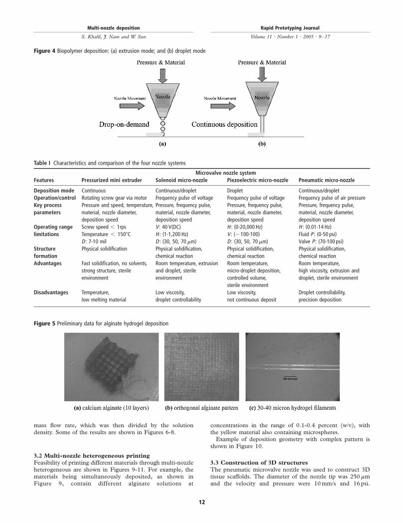
In general there are two modes of biopolymer deposition theextrusion mode and the droplet mode In the extrusion modethe material is extruded out of the nozzle tip under an appliedpressure This mode can basically lay down the material in theform of line structures to create the desired model by movingthe nozzle tip over a substrate in the designed path Thisprocess can be repeated layer by layer to develop a freeformfabricated part In the droplet mode the material is depositedin the form of droplets that is controlled by using a frequencyfunction and key parameters in the nozzle system settingsThe droplet mode can form a structured layer by depositingmultiple droplets at desired locations on a substrate Similarlythis process can be repeated to fabricate a 3D structureFigure 4 shows a schematic diagram of the extrusion anddroplet mode for the depositionEach nozzle system is unique in its method of operation
which makes each system have its advantages and limitationsover the others All nozzle systems have a material deliverysystem however the detailed set-up for each system isdifferent and the material delivery system parameters such asair pressure are not controlled by in-house software
Characteristics and comparison of the four nozzle systemswith their advantages and limitations is shown in Table I
3 Preliminary results
31 Preliminary data on biopolymer deposition
We have conducted some preliminary studies on using themulti-nozzle biopolymer deposition system to construct 3Dbiopolymer based tissue scaffolds For example shown inFigure 5 are several 3D hydrogel scaffolds (ten layerscalcium alginate) extruded as a 3 percent (wv) alginatefilament within a cross-linking solution (Figure 5(a)) andsimple alginate geometrical pattern (Figure 5(b)) Dependingupon the size of the syringe nozzle the pressures used andthe type of deposition method (extrusion) we were able tocreate alginate filaments within the 30-40 micron range(Figure 5(c)) for 3 percent (wv) sodium alginate solutionwith a 5 percent (wv) calcium chloride cross-linking solutionat 050 psiThe deposition flow rates were calculated by weighing the
solution mass on a balance at a time interval to obtain the
Figure 2 Schematic of system set-up for biopolymer depositions
Figure 3 Schematic diagram of a pneumatic microvalve system
Multi-nozzle deposition
S Khalil J Nam and W Sun
Rapid Prototyping Journal
Volume 11 middot Number 1 middot 2005 middot 9ndash17
11
mass flow rate which was then divided by the solutiondensity Some of the results are shown in Figures 6-8
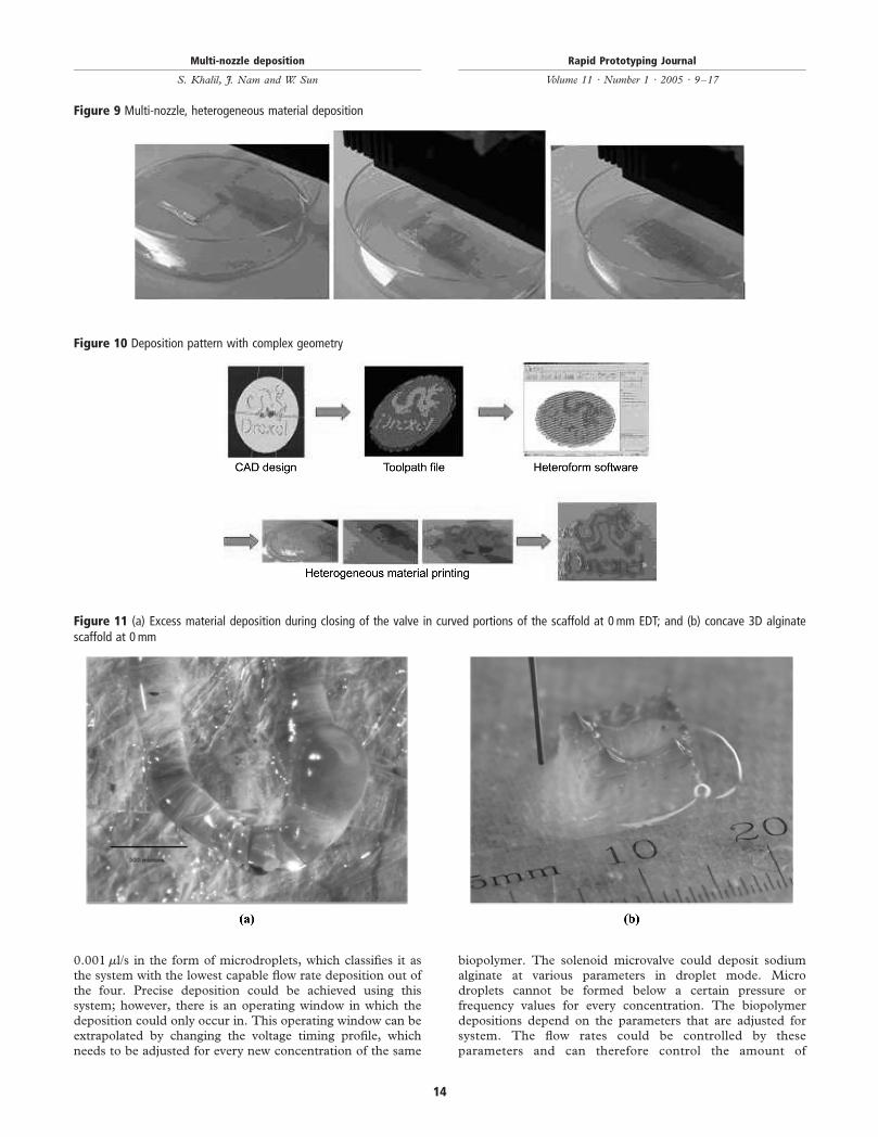
32 Multi-nozzle heterogeneous printing
Feasibility of printing different materials through multi-nozzleheterogeneous are shown in Figures 9-11 For example thematerials being simultaneously deposited as shown inFigure 9 contain different alginate solutions at
concentrations in the range of 01-04 percent (wv) withthe yellow material also containing microspheresExample of deposition geometry with complex pattern is
shown in Figure 10
33 Construction of 3D structures
The pneumatic microvalve nozzle was used to construct 3Dtissue scaffolds The diameter of the nozzle tip was 250mmand the velocity and pressure were 10mms and 16psi
Table I Characteristics and comparison of the four nozzle systems
Microvalve nozzle system
Features Pressurized mini extruder Solenoid micro-nozzle Piezoelectric micro-nozzle Pneumatic micro-nozzle
Volume 11 middot Number 1 middot 2005 middot 9ndash17
12
Experiments were set to investigate the effect of the earlydeposition termination (EDT) on the geometrical formationof the 3D scaffolds The first EDT tested was at 0mmswhich means that the valve was closed at the very end of theformation of every straight line The results were analyzedusing an optical digital camera that viewed that excessmaterial was deposited at the curved line structures at theedge perimeter of each layer It was observed that thediameter of the curved sections (310mm) were much larger
than the straight lines (270mm) shown in Figure 11(a) As aresult the 3D alginate structure was thicker around the edgesand caused concaving of the scaffold after 20 layers as shownin Figure 11(b)The second EDT tested was at 1mms so that the valve
would close 1mm prior to the end point of the straight lineformation The image shown in Figure 12(a) reveals thatthe diameter of the curved portion of the alginate strand isalmost equal to the straight strands As a result the 3Dalginate structure is fabricated uniformly up to 40 layerswithout geometrical defects as shown in Figure 12(b)Figure 14(a) and (b) shows the third and last EDTexperiment for the valve close 2mm prior to the end pointof the straight line formation It was observed through theimages in Figure 13 that the lines formed were not placed aswas originally designed and as a result constructed anirregularly shaped alginate 3D scaffold This resulted from therelatively large EDT (2mms) that caused dragging of thealginate strands every time the valve was closed The EDT atthis parameter forced the nozzle tip without any deposition fora particular distance The alginate strand was attached to thenozzle tip because of the calcium chloride crosslinkingsolution during the time where the nozzle was travelingwithout any deposition An analytical model for the EDT iscurrently under developmentThe distance recorded between the struts was found to be
920mm and the length of the square pore was found to be650mm for the scaffolds fabricated with EDT at 1mm Thestruts showed uniform geometry at a diameter of 270mm asshown in Figure 14
34 Preliminary results on deposition of cell-hydrogel
threads
Preliminary cell depositionextrusion studies were conductedby extruding alginate hydrogel mixed with human endothelialcells (Figure 15) and fibroblast cells (Figure 16) at a cellconcentration 750000 cellsml with sodium alginate15 percent (wv) nozzle pneumatic 200mm at pressure2 psi deposition speed 10mms and calcium chloride5 percent (wv) (Figure 15) As shown in the figures cellswere able to be deposited with alginate solution to formcell-embedded hydrogel threads The survivability of the cellsafter the deposition under the given process condition wasaveragely above 85 percent and the cells were cultured andproliferated in 7 days after the deposition Detailedexperiments for the effect of the process parameters on thescaffold cellular biological behavior is currently conducted
4 Conclusion
A multi-nozzle system was integrated into a 3D motionsystem for SFF of tissue engineering applications Thefreeform SFF system also accommodated a material deliverysystem of various arrangements to accustom each nozzlesystem The four nozzle systems were the pneumaticmicrovalve piezoelectric nozzle solenoid microvalve andthe PED The former three were capable of depositing sodiumalginate aqueous solutions of various viscosity rangesThe pneumatic nozzle system is a robust system that is fairy
simple to operate with few parameters to deal with It canoperate at relatively high pressures that allow it to depositbiopolymers with viscosities over 2000 cP The piezoelectricnozzle system was capable of depositing flow rates as low as
Figure 8 Frequency vs flow rate for 04 percent (wv) sodium alginateaqueous solution at 3 mills using the solenoid nozzle system
Figure 6 Pressure vs flow rate for 3 percent (wv) sodium alginatesolution (using the pneumatic nozzle system)
Figure 7 Frequency vs flow rate for 04 percent (wv) sodium alginateaqueous solution (using the piezoelectric nozzle system)
Multi-nozzle deposition
S Khalil J Nam and W Sun
Rapid Prototyping Journal
Volume 11 middot Number 1 middot 2005 middot 9ndash17
13
0001mls in the form of microdroplets which classifies it asthe system with the lowest capable flow rate deposition out ofthe four Precise deposition could be achieved using thissystem however there is an operating window in which thedeposition could only occur in This operating window can beextrapolated by changing the voltage timing profile whichneeds to be adjusted for every new concentration of the same
biopolymer The solenoid microvalve could deposit sodiumalginate at various parameters in droplet mode Microdroplets cannot be formed below a certain pressure orfrequency values for every concentration The biopolymerdepositions depend on the parameters that are adjusted forsystem The flow rates could be controlled by theseparameters and can therefore control the amount of
Figure 11 (a) Excess material deposition during closing of the valve in curved portions of the scaffold at 0 mm EDT and (b) concave 3D alginatescaffold at 0 mm
Figure 9 Multi-nozzle heterogeneous material deposition
Figure 10 Deposition pattern with complex geometry
Multi-nozzle deposition
S Khalil J Nam and W Sun
Rapid Prototyping Journal
Volume 11 middot Number 1 middot 2005 middot 9ndash17
14
Figure 12 (a) Moderate material deposition during closing of the valve in curved portions of the scaffold at 1 mm EDT and (b) uniformly shaped 3Dalginate scaffold at 1 mm
Figure 13 (a) Irregular material deposition placement of the scaffold at 2 mm EDT and (b) irregularly shaped 3D alginate scaffold at 2 mm
Figure 14 (a) Close up image of pore for 3D alginate scaffold at 1 mm EDT and (b) overall image of 3D alginate scaffold at 1 mm EDT
Multi-nozzle deposition
S Khalil J Nam and W Sun
Rapid Prototyping Journal
Volume 11 middot Number 1 middot 2005 middot 9ndash17
15
material that is desired to for a structure at specific locationsgiven by the freeform fabrication design modelOn-going research is conducted to establish the process
model and to investigate the effect of the process parameterson the polymer deposition and structure formation of 3Dtissue scaffolds Biological studies are also carrying on forunderstanding cell survivability under the deposition and thecellular behavior of cell proliferation migration and cell-tissue formation after the deposition
References
Bhatia SN and Chen CS (1999) ldquoTissue engineering atthe microscalerdquo Biomedical Microdevices Vol 2 No 2pp 131-44
Calvert P and Liu Z (1998) ldquoFreeform fabrication ofhydrogelsrdquo Acta Materialia Vol 46 No 7 pp 2565-71
Ciapetti G Ambrosio L Savarino L Granchi D CenniE Baldini N Pagani S Guizzardi S Causa F andGiunti A (2003) ldquoOsteoblast growth and function inporous poly e-caprolactone matrices for bone repair apreliminary studyrdquo Biomaterials Vol 24 pp 3815-24
Hoffman AS (2002) ldquoHydrogels for biomedicalapplicationsrdquo Advanced Drug Delivery Reviews Vol 43pp 3-12
Khalil S Nam J Darling A and Sun W (2004) ldquoMulti-nozzle biopolymer deposition and freeform fabrication oftissue scaffoldsrdquo Proceedings of 14th Interdisciplinary ResearchConference on Biomaterials Limoges France 25-26 March
Landers R Hubner U Schmelzeisen R and MulhaurptR (2002) ldquoRapid prototyping of scaffolds derived fromthermoreversible hydrogels and tailored for applications intissue engineeringrdquo Biomaterials Vol 23 pp 4437-47
Langer R (2000) ldquoTissue engineeringrdquo Molecular TherapyVol 1 No 1
Langer R and Vacanti JP (1993) ldquoTissue engineeringrdquoScience Vol 260 pp 920-6
Lee KY and Mooney DJ (2001) ldquoHydrogels for tissueengineeringrdquo Chemical Reviews Vol 101 No 7
Leong KF Cheah CM and Chua CK (2003) ldquoSolidfreeform fabrication of three-dimensional scaffolds forengineering replacements tissues and organsrdquoBiomaterials Vol 42 No 13 pp 2363-78
Poncelet D Neufeld RJ Goosen MFA Burgarski Band Babak V (1999) ldquoFormation of microgel beads byelectric dispersionsrdquo American Institute of ChemicalEngineers Vol 45 No 9 pp 2018-23
Volume 11 middot Number 1 middot 2005 middot 9ndash17
16
Sun W Darling A Starly B and Nam J (2004a)ldquoComputer-aided tissue engineering part I overview scopeand challengesrdquo J Biotechnology and Applied BiochemistryVol 39 No 1 pp 29-47
Sun W Nam J Khalil S and Darling A (2003) ldquoMulti-nozzle biopolymer deposition for tissue engineeringapplicationrdquo paper presented at the 6th InternationalConference on Tissue Engineering Orlando FL 10-13December
Sun W Nam J Darling A and Kahlil S (2004b)ldquoMethod and apparatus of computer-aided tissueengineering for modeling design and freeform fabricationof tissue scaffolds constructs and devicesrdquo US ProvisionalPatent 60520672 (2003) International PatentApplication PCTUS2004015316
Vacanti JP and Lange R (1999) ldquoTissue engineering thedesign and fabrication of living replacement devices forsurgical reconstruction and transplantationrdquo LancetVol 354 pp 32-4
Vozzi G Previti A de Rossi D and Ahluwalia A (2002)ldquoMicrosyringe-based deposition of two-dimensional andthree-dimensional polymers scaffolds with a well-definedgeometry for application to tissue engineeringrdquo TissueEngineering Vol 8 No 6 pp 1089-98
Wang F Shor L Darling A Khalil S Sun W GuceriS and Lau A (2004) ldquoPrecision extruding deposition andcharacterization of cellualr poly-1-caprolactone tissuescaffoldsrdquo Rapid Prototyping Journal Vol 10 No 1pp 42-9
Zein I Hutmacher DW Tan KC and Teoh SH (2002)ldquoFused deposition modeling of novel scaffold architecturesfor tissue engineering applicationsrdquo Biomaterials Vol 23pp 1169-85
About the authors
S Khalil is a PhD student at the Laboratory for Computer-Aided Tissue Engineering in the Department of MechanicalEngineering and Mechanics at Drexel University Hisresearch is in biopolymer disposition for freeform fabricationof tissue scaffolds process modeling cell viability study andSolid Freeform Fabrication
J Nam is a PhD student at the Laboratory for Computer-Aided Tissue Engineering in the Department of MechanicalEngineering and Mechanics at Drexel University Hisresearch is in the freeform fabrication of bioactive tissueconstructs cellular biological behavior of 3D cell-embeddedtissue structure and Computer-Aided Tissue Engineering
W Sun is Associate Professor in the Department ofMechanical Engineering and Mechanics at DrexelUniversity He received his PhD in MechanicalEngineering from Drexel University His research interestsinclude Computer-Aided Tissue Engineering Design andManufacturing CADCAM and Solid Freeform Fabrication
Multi-nozzle deposition
S Khalil J Nam and W Sun
Rapid Prototyping Journal
Volume 11 middot Number 1 middot 2005 middot 9ndash17
17
Hydrogels based on both natural and synthetic polymershave been of interest for encapsulation of cells and mostrecently such hydrogels have become especially attractive tothe new field of tissue engineering as matrices (Hoffman2002 Lee and Mooney 2001) PCL has also proven to be agood candidate for scaffolds (Ciapetti et al 2003 Zein et al2002) The deposition of polymers can be controlled throughvarious techniques for the fabrication process (Poncelet et al1999 Calvert and Liu 1998 Landers et al 2002 Vozziet al 2002 Wang et al 2004 Sun et al nd) This paperpresents three types of nozzle systems that can be usedto deposit sodium alginate solutions and one to depositPCL for the fabrication of three-dimensional structuresA comparison of the four systems is also presented inaddition to the operating parameters and performance of thesystems
2 System configuration
We have developed a multi-nozzle biopolymer depositionsystem which is capable of extruding biopolymer solutionsand living cells for freeform construction of 3D tissuescaffolds (Sun et al 2004b 2003 nd Khalil et al 2004)The deposition process is biocompatible and occurs at roomtemperature and low pressures to reduce damage to cellsOther SFF manufacturing methods utilize harsh solventshigh pressures or temperatures or post-processing methodsthat are not suited for working with bioactive materialsBy contrast our system is capable of simultaneously with thescaffold construction depositing controlled amount of cellsgrowth factors or other bioactive compounds with precisespatial position to form well-defined cell-seeded tissueconstructs This process may solve the problem of cellloading of preformed scaffolds which hitherto has been asignificant barrier in tissue engineeringAn information pipeline of multi-nozzle biopolymer
deposition system for freeform fabrication of tissueconstructs is shown in Figure 1 As shown in the figure thedata processing system processes the designed scaffold modeland converts it into a layered process tool path The motioncontrol system is driven by the layered manufacturingtechnique the material delivery system consists of multiple
nozzles with different types and sizes thus enabling thedeposition of specified hydrogels with different viscosities forconstructing 3D tissue scaffolds Four types of the nozzles areused in the system solenoid-actuated nozzles piezoelectricglass capillary nozzles pneumatic syringe nozzles and spraynozzles with size ranges varying from 30 to 500mm Thesystem can continuously extrude hydrogel gels or formhydrogels in single droplets with picoliter volumes Themultiple nozzle capability allows us to simultaneously depositcells growth factors and scaffold materials thus enabling theconstruction of heterogeneous scaffolds with bioactivecompounds or establishing functional gradient scaffoldswith different mechanicalstructural properties in differentscaffold regionsThe multi-nozzle biopolymer deposition system consists of
four different micro-nozzles pneumatic microvalvepiezoelectric nozzle solenoid valve and precision extrusiondeposition (PED) nozzle as shown in Figure 2 A materialdelivery system was assembled to supply the nozzles with theappropriate biopolymer The system consisted of an airpressure supply both positive and negative a materialcontainer or reservoir and a material delivery tube Eachnozzle system had its independent parameters adjusted asrequired such as the air pressure and biopolymerconcentrationAs an example Figure 3 shows a schematic diagram of
pneumatic microvalve system Pneumatic microvalve is atypical mechanical valve that opens and closes the valve viaan applied air pressure and is regulated by a controller Thesystem could work in extrusion or droplet mode Inextrusion mode the controller applies pressure to open thevalve by lifting the piston against the spring that lifts theneedle from the needle seat The biopolymer material is thenextruded out of the nozzle tip under an applied pressure thatis adjusted through the material delivery system Theextrusion is ended when the controller shuts the valve byplacing the needle back to the needle seat The pneumaticvalve could perform in droplet mode by repeating thecontinuous mode in a cyclic manner Multiple pneumaticvalves were simultaneously operated for performingheterogeneous deposition in the development of the 3Dalginate scaffolds
Figure 1 Configuration of biopolymer deposition system
Multi-nozzle deposition
S Khalil J Nam and W Sun
Rapid Prototyping Journal
Volume 11 middot Number 1 middot 2005 middot 9ndash17
10
In general there are two modes of biopolymer deposition theextrusion mode and the droplet mode In the extrusion modethe material is extruded out of the nozzle tip under an appliedpressure This mode can basically lay down the material in theform of line structures to create the desired model by movingthe nozzle tip over a substrate in the designed path Thisprocess can be repeated layer by layer to develop a freeformfabricated part In the droplet mode the material is depositedin the form of droplets that is controlled by using a frequencyfunction and key parameters in the nozzle system settingsThe droplet mode can form a structured layer by depositingmultiple droplets at desired locations on a substrate Similarlythis process can be repeated to fabricate a 3D structureFigure 4 shows a schematic diagram of the extrusion anddroplet mode for the depositionEach nozzle system is unique in its method of operation
which makes each system have its advantages and limitationsover the others All nozzle systems have a material deliverysystem however the detailed set-up for each system isdifferent and the material delivery system parameters such asair pressure are not controlled by in-house software
Characteristics and comparison of the four nozzle systemswith their advantages and limitations is shown in Table I
3 Preliminary results
31 Preliminary data on biopolymer deposition
We have conducted some preliminary studies on using themulti-nozzle biopolymer deposition system to construct 3Dbiopolymer based tissue scaffolds For example shown inFigure 5 are several 3D hydrogel scaffolds (ten layerscalcium alginate) extruded as a 3 percent (wv) alginatefilament within a cross-linking solution (Figure 5(a)) andsimple alginate geometrical pattern (Figure 5(b)) Dependingupon the size of the syringe nozzle the pressures used andthe type of deposition method (extrusion) we were able tocreate alginate filaments within the 30-40 micron range(Figure 5(c)) for 3 percent (wv) sodium alginate solutionwith a 5 percent (wv) calcium chloride cross-linking solutionat 050 psiThe deposition flow rates were calculated by weighing the
solution mass on a balance at a time interval to obtain the
Figure 2 Schematic of system set-up for biopolymer depositions
Figure 3 Schematic diagram of a pneumatic microvalve system
Multi-nozzle deposition
S Khalil J Nam and W Sun
Rapid Prototyping Journal
Volume 11 middot Number 1 middot 2005 middot 9ndash17
11
mass flow rate which was then divided by the solutiondensity Some of the results are shown in Figures 6-8
32 Multi-nozzle heterogeneous printing
Feasibility of printing different materials through multi-nozzleheterogeneous are shown in Figures 9-11 For example thematerials being simultaneously deposited as shown inFigure 9 contain different alginate solutions at
concentrations in the range of 01-04 percent (wv) withthe yellow material also containing microspheresExample of deposition geometry with complex pattern is
shown in Figure 10
33 Construction of 3D structures
The pneumatic microvalve nozzle was used to construct 3Dtissue scaffolds The diameter of the nozzle tip was 250mmand the velocity and pressure were 10mms and 16psi
Table I Characteristics and comparison of the four nozzle systems
Microvalve nozzle system
Features Pressurized mini extruder Solenoid micro-nozzle Piezoelectric micro-nozzle Pneumatic micro-nozzle
Volume 11 middot Number 1 middot 2005 middot 9ndash17
12
Experiments were set to investigate the effect of the earlydeposition termination (EDT) on the geometrical formationof the 3D scaffolds The first EDT tested was at 0mmswhich means that the valve was closed at the very end of theformation of every straight line The results were analyzedusing an optical digital camera that viewed that excessmaterial was deposited at the curved line structures at theedge perimeter of each layer It was observed that thediameter of the curved sections (310mm) were much larger
than the straight lines (270mm) shown in Figure 11(a) As aresult the 3D alginate structure was thicker around the edgesand caused concaving of the scaffold after 20 layers as shownin Figure 11(b)The second EDT tested was at 1mms so that the valve
would close 1mm prior to the end point of the straight lineformation The image shown in Figure 12(a) reveals thatthe diameter of the curved portion of the alginate strand isalmost equal to the straight strands As a result the 3Dalginate structure is fabricated uniformly up to 40 layerswithout geometrical defects as shown in Figure 12(b)Figure 14(a) and (b) shows the third and last EDTexperiment for the valve close 2mm prior to the end pointof the straight line formation It was observed through theimages in Figure 13 that the lines formed were not placed aswas originally designed and as a result constructed anirregularly shaped alginate 3D scaffold This resulted from therelatively large EDT (2mms) that caused dragging of thealginate strands every time the valve was closed The EDT atthis parameter forced the nozzle tip without any deposition fora particular distance The alginate strand was attached to thenozzle tip because of the calcium chloride crosslinkingsolution during the time where the nozzle was travelingwithout any deposition An analytical model for the EDT iscurrently under developmentThe distance recorded between the struts was found to be
920mm and the length of the square pore was found to be650mm for the scaffolds fabricated with EDT at 1mm Thestruts showed uniform geometry at a diameter of 270mm asshown in Figure 14
34 Preliminary results on deposition of cell-hydrogel
threads
Preliminary cell depositionextrusion studies were conductedby extruding alginate hydrogel mixed with human endothelialcells (Figure 15) and fibroblast cells (Figure 16) at a cellconcentration 750000 cellsml with sodium alginate15 percent (wv) nozzle pneumatic 200mm at pressure2 psi deposition speed 10mms and calcium chloride5 percent (wv) (Figure 15) As shown in the figures cellswere able to be deposited with alginate solution to formcell-embedded hydrogel threads The survivability of the cellsafter the deposition under the given process condition wasaveragely above 85 percent and the cells were cultured andproliferated in 7 days after the deposition Detailedexperiments for the effect of the process parameters on thescaffold cellular biological behavior is currently conducted
4 Conclusion
A multi-nozzle system was integrated into a 3D motionsystem for SFF of tissue engineering applications Thefreeform SFF system also accommodated a material deliverysystem of various arrangements to accustom each nozzlesystem The four nozzle systems were the pneumaticmicrovalve piezoelectric nozzle solenoid microvalve andthe PED The former three were capable of depositing sodiumalginate aqueous solutions of various viscosity rangesThe pneumatic nozzle system is a robust system that is fairy
simple to operate with few parameters to deal with It canoperate at relatively high pressures that allow it to depositbiopolymers with viscosities over 2000 cP The piezoelectricnozzle system was capable of depositing flow rates as low as
Figure 8 Frequency vs flow rate for 04 percent (wv) sodium alginateaqueous solution at 3 mills using the solenoid nozzle system
Figure 6 Pressure vs flow rate for 3 percent (wv) sodium alginatesolution (using the pneumatic nozzle system)
Figure 7 Frequency vs flow rate for 04 percent (wv) sodium alginateaqueous solution (using the piezoelectric nozzle system)
Multi-nozzle deposition
S Khalil J Nam and W Sun
Rapid Prototyping Journal
Volume 11 middot Number 1 middot 2005 middot 9ndash17
13
0001mls in the form of microdroplets which classifies it asthe system with the lowest capable flow rate deposition out ofthe four Precise deposition could be achieved using thissystem however there is an operating window in which thedeposition could only occur in This operating window can beextrapolated by changing the voltage timing profile whichneeds to be adjusted for every new concentration of the same
biopolymer The solenoid microvalve could deposit sodiumalginate at various parameters in droplet mode Microdroplets cannot be formed below a certain pressure orfrequency values for every concentration The biopolymerdepositions depend on the parameters that are adjusted forsystem The flow rates could be controlled by theseparameters and can therefore control the amount of
Figure 11 (a) Excess material deposition during closing of the valve in curved portions of the scaffold at 0 mm EDT and (b) concave 3D alginatescaffold at 0 mm
Figure 9 Multi-nozzle heterogeneous material deposition
Figure 10 Deposition pattern with complex geometry
Multi-nozzle deposition
S Khalil J Nam and W Sun
Rapid Prototyping Journal
Volume 11 middot Number 1 middot 2005 middot 9ndash17
14
Figure 12 (a) Moderate material deposition during closing of the valve in curved portions of the scaffold at 1 mm EDT and (b) uniformly shaped 3Dalginate scaffold at 1 mm
Figure 13 (a) Irregular material deposition placement of the scaffold at 2 mm EDT and (b) irregularly shaped 3D alginate scaffold at 2 mm
Figure 14 (a) Close up image of pore for 3D alginate scaffold at 1 mm EDT and (b) overall image of 3D alginate scaffold at 1 mm EDT
Multi-nozzle deposition
S Khalil J Nam and W Sun
Rapid Prototyping Journal
Volume 11 middot Number 1 middot 2005 middot 9ndash17
15
material that is desired to for a structure at specific locationsgiven by the freeform fabrication design modelOn-going research is conducted to establish the process
model and to investigate the effect of the process parameterson the polymer deposition and structure formation of 3Dtissue scaffolds Biological studies are also carrying on forunderstanding cell survivability under the deposition and thecellular behavior of cell proliferation migration and cell-tissue formation after the deposition
References
Bhatia SN and Chen CS (1999) ldquoTissue engineering atthe microscalerdquo Biomedical Microdevices Vol 2 No 2pp 131-44
Calvert P and Liu Z (1998) ldquoFreeform fabrication ofhydrogelsrdquo Acta Materialia Vol 46 No 7 pp 2565-71
Ciapetti G Ambrosio L Savarino L Granchi D CenniE Baldini N Pagani S Guizzardi S Causa F andGiunti A (2003) ldquoOsteoblast growth and function inporous poly e-caprolactone matrices for bone repair apreliminary studyrdquo Biomaterials Vol 24 pp 3815-24
Hoffman AS (2002) ldquoHydrogels for biomedicalapplicationsrdquo Advanced Drug Delivery Reviews Vol 43pp 3-12
Khalil S Nam J Darling A and Sun W (2004) ldquoMulti-nozzle biopolymer deposition and freeform fabrication oftissue scaffoldsrdquo Proceedings of 14th Interdisciplinary ResearchConference on Biomaterials Limoges France 25-26 March
Landers R Hubner U Schmelzeisen R and MulhaurptR (2002) ldquoRapid prototyping of scaffolds derived fromthermoreversible hydrogels and tailored for applications intissue engineeringrdquo Biomaterials Vol 23 pp 4437-47
Langer R (2000) ldquoTissue engineeringrdquo Molecular TherapyVol 1 No 1
Langer R and Vacanti JP (1993) ldquoTissue engineeringrdquoScience Vol 260 pp 920-6
Lee KY and Mooney DJ (2001) ldquoHydrogels for tissueengineeringrdquo Chemical Reviews Vol 101 No 7
Leong KF Cheah CM and Chua CK (2003) ldquoSolidfreeform fabrication of three-dimensional scaffolds forengineering replacements tissues and organsrdquoBiomaterials Vol 42 No 13 pp 2363-78
Poncelet D Neufeld RJ Goosen MFA Burgarski Band Babak V (1999) ldquoFormation of microgel beads byelectric dispersionsrdquo American Institute of ChemicalEngineers Vol 45 No 9 pp 2018-23
Volume 11 middot Number 1 middot 2005 middot 9ndash17
16
Sun W Darling A Starly B and Nam J (2004a)ldquoComputer-aided tissue engineering part I overview scopeand challengesrdquo J Biotechnology and Applied BiochemistryVol 39 No 1 pp 29-47
Sun W Nam J Khalil S and Darling A (2003) ldquoMulti-nozzle biopolymer deposition for tissue engineeringapplicationrdquo paper presented at the 6th InternationalConference on Tissue Engineering Orlando FL 10-13December
Sun W Nam J Darling A and Kahlil S (2004b)ldquoMethod and apparatus of computer-aided tissueengineering for modeling design and freeform fabricationof tissue scaffolds constructs and devicesrdquo US ProvisionalPatent 60520672 (2003) International PatentApplication PCTUS2004015316
Vacanti JP and Lange R (1999) ldquoTissue engineering thedesign and fabrication of living replacement devices forsurgical reconstruction and transplantationrdquo LancetVol 354 pp 32-4
Vozzi G Previti A de Rossi D and Ahluwalia A (2002)ldquoMicrosyringe-based deposition of two-dimensional andthree-dimensional polymers scaffolds with a well-definedgeometry for application to tissue engineeringrdquo TissueEngineering Vol 8 No 6 pp 1089-98
Wang F Shor L Darling A Khalil S Sun W GuceriS and Lau A (2004) ldquoPrecision extruding deposition andcharacterization of cellualr poly-1-caprolactone tissuescaffoldsrdquo Rapid Prototyping Journal Vol 10 No 1pp 42-9
Zein I Hutmacher DW Tan KC and Teoh SH (2002)ldquoFused deposition modeling of novel scaffold architecturesfor tissue engineering applicationsrdquo Biomaterials Vol 23pp 1169-85
About the authors
S Khalil is a PhD student at the Laboratory for Computer-Aided Tissue Engineering in the Department of MechanicalEngineering and Mechanics at Drexel University Hisresearch is in biopolymer disposition for freeform fabricationof tissue scaffolds process modeling cell viability study andSolid Freeform Fabrication
J Nam is a PhD student at the Laboratory for Computer-Aided Tissue Engineering in the Department of MechanicalEngineering and Mechanics at Drexel University Hisresearch is in the freeform fabrication of bioactive tissueconstructs cellular biological behavior of 3D cell-embeddedtissue structure and Computer-Aided Tissue Engineering
W Sun is Associate Professor in the Department ofMechanical Engineering and Mechanics at DrexelUniversity He received his PhD in MechanicalEngineering from Drexel University His research interestsinclude Computer-Aided Tissue Engineering Design andManufacturing CADCAM and Solid Freeform Fabrication
Multi-nozzle deposition
S Khalil J Nam and W Sun
Rapid Prototyping Journal
Volume 11 middot Number 1 middot 2005 middot 9ndash17
17
Sun W Darling A Starly B and Nam J (2004a)ldquoComputer-aided tissue engineering part I overview scopeand challengesrdquo J Biotechnology and Applied BiochemistryVol 39 No 1 pp 29-47
Sun W Nam J Khalil S and Darling A (2003) ldquoMulti-nozzle biopolymer deposition for tissue engineeringapplicationrdquo paper presented at the 6th InternationalConference on Tissue Engineering Orlando FL 10-13December
Sun W Nam J Darling A and Kahlil S (2004b)ldquoMethod and apparatus of computer-aided tissueengineering for modeling design and freeform fabricationof tissue scaffolds constructs and devicesrdquo US ProvisionalPatent 60520672 (2003) International PatentApplication PCTUS2004015316
Vacanti JP and Lange R (1999) ldquoTissue engineering thedesign and fabrication of living replacement devices forsurgical reconstruction and transplantationrdquo LancetVol 354 pp 32-4
Vozzi G Previti A de Rossi D and Ahluwalia A (2002)ldquoMicrosyringe-based deposition of two-dimensional andthree-dimensional polymers scaffolds with a well-definedgeometry for application to tissue engineeringrdquo TissueEngineering Vol 8 No 6 pp 1089-98
Wang F Shor L Darling A Khalil S Sun W GuceriS and Lau A (2004) ldquoPrecision extruding deposition andcharacterization of cellualr poly-1-caprolactone tissuescaffoldsrdquo Rapid Prototyping Journal Vol 10 No 1pp 42-9
Zein I Hutmacher DW Tan KC and Teoh SH (2002)ldquoFused deposition modeling of novel scaffold architecturesfor tissue engineering applicationsrdquo Biomaterials Vol 23pp 1169-85
About the authors
S Khalil is a PhD student at the Laboratory for Computer-Aided Tissue Engineering in the Department of MechanicalEngineering and Mechanics at Drexel University Hisresearch is in biopolymer disposition for freeform fabricationof tissue scaffolds process modeling cell viability study andSolid Freeform Fabrication
J Nam is a PhD student at the Laboratory for Computer-Aided Tissue Engineering in the Department of MechanicalEngineering and Mechanics at Drexel University Hisresearch is in the freeform fabrication of bioactive tissueconstructs cellular biological behavior of 3D cell-embeddedtissue structure and Computer-Aided Tissue Engineering
W Sun is Associate Professor in the Department ofMechanical Engineering and Mechanics at DrexelUniversity He received his PhD in MechanicalEngineering from Drexel University His research interestsinclude Computer-Aided Tissue Engineering Design andManufacturing CADCAM and Solid Freeform Fabrication
Multi-nozzle deposition
S Khalil J Nam and W Sun
Rapid Prototyping Journal
Volume 11 middot Number 1 middot 2005 middot 9ndash17