19
THE NEED FOR TRANSPORT - ANIMALS Multicellular Organisms - Section 6
| Date post: | 23-Dec-2015 |
| Category: |
Documents |
| Upload: | jonas-glenn |
| View: | 213 times |
| Download: | 0 times |

THE NEED FOR TRANSPORT - ANIMALS
Multicellular Organisms- Section 6

Animal transport & exchange systems
A large animal has a small surface area relative to its volume
It needs additional absorbing surfaces to take in oxygen & food
Once these substances have entered the body, they need to be carried to each of the cells
- this requires a circulatory system

Learning Outcomes
By the end of this section you will be able to:
- describe the pathway of blood flow through the circulatory system
- identify the chambers of the heart - identify the key structural features of
the three types of blood vessel - describe the components of blood

Structure of the heart• Divided into 4 chambers• - 2 atria & 2 ventricles• Right atrium receives
deoxygenated blood from the body
• - via the vena cava• This blood passes through
the heart• Leaves via the pulmonary
artery• Picks up oxygen in the
lungs• Returns to the heart via
the pulmonary veins• Passes through the heart,
and leaves via the aorta

Heart wall and valves• Left ventricle wall is very
thick and muscular• - to pump blood around
the body• Right ventricle wall is
thinner• - only pumping to the
lungs• The heart has 4 valves• They open and close to
let blood flow through• - ensure blood only
travels in one direction• - stops blood flowing
backwards

Blood Vessels• Arteries • - carry blood AWAY from the
heart• - have a thick muscular wall (to
withstand high pressure)• - usually carry oxygenated blood
(exception is pulmonary artery)• Veins• - carry blood TO the heart• - carry blood at low pressure• - thin walled, but have valves• - valves prevent backflow• Capillaries• - tiny vessels (1 cell thick)• - exchange materials between
blood and cells
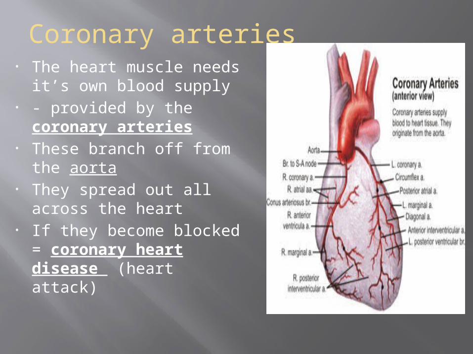
Coronary arteries• The heart muscle needs
it’s own blood supply• - provided by the
coronary arteries• These branch off from the
aorta• They spread out all across
the heart• If they become blocked =
coronary heart disease (heart attack)
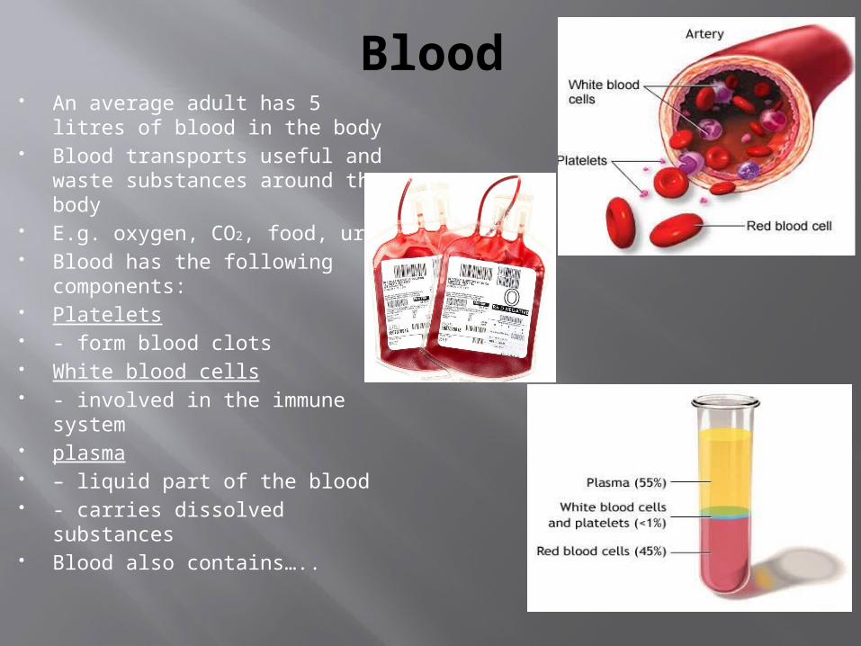
Blood An average adult has 5 litres
of blood in the body Blood transports useful and
waste substances around the body
E.g. oxygen, CO2, food, urea Blood has the following
components: Platelets - form blood clots White blood cells - involved in the immune
system plasma – liquid part of the blood - carries dissolved substances Blood also contains…..

Red Blood Cells
-Very small and numerous (5.5 million/ml of blood)
- Have a biconcave shape- - provides a larger surface
area- Small and flexible- - can easily squeeze through
narrow capillaries- Cytoplasm is rich in
haemoglobin- - in the lungs, combines with
oxygen to form oxy-haemoglobin
- - at the tissues, oxygen is released

Re-Cap questions
1) Name the four chambers of the heart
2) Name blood vessel A 3) Where has the blood in
vessel A come from? 4) Name the three types of
blood vessel, and describe the differences in their structure
5) Name the pigment in red blood cells, that helps carry oxygen
A

Structure & function of lungs
• Organs of gas exchange• Air passes in through the nose
or mouth• Then passes through the
trachea (windpipe), bronchi, bronchioles
• - and finally into the alveoli (air sacs)
• Trachea and bronchi are lined by tiny hair-like cilia
• - and cells that secrete mucus• - which traps dirt and micro-
organisms• The cilia sweep the mucus
away from the lungs

Gas Exchange• Alveoli are
surrounded by many capillaries
• Alveolus lining is very thin and moist
• Large surface area for exchange
• Breathing in:• - air enters alveoli• - oxygen dissolves in
the moist lining• - then diffuses into
the blood• - blood is now
oxygenated• - CO2 moves in the
opposite direction

Need For Digestion Food is needed for cells to: Give fuel for energy Provide building materials for - growth - repair - fighting infection (antibody
production) Digestion breaks down large
insoluble molecules - into small soluble molecules - much of the digestion is done by
enzymes These molecules can then be
absorbed into the bloodstream

Alimentary Canal A long tube running
from mouth to anus Has several
associated organs connected to it
e.g. liver, pancreas, salivary glands
These organs are connected by tubes or ducts
Pharynx (throat)
Oesophagus

Oesophagus & Peristalsis Oesophagus is a muscular
tube - connects the mouth to the
stomach It’s wall is lined with circular
muscle Contraction & relaxation of
this muscle pushes food along
This is known as Peristalsis This process continues all the
way along the alimentary canal

Small Intestine Where the majority of digestion (&
all absorption) takes place Structure: - very long - folded inner lining - covered in finger-like villi These create a large surface area Each villus has a blood capillary
and lacteal Lining is only 1 cell thick Glucose & amino acids pass into
the capillary Fat digestion products pass into the
lacteal Digestion products absorbed into
the bloodstream

A balanced diet The major food groups
that make up a balanced diet are carbohydrates, fats and proteins
Carbohydrates – provide energy for cell
activities Fats – Energy storage &
insulation Proteins – Provide materials for
growth & repair

Vitamins & Minerals
The lack of a certain vitamin or mineral in the diet can result in a deficiency disease
Vitamin
Food source
Function Effect of Vitamin
deficiency
A Dairy products
& fish
Healthy skin, good vision, strong
immune system
Night blindness
C Fruit & Veg
Growth & repair of skin
Scurvy
D Oily fish & eggs
Calcium absorption – for bone growth
Rickets
Mineral Food source
Function Effect of deficienc
y
Iron Meat & eggs
Helps form haemoglobin
Anaemia
Calcium Dairy products
Hardening bones & teeth
Rickets
Sodium /Potassium
Most foods Muscle contraction /
nerve impulses
Impaired movement

Re-cap questions
1) What is the purpose of : - the cilia - mucus 2) Explain the role of alveoli 3) Describe the mechanism of peristalsis,
with reference to a mouthful of food having been swallowed
4) State three ways the small intestine is suited to it’s role of absorbing digested food


