1 Multifunctional Hybrid Silica Nanoparticles for Nanomedicine Bioconjugation with DNA for Gene Therapy Extended Abstract Rui Manuel de Azevedo Pedrosa de Brito Colaço Dissertaªo para obtenªo do Grau de Mestre em Engenharia de Materiais Orientadoras: Prof“. Dr“. Maria Clara Henriques Batista Gonalves Prof“. Dr“. Maria BÆrbara dos Anjos Figueira Martins Maio de 2011
Transcript
1
Multifunctional Hybrid Silica Nanoparticles for Nanomedicine
Bioconjugation with DNA for Gene Therapy
Extended Abstract
Rui Manuel de Azevedo Pedrosa de Brito Colaço
Dissertação para obtenção do Grau de Mestre em Engenharia de Materiais
Orientadoras: Profª. Drª. Maria Clara Henriques Batista Gonçalves
Profª. Drª. Maria Bárbara dos Anjos Figueira Martins
Maio de 2011
1
Abstract
Nanomedicine is an emerging new field combining nanotechnology and medicine. Silica
nanoparticles are chemical and biologically inert, optically transparent and can be doped with imaging
agents and/or functionalised to promote its bioconjugation with different therapeutic molecules. Silica
nanoparticles can be engineered to improve diagnosis, treatment and follow-up of diseases. A
combination of diagnosis devices and therapeutics (theragnosis) would be beneficial for patients.
In this work, amino-, methyl-, vinyl- and phenyl-functionalised silica nanoparticles as drug
carriers or non-viral vectors for gene delivery were prepared via different modified Stöber sol-gel
processes. Core-shell structures, using superparamagnetic iron oxide or silica nanoparticles as core,
coated with organically modified silica (ORMOSIL) shell were synthesised, as well as plain ORMOSIL
nanoparticles fabricated directly using sodium silicate solution as nucleating agent.
Dynamic light scattering, transmission electron microscopy and Fourier transformed infrared
spectroscopy were used to characterise the hybrid nanospheres. Synthesis parameters were studied
and fabrication methods have been optimised. Monodisperse spherical nanoparticles with desired size
and functionality have been obtained. Nanoparticle-DNA complexes were successfully obtained at
different nanoparticle / pDNA ratios and confirmed by agarose gel electrophoresis and ethidium
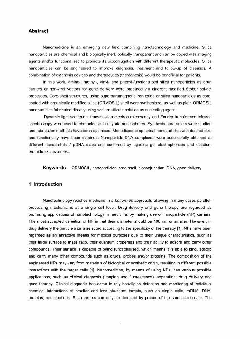
Characterisation by DLS, TEM and FTIR was performed, thus confirming the presence of the
organic functional groups in the plain ORMOSIL NPs. The fabrication process was studied: Plain
ORMOSIL NPs size was observed to increase with APTES/TEOS ratio: to optimise the number of
surface amino groups, a balance between the amino surface density (increase with APTES/TEOS
ratio) and NP size (decrease with TEOS/APTES ratio) should be established. The amount of seeds
(SSS) was also subject of study. By increasing the number of nucleation sites, more and smaller NPs
were expected. Further studies are required to assess the effect of SSS in the plain ORMOSIL NP
characteristics. By decreasing sol-gel precursor concentrations (keeping constant TEOS:APTES
ratio), a decrease in NP size and a narrow NP size distribution was observed. Using methanol instead
of ethanol was advantageous to achieve smaller and more monodisperse plain ORMOSIL NPs,
probably due to a faster hydrolysis rate. Monodisperse plain ORMOSIL NPs with sizes between 100
and 200 nm were fabricated, with different functional organic groups for future bioconjugation. By
combining different ORMOSIL precursors, multifunctional NPs were achieved. This is promising for
bioconjugation with different specific biomolecules. The combination of VTES, TEOS, APTES and
DETA, allows synthesising rougher (and probably more porous) monodisperse plain NPs. Such
rugosity can be useful for increasing loading with drugs for theragnosis in nanomedicine.
Summing up, the process was studied and optimised. ORMOSIL NPs can be synthesised with
the controlled size, size distribution, surface morphology and surface chemistry, which can be chosen
to fulfil the requirements of biomedical applications.
Table 2 – Optimised plain ORMOSIL NPs: functionality; precursors used for the synthesis; diameter measured by TEM; hydrodynamic diameter (HD) and polydispersion index (PdI).
* Synthesised using methanol as co-solvent
7
6. Bioconjugation with DNA
DNA loading of amino-functionalised ORMOSIL NPs was accomplished by direct incubation of
the desired plasmid with the NPs, with varying weight ratios (NP/pDNA), for 30 min at room
temperature. The conjugation is done by electrostatic interaction between the cationic amino groups of
the ORMOSIL NPs and the anionic phosphate groups of the plasmid. The binding of pDNA with NPs
was determined by 1% agarose (low melting point) gel electrophoresis. A series of different NPs to
pDNA weight ratios was loaded (20 μl of the sample containing 0,2 μg of pDNA). A 1:6 dilution of
loading dye was added to each and electrophoresis was carried out at a constant voltage of 100 V for
1 h in TBE buffer (4.45 mM Tris–base, 1 mM sodium EDTA, 4.45 mM boric acid, pH 8,3) containing
0,5 μg/ml ethidium bromide. The pDNA bands were then visualised under a UV transilluminator at a
wavelength of 365 nm. DNA condensation was measured by quenching of ethidium bromide (EtBr)
fluorescence. Briefly, quadruplicates of 0,2 μg of pDNA were complexed with increasing amounts of
NPs in 96-well plates in 10 mM PBS buffer at pH 7,4. After 10 min incubation time, 20 μl EtBr solution
(0.1 mg/ml) were added. The fluorescence was measured on a fluorescence plate reader (Tecan
Infinite M200, Austria) at excitation wavelength of 518 nm and at emission wavelength emission of 605
nm.
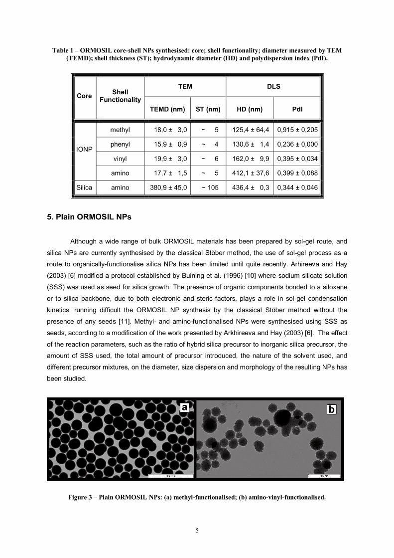
Figure 4 – Optimised plain amino-functionalized ORMOSIL NPs and respective agarose gel electrophoresis test after bioconjugation with plasmid-DNA.
Incubating the amino-functionalised ORMOSIL NPs (Plain and CS) with plasmid-DNA and
running agarose gel electrophoresis tests confirmed their capability of successfully forming NP/pDNA
complexes. Plain ORMOSIL NPs synthesised using sodium silicate solution as seeds were proven
efficient, despite the percentage of ORMOSIL precursor used in their synthesis, the chemical nature of
the solvent used (ethanol or methanol) and their final NP size and size dispersion. Amino-
functionalised CS structures also showed affinity to DNA, both with iron oxide and silica cores. Non-
functionalised NPs did not attach DNA, thus confirming the amino groups are essential for creating
NP/pDNA complexes. The amino-functionalised NPs fabricated for this work have potential for future
applications of gene delivery and therapy, which will be confirmed in future in vitro and in vivo tests.
8
7. Conclusions
In this work, hybrid silica NPs were synthesised by several sol-gel procedures, with controlled
dimensions, size dispersion, surface chemistry and roughness, among other properties. Core-shell
structures were fabricated, using either amorphous silica NPs (~ 170 nm) or superparamagnetic iron
oxide NPs (~ 8 nm) as core, coated with an ORMOSIL layer with the desired functional organic
groups: amino, methyl, vinyl or phenyl. Plain ORMOSIL NPs were also synthesised using aqueous
sodium silicate solution as a seed for growth, functionalised with methyl, amino, vinyl groups alone or
combined. Synthesis parameters were studied, such as the amount of catalyser (ammonia), the
chemical nature of the solvent (ethanol or methanol), the amount of precursors introduced, how they
were introduced and the proportion between them, or the quantity of nucleation sites. Processes were
optimised to achieve the desired final properties. Core-shell (superparamagnetic and non-
superparamagnetic) and plain ORMOSIL NPs functionalised with amino groups were complexed with
plasmid-DNA, revealing their potential for using in gene therapy.
Because of the characteristics of the NPs synthesised, they are considered to be a good
scaffold for multifunctional applications in the area of nanomedicine. Their intrinsic mesoporosity could
allow them to be loaded with one or more types of drugs, for therapy by drug delivery, or even
complexed with a fluorophore, for real time imaging and diagnosis. Their superparamagnetic
properties could permit this drug delivery to be remotely controlled by the application of a magnetic
field in the diseased tissue. By synthesising ORMOSIL NPs with the appropriate surface functional
groups, such NPs could be conjugated with several types of biomolecules, such as specific linkers,
fluorescent labels, antibodies or DNA, giving them simultaneous diagnostic and therapeutic
(theragnosis) capabilities.
9
8. References
[1] De Jong, W.H.; Borm, P.J.A. “Drug Delivery and Nanoparticles: Applications and hazards.”
International Journal of Medicine: vol. 3(2), pp. 133-149, 2008
[2] Wang, L.; Zhao, W.; Tan, W. “Bioconjugated Silica Nanoparticles: Development and
Applications.” Nano Research: vol. 1, pp. 99-115, 2008
[3] Davis, S.S. “Biomedical applications of nanotechnology – implications for drug targeting
and gene therapy.” Trends in Biotechnology: vol. 15, pp. 217-224, 1997
[4] Bharali, D.J; Klejbor, I., et al. ”Organically modified silica Nanoparticles: A nonviral vector
for in vivo gene delivery and expression in the brain.” Proceedings of the National Academy of
Sciences: vol. 102, pp. 11539-11544, 2005
[5] Luo, D.; Saltzman, W.M. “Thinking of silica.” Gene Therapy: vol. 13, pp. 585-586, 2006
[6] Arkhireeva, A. and J. N. Hay. "Synthesis of sub-200 nm silsesquioxane particles using a
modified Stöber sol–gel route." Journal of Materials Chemistry: vol 13, pp. 3122-3127, 2003
[7] Huang, Y. and J. E. Pemberton. "Synthesis of uniform, spherical sub-100nm silica particles
using a conceptual modification of the classic LaMer model." Colloids and Surfaces A:
Physicochemical and Engineering Aspects: vol. 360: pp. 175-183, 2010
[8] Bertoluzza, A., Fagnano, C., Morelli, M.A. "Raman and infrared spectra on silica gel
evolving toward glass." Journal of Non-Crystalline Solids: vol. 48, pp. 117-128, 1982
[9] Hsiao, V.K.S., Waldeisen, J.R. et al. "Aminopropyltriethoxysilane (APTES)-functionalized
nanoporous polymeric gratings: fabrication and application in biosensing." Journal of Materials
Chemistry: vol. 17, pp. 4896-4901, 2007
[10] Buining, P.A., Liz-Marzan L.M., Philipse, A.P. "A Simple Preparation of Small, Smooth
Silica Spheres in a Seed Alcosol for Stöber Synthesis." Journal of Colloid and Interface Science: vol.
179, pp. 318-321, 1996
[11] Iler, R. K. (1979). “The chemistry of silica: solubility, polymerization, colloid and surface
properties, and biochemistry.” Lavoisier.
[12] Ge, J., Yin, Y. "Magnetically tunable colloidal photonic structures in alkanol solutions."
Advanced Materials: vol. 20, pp. 3485-3491, 2008
[13] McComb, D. W., Treble, B. M., et al. "Synthesis and characterisation of photonic crystals."
Journal of Materials Chemistry: vol. 11, pp. 142-148, 2001
[14] Stöber, W., Fink, A., Bohn, E. “Controlled growth of monodisperse silica spheres in micron
size range” Journal of Colloid Interface Science: vol. 26, pp. 62-69, 1968
[15] Qu, S., Yang, H., et al. "Magnetite nanoparticles prepared by precipitation from partially
reduced ferric chloride aqueous solutions." Journal of Colloid and Interface Science: vol. 215, pp. 190-


![Nonporous silica nanoparticles for nanomedicine application...Synthesis of size-controlled silica NPs was first reported by Stöber et al. in 1968 [19]. Monodisperse silica spheres](https://static.documents.pub/doc/80x56/609b60c36163736c4d72f238/nonporous-silica-nanoparticles-for-nanomedicine-application-synthesis-of-size-controlled.jpg)
