Original Article Multilayered Electrospun Scaffolds for Tendon Tissue Engineering Abby Chainani, 1,2, * Kirk J. Hippensteel, MD, 1, * Alysha Kishan, BS, 1,2 N. William Garrigues, PhD, 1,2 David S. Ruch, MD, 1 Farshid Guilak, PhD, 1,2 and Dianne Little, BVSc, PhD 1 Full-thickness rotator cuff tears are one of the most common causes of shoulder pain in people over the age of 65. High retear rates and poor functional outcomes are common after surgical repair, and currently available extracellular matrix scaffold patches have limited abilities to enhance new tendon formation. In this regard, tissue-engineered scaffolds may provide a means to improve repair of rotator cuff tears. Electrospinning pro- vides a versatile method for creating nanofibrous scaffolds with controlled architectures, but several challenges remain in its application to tissue engineering, such as cell infiltration through the full thickness of the scaffold as well as control of cell growth and differentiation. Previous studies have shown that ligament-derived extra- cellular matrix may enhance differentiation toward a tendon or ligament phenotype by human adipose stem cells (hASCs). In this study, we investigated the use of tendon-derived extracellular matrix (TDM)-coated electrospun multilayered scaffolds compared to fibronectin (FN) or phosphate-buffered saline (PBS) coating for use in rotator cuff tendon tissue engineering. Multilayered poly(e-caprolactone) scaffolds were prepared by sequentially collecting electrospun layers onto the surface of a grounded saline solution into a single scaffold. Scaffolds were then coated with TDM, FN, or PBS and seeded with hASCs. Scaffolds were maintained without exogenous growth factors for 28 days in culture and evaluated for protein content (by immunofluorescence and biochemical assay), markers of tendon differentiation, and tensile mechanical properties. The collagen content was greatest by day 28 in TDM-scaffolds. Gene expression of type I collagen, decorin, and tenascin C increased over time, with no effect of scaffold coating. Sulfated glycosaminoglycan and dsDNA contents increased over time in culture, but there was no effect of scaffold coating. The Young’s modulus did not change over time, but yield strain increased with time in culture. Histology demonstrated cell infiltration through the full thickness of all scaffolds and immunofluorescence demonstrated greater expression of type I, but not type III collagen through the full thickness of the scaffold in TDM-scaffolds compared to other treatment groups. Together, these data suggest that nonaligned multilayered electrospun scaffolds permit tenogenic differentiation by hASCs and that TDM may promote some aspects of this differentiation. Introduction S houlder pain is the leading cause of musculoskeletal pain in people over the age of 65, 1 and rotator cuff and subacromial bursa pathologies contribute most to shoulder pain. 2 The functional rotator cuff is a composite of several tendons, ligaments, and muscles, but tears involving the supraspinatus and infraspinatus tendons are the most com- monly identified injuries. 3 Rotator cuff tears account for over 4 million physician visits and 300,000 surgical repairs an- nually, 4,5 and have a similar impact on individual quality of life as diseases such as congestive heart failure, diabetes, or clinical depression. 6 Suture repair to reattach the torn tendon to bone using an arthroscopic or open approach is the most common repair technique and is highly cost effective. 7 However, up to 30% of rotator cuff tears are irreparable, 8 and retear rates of 34%–94% are reported after suture repair. 9 Efforts to improve clinical outcomes have focused on the use of extracellular matrix scaffold allograft or xenograft patches for mechanical augmentation of rotator cuff repair. These patches are thought to improve healing in the critical early postoperative period because they contain bioactive mole- cules. However, these approaches have limitations for a va- riety of reasons, including poor cellularization, immunogenic Abstract previously presented by Hippensteel KJ, Chainani A, Kishan A, Garrigues NW, Ruch DS, Guilak F, Little D. Tendon-Derived Matrix Coated Multi-Layered Hydrospun Scaffolds for Tendon Tissue Engineering. Trans Orthop Res Soc 2012. Departments of 1 Orthopaedic Surgery and 2 Biomedical Engineering, Duke University Medical Center, Durham, North Carolina. *These authors are cofirst authors. TISSUE ENGINEERING: Part A Volume 00, Number 00, 2013 ª Mary Ann Liebert, Inc. DOI: 10.1089/ten.tea.2013.0165 1
Transcript
Original Article
Multilayered Electrospun Scaffolds for TendonTissue Engineering
Abby Chainani,1,2,* Kirk J. Hippensteel, MD,1,* Alysha Kishan, BS,1,2 N. William Garrigues, PhD,1,2
David S. Ruch, MD,1 Farshid Guilak, PhD,1,2 and Dianne Little, BVSc, PhD1
Full-thickness rotator cuff tears are one of the most common causes of shoulder pain in people over the age of 65.High retear rates and poor functional outcomes are common after surgical repair, and currently availableextracellular matrix scaffold patches have limited abilities to enhance new tendon formation. In this regard,tissue-engineered scaffolds may provide a means to improve repair of rotator cuff tears. Electrospinning pro-vides a versatile method for creating nanofibrous scaffolds with controlled architectures, but several challengesremain in its application to tissue engineering, such as cell infiltration through the full thickness of the scaffold aswell as control of cell growth and differentiation. Previous studies have shown that ligament-derived extra-cellular matrix may enhance differentiation toward a tendon or ligament phenotype by human adipose stemcells (hASCs). In this study, we investigated the use of tendon-derived extracellular matrix (TDM)-coatedelectrospun multilayered scaffolds compared to fibronectin (FN) or phosphate-buffered saline (PBS) coating foruse in rotator cuff tendon tissue engineering. Multilayered poly(e-caprolactone) scaffolds were prepared bysequentially collecting electrospun layers onto the surface of a grounded saline solution into a single scaffold.Scaffolds were then coated with TDM, FN, or PBS and seeded with hASCs. Scaffolds were maintained withoutexogenous growth factors for 28 days in culture and evaluated for protein content (by immunofluorescence andbiochemical assay), markers of tendon differentiation, and tensile mechanical properties. The collagen contentwas greatest by day 28 in TDM-scaffolds. Gene expression of type I collagen, decorin, and tenascin C increasedover time, with no effect of scaffold coating. Sulfated glycosaminoglycan and dsDNA contents increased overtime in culture, but there was no effect of scaffold coating. The Young’s modulus did not change over time, butyield strain increased with time in culture. Histology demonstrated cell infiltration through the full thickness ofall scaffolds and immunofluorescence demonstrated greater expression of type I, but not type III collagenthrough the full thickness of the scaffold in TDM-scaffolds compared to other treatment groups. Together, thesedata suggest that nonaligned multilayered electrospun scaffolds permit tenogenic differentiation by hASCs andthat TDM may promote some aspects of this differentiation.
Introduction
Shoulder pain is the leading cause of musculoskeletalpain in people over the age of 65,1 and rotator cuff and
subacromial bursa pathologies contribute most to shoulderpain.2 The functional rotator cuff is a composite of severaltendons, ligaments, and muscles, but tears involving thesupraspinatus and infraspinatus tendons are the most com-monly identified injuries.3 Rotator cuff tears account for over4 million physician visits and 300,000 surgical repairs an-nually,4,5 and have a similar impact on individual quality oflife as diseases such as congestive heart failure, diabetes, or
clinical depression.6 Suture repair to reattach the torn tendonto bone using an arthroscopic or open approach is the mostcommon repair technique and is highly cost effective.7
However, up to 30% of rotator cuff tears are irreparable,8 andretear rates of 34%–94% are reported after suture repair.9
Efforts to improve clinical outcomes have focused on the useof extracellular matrix scaffold allograft or xenograft patchesfor mechanical augmentation of rotator cuff repair. Thesepatches are thought to improve healing in the critical earlypostoperative period because they contain bioactive mole-cules. However, these approaches have limitations for a va-riety of reasons, including poor cellularization, immunogenic
Abstract previously presented by Hippensteel KJ, Chainani A, Kishan A, Garrigues NW, Ruch DS, Guilak F, Little D. Tendon-DerivedMatrix Coated Multi-Layered Hydrospun Scaffolds for Tendon Tissue Engineering. Trans Orthop Res Soc 2012.
Departments of 1Orthopaedic Surgery and 2Biomedical Engineering, Duke University Medical Center, Durham, North Carolina.*These authors are cofirst authors.
TISSUE ENGINEERING: Part AVolume 00, Number 00, 2013ª Mary Ann Liebert, Inc.DOI: 10.1089/ten.tea.2013.0165
1
responses, insufficient mechanical properties to supportphysiological loading postoperatively, and poor sutureretention.10–17
Tissue engineering affords the opportunity to overcomesome of these limitations by providing functional, cell-basedscaffolds for rotator cuff repair. In particular, nanofibrousscaffolds are suited for this application because they can beengineered to mimic the ultrastructure and properties of thenative tendon, using techniques such as electrospinning.10
Several such approaches are under investigation, includingthe use of nanofibrous scaffolds for interfacial regenerationof the tendon-to-bone interface18–21 as an over-the-top ten-don augmentation scaffold,22,23 and as interposition grafts.24
One challenge in the use of electrospun scaffolds for rotatorcuff repair has been the lack of cell infiltration through thefull thickness of the scaffold.25–27 Several approaches havebeen taken to mitigate this problem. For example, electro-spun scaffolds based on biological materials, such as colla-gen, demonstrate more rapid cell infiltration than syntheticpolymers.27 However, mechanical properties of pure bio-polymers are generally inferior to synthetic polymers,25,28,29
although composite fibers of synthetic and biopolymers mayhave improved tensile properties compared to the syntheticpolymer especially at low concentrations.30–32 Physicalmethods have been used to improve the porosity andtherefore cell infiltration, including the use of sacrificial fi-bers, which enhance cell infiltration and matrix depositionin vitro and in subcutaneous pouch models.25,26 However, inan over-the-top rotator cuff augmentation animal model, thisapproach was less successful possibly because the scaffoldwith sacrificial fibers was less resistant to compression dur-ing handling and in a challenging in vivo environment.23
Other methods to improve porosity include the use of saltleaching techniques,33 combined micro- and nanofiber scaf-folds,34,35 and laser ablation.36
Multilayered electrospinning has been reported previ-ously, either using a layer-by-layer approach with simulta-neous wet electrospinning and cell-seeding,37 by alternatinglayers of nanofibers and microfibers,38 by alternating poly-mers,39 or by alternately coating electrospun layers withcollagen.40 With wet electrospinning, electrospun fibers canbe aligned by depositing nonaligned fibers onto an aqueouscollecting medium, and then drawing out the fibers using arotating mandrel.41–43 Alternatively, others have reportedintermittent collection of nonaligned layers of electrospunbiopolymers from the surface of a saline bath, termed ‘‘hy-drospinning,’’ followed by vacuum desiccation of the scaf-fold.44 Cell-seeding by direct deposition onto the surface ofthese scaffolds after vacuum desiccation resulted in rapid cellinfiltration throughout the scaffold, in contrast to similarscaffolds prepared by standard electrospinning techniquesonto a solid ground plate.44
In addition to early and complete cell infiltration into thescaffolds, the rapid synthesis and accumulation of the ten-don-specific extracellular matrix is also likely to be crucial tothe ultimate success of a patch designed for rotator cuffaugmentation or interposition. We have previously shownthat a ligament-derived matrix can enhance development ofa ligamentous phenotype by human adipose stem cells(hASCs) compared to type I collagen gel alone,45 and othershave found that the engineered tendon matrix enhancesproliferation, maintenance of tendon stem cell stemness, and
tendon-related gene expression compared to tissue cultureplastic.46 These studies align with work in other musculo-skeletal fields, where tissue-specific extracellular matriceshave potent ability to induce tissue-specific neotissue for-mation without the use of exogenous growth factors.47–51
Extracellular matrix proteins such as small intestinal sub-mucosa, collagen, and silk fibroin have been incorporatedinto electrospun synthetic polymer fibers to improve hy-drophilicity, cell attachment, early proliferation, and neo-tissue organization.52–54 However, there is controversy as tohow well the native structure of collagen is preserved duringelectrospinning,28,55 and recent work confirms that collagenis unfolded in fluorinated solvents and only partially re-folded during the electrospinning process itself.56 As an al-ternative, coating of electrospun fibers with collagen hasbeen used to circumvent the problem of collagen denatur-ation, while retaining the beneficial effects of collagen on cellattachment.40
The primary objective of this study was to develop andevaluate the use of multilayered electrospun scaffolds fabri-cated by sequential collection of individual layers from thesurface of an aqueous ground substrate as a single scaffoldfor potential use in rotator cuff tissue engineering. A secondaim of the project was to evaluate if there was additionalbenefit to coating the scaffolds with the tendon-derived ex-tracellular matrix (TDM) compared to fibronectin (FN), pre-viously used to improve cell attachment44 or phosphate-buffered saline (PBS)-coated controls.
Materials and Methods
Tendon-derived matrix preparation
Fresh digital flexor and extensor tendons were dissectedfrom adult female porcine hindlimbs (n = 10) obtained from aslaughterhouse. Tissue was minced, lyophilized (LabconcoFreezone 2.5L; Labconco), and pulverized using a 6750 SpexSamplePrep Freezer Mill (Spex CertiPrep). The resultingpowder was sieved to pass through 106-mm wire meshopenings and stored at - 80�C until use.
Multilayered electrospun scaffold preparation
Multilayered electrospun scaffolds were prepared in asimilar manner as previously described.44 Poly(e-capro-lactone) (PCL) (Mn = 80,000) (Sigma-Aldrich, St. Louis, MO)was dissolved at 100 mg/mL in 70% dichloromethane/30%ethanol for 24 h before use. The resulting solution was elec-trospun through a 25G needle fitted with a round wire meshfocusing cage (3 cm diameter, needle tip protruding 4 mmfrom bottom of cage) at 2.5 mL/h and 17 kV with a 17-cmneedle-ground distance. The ground collector was a salinebath (1.25 g/L NaCl in distilled water), and nonalignedlayers were collected sequentially from the surface of thesaline bath every 90 s using a 5 · 7.5-cm glass slide, for a totalof 70 layers/scaffold. Relative humidity was 20%–40% andambient temperature was 18�C–25�C. Each scaffold was al-lowed to dry at room temperature, and then stored at roomtemperature and protected from light until use. Scaffoldswere cut into individual test strips and sutured to a 3.2-mm-outer diameter Teflon ring-shaped holder to maintain statictension and suspension in media. Each scaffold was rehy-drated and sterilized in a graded series of ethanol baths
2 CHAINANI ET AL.
before a final 30-min rinse in PBS pH7.4. Scaffolds wererandomized into three treatment groups: PBS coated, FNcoated, or TDM coated. Scaffolds were coated on each sideby direct pipetting with human FN in PBS (BD Biosciences)at 4mg/cm2, TDM in PBS at 2 mg/cm2, or an equal volume ofPBS and allowed to dry. The surfaces of scaffolds weresterilized under ultraviolet light for 10 min on each side andprewetted before cell seeding.
Analysis of fiber diameter
One 6-mm biopsy punch was harvested from the center ofeach of three representative scaffolds, critical point dried inCO2, and then sputter coated with gold. Each sample wasviewed with a Philips 501 Scanning Electron Microscope. Threerepresentative images were taken of each sample and the di-ameter of 50 fibers within each image was measured usingGNU Image Manipulation Program (GIMP) 2.8.4 freeware.
Cell culture and seeding
The hASCs used in this study were isolated by collagenasedigestion57 of lipoaspirate surgical waste from five dei-dentified female donors (age 36–59, body mass index 19.6–33.1) with approval of the Duke University InstitutionalReview Board. Cells were expanded in monolayer on tissueculture plastic through passage 4 as described previously.45
Cells were used at passage 4 and seeded at a density of 0 or1 · 106 hASCs/cm2 for each treatment group (scaffoldscoated with PBS, FN, or TDM). Scaffolds were seeded on oneside of the scaffold with half of the cells and allowed toincubate for 15 min. Scaffolds were then turned over andseeded with the remaining cells. The specimens were thenincubated for a further 15 min before transfer to 6-well platescoated with 2% agarose, and addition of culture media.Scaffolds were maintained without growth factors at 37�Cand 5% CO2 in Advanced DMEM (Life Technologies), 10%fetal bovine serum (Zen-Bio), 1% penicillin–streptomycin–fungizone (Life Technologies), 4 mM l-glutamine (LifeTechnologies), and 15 mM l-ascorbic acid-2-phosphate (Sig-ma-Aldrich), which was changed every other day for thedesignated culture periods.
Biochemical assays
On days 0 (unseeded), 1, 14, and 28 (hASC-seeded), scaf-folds from each treatment group (PBS, FN, and TDM) (n = 5)were harvested and lyophilized to obtain dry weight. Sam-ples were pulverized using a freezer mill, and digested for 1week in a papain solution (125 mg/mL) at 60�C. The dsDNAcontent was quantified using the Picogreen Assay (LifeTechnologies). The sulfated glycosaminoglycan (s-GAG)content was quantified spectrophotometrically using the 1,9-dimethylmethylene blue (DMMB) dye (pH 3.0).58 The hy-droxyproline assay was used to determine the total collagencontent using a conversion factor of 1:7.46 to convert hy-droxyproline to collagen.59 All results were normalized todry weight (mean – SD).
RNA isolation and real time quantitative polymerasechain reaction
RNA was extracted from hASC-seeded scaffolds in each ofthe three treatment groups (n = 5) after 4, 7, and 14 days of
cell culture. Scaffolds were pulverized and RNA extractionwas performed using the QiaShredder column (Qiagen) fol-lowed by the RNeasy Mini kit (Qiagen) with on-columnDNAase treatment. Equal amounts of RNA were reversetranscribed using the Superscript VILO cDNA Synthesis Kit(Life Technologies). Real-time polymerase chain reaction wasperformed on an iCycler (Biorad) using Express qPCR Su-perMix (Life Technologies). Commercially available primersand probes (Applied Biosystems) were used to comparetranscript levels for seven different genes: 18S rRNA (en-dogenous control, assay ID Hs99999901_s1), type I collagen(COL1A1, assay ID Hs00164004_m1), type III collagen(COL3A1, assay ID Hs00164103_m1), biglycan (BGN, assayID Hs00959143_m1), decorin (DCN, assay ID Hs00266491_m1), tenomodulin (TNMD, assay ID Hs00223332_m1), andtenascin C (TNC, assay ID Hs00233648_m1). Data from eachgene of interest for each sample were corrected for efficiencyand normalized to expression of 18s. These data were thenexpressed as fold-change relative to the level of gene ex-pression in 1 million P4 hASCs before cell seeding from eachdonor at day 0.60
Histology
Unseeded and hASC-seeded scaffolds from each of thethree treatment groups (n = 5) were harvested after 28 days ofculture, embedded in optimal cutting temperature gel (Sa-kura), and frozen at - 80�C. Samples were cut into 7–10-mmsections and mounted on slides and evaluated by light mi-croscopy after staining with hematoxylin and eosin andsafranin-O/fast green or examined under a Zeiss LSM 510Confocal Microscope (Carl Zeiss) after immunofluorescencelabeling of human type I and III collagen, as described pre-viously.45
Mechanical testing
After harvest at day 0 or 28, hASC-seeded dog-bonesamples (n = 5) were wrapped in gauze soaked in PBS andstored at - 80�C until analysis. Samples were marked in thecenter and at 5 mm proximal and distal to the central markusing India ink to allow regional strain analysis of the centralcentimeter of the dog-bone. The ends of each sample weresandwiched and glued between fine-gauge waterproofsandpaper and then clamped and mounted in the loadframe. The specimens were tested wet in tension at a strainrate of 1%/s with 0.5 g preload using an electromechanicaltesting system (Bose Enduratec Smart Test Series; BoseCorporation) with a 2.27 kg load cell (Sensotec Model 31;Honeywell International), using the full displacement limitsof the transducer. Initial scaffold thickness was measuredusing a digital camera (Allied Vision Technologies, Inc.) anddigital calipers in GIMP 2.8.4 freeware. Midsubstance strainswere calculated from digital images acquired at 20 Hz andinterpolated to load frame data using custom MATLAB(MathWorks) code. The Young’s modulus of the linear re-gion and stress and strain at yield were calculated in Excel(Microsoft Office).
Statistical analysis
Data are reported as mean – SD, tested for normality,transformed using Box-Cox transformation if necessary,
MULTILAYERED SCAFFOLDS FOR TENDON TISSUE ENGINEERING 3
and then evaluated for the effect of scaffold coating andtime using factorial analysis of variance (ANOVA). TheNewman–Keuls post hoc test was used to determine differ-ences between treatments following ANOVA. Significancewas reported at the 95% confidence level for all analyses(a = 0.05).
Results
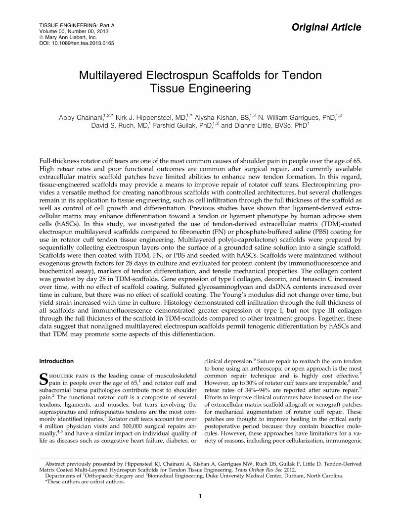
Scanning electron micrographs of surface and edgeof unseeded and hASC-seeded scaffolds after 28 days ofculture are shown in Figure 1. The thickness of the PBS-, FN-,and TDM-scaffolds were 0.80 – 0.06, 0.76 – 0.07, and0.73 – 0.04 mm, respectively, at day 0 and did not differamong groups. The median fiber diameter was 2235 nm(interquartile range 1998–2468 nm). The DNA content of allscaffolds significantly increased after cell seeding and withculture until day 14, after which the DNA content did notincrease further; there was no effect of scaffold coating onDNA content at any time point (Fig. 2A). The s-GAG contentsignificantly increased after cell seeding at all time points,but there was no effect of scaffold coating (Fig. 2B). At day 0,TDM-scaffolds had significantly increased collagen contentcompared to the other groups, but by day 1, there was noeffect of coating on collagen content between groups. Thecollagen content returned to day 0 levels in the TDM-coatedgroups by day 14, and by day 28, the collagen content ofTDM-scaffolds was greater than FN- or PBS-scaffolds and allscaffolds at all other time points (Fig. 2C).
COL1A1 expression was significantly increased comparedto P4 cells from day 4 and remained elevated (Fig. 3).COL3A1 expression was increased at day 7 of culture only,and DCN expression increased in all scaffold groups fromday 7 after seeding (Fig. 3), but BGN expression levels did
not change (data not shown). TNMD expression showed atrend ( p = 0.09) to increase at day 4 compared to other timepoints (data not shown). TNC expression increased in allscaffold groups at day 4 and remained elevated (Fig. 3).There was no significant effect of scaffold coating on geneexpression.
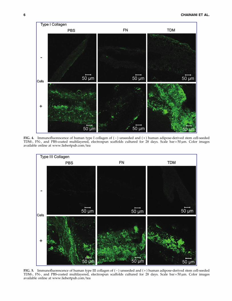
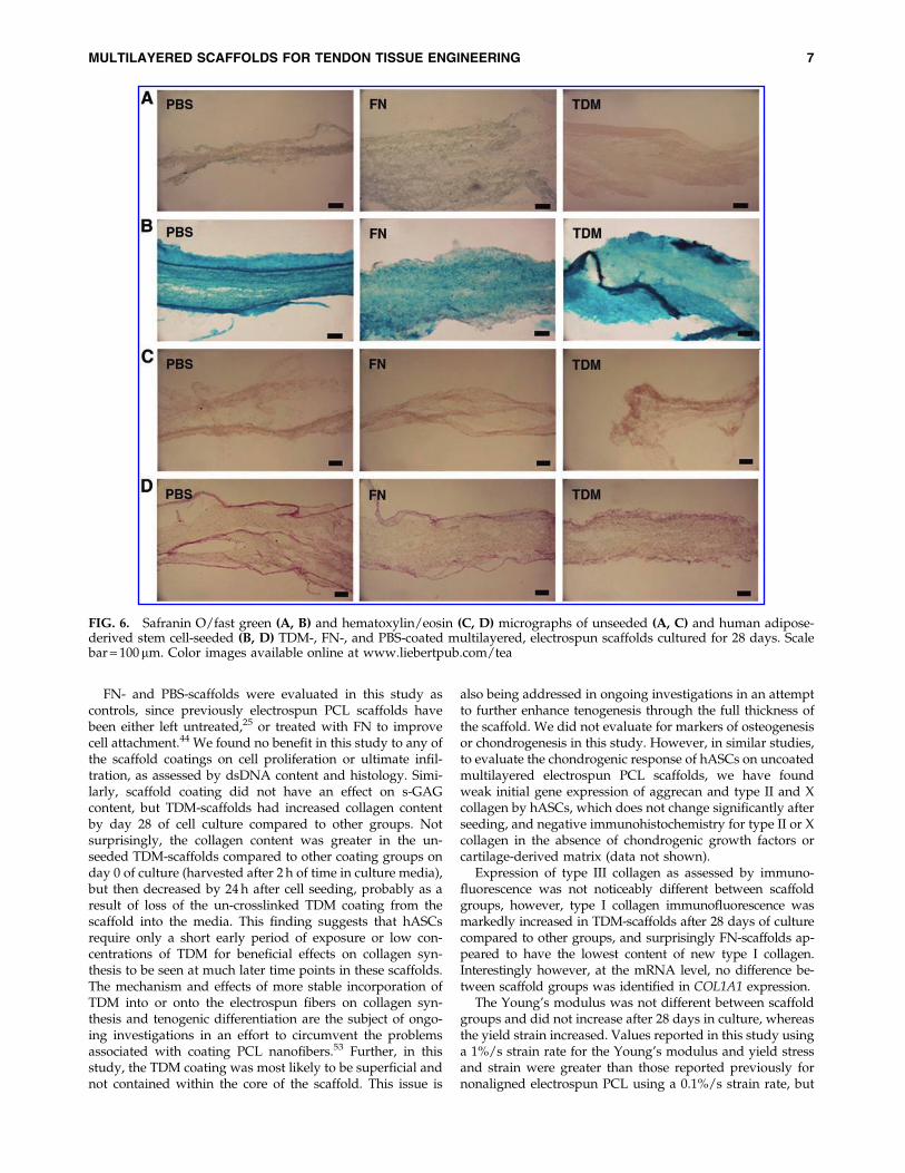
Immunofluorescence for human type I collagen revealed apositive signal through the full thickness of the scaffold in allthree cell-seeded treatment groups, but appeared to be morerobust in the TDM-scaffolds (Fig. 4). Immunofluorescencefor human type III collagen demonstrated robust positivesignals through the full thickness in all scaffold groups(Fig. 5). Safranin O/fast green of unseeded and seededscaffolds demonstrated synthesis of a new extracellular ma-trix through the full thickness of the scaffold in seededscaffolds (Fig. 6A, B), and hematoxylin and eosin stainingconfirmed cellular infiltration through the full thickness ofthe scaffold (Fig. 6C, D).
There was no effect of time in culture or scaffold treatmenton Young’s modulus (Fig. 7A); however, yield strain in-creased from day 0 to 28 (Fig. 7B). Yield stress increased overtime in culture for the FN-scaffold group only (Fig. 7C). Noscaffold failed within the maximum displacement limitspermitted by the actuator and transducer used in this study,representing a strain in excess of 2.5, well beyond the limitsof physiological relevance, thus failure properties and modewere not determined.
Discussion
The findings of this study show that multilayered elec-trospun PCL scaffolds formed by electrospinning onto a sa-line ground solution and sequential collection of multiplelayers as a single scaffold permitted complete cellular
FIG. 1. Scanning electronmicrographs of edge (A, B)and surface (C, D) of un-seeded (A, C) scaffolds, andphosphate-buffered saline(PBS)-coated scaffolds seededwith human adipose stemcells and cultured for 28 days(B, D).
infiltration and formation of the tendon-like extracellularmatrix by hASCs by measures of gene expression, proteinsynthesis, biochemical assays, and immunofluorescence.Additionally, coating each side of these scaffolds with TDMenhanced the synthesis and accumulation of collagen ascompared to FN- or PBS-scaffolds. The mechanism of en-
hanced induction of tenogenesis by TDM in the absence ofexogenous growth factors remains to be determined. Multi-layered scaffolds containing TDM may solve current chal-lenges associated with achieving cell infiltration through thefull thickness of electrospun scaffolds and therefore representa novel approach for rotator cuff tendon tissue engineering.
FIG. 2. DNA (A), sulfated gly-cosaminoglycan (s-GAG) (B), andcollagen (C) content (normalized todry weight) of tendon-derived ex-tracellular matrix (TDM)-, fibro-nectin (FN)-, and PBS-coatedmultilayered electrospun scaffoldsat day 0 (unseeded), 1, 14, and 28,after seeding with human adipose-derived stem cells. Groups havingdifferent letters are significantlydifferent from each other ( p £ 0.05,n = 5).
FIG. 3. Expression of type 1 col-lagen (COL1A1), type III collagen(COL3A1), decorin (DCN), and te-nascin C (TNC) by human adipose-derived stem cells after seeding onTDM-, FN-, and PBS-coated multi-layered electrospun scaffolds at day4, 7, and 14 of culture, normalizedto 18S expression and unseeded cellpellets at day 0. Groups havingdifferent letters are significantlydifferent from each other ( p £ 0.05,n = 5).
MULTILAYERED SCAFFOLDS FOR TENDON TISSUE ENGINEERING 5
FIG. 4. Immunofluorescence of human type I collagen of ( - ) unseeded and ( + ) human adipose-derived stem cell-seededTDM-, FN-, and PBS-coated multilayered, electrospun scaffolds cultured for 28 days. Scale bar = 50 mm. Color imagesavailable online at www.liebertpub.com/tea
FIG. 5. Immunofluorescence of human type III collagen of ( - ) unseeded and ( + ) human adipose-derived stem cell-seededTDM-, FN-, and PBS-coated multilayered, electrospun scaffolds cultured for 28 days. Scale bar = 50 mm. Color imagesavailable online at www.liebertpub.com/tea
FN- and PBS-scaffolds were evaluated in this study ascontrols, since previously electrospun PCL scaffolds havebeen either left untreated,25 or treated with FN to improvecell attachment.44 We found no benefit in this study to any ofthe scaffold coatings on cell proliferation or ultimate infil-tration, as assessed by dsDNA content and histology. Simi-larly, scaffold coating did not have an effect on s-GAGcontent, but TDM-scaffolds had increased collagen contentby day 28 of cell culture compared to other groups. Notsurprisingly, the collagen content was greater in the un-seeded TDM-scaffolds compared to other coating groups onday 0 of culture (harvested after 2 h of time in culture media),but then decreased by 24 h after cell seeding, probably as aresult of loss of the un-crosslinked TDM coating from thescaffold into the media. This finding suggests that hASCsrequire only a short early period of exposure or low con-centrations of TDM for beneficial effects on collagen syn-thesis to be seen at much later time points in these scaffolds.The mechanism and effects of more stable incorporation ofTDM into or onto the electrospun fibers on collagen syn-thesis and tenogenic differentiation are the subject of ongo-ing investigations in an effort to circumvent the problemsassociated with coating PCL nanofibers.53 Further, in thisstudy, the TDM coating was most likely to be superficial andnot contained within the core of the scaffold. This issue is
also being addressed in ongoing investigations in an attemptto further enhance tenogenesis through the full thickness ofthe scaffold. We did not evaluate for markers of osteogenesisor chondrogenesis in this study. However, in similar studies,to evaluate the chondrogenic response of hASCs on uncoatedmultilayered electrospun PCL scaffolds, we have foundweak initial gene expression of aggrecan and type II and Xcollagen by hASCs, which does not change significantly afterseeding, and negative immunohistochemistry for type II or Xcollagen in the absence of chondrogenic growth factors orcartilage-derived matrix (data not shown).
Expression of type III collagen as assessed by immuno-fluorescence was not noticeably different between scaffoldgroups, however, type I collagen immunofluorescence wasmarkedly increased in TDM-scaffolds after 28 days of culturecompared to other groups, and surprisingly FN-scaffolds ap-peared to have the lowest content of new type I collagen.Interestingly however, at the mRNA level, no difference be-tween scaffold groups was identified in COL1A1 expression.
The Young’s modulus was not different between scaffoldgroups and did not increase after 28 days in culture, whereasthe yield strain increased. Values reported in this study usinga 1%/s strain rate for the Young’s modulus and yield stressand strain were greater than those reported previously fornonaligned electrospun PCL using a 0.1%/s strain rate, but
FIG. 6. Safranin O/fast green (A, B) and hematoxylin/eosin (C, D) micrographs of unseeded (A, C) and human adipose-derived stem cell-seeded (B, D) TDM-, FN-, and PBS-coated multilayered, electrospun scaffolds cultured for 28 days. Scalebar = 100mm. Color images available online at www.liebertpub.com/tea
MULTILAYERED SCAFFOLDS FOR TENDON TISSUE ENGINEERING 7
values for yield strain were similar to those reported previ-ously.29 Fiber diameters in the current study (*2 mm) weregreater than in the previously reported study ( < 1.5 mm).29
Previous studies have found an increase in the modulus withan increasing fiber diameter, but opposite effects on elon-gation at break and yield strength for aligned61 and non-aligned62 fiber patterns. Further, the degree of strain ratedependence on tensile mechanical properties in electrospunnanoscaffolds is unknown, and may account for some of thedifferences in mechanical properties observed, as has beenseen in other tissues.63
The degradation profile of the multilayered electrospunPCL scaffolds in the current study is not yet known; how-ever, decreases in the Young’s modulus and elongation atbreak were detected in 15-mm-thick PCL nanofibrous scaf-folds after 1-month of incubation in a physiological solu-tion.62 Therefore, in the current study, it is possible that thelack of change in Young’s modulus over time in culture re-presented load-sharing between the newly formed tendon-like tissue and the degrading electrospun PCL scaffold. Notsurprisingly, the Young’s modulus of this nonaligned scaf-fold was lower than all regions of the human rotator cuff,except for the relatively nonaligned posterior aspect of thesupraspinatus tendon.64 Therefore, we are continuing to ex-plore ways to improve initial mechanical properties using thiselectrospinning technology. Aligned electrospun scaffolds aremore attractive for tendon tissue engineering than nonalignedscaffolds because they recapitulate the mechanical anisotropyof the target tissue,65 and enhance collagen production.66
However, there are conflicting reports as to the benefit ofalignment on tendon-related gene expression. Compared torandomly aligned nanofibers, expression of COL1A1, DCN,and TNMD by mesenchymal stem cells was increased onaligned nanofibers,67 but conversely, there was no beneficial
effect of alignment on the expression of COL1A1, DCN, orscleraxis (SCX) by embryonic fibroblasts.68 Further, the hu-man supraspinatus tendon demonstrates heterogeneous re-gional anisotropy, being highly anisotropic medially, but moreisotropic on the bursal side close to its insertion.69 Regionalmatching of augmentation patch properties to the underlyingrotator cuff is likely to be important, since mismatch of patchmaterial properties to the underlying tendon may result instress concentrations and increased likelihood of postoperativerepair failure.70 Thus, given the problems associated withcellular infiltration throughout aligned nanoscaffolds, and theregional anisotropy of the target tissue, multilayered electro-spun scaffolds afford the possibility of introducing regionallamellar anisotropy to improve baseline mechanical propertiesand maintain collagen production, without a negative impacton the overall ability of cells seeded on the scaffolds to dif-ferentiate toward a tendon phenotype or to fully infiltrate.Lamellar regional anisotropy can be introduced within anelectrospun scaffold, using several techniques,71–73 and futurestudies will evaluate this potential in multilayered electrospunscaffolds.
The fiber diameters obtained in this study were larger thanthe 800-nm-diameter fibers previously reported for nanofi-brous scaffolds produced using this technique.44 This largerfiber size is likely attributable to the greater flow rate and thedifferent solvents used in this study, since the needle sizeand electric field strength were comparable. The effect of thelarger fiber diameters (*2mm) observed in this study on thedegree of cell infiltration and differentiation compared tosmaller fiber diameters is unknown, although larger fiberdiameters can improve cell infiltration in electrospun scaf-folds.38 Nonetheless, the sequential collection of multiplelayers into a single scaffold likely still improves cell infil-tration compared to scaffolds composed of similar diameter
FIG. 7. The Young’s modulus oflinear region (A), yield strain (B),and yield stress (C) of human adi-pose-derived stem-cell seededTDM-, FN-, and PBS-coated multi-layered electrospun scaffolds at day0 and after 28 days of culture.Groups having different letters aresignificantly different from eachother ( p £ 0.05, n = 5).
fibers collected as a single layer, since culture in a flowperfusion bioreactor was required to achieve complete cellinfiltration in scaffolds composed of 5-mm fibers.38 The effectof the fiber diameter on tenogenic differentiation of rotatorcuff fibroblasts, mesenchymal stem cells, and embryonicfibroblasts has recently been examined.61,67,68 As aligned fiberdiameter increases to the microfiber scale, rotator cuff fibro-blasts elongate and align to a greater extent, demonstrateincreased tendon-like gene expression, and form a more ten-don-like tissue in contrast to the scar-like tissue formed onnanofibers.61 Similarly, as the diameter increased on non-aligned fibers, the aspect ratio also increased for mesenchymalstem cells, and expression of COL1A1, DCN, and TNMD orSCX increased at later time points.67,68 These studies all founda detrimental effect on cell numbers associated with micro-fibers compared to nanofibers, but in most cases, differences incell numbers resolved after several weeks in culture.61,67,68
The reason for the beneficial effect of TDM treatment onthe total collagen content after 28 days of culture and on typeI collagen production through the full thickness of the scaf-fold is unknown, but is consistent with similar findingspreviously using hASCs seeded in a type I collagen gelsystem.45 The extracellular matrix provides a complex envi-ronment that regulates many aspects of cellular behavior.74
Therefore, the most likely mechanism is via matricellularinteractions with tendon-specific components of the extra-cellular matrix.45,75–79 Importantly, however, the use of tis-sue-specific extracellular matrix avoids the use of exogenousgrowth factors for tenogenic differentiation, where identify-ing the safest and most appropriate physiological dose torealize the preferred biomechanical and morphologicalproperties at the repair site may be challenging,80 and likelyto be more difficult where combinations of growth factorsare used. In this study, the enhanced type I collagen pro-duction exhibited on TDM-scaffolds did not improve themechanical properties of the constructs. We did not evaluateneocollagen alignment, which may have been disorganizedin the static loading conditions used in this study. Thus,future studies may wish to examine the synergistic effect ofTDM and dynamic loading on the collagen content,81 andpotentially, the development of mechanical properties.
In summary, our findings indicate that multilayeredelectrospun scaffolds incorporated with TDM show in-creased levels of collagen accumulation by ASCs as com-pared to FN- or PBS-coated scaffolds. This approachprovides a novel means for tissue engineering of the rotatorcuff. While the exact mechanism of action of TDM is underinvestigation, future studies will additionally focus on im-proving the stability of TDM within the scaffolds, reducingpotential immunogenicity,82 and on developing regionalanisotropy and alignment, while maintaining cellular infil-tration and tenogenic differentiation.
Acknowledgments
This work was supported by NIH grants AR59784 (D.L.),AR48852 (F.G.), AG15768 (F.G.), AR48182 (F.G.), andAR50245 (F.G.) and by Synthes USA (D.L., D.S.R., F.G.).
Disclosure Statement
No competing financial interests exist.
References
1. Taylor, W. Musculoskeletal pain in the adult New Zealandpopulation: prevalence and impact. N Z Med J 118, U1629, 2005.
236, 2009.3. Patte, D. Classification of rotator cuff lesions. Clin Orthop
Relat Res 254, 81, 1990.4. Physician Visits—National Ambulatory Medical Care Sur-
vey 1998–2006. US Department of Health and Human Ser-vices, Centers for Disease Control and Prevention, NationalCenter for Health Statistics. http://www.cdc.gov/nchs/ahcd/ahcd_reports.htm. Accessed August 5, 2013.
5. Hospitalizations—National Hospital Discharge Survey1998–2006. US Department of Health and Human Services,Centers for Disease Control and Prevention, National Centerfor Health Statistics. http://www.cdc.gov/nchs/data/nhds/10Detaileddiagnosesprocedures/2006det10_numberalldiagnoses.pdf. Accessed August 5, 2013.
6. Gartsman, G.M., Brinker, M.R., Khan, M., and Karahan, M.Self-assessment of general health status in patients withfive common shoulder conditions. J Shoulder Elbow Surg 7,
10. Zhang, X., Bogdanowicz, D., Erisken, C., Lee, N.M., and Lu,H.H. Biomimetic scaffold design for functional and inte-grative tendon repair. J Shoulder Elbow Surg 21, 266, 2012.
11. Cheung, E.V., Silverio, L., and Sperling, J.W. Strategies inbiologic augmentation of rotator cuff repair: a review. ClinOrthop Relat Res 6, 1476, 2010.
12. Ricchetti, E.T., Aurora, A., Iannotti, J.P., and Derwin, K.A.Scaffold devices for rotator cuff repair. J Shoulder ElbowSurg 21, 251, 2012.
13. Longo, U.G., Lamberti, A., Maffulli, N., and Denaro, V.Tendon augmentation grafts: a systematic review. Br MedBull 94, 165, 2010.
14. Shea, K.P., McCarthy, M.B., Ledgard, F., Arciero, C., Cho-waniec, D., and Mazzocca, A.D. Human tendon cell re-sponse to 7 commercially available extracellular matrixmaterials: an in vitro study. Arthroscopy 26, 1181, 2010.
15. McCarron, J.A., Derwin, K.A., Bey, M.J., Polster, J.M., Schils,J.P., Ricchetti, E.T., and Iannotti, J.P. Failure with continuity inrotator cuff repair ‘‘healing’’. Am J Sports Med 41, 134, 2013.
16. Shea, K.P., Obopilwe, E., Sperling, J.W., and Iannotti, J.P. Abiomechanical analysis of gap formation and failure me-chanics of a xenograft-reinforced rotator cuff repair in a ca-daveric model. J Shoulder Elbow Surg 21, 1072, 2012.
18. Moffat, K.L., Kwei, A.S., Spalazzi, J.P., Doty, S.B., Levine,W.N., and Lu, H.H. Novel nanofiber-based scaffold for rotatorcuff repair and augmentation. Tissue Eng Part A 15, 115, 2009.
19. Moffat, K.L., Zhang, X., Greco, S., Boushell, M.B., Guo, X.E.,Doty, S.B., Soslowsky, L.J., Levine, W.N., and Lu, H.H. Invitro and in vivo evaluation of a bi-phasic nanofiber scaffoldfor integrative rotator cuff repair. Trans Orthop Res Soc 35,
482, 2011.
MULTILAYERED SCAFFOLDS FOR TENDON TISSUE ENGINEERING 9
20. Xie, J., Li, X., Lipner, J., Manning, C.N., Schwartz, A.G.,Thomopoulos, S., and Xia, Y. ‘‘Aligned-to-random’’ nanofiberscaffolds for mimicking the structure of the tendon-to-boneinsertion site. Nanoscale 2, 923, 2010.
21. Xie, J., Ma, B., Michael, P.L., and Shuler, F.D. Fabrication ofnanofiber scaffolds with gradations in fiber organization andtheir potential applications. Macromol Biosci 12, 1336, 2012.
23. Beason, D.P., Connizzo, B.K., Dourte, L.M., Mauck, R.L.,Soslowsky, L.J., Steinberg, D.R., and Bernstein J. Fiber-aligned polymer scaffolds for rotator cuff repair in a ratmodel. J Shoulder Elbow Surg 21, 245, 2012.
24. Inui, A., Kokubu, T., Mifune, Y., Sakata, R., Nishimoto, H.,Nishida, K., Akisue, T., Kuroda, R., Satake, M., Kaneko, H.,and Fujioka, H. Regeneration of rotator cuff tear usingelectrospun poly(d,l-lactide-co-glycolide) scaffolds in a rab-bit model. Arthroscopy 28, 1790, 2012.
25. Baker, B.M., Gee, A.O., Metter, R.B., Nathan, A.S., Marklein,R.A., Burdick, J.A., and Mauck, R.L. The potential to im-prove cell infiltration in composite fiber-aligned electrospunscaffolds by the selective removal of sacrificial fibers. Bio-materials, 29, 2348, 2008.
26. Baker, B.M., Shah, R.P., Silverstein, A.M., Esterhai, J.L.,Burdick, J.A., and Mauck, R.L. Sacrificial nanofibrous com-posites provide instruction without impediment and enablefunctional tissue formation. Proc Natl Acad Sci USA 109,
E.D., Cohen, N., Baumgarten, C.M., Mathews, J., and Simp-son, D.G. Regulation of cellular infiltration into tissue engi-neering scaffolds composed of submicron diameter fibrilsproduced by electrospinning. Acta Biomater 1, 377, 2005.
28. Matthews, J.A., Wnek, G.E., Simpson, D.G., and Bowlin,G.L. Electrospinning of collagen nanofibers. Biomacromole-cules 3, 232, 2002.
29. Li, W.J., Cooper, J.A., Jr., Mauck, R.L., and Tuan, R.S. Fab-rication and characterization of six electrospun poly(alpha-hydroxy ester)-based fibrous scaffolds for tissue engineeringapplications. Acta Biomater 2, 377, 2006.
30. Hong, S., and Kim, G. Electrospun micro/nanofibrousconduits composed of poly(epsilon-caprolactone) and smallintestine submucosa powder for nerve tissue regeneration.J Biomed Mater Res B Appl Biomater 94, 421, 2010.
31. Lee, H., and Kim, G. Biocomposites electrospun withpoly(epsilon-caprolactone) and silk fibroin powder for bio-medical applications. J Biomater Sci Polym Ed 21, 1687, 2010.
32. Lee, S.J., Liu, J., Oh, S.H., Soker, S., Atala, A., and Yoo, J.J.Development of a composite vascular scaffolding systemthat withstands physiological vascular conditions. Bioma-terials 29, 2891, 2008.
33. Nam, J., Huang, Y., Agarwal, S., and Lannutti, J. Improvedcellular infiltration in electrospun fiber via engineered po-rosity. Tissue Eng 13, 2249, 2007.
34. Levorson, E.J., Raman Sreerekha, P., Chennazhi, K.P., Kas-per, F.K., Nair, S.V., and Mikos, A.G. Fabrication and char-acterization of multiscale electrospun scaffolds for cartilageregeneration. Biomed Mater 8, 014103, 2013.
35. Shalumon, K.T., Chennazhi, K.P., Tamura, H., Kawahara,K., Nair, S.V., and Jayakumar, R. Fabrication of three-dimensional nano, micro and micro/nano scaffolds ofporous poly(lactic acid) by electrospinning and comparison
of cell infiltration by Z-stacking/three-dimensional projec-tion technique. IET Nanobiotechnol 6, 16–25, 2012.
36. McCullen, S.D., Miller, P.R., Gittard, S.D., Gorga, R.E.,Pourdeyhimi, B., Narayan, R.J., and Loboa, E.G. In situ col-lagen polymerization of layered cell-seeded electrospunscaffolds for bone tissue engineering applications. TissueEng Part C Methods 16, 1095, 2010.
37. Yang, X., Shah, J.D., and Wang, H. Nanofiber enabled layer-by-layer approach toward three-dimensional tissue forma-tion. Tissue Eng Part A 15, 945, 2009.
38. Pham, Q.P., Sharma, U., and Mikos, A.G. Electrospun poly(ep-silon-caprolactone) microfiber and multilayer nanofiber/micro-fiber scaffolds: characterization of scaffolds and measurement ofcellular infiltration. Biomacromolecules 7, 2796, 2006.
39. Kidoaki, S., Kwon, I.K., and Matsuda, T. Mesoscopic spatialdesigns of nano- and microfiber meshes for tissue-engineeringmatrix and scaffold based on newly devised multilayering andmixing electrospinning techniques. Biomaterials 26, 37–46, 2005.
41. Khil, M.S., Bhattarai, S.R., Kim, H.Y., Kim, S.Z., and Lee, K.H.Novel fabricated matrix via electrospinning for tissue engi-neering. J Biomed Mater Res B Appl Biomater 72, 117, 2005.
42. Shang, S., Yang, F., Cheng, X., Walboomers, X.F., and Jan-sen, J.A. The effect of electrospun fibre alignment on thebehaviour of rat periodontal ligament cells. Eur Cell Mater19, 180, 2010.
43. Smit, E., Buttner, U., and Sanderson, R.D. Continuous yarnsfrom electrospun fibers. Polymer 46, 2419, 2005.
44. Tzezana, R., Zussman, E., and Levenberg, S. A layered ultra-porous scaffold for tissue engineering, created via a hydro-spinning method. Tissue Eng Part C Methods 14, 281, 2008.
45. Little, D., Guilak, F., and Ruch, D.S. Ligament-derived ma-trix stimulates a ligamentous phenotype in human adipose-derived stem cells. Tissue Eng Part A 16, 2307, 2010.
46. Zhang, J., Li, B., and Wang, J.H. The role of engineeredtendon matrix in the stemness of tendon stem cells in vitroand the promotion of tendon-like tissue formation in vivo.Biomaterials 32, 6972, 2011.
47. Urist, M.R. Bone: formation by autoinduction. Science 150,
893, 1965.48. Covey, D.C., and Albright, J.A. Clinical induction of bone
repair with demineralized bone matrix or a bone morpho-genetic protein. Orthop Rev 18, 857, 1989.
49. Cheng, N.C., Estes, B.T., Awad, H.A., and Guilak, F. Chon-drogenic Differentiation of adipose-derived adult stem cellsby a porous scaffold derived from native articular cartilageextracellular matrix. Tissue Eng Part A 15, 231, 2009.
50. Yang, Q., Peng, J., Guo, Q., Huang, J., Zhang, L., Yao, J.,Yang, F., Wang, S., Xu, W., Wang, A., and Lu, S. A cartilageECM-derived 3-D porous acellular matrix scaffold for in vivocartilage tissue engineering with PKH26-labeled chondro-genic bone marrow-derived mesenchymal stem cells. Bio-materials 29, 2378, 2008.
51. Cheng, N.C., Estes, B.T., Young, T.H., and Guilak, F. Geni-pin-crosslinked cartilage-derived matrix as a scaffold forhuman adipose-derived stem cell chondrogenesis. TissueEng Part A 19, 484, 2013.
52. Yoon, H., and Kim, G. Micro/nanofibrous scaffolds elec-trospun from PCL and small intestinal submucosa. J Bio-mater Sci Polym Ed 21, 553, 2010.
53. Lim, J.S., Ki, C.S., Kim, J.W., Lee, K.G., Kang, S.W., Kweon,H.Y., and Park, Y.H. Fabrication and evaluation of
54. Powell, H.M., and Boyce, S.T. Engineered human skin fab-ricated using electrospun collagen-PCL blends: morpho-genesis and mechanical properties. Tissue Eng Part A 15,
Y.W., Yung, L.Y., Hutmacher, D.W., Sheppard, C., and Ra-ghunath, M. Electro-spinning of pure collagen nano-fibres—just an expensive way to make gelatin? Biomaterials29, 2293, 2008.
56. Burck, J., Heissler, S., Geckle, U., Ardakani, M.F., Schneider,R., Ulrich, A.S., and Kazanci, M. Resemblance of electrospuncollagen nanofibers to their native structure. Langmuir 29,
1562, 2013.57. Estes, B.T., Diekman, B.O., Gimble, J.M., and Guilak, F.
Isolation of adipose-derived stem cells and their induction toa chondrogenic phenotype. Nat Protoc 5, 1294, 2010.
58. Enobakhare, B.O., Bader, D.L., and Lee, D.A. Quantificationof sulfated glycosaminoglycans in chondrocyte/alginatecultures, by use of 1,9-dimethylmethylene blue. Anal Bio-chem 243, 189, 1996.
59. Neidert, M.R., Lee, E.S., Oegema, T.R., and Tranquillo, R.T.Enhanced fibrin remodeling in vitro with TGF-beta1, insulinand plasmin for improved tissue-equivalents. Biomaterials23, 3717, 2002.
60. Pfaffl, M.W. A new mathematical model for relative quanti-fication in real-time RT-PCR. Nucleic Acids Res 29, e45, 2001
61. Erisken, C., Zhang, X., Moffat, K.L., Levine, W.N., and Lu,H.H. Scaffold fiber diameter regulates human tendon fi-broblast growth and differentiation. Tissue Eng Part A 19,
519, 2013.62. Bolgen, N., Menceloglu, Y.Z., Acatay, K., Vargel, I., and Pi-
skin, E. In vitro and in vivo degradation of non-woven ma-terials made of poly(epsilon-caprolactone) nanofibersprepared by electrospinning under different conditions. JBiomater Sci Polym Ed 16, 1537, 2005.
63. Clemmer, J., Liao, J., Davis, D., Horstemeyer, M.F., andWilliams, L.N. A mechanistic study for strain rate sensitivityof rabbit patellar tendon. J Biomech 43, 2785, 2010.
64. Lake, S.P., Miller, K.S., Elliott, D.M., and Soslowsky, L.J.Effect of fiber distribution and realignment on the nonlinearand inhomogeneous mechanical properties of human su-praspinatus tendon under longitudinal tensile Loading. JOrthop Res 27, 1596, 2009.
65. Li, W.J., Mauck, R.L., Cooper, J.A., Yuan, X., and Tuan, R.S.Engineering controllable anisotropy in electrospun biode-gradable nanofibrous scaffolds for musculoskeletal tissueengineering. J Biomech 40, 1686, 2007.
66. Lee, C.H., Shin, H.J., Cho, I.H., Kang, Y.M., Kim, I.A., Park,K.D., and Shin, J.W. Nanofiber alignment and direction ofmechanical strain affect the ECM production of human ACLfibroblast. Biomaterials 26, 1261, 2005.
67. Bashur, C.A., Shaffer, R.D., Dahlgren, L.A., Guelcher, S.A.,and Goldstein, A.S. Effect of fiber diameter and alignment ofelectrospun polyurethane meshes on mesenchymal progen-itor cells. Tissue Eng Part A 15, 2435, 2009.
68. Cardwell, R.D., Dahlgren, L.A., and Goldstein, A.S. Elec-trospun fibre diameter, not alignment, affects mesenchymalstem cell differentiation into the tendon/ligament lineage. JTissue Eng Regen Med 2012. DOI: 10.1002/term.1589.
69. Lake, S.P., Miller, K.S., Elliott, D.M., and Soslowsky, L.J.Tensile properties and fiber alignment of human supraspi-natus tendon in the transverse direction demonstrate inho-
mogeneity, nonlinearity, and regional isotropy. J Biomech43, 727, 2010.
71. Garrigues, N.W., Little, D., O’Conor, C.J., and Guilak, F. Useof an insulating mask for controlling anisotropy in multi-layer electrospun scaffolds for tissue engineering. J MaterChem 20, 8962, 2010.
72. Nerurkar, N.L., Mauck, R.L., and Elliott, D.M. Modelinginterlamellar interactions in angle-ply biologic laminates forannulus fibrosus tissue engineering. Biomech Model Me-chanobiol 10, 973, 2011.
73. Nerurkar, N.L., Baker, B.M., Sen, S., Wible, E.E., Elliott,D.M., and Mauck, R.L. Nanofibrous biologic laminatesreplicate the form and function of the annulus fibrosus. NatMater 8, 986, 2009
74. Huxley-Jones, J., Pinney, J.W., Archer, J., Robertson, D.L.,and Boot-Handford, R.P. Back to basics—how the evolutionof the extracellular matrix underpinned vertebrate evolu-tion. Int J Exp Pathol 90, 95, 2009.
75. Maquart, F.X., Bellon, G., Pasco, S., and Monboisse, J.C.Matrikines in the regulation of extracellular matrix degra-dation. Biochimie 87, 353, 2005.
76. Maquart, F.X., Pasco, S., Ramont, L., Hornebeck, W., andMonboisse, J.C. An introduction to matrikines: extracellularmatrix-derived peptides which regulate cell activity. Implica-tion in tumor invasion. Crit Rev Oncol Hematol 49, 199, 2004.
77. Rapraeger, A.C., Krufka, A., and Olwin, B.B. Requirement ofheparan sulfate for bFGF-mediated fibroblast growth andmyoblast differentiation. Science 252, 1705, 1991.
78. Swindle, C.S., Tran, K.T., Johnson, T.D., Banerjee, P., Mayes,A.M., Griffith, L., and Wells, A. Epidermal growth factor(EGF)-like repeats of human tenascin-C as ligands for EGFreceptor. J Cell Biol 154, 459, 2001.
79. Walker, A., Turnbull, J.E., and Gallagher, J.T. Specific he-paran sulfate saccharides mediate the activity of basic fi-broblast growth factor. J Biol Chem 269, 931, 1994.
80. Hee, C.K., Dines, J.S., Dines, D.M., Roden, C.M., Wisner-Lynch, L.A., Turner, A.S., McGilvray, K.C., Lyons, A.S.,Puttlitz, C.M., and Santoni, B.G. Augmentation of a rotatorcuff suture repair using rhPDGF-BB and a type I bovinecollagen matrix in an ovine model. Am J Sports Med 39,
1630, 2011.81. Kuo, C.K. and Tuan, R.S. Mechanoactive tenogenic differ-
entiation of human mesenchymal stem cells. Tissue Eng PartA 14, 1615, 2008.
82. Rowland, C.R., Little, D., and Guilak, F. Factors influencingthe long-term behavior of extracellular matrix-derived scaf-folds for musculoskeletal soft tissue repair. J Long Term EffMed Implants 22, 181, 2012.