The program is supported through educational grants from Bristol-Myers Squibb, EMD Serono, Inc., Greenwich Biosciences, Inc., Novartis Pharmaceuticals Corporation and Sanofi Genzyme. MULTIPLE SCLEROSIS OVERVIEW AND DIAGNOSIS Aliza Ben-Zacharia, PhD, DNP, ANP-BC Assistant Professor, Co-Director Research/EBP, Mount Sinai Assistant Professor, Hunter Bellevue School of Nursing
Transcript
The program is supported through educational grants from Bristol-Myers Squibb, EMD Serono, Inc., Greenwich Biosciences,
Inc., Novartis Pharmaceuticals Corporation and Sanofi Genzyme.
MULTIPLE SCLEROSISOVERVIEW AND DIAGNOSIS
Aliza Ben-Zacharia, PhD, DNP, ANP-BCAssistant Professor, Co-Director Research/EBP, Mount Sinai
Assistant Professor, Hunter Bellevue School of Nursing
MS Overview and Diagnosis
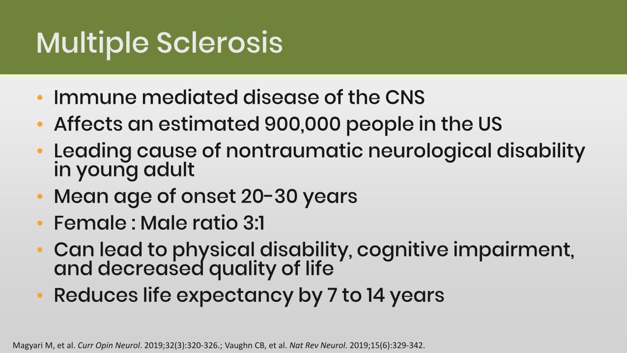
Multiple Sclerosis
• Immune mediated disease of the CNS• Affects an estimated 900,000 people in the US• Leading cause of nontraumatic neurological disability
in young adult• Mean age of onset 20-30 years• Female : Male ratio 3:1• Can lead to physical disability, cognitive impairment,
and decreased quality of life• Reduces life expectancy by 7 to 14 years
Magyari M, et al. Curr Opin Neurol. 2019;32(3):320-326.; Vaughn CB, et al. Nat Rev Neurol. 2019;15(6):329-342.
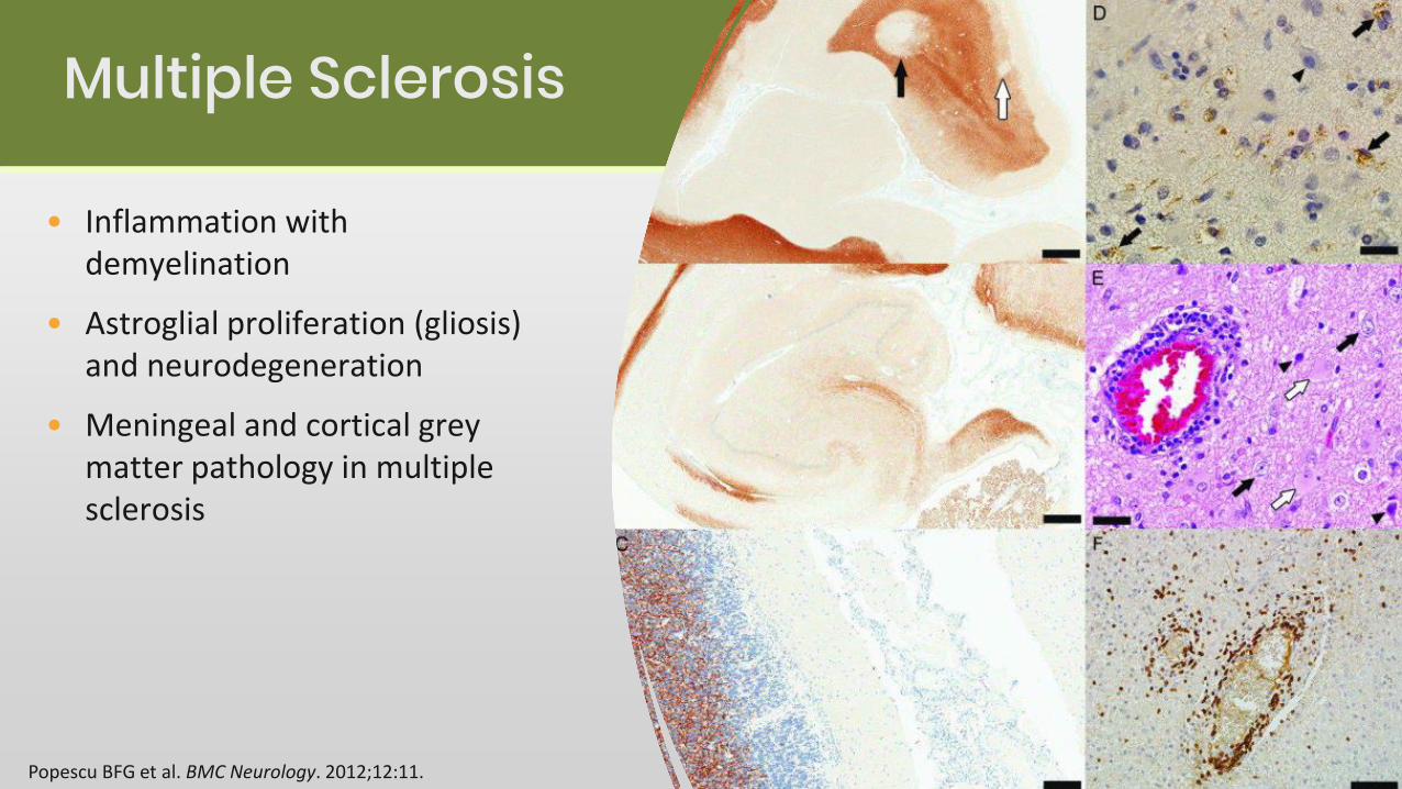
Multiple Sclerosis
• Inflammation with demyelination
• Astroglial proliferation (gliosis) and neurodegeneration
• Meningeal and cortical grey matter pathology in multiple sclerosis
Popescu BFG et al. BMC Neurology. 2012;12:11.
MS as a Silent Disease: Topographical Model
Krieger SC. Poster presented at: 2015 Meeting of the CMSC; May 29, 2015; Indianapolis, IN. Poster DX47.
Waubant E et al. Ann Clin Transl Neurol. 2019;6(9):1905-1922.
• Around 20% of the heritability risk is attributable to common genetic variants• HLA DRB15:01 haplotype (odds
ratio (OR) of ~3)• Smoking• Obesity• Low sun exposure
• Vitamin D deficiency
Changes to lung microenvironment
Gut microbiome alterations
Metabolic dysregulation
Low physical activity
Poor sleep
Diet
Smoking
Obesity
Vitamin D/sunlight
Air pollution
Maternal illness
PesticidesEBV
Gene transcription
activation
Pro-inflammatory cytokines
Fibrinogen pathway
Epigenetic changes
Aryl hydrocarbon
receptor pathway
Oxidative pathway
MS onset Relapses Disability progression
CNSDemyelination
Autoimmune process,
inflammation
ATP production unable to meet Na+/K+ demands
Calcium influx
Axonal damageReactive oxygen species
Gut microbiome (diet, obesity)
Mitochondrialdysfunction
Prodromal MS
Adapted from: Tremlett H et al. Mult Scler. 2021;27(1):6-12.
GeneticFactors
EnvironmentalExposures
RIS
DiseaseInitiation
ProdromalPeriod
MSdiagnosis
Risk factors for MS
Prodrome:Fatigue, Pain, Headache,
Low mood, Anxiety,Bladder issues,
Infections
CIS
Natural History of MS Pre-treatment EraHauser and Cree American Journal of Medicine 2020
Hauser SL et al. Am J Med. 2020;133(12):1380-1390.CIS – Clinically Isolated Syndrome; EDSS - Expanded Disability Status Scale
Relapsing MSCIS
EDSS
RELAPSES
Progressive MS
NA
TUR
AL
HIS
TOR
Y
TIME (years) Onset 5 10 15 30
8
6
4
2
MS Diagnosis• MS is diagnosed on the basis of clinical findings and
supporting evidence from ancillary tests• Magnetic resonance imaging: The imaging procedure of
choice for confirming MS and monitoring disease progression in the CNS
• Evoked potentials: Used to identify subclinical lesions; results are not specific for MS
• Lumbar puncture: May be useful to support DIT; CSF is evaluated for oligoclonal bands and intrathecal immunoglobulin G (IgG) production
https://cdn.ymaws.com/mscare.site-ym.com/resource/collection/9C5F19B9-3489-48B0-A54B-623A1ECEE07B/2018MRIGuidelines_booklet_with_final_changes_0522.pdf. Accessed May 14, 2021.
DIT – dissemination in time
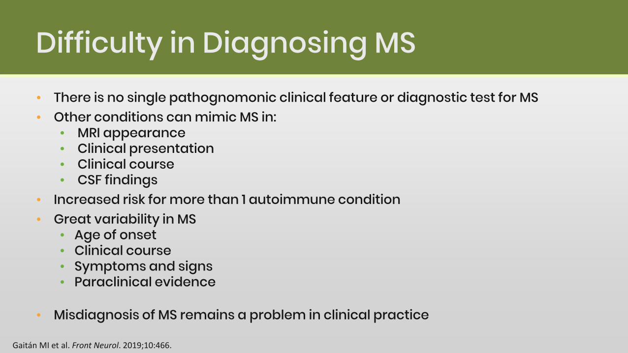
Difficulty in Diagnosing MS• There is no single pathognomonic clinical feature or diagnostic test for MS• Other conditions can mimic MS in:
• Other • Spinal stenosis; central pontine myelinolysis; radiation therapy• Medications: adalimumab
https://www.nationalmssociety.org/Symptoms-Diagnosis/Other-Conditions-to-Rule-Out. Accessed May 14, 2021.
Multiple Sclerosis Criteria
Thompson AJ. Lancet. 2018;391(10130):1622-1636.; Partucco L. Mult Scler J Exp Transl Clin. 2017;3(3):2055217317721943.
18381st
drawing
1868 Charcot
1969 Schumacher
1983 Poser2001
McDonald Criteria
2005 McDonald
Criteria
2010 McDonald
Criteria
2017 McDonald
Criteria
MRI as a Paraclinical tool
1800’s 20th century 21st century 2018
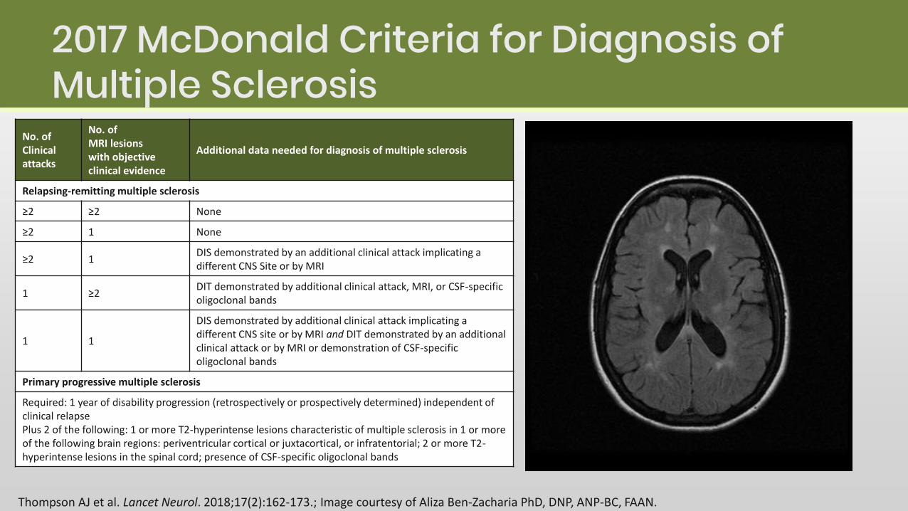
2017 McDonald Criteria for Diagnosis of Multiple Sclerosis
Thompson AJ et al. Lancet Neurol. 2018;17(2):162-173.; Image courtesy of Aliza Ben-Zacharia PhD, DNP, ANP-BC, FAAN.
No. of Clinical attacks
No. ofMRI lesionswith objectiveclinical evidence
Additional data needed for diagnosis of multiple sclerosis
Relapsing-remitting multiple sclerosis
≥2 ≥2 None
≥2 1 None
≥2 1DIS demonstrated by an additional clinical attack implicating a different CNS Site or by MRI
1 ≥2DIT demonstrated by additional clinical attack, MRI, or CSF-specific oligoclonal bands
1 1
DIS demonstrated by additional clinical attack implicating a different CNS site or by MRI and DIT demonstrated by an additional clinical attack or by MRI or demonstration of CSF-specific oligoclonal bands
Primary progressive multiple sclerosis
Required: 1 year of disability progression (retrospectively or prospectively determined) independent of clinical relapse Plus 2 of the following: 1 or more T2-hyperintense lesions characteristic of multiple sclerosis in 1 or more of the following brain regions: periventricular cortical or juxtacortical, or infratentorial; 2 or more T2-hyperintense lesions in the spinal cord; presence of CSF-specific oligoclonal bands
Key changes made to the McDonald Criteria in 2017• Brain stem and cord lesions can now be counted among
the 2 lesions disseminated in space and time• CSF oligoclonal bands can now be used to substitute for
demonstration of dissemination in time in some settings• Both asymptomatic and now symptomatic MRI lesions
can be considered in determining dissemination in space (optic nerve lesions are still excluded).
• Cortical lesions have been added to juxtacortical lesions as determinant for dissemination in space
Thompson AJ et al. Lancet Neurol. 2018;17(2):162-173.
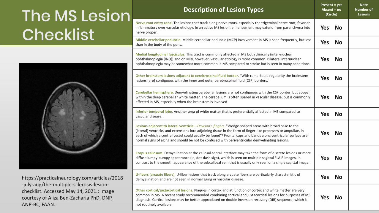
The MS Lesion Checklist
https://practicalneurology.com/articles/2018-july-aug/the-multiple-sclerosis-lesion-checklist. Accessed May 14, 2021.; Image courtesy of Aliza Ben-Zacharia PhD, DNP, ANP-BC, FAAN.
Description of Lesion Types Present = yesAbsent = no
(Circle)
NoteNumber of
Lesions
Nerve root entry zone. The lesions that track along nerve roots, especially the trigeminal nerve root, favor an inflammatory over vascular etiology. In an active MS lesion, enhancement may extend from parenchyma into nerve proper.
Yes No
Middle cerebellar peduncle. Middle cerebellar peduncle (MCP) involvement in MS is seen frequently, but less than in the body of the pons. Yes No
Medial longitudinal fasciculus. This tract is commonly affected in MS both clinically (inter-nuclear ophthalmoplegia [INO]) and on MRI, however, vascular etiology is more common. Bilateral internuclear ophthalmoplegia may be somewhat more common in MS compared to stroke but is seen in many conditions.
Yes No
Other brainstem lesions adjacent to cerebrospinal fluid border. "With remarkable regularity the brainstem lesions [are] contiguous with the inner and outer cerebrospinal fluid (CSF) borders.” Yes No
Cerebellar hemisphere. Demyelinating cerebellar lesions are not contiguous with the CSF border, but appear within the deep cerebellar white matter. The cerebellum is often spared in vascular disease, but is commonly affected in MS, especially when the brainstem is involved.
Yes No
Inferior temporal lobe. Another area of white matter that is preferentially affected in MS compared to vascular disease. Yes No
Lesions adjacent to lateral ventricle—Dawson's fingers. "Wedge-shaped areas with broad base to the [lateral] ventricle, and extensions into adjoining tissue in the form of finger-like processes or ampullae, in each of which a central vessel could usually be found"3 Frontal caps and bands along ventricular surface are normal signs of aging and should be not be confused with periventricular demyelinating lesions.
Yes No
Corpus callosum. Demyelination at the callosal-septal interface may take the form of discrete lesions or more diffuse lumpy-bumpy appearance (ie, dot-dash sign), which is seen on multiple sagittal FLAIR images, in contrast to the smooth appearance of the subcallosal vein that is usually only seen on a single sagittal image.
Yes No
U-fibers (arcuate fibers). U-fiber lesions that track along arcuate fibers are particularly characteristic of demyelination and are not seen in normal aging or vascular disease. Yes No
Other cortical/juxtacortical lesions. Plaques in cortex and at junction of cortex and white matter are very common in MS. A recent study recommended combining cortical and juxtacortical lesions for purposes of MS diagnosis. Cortical lesions may be better appreciated on double inversion recovery (DIR) sequence, which is not routinely available.
• Spinal cord lesions• <3 vertebral segments• Only part of cross-section
of the cord• No extensive cord swelling
• Gad enhancement• Initially nodular• Can evolve to a ring or an arc
• T1 hypointense center• Opening of ring points
toward the cortex
Neema M et al. Neurother. 2007;4 (4): 602-617.
Demyelination and Axonal Transection on MRI
Courtesy of Bruce D. Trapp.
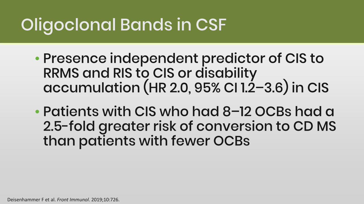
Oligoclonal Bands in CSF
• Presence independent predictor of CIS to RRMS and RIS to CIS or disability accumulation (HR 2.0, 95% CI 1.2–3.6) in CIS
• Patients with CIS who had 8–12 OCBs had a 2.5-fold greater risk of conversion to CD MS than patients with fewer OCBs
Deisenhammer F et al. Front Immunol. 2019;10:726.
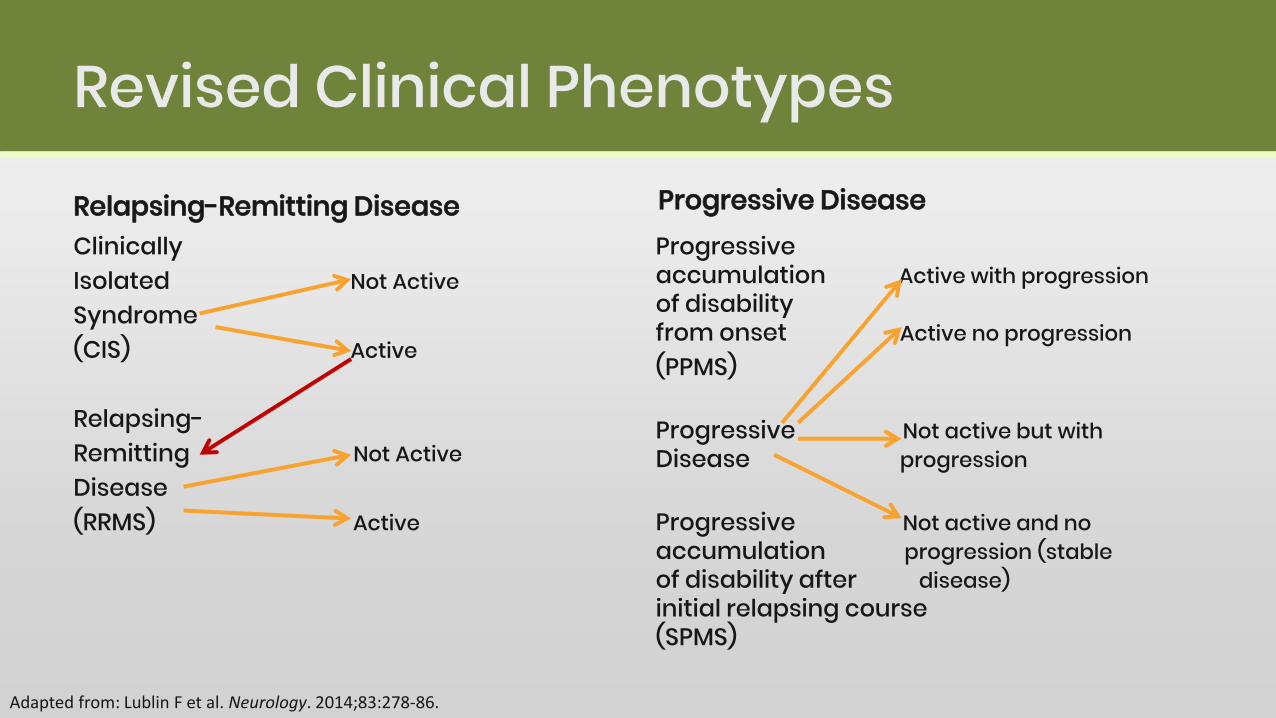
Revised Clinical Phenotypes
Relapsing-Remitting DiseaseClinicallyIsolated Not ActiveSyndrome(CIS) Active
Relapsing-Remitting Not ActiveDisease(RRMS) Active
Progressive DiseaseProgressive accumulation Active with progressionof disabilityfrom onset Active no progression(PPMS)
Progressive Not active but withDisease progression
Progressive Not active and noaccumulation progression (stableof disability after disease)initial relapsing course(SPMS)
Adapted from: Lublin F et al. Neurology. 2014;83:278-86.
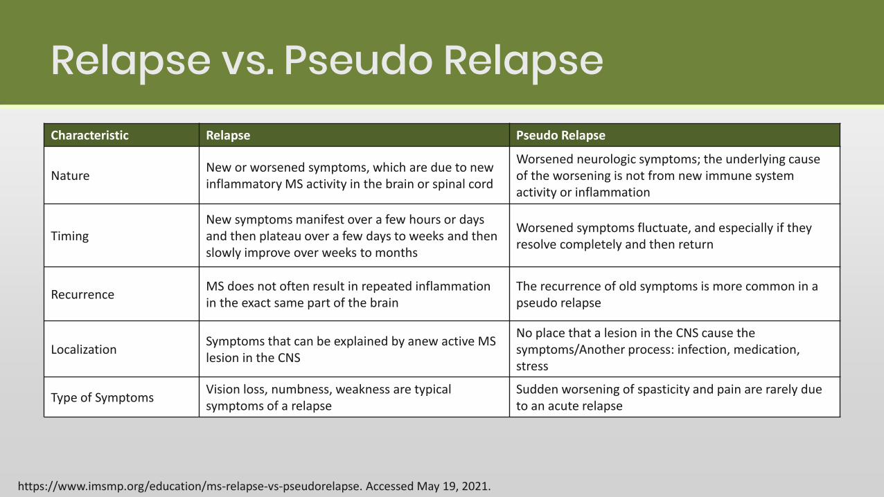
Relapse vs. Pseudo Relapse Characteristic Relapse Pseudo Relapse
NatureNew or worsened symptoms, which are due to new inflammatory MS activity in the brain or spinal cord
Worsened neurologic symptoms; the underlying cause of the worsening is not from new immune system activity or inflammation
TimingNew symptoms manifest over a few hours or days and then plateau over a few days to weeks and then slowly improve over weeks to months
Worsened symptoms fluctuate, and especially if they resolve completely and then return
RecurrenceMS does not often result in repeated inflammation in the exact same part of the brain
The recurrence of old symptoms is more common in a pseudo relapse
LocalizationSymptoms that can be explained by anew active MS lesion in the CNS
No place that a lesion in the CNS cause the symptoms/Another process: infection, medication, stress
Type of SymptomsVision loss, numbness, weakness are typical symptoms of a relapse
Sudden worsening of spasticity and pain are rarely due to an acute relapse
https://www.imsmp.org/education/ms-relapse-vs-pseudorelapse. Accessed May 19, 2021.
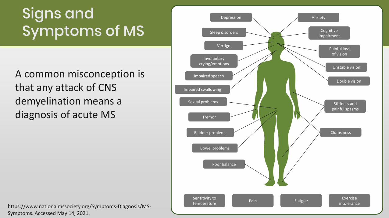
A common misconception is that any attack of CNS demyelination means a diagnosis of acute MS
Signs and Symptoms of MS
https://www.nationalmssociety.org/Symptoms-Diagnosis/MS-Symptoms. Accessed May 14, 2021.
Depression Anxiety
CognitiveImpairment
Painful lossof vision
Unstable vision
Double vision
Stiffness and painful spasms
Clumsiness
Exercise intolerance
FatiguePainSensitivity to temperature
Poor balance
Bowel problems
Bladder problems
Tremor
Sexual problems
Impaired swallowing
Impaired speech
Involuntary crying/emotions
Vertigo
Sleep disorders
Confirmed MS Diagnosis
Initiate DMT
https://cdn.ymaws.com/mscare.site-ym.com/resource/collection/9C5F19B9-3489-48B0-A54B-623A1ECEE07B/2018MRIGuidelines_booklet_with_final_changes_0522.pdf. Accessed May 14, 2021.
New or worsened neurologic symptoms lasing > 24 hrs
Fever, clinical and/or Lab signs of infection?Evaluate to
confirm or rule out MS relapse
Treat infection and re-evaluate in 7-10 days
Once MS relapse clinically confirmed – in most cases initiate systemic steroids (typically IVMP 1g for 5days)
No response or intolerability to MP – consider repository corticotropin injection
(typically 80 U IM or SQ for 5-14 days)
No response and/or severe disablingsymptoms – consider plasmapheresis
• Diagnosis of RIS occurs during diagnosis of another unrelated condition, such as migraine headaches or trauma to the area
• Typical MRI MS lesions without clinical presentation
• Two-year period, one third of patients with RIS develop a neurological event and are diagnosed with MS, one third develop a new finding on MRI without any symptoms, and one third show no change
Okuda, DT et al. PloS one. 2014;9(3):e905.; Image courtesy of Aliza Ben-Zacharia, PhD, DNP, ANP-BC, FAAN.
Clinically Isolated Syndrome
• CIS is a first episode of neurologic symptoms caused by inflammation and demyelination in the CNS
• The episode, must last for at least 24 hours, is characteristic of multiple sclerosis but does not yet meet the criteria for a MS diagnosis because people who experience a CIS may or may not go on to develop MS
• The 2017 McDonald criteria make it possible to diagnose MS in a person with CIS who also has specific findings on brain MRI
https://www.nationalmssociety.org/What-is-MS/Types-of-MS/Clinically-Isolated-Syndrome-(CIS). Accessed May 14, 2021.
MS Endophenotypes
Giovannoni G. Lancet Neurol. 2017;16(6):413-414.
Multiple sclerosis endophenotype
Presymptomatic Symptomatic
Prediagnostic phase
At riskAsymptomatic multiple
sclerosis (RIS)Prodromal
multiple sclerosisCIS Multiple Sclerosis
Diagnostic phase
Disease state (focal multiple sclerosis pathology)Inflammation, demyelination, neuroaxonal loss,
gliosis, and intrathecal synthesis of oligoclonal IgG
Predisease state
Relapsing Remitting Multiple Sclerosis• Relapses and remissions• Transforms into SPMS• Attacks of new or increasing
neurologic symptoms• Relapses lead to disability
accumulation/EDSS • RRMS active (with relapses and/or
evidence of new MRI activity) • RRMS not active, worsening (a
confirmed increase in disability following a relapse) or not worsening
https://www.nationalmssociety.org/What-is-MS/Types-of-MS/Relapsing-remitting-MS. Accessed May 14, 2021.
Dis
abili
ty
Time
RRMS
Relapse
Active without worsening
Worsening (incomplete recoveryfrom relapse)
Stable without activity
New MRI activity
Secondary Progressive MS
• SPMS follows an initial RRMS• SPMS a progressive worsening of
neurologic function (accumulation of disability) over time
• SPMS active - with relapses and/or evidence of new MRI activity
• SPMS not active, with progression (evidence of disability accumulation over time, with or without relapses or new MRI activity) or without progression
https://www.nationalmssociety.org/What-is-MS/Types-of-MS/Secondary-progressive-MS. Accessed May 14, 2021.
Dis
abili
ty
Time
SPMS
RRMS
Active (relapse or new MRI activity)with progression
Not active with progression
Not active without progression (stable)
New MRI activity
Active (relapse or MRI activity)without progression
function (accumulation of disability) from the onset of symptoms, without early relapses or remissions
• PPMS active (with an occasional relapse and/or evidence of new MRI activity over a specified period of time)
• PPMS not active, with progression (evidence of disability accumulation over time, with or without relapse or new MRI activity) or without progression
https://www.nationalmssociety.org/What-is-MS/Types-of-MS/Primary-progressive-MS. Accessed May 14, 2021.
Dis
abili
ty
Time
PPMS
Active (relapse or new MRI activity)with progression
Not active without progression (stable)
Not active with progression
Active without progression
New MRI activity
Multiple Sclerosis Prognosis
Rotstein D et al. Nat Rev Neurol. 2019;15:287-300.
Demographic and environmental factors
• Older age
• Male sex
• Not of European descent
• Low vitamin D levels
• Smoking
• Comorbid conditions
Clinical factors• Primary progressive disease subtype• A high relapse rate• A shorter interval between the first and second
relapses• Brainstem, cerebellar or spinal cord onset• Poor recovery from the first relapse• A higher Expanded Disability Status Scale score at
diagnosis• Polysymptomatic onset• Early cognitive deficits
MRI observations• A high number of T2 lesions• A high T2 lesion volume• The presence of gadolinium-enhancing lesions• The presence of infratentorial lesions• The presence of spinal cord lesions• Whole brain atrophy• Grey matter atrophy
Biomarkers• A high number of T2 lesions• The presence of IgG and IgM oligoclonal bands in
the CSF• High levels of neurofilament light chain in the CSF
and serum• High levels of chitinase in the CSF• Retinal nerve fibre layer thinning detected with
optical coherence tomography
Poor prognosis
Clinical Case• 25-year-old Hispanic female • New onset: weakness of left arm,
Numbness• Medical History: Optic neuritis 3 years
ago, depression, smoker • Current Medications: Vitamin D,
partially adherent• Cultural Considerations: her mother
has never heard of the disease• BRAIN MRI 3 years ago
Courtesy of Aliza Ben-Zacharia, PhD, DNP, ANP-BC, FAAN.
Meet Criteria?
Courtesy of Aliza Ben-Zacharia, PhD, DNP, ANP-BC, FAAN.
No. of Clinical attacks
No. ofMRI lesionswith objectiveclinical evidence
Additional data needed for diagnosis of multiple sclerosis
Relapsing-remitting multiple sclerosis
≥2 ≥2 None
Conclusion• MS is a complex disease with multiple endophenotypes• High-risk RIS and prodrome may become a part of the MS
spectrum in the next version of the McDonald criteria• Many patients previously labelled as CIS now receive the
diagnosis of MS, making the prognosis of both CIS and RRMS milder
• Important to diagnose early and treat early• Once diagnosed, important to assess the presence of poor
prognostic indicators, symptoms, treating exacerbations, starting DMT and managing comorbidities