Page 1
Steven M. Thompson Manuscript for Multiple Sequence Alignment and Analysis Page 1 3/31/04
Multiple Sequence Alignment and Analysis: Part I —
An Introduction to the Theory and Application of
Multiple Sequence Analysis.
author: Steven M. Thompson
Florida State UniversitySchool of Computational Science and Information TechnologyTallahassee, Florida 32306-4120telephone: 850-644-1010fax: 850-644-0098
corresponding address:
Steve ThompsonBioInfo 4U2538 Winnwood CircleValdosta, Georgia, 31601-7953telephone: [email protected]
Page 2
Steven M. Thompson Manuscript for Multiple Sequence Alignment and Analysis Page 2 3/31/04
¥GCG is the Genetics Computer Group, the producer of the Wisconsin Package for sequence analysis
and a part of Accelrys Inc., a subsidiary of Pharmacopeia Inc.
2003 BioInfo 4U
Page 3
Steven M. Thompson Manuscript for Multiple Sequence Alignment and Analysis Page 3 3/31/04
Abstract.
I introduce the foundations, principles, and applications of multiple sequence analysis in this chapter, with a
beginners perspective in mind. I begin with a general introduction to the principles of pairwise sequence
comparison, scoring matrices, and the dynamic programming algorithm. The concepts of similarity,
significance, and homology are next discussed. These principles are then extended to multiple sequence
alignment and analysis and its varied applications, specifically motif, profile, and phylogenetic techniques. A
brief discussion of multiple sequence alignment related to protein structure prediction concludes the chapter.
These concepts are all illustrated in Part II’s (Chapter 4) practical session using the Accelrys Wisconsin
Package software.
Contents.
1. Introduction.
2. Dynamic Programming.
3. Scoring Matrices.
4. Similarity and Significance.
5. Applicability?
6. Multiple Sequence Dynamic Programming.
6.1. How the Algorithm Works.
7. Motif Definition: What is a Motif?
8. Profile Analysis: Position Specific, Weighted Score Matrices of Multiple Sequence Alignments.
8.1. Hidden Markov Modeling and Profiles.
9. Multiple Sequence Alignment and Structure Prediction.
10. Conclusions and Reliability?
1. Introduction.
What can we learn about a biological molecule given its nucleotide or amino acid sequence? We can uncover
some of the underlying information in sequences by searching for patterns that may reflect some constrain on
the molecule. These can be catalogued motifs or domains, secondary structure predictions, physical
attributes such as hydrophobicity, or even the content of DNA itself, as in some gene finding techniques. But
what about comparisons with other sequences? Can we learn about one molecule by comparing it to
another? Yes, naturally we can; inference through homology is fundamental to all the biological sciences. We
can learn a tremendous amount by comparing our sequence against others.
The power and sensitivity of sequence based computational methods dramatically increases with the addition
of more data. More data yields stronger analyses — if done carefully! Otherwise, it can confound the issue.
The patterns of conservation become clearer by comparing the conserved portions of sequences amongst a
larger and larger dataset. Those areas most resistant to change are structurally and functionally the most
important to the molecule. The basic assumption is that those portions of sequence of crucial structural, and
hence functional value, are most constrained against evolutionary change. They will not tolerate many
mutations. Not that mutations do not occur in these portions, just that most mutations in the region are lethal
so we never see them. Other areas of sequence are able to drift more readily, being less subject to
Page 4
Steven M. Thompson Manuscript for Multiple Sequence Alignment and Analysis Page 4 3/31/04
evolutionary pressure. Therefore, sequences end up a mosaic of quickly and slowly changing regions over
evolutionary time. However, in order to learn anything by comparing sequences, we need to know how to
compare them. We can use those constrained portions as ‘anchors’ to create a sequence alignment allowing
comparison, but this brings up the alignment problem and ‘similarity’. It is easy to see that two sequences are
aligned when they have identical symbols at identical positions, but what happens when symbols are not
identical or the sequences are not the same length. How can we know that the most similar portions of our
sequences are aligned, when is an alignment optimal, and does optimal mean biologically correct? How can
anybody figure any of this out?
A ‘brute force’ approach just won’t work. Even without considering the introduction of gaps, the
computation required to compare all possible alignments between two sequences requires time proportional
to the product of the lengths of the two sequences. Therefore, if the two sequences are approximately the
same length (N), this is a N2 problem. To include gaps, we would have to repeat the calculation 2N times to
examine the possibility of gaps at each possible position within the sequences, now a N4N problem. Michael
Waterman illustrated the problem in 1989 stating that to align two sequences 300 characters long, 1088
comparisons would be required, about the same number as the number of elementary particles estimated to
exist in the universe! Part of the solution to this problem is the dynamic programming algorithm.
2. Dynamic Programming.
Let’s begin with a review of pairwise dynamic programming. In a simplistic illustration of dynamic
programming we will consider matching symbols to be worth one point and non-matching symbols to be
worth zero points. We will also impose a very simple gap penalty function — we will penalize the scoring
scheme by subtracting one point for every gap inserted, unless at the beginning or end of the sequence. In
other words, end gaps will not be penalized, i.e. both sequences do not have to begin or end at the same point
in the alignment. This zero penalty end-weighting scheme is the default for most alignment programs, but
can often be changed with a program option, if desired. However, the gap function described here and used
in the example is a much simpler gap penalty function than normally used in alignment programs. Normally
an ‘affine’, i.e. a linear, function is used; the standard y = mx + b equation:
total penalty = gap opening penalty + ([length of gap] * [gap extension penalty]).
To run most alignment programs with the type of simple DNA gap penalty used in this example, you would
have to designate a gap ‘creation’ or ‘opening’ penalty of zero and a gap ‘extension’ or ‘length’ penalty of
whatever counts in that particular program’s scoring matrix as an identical base match for DNA sequences.
One way to visualize the process works through the cells of a matrix. The solution occurs in two stages. The
first begins very much like dot plot methods; the second is totally different. I will further simplify my
illustration. Instead of calculating the ‘score matrix’ on the fly, as is often taught as one proceeds through the
graph, I like to completely fill in an original ‘match matrix’ first, and then add points to those positions which
produce favorable alignments next. Points are added based on a “looking back over-your-left-shoulder”
algorithm rule, where the only allowable trace-back is diagonally behind and above.
Page 5
Steven M. Thompson Manuscript for Multiple Sequence Alignment and Analysis Page 5 3/31/04
My example in Table 1 uses two sequences that represent the TATA consensus regions of Eukaryotes and
Bacteria. The most conserved bases within the consensus are capitalized. The Eukaryote promoter sequence
is along the X-axis; the Bacteria promoter sequence is along the Y-axis.
There may be more than one best path through the matrix. This time, starting at the top and working down
as we did, then tracing back, I found one optimum alignment, but there’s probably more:
cTATAtAagg
| |||||
cg.TAtAaT.
This alignment has a final score of 5. This is the number optimized by the algorithm, not any type of a
similarity or identity percentage! The software will arbitrarily (based on some rule) choose one optimal
solution. To help explore potential solution space the decision can be partly controlled in the Accelrys
Wisconsin Package (GCG) programs BestFit and Gap with the -HighRoad/-LowRoad options. The above
solution is the GCG -HighRoad solution found when running the program Gap with the above example’s
parameter settings. This is seen in Table 2. Do you have any idea about how other alignments, such as
GCG’s -LowRoad solution, could be discovered? Answer: Often if you reverse the solution of the entire
dynamic programming process, other solutions are found! In other words, reverse the sequences in software
programs to see alternative alignments.
To recap, the dynamic programming algorithm discovers an optimal pairwise alignment, where optimal is
defined as an arrangement of two sequences, 1 of length i and 2 of length j, such that:
1) you maximize the number of matching symbols between 1 and 2;
2) you minimize the number of gaps within 1 and 2; and
3) you minimize the number of mismatched symbols between 1 and 2.
Therefore, the actual solution can be represented by:
Si-1 j-1 or
max Si-x j-1 + wx-1 or
Sij = sij + max 2 < x < i
max Si-1 j-y + wy-1
2 < y < i
where Sij is the score for the alignment ending at i in sequence 1 and j in sequence 2,
sij is the score for aligning i with j,
wx is the score for making a x long gap in sequence 1,
wy is the score for making a y long gap in sequence 2,
allowing gaps to be any length in either sequence.
However, as we’ve seen, just because dynamic programming guarantees an optimal alignment, it is not
necessarily the only optimal alignment. Furthermore, the optimal alignment is not necessarily the ‘right’ or
biologically relevant alignment! As always, question the results of any computerized solution based on what
Page 6
Steven M. Thompson Manuscript for Multiple Sequence Alignment and Analysis Page 6 3/31/04
you know about the biology of the system. The above example illustrates the Needleman and Wunsch (1970)
global solution. Later refinements (Smith and Waterman, 1981) demonstrated how dynamic programming
could also be used to find optimal local alignments. To solve dynamic programming using local alignment
(without going into all the gory details) programs use the following two tricks:
• A scoring match matrix using negative numbers for mismatches is incorporated. Therefore, bad
paths quickly become very bad. This leads to a trace-back path matrix with many alternative paths,
most of which do not extend the full length of the graph.
• The best trace-back within the graph is chosen. This does not have to begin or end at the edges of the
graph — it looks for the best segment of alignment.
The Wisconsin Package has three pairwise dynamic programming implementations. Gap is a ‘global’ (i.e.
Needleman and Wunsch, 1970) alignment program and BestFit is a ‘local’ (i.e. Smith and Waterman, 1981)
alignment program, both between two sequences of the same type, whereas FrameAlign can be global or
local depending on the options that you set, but it always aligns DNA to protein. Using one versus the other
implies that you are looking for distinctly different relationships. If you already know that the full length of
two sequences of the same type are pretty close, that they probably belong to the same family, then Gap is the
program for you; if you only suspect an area of one is similar to an area of another, then you should use
BestFit. To force BestFit to be even more local, you can specify a more stringent alternative symbol
comparison table, such as pam250.cmp or blosum100.cmp. If you suspect that a DNA frame shift sequencing
error is affecting the alignment, then FrameAlign is the program to use. It uses dynamic programming to
align a protein to a DNA sequence with the allowance of frame shifts. Frame shift errors will appear in the
output alignment as gaps that are not multiples of three.
3. Scoring Matrices.
But what about protein sequences — conservative replacements and similarities, as opposed to identities?
This is definitely an additional complication to consider. Certain amino acids are very much alike,
structurally, chemically, and genetically. How can we take advantage of the similarity of amino acids in our
alignments? People have been struggling with this problem since the late 1960’s.
Margaret Dayhoff (Schwartz and Dayhoff, 1979) unambiguously aligned closely related protein datasets (no
more than 15% difference) available at that point in time and noticed that certain residues, if they mutate at
all, are prone to change into certain other residues. As it works out, these propensities for change fell into the
same chemical and structural amino acid categories that chemists had known for years — conserved through
the evolutionary constraints of natural selection. However, Dayhoff’s empirical observation quantified these
changes. Based on the unequivocal multiple sequence alignments that she created, the assumption that
estimated mutation rates in closely related proteins can be extrapolated to more distant relationships, and on
matrix and logarithmic mathematics to smooth the statistics of the system, she was able to empirically specify
the relative probabilities at which different residues mutate into other residues through evolutionary history
as appropriate within some level of divergence between the sequences considered. This is the basis of the
famous PAM (corrupted acronym of accepted point mutation) 250 (meaning that the matrix has been
multiplied by itself 250 times) log-odds matrix. Since Dayhoff’s time other biomathematicians (esp. see
Page 7
Steven M. Thompson Manuscript for Multiple Sequence Alignment and Analysis Page 7 3/31/04
Henikoff and Henikoff’s [1992] BLOSUM series of tables, and Gonnet et al. [1992]) have created newer
matrices with more or less success than Dayhoff’s original, but the concept remains the same, and Dayhoff’s
original PAM 250 table remains a classic as historically the most widely used. This chapter will not cover the
mathematics of how these matrices are created, but I encourage you read the primary references to gain some
appreciation of the process. Collectively these types of tables are known as symbol comparison tables, log-
odds matrices, or scoring matrices, and they are fundamental to all sequence comparison techniques.
The default amino acid substitution scoring matrix for many protein similarity comparison programs is now
the BLOSUM62 table (Henikoff and Henikoff, 1992). It is shown in Table 3; the main identity diagonal is
highlighted with outline characters to make it easier to recognize, as are absolute values ±4. Notice that
positive values for identity range from 4 to 11 and negative values for those substitutions that rarely occur go
as low as –4. The most conserved residue is tryptophan with an identity score of 11; cysteine is next with a
score of 9; histidine gets 8; both proline and tyrosine get scores of 7. Also check out the hydrophobic
substitution triumvirate — isoleucine, leucine, valine, and to a lesser extent methionine — all easily swap
places. So rather than using the one/zero match function that we used in the simple TATA dynamic
programming example above, protein sequence alignments use the match function provided by a scoring
matrix such as this. The concept of similarity becomes very important with some amino acids being way
‘more similar’ than others!
4. Similarity and Significance.
People are often confused by the distinction between homology and similarity: There is a huge difference!
Similarity is merely a statistical parameter that describes how much two sequences, or portions of them, are
alike according to some set scoring criteria. It can be normalized to ascertain statistical significance, but it’s
still just a number. Homology, in contrast and by definition, implies an evolutionary relationship — more
than just the fact that we have all evolved from the same old primordial ‘ooze’. You need to place the
organisms or genes of interest in a phylogenetic framework amongst their relatives to claim homology. Better
yet, demonstrate experimental evidence — structural, morphological, genetic, or fossil — that corroborates
your assertion. There really is no such thing as percent homology; something is either homologous or it is
not. Walter Fitch is credited with the joke “homology is like pregnancy — you can’t be 45% pregnant, just
like something can’t be 45% homologous. You either are or you are not”. Do not make the commonly made
mistake of calling any old sequence similarity homology. Highly significant similarity can argue for
homology, but never the other way around.
So, how do you tell if a similarity, in other words, an alignment discovered by some program, means
anything? Is it statistically significant, is it truly homologous, and even more importantly, does it have
anything to do with real biology? Many of the programs generate percentage scores, but these really don’t
mean a whole lot. Do not use percent similarities or identities to compare sequences except in the roughest
way. They are not optimized or normalized in any manner, and they don’t reflect the length of the alignment
at all. The ‘raw’ similarity scores, opt, S, or quality, depending on the program, all mean a lot more but can be
confusing. At least they take the length of similarity, all of the necessary introduced gaps, and the matching
of symbols all into account, but they are only relevant within the context of a particular comparison or search
with a particular scoring matrix and specific gap penalties. Some programs generate histograms of score
Page 8
Steven M. Thompson Manuscript for Multiple Sequence Alignment and Analysis Page 8 3/31/04
distributions; this helps some. To get a better handle on what the various scores mean, read the original
papers, textbook and review summaries, and the relevant algorithm sections of the GCG Program Manual —
statistics can be confusing but the more you read, the better you’ll understand.
A traditional way of deciding alignment significance relies on an old statistics trick — Monte Carlo
simulations. This type of significance estimation has many implicit statistical problems; however, few
practical alternatives exist for comparing just two sequences. Monte Carlo methods compare an actual score,
in this case the similarity score of an alignment, against the distribution of scores of alignments against a
randomized sequence. Therefore, one way of estimating alignment significance is to take advantage of the
Monte Carlo style randomizations option available in the GCG programs Gap and BestFit. To utilize this
strategy, compare two sequences using the appropriate algorithm, depending on whether you’re trying to
compare the entire length of each sequence, or only the best regions of similarity of each, respectively, and
specify the command line option “-Randomizations=100”. This option jumbles the second sequence of the
comparison 100 times after the initial alignment is produced and then generates scores and a standard
deviation based on the jumbled matches. Comparing the quality scores of the randomized alignments to the
initial alignment can help give a feeling for the relative meaning of the score.
You can compare the mean of the random scores to the unjumbled score using a ‘Z score’ to help decide
significance. An old ‘rule-of-thumb’ that people often use is, if the actual score is much more than three
standard deviations above the mean of the randomized scores, the analysis may be significant; if it is much
more than five, than it probably is significant; and if it is above nine, than it definitely is significant. Many Z
scores measure the distance from a mean using this simplistic Monte Carlo model assuming a normal (i.e.
Gaussian) distribution, in spite of the fact that ‘sequence-space’ actually follows what is know as the ‘extreme
value distribution;’ however, the method does approximate significance estimates quite well and is calculated
with the following formula:
Z score = [ ( actual score ) - ( mean of randomized scores ) ]
( standard deviation of randomized score distribution )
When the two TATA sequences from the previous dynamic programming example are compared to one
another using the same scoring parameters as before, but incorporating a Monte Carlo Z score calculation,
their similarity is found, surprisingly, to be not at all significant, in spite of being 75% identical. It is merely a
reflection of the compositional bias of the two sequences to contain lots of T’s and A’s. Those results follow:
Average quality based on 100 randomizations: 41.8 +/- 7.4. Plugged into the formula: ( 50 – 41.8 ) / 7.4
= 1.11, i.e. no significance. Composition can make a huge difference!
Sometimes a seemingly decent alignment will not be significant upon further inspection — do not blindly
accept the output of any computer program! Always investigate further for similarities can be strictly
artifactual. Comparisons can be insignificant in spite of what seems to be, upon first inspection, very good
alignments with high percent identities. A Monte Carlo style Z-test below around 3.5, near the bottom of
Russell Doolittle’s “Twilight Zone” (1986), can suggest that the similarity is not significant, that it is merely
the result of compositional bias.
Page 9
Steven M. Thompson Manuscript for Multiple Sequence Alignment and Analysis Page 9 3/31/04
The FastA (Pearson and Lipman, 1988; and Pearson, 1998), BLAST (Altschul et al. 1990 and 1997),
ProfileSearch (Gribskov, et al., 1987 and 1989), and HMMerSearch (Eddy, 1996 and 1998) database similarity
searching suites all use a similar approach, but they base their statistics on the distance of alignment scores
from their mean using a more realistic model than Monte Carlo style Z scores do. They use the actual, or a
simulated, ‘extreme value distribution’ of ‘insignificantly similar’ alignment scores from the database being
searched. BLAST, FastA, and HMMerSearch all generate Expectation, “E”, values in this manner;
ProfileSearch returns Z scores, which follow the same guidelines as mentioned above. Expectation values are
printed in scientific notation, and the smaller the number, i.e. the closer it is to zero, the more significant the
match is, and the higher its Z score will be. The higher the E value is, the more probable the observed match
is due to chance, and the lower its Z score will be, i.e. the score is not significant. Expectation values show us
how often we would expect that particular alignment match to occur merely by chance alone in a search of
that size database; or from another perspective, they describe the number of search set sequences that would
be needed to obtain an alignment score greater than or equal to that obtained in any particular search purely
by chance. Often you can see a demarcation where the Expectation values drop off between the significant
hits and background noise. True homologues often segregate from other sequences that only contain similar
modules or domains and these will segregate from the rest of ‘sequence space’. The E value is the number
that really matters, that you need to pay attention to, not the raw ‘scores’. Conservative, ‘rule-of-thumb’
guidelines for Z scores and Expectation values from a typical protein search are shown in Table 4.
Even though Monte Carlo style Z scores follow E values fairly well, be very careful with any guidelines such
as those in Table 4. They are entirely dependent on the query sequence’s composition, and on both the size
and content of the database being searched, as well as on how often you perform the search! Think about it
— the odds are way different for rolling a “Yahtzee” depending on how many dice you roll, whether they are
‘loaded’ or not, and how often you try. The programs Xnu and Seg are available in the Wisconsin Package
outside of BLAST for prefiltering your sequences of the type of repeat and low complexity regions that can
cause compositional biases with the potential to confound search algorithms.
Another very powerful empirical method of determining significance is to repeat a database search with the
entry in question. If that entry finds more significant ‘hits’ with the same sorts of sequences as the original
search, then the entry in question is undoubtedly homologous to the original entry. That is, homology is
transient. If it finds entirely different types of sequences, then it probably is not a true homologue. Modular
proteins with distinctly separate domains confuse issues, but the principles remain the same, and can be
explained through domain swapping and nonvertical transmission. And, finally, the ‘Gold-standard’ of
homology is shared structural folds — if you can demonstrate that two proteins have the same structural
fold, then, regardless of similarity, at least that particular domain is homologous between the two.
Furthermore, all alignment, regardless of the algorithm used, is far more sensitive at the amino acid level than
at the DNA level. This is because proteins have twenty match criteria versus DNA’s four and those four
DNA bases are usually identical, not similar, to each other; and many DNA base changes (especially third
position changes) do not change the encoded protein. All of these factors drastically increase the ‘noise’ level
of a DNA against DNA search, and gives protein searches a much greater ‘look-back’ time, doubling it or
Page 10
Steven M. Thompson Manuscript for Multiple Sequence Alignment and Analysis Page 10 3/31/04
more. Therefore, whenever dealing with coding sequence, always search at the protein level, either directly
or with programs that translate nucleotide sequences ‘on-the-fly’.
5. Applicability?
So what’s so great about multiple sequence alignments; why would anyone want to bother? They are:
• very useful in the development of PCR primers and hybridization probes;
• great for producing annotated, publication quality, graphics and illustrations;
• invaluable in structure/function studies through homology inference;
• essential for building “profiles” for remote homology similarity searching; and
• required for molecular evolutionary phylogenetic inference programs.
A multiple sequence alignment is useful for probe and primer design by allowing you to visualize the most
conserved regions of an alignment. This technique is great for designing phylogenetic specific probes as it
clearly localizes areas of high conservation and high variability in an alignment. Depending on the dataset
that you analyze, any level of phylogenetic specificity can be achieved. Pick areas of high variability in the
overall dataset that correspond to areas of high conversation in phylogenetic category subset datasets to
differentiate between universal and specific potential probe sequences. After localizing general target areas
on the sequence, you can then use a primer discovery program to find the best primers within those regions
and to test those potential probes for common PCR conditions and problems.
Graphics prepared from multiple sequence alignments can dramatically illustrate functional and structural
conservation. Alignments, or portions thereof, can take many forms — shaded or colored boxes or letters for
each residue, cartoon representations of features, running line graphs of overall similarity, overlays of
attributes, various consensus representations — all can be printed with high-resolution equipment, in color or
gray tones. These can make a big impact in a manuscript or poster presentation.
Conserved regions of an alignment are structurally and functionally important. In addition to the
conservation of primary sequence, secondary and even tertiary structure is conserved in these crucial regions.
Recognizable structural conservation between true homologues extends way beyond statistically significant
sequence similarity. This is why statistically insignificant similarity can not negate homology. An oft-cited
example is in the serine protease superfamily. S. griseus protease A demonstrates remarkably little similarity
when compared to the rest of the superfamily (Expectation values E() 101.8 in a typical search) yet its three-
dimensional structure clearly shows its allegiance to the serine proteases (Pearson, W.R., personal
communication). These principles are the premise of ‘homology modeling’, which works remarkably well.
Profiles are position specific weight matrix descriptions of an alignment or a portion thereof. Gap insertion is
penalized more heavily in conserved areas than in variable regions, and the more highly conserved a residue
is, the more important it becomes. Originally described by Gribskov, et al. (1987 and 1989), later refinements
have added statistical rigor (see e.g. Eddy’s Hidden Markov Model profiles [1996 and 1998]). Several profile
methods will be described in this chapter. Profiles are used to search databases for remote sequence
Page 11
Steven M. Thompson Manuscript for Multiple Sequence Alignment and Analysis Page 11 3/31/04
similarities, and to create larger and larger alignments. Profile searching is tremendously powerful and can
provide the most sensitive, albeit extremely computationally intensive, database similarity search possible.
Finally, multiple sequence alignment is a necessary prerequisite for sequence based phylogenetic inference,
and phylogenetic inference guides our understanding of molecular evolution. The famous Darwinian
Theodosius Dobzhansky summed it up succinctly in 1973, provided as an inscription on the inner cover of the
classic organic evolution text Evolution: “Nothing in biology makes sense except in the light of evolution”
(Dobzhansky, et al., 1977). These words ring true — evolution provides the single, unifying, cohesive force
that explains all life. It is to the life sciences what the ‘holy grail’ of the unified field theory is to astrophysics.
Based on the assertion of homologous positions in an alignment, we can estimate the most reasonable
evolutionary tree for that alignment (see e.g. PAUP* (Phylogenetic Analysis Using Parsimony [and other
methods]) [Swofford, 1989–2003] and PHYLIP (PHYLogeny Inference Package) [Felsenstein, 1980–2003]).
This is a huge, complicated, and highly contentious field. However, always remember that regardless of
algorithm used, parsimony, any distance method, maximum likelihood, or even Bayesian techniques, all
molecular sequence phylogenetic inference programs make the absolute validity of your input alignment
their first and most critical assumption. The accuracy of your alignment is the most important factor in
inferring reliable phylogenies; the results are utterly dependent on its quality. Do not use any questionable
parts. Only analyze those portions that assuredly align. If any portions of the alignment are in doubt,
exclude them. This usually means trimming down or masking the alignment’s terminal ends and may
require internal trimming or masking as well (see masking explained in the next chapter’s section 9).
Biocomputing is always a delicate balance — signal against noise — and sometimes it can be quite the
balancing act!
6. Multiple Sequence Dynamic Programming.
As seen in pairwise dynamic programming, looking at every possible position by sliding one sequence along
every other sequence, just will not work for alignment. Therefore, dynamic programming reduces the
problem back down to N2. But how do you work with more than just two sequences at a time? It becomes a
much harder problem. You could painstakingly manually align all your sequences using an editor, and many
people do just that, but some type of an automated solution is desirable, at least as a starting point to manual
alignment. However, solving the dynamic programming algorithm for more than just two sequences rapidly
becomes intractable. Dynamic programming’s complexity, and hence its computational requirements,
increases exponentially with the number of sequences in the dataset being compared (complexity=[sequence
length]number of sequences). Mathematically this is an N-dimensional matrix, quite complex indeed. As
seen, pairwise dynamic programming solves a two-dimensional matrix, and the complexity of the solution is
equal to the length of the longest sequence squared. Well, a three member standard dynamic programming
sequence comparison would be a matrix with three axes, the length of the longest sequence cubed, and so
forth. You can at least draw a three-dimensional matrix, but more than that becomes impossible to even
visualize. It quickly boggles the mind!
Page 12
Steven M. Thompson Manuscript for Multiple Sequence Alignment and Analysis Page 12 3/31/04
Several different heuristics have been employed over the years to simplify the complexity of the problem.
One program, MSA (Gupta et al., 1995), does attempt to globally solve the N-dimensional matrix equation
using a bounding box trick. However, the algorithm’s complexity precludes its use in most situations, except
with very small datasets. One way to still globally solve the algorithm and yet reduce its complexity is to
restrict the search space to only the most conserved ‘local’ portions of all the sequences involved. This
approach is used by the program PIMA (Smith and Smith, 1992). You can run MSA and PIMA at several
bioinformatics server sites on the Internet (in particular the Baylor College of Medicine’s Search Launcher at
http://searchlauncher.bcm.tmc.edu/ Smith et al., 1996).
6.1. How the Algorithm Works.
The most common implementations of automated multiple alignment modify dynamic programming by
establishing a pairwise order in which to build the alignment. This modification is known as pairwise,
progressive dynamic programming. Originally attributed to Feng and Doolittle (1987), this variation of the
dynamic programming algorithm generates a global alignment, but restricts its search space at any one time
to a local neighborhood of the full length of only two sequences. Consider a group of sequences. First all are
compared to each other, pairwise, using normal dynamic programming. This establishes an order for the set,
most to least similar. Subgroups are clustered together similarly. Then take the top two most similar
sequences and align them using normal dynamic programming. Now create a consensus of the two and align
that consensus to the third sequence using standard dynamic programming. Now create a consensus of the
first three sequences and align that to the forth most similar. This process continues until it has worked its
way through all sequences and/or sets of clusters. The pairwise, progressive solution is implemented in
several programs. Perhaps the most popular is Thompson et al.’s ClustalW (1994) and its multiplatform GUI
ClustalX (Thompson, et al., 1997). The ClustalX homesite guarantees the latest version: ftp://ftp-igbmc.u-
strasbg.fr/pub/ClustalX/. The Wisconsin Package program PileUp implements a very similar method and is
thoroughly explored in Part II, Chapter 4, Section 6.
As with pairwise alignments and sequence database similarity searching, all of this is much easier with
protein sequences versus nucleotide sequences. Twenty symbols are just much easier to align then only four;
the signal to noise ratio is so much better. And, as in database searching, the concept of similarity applies to
amino acids but generally not to nucleotides. Therefore, just like in database searching, multiple sequence
alignment should always be done on a protein level if at all possible, unless the DNA sequences are so similar
as to not cause any problem. Therefore, translate nucleotide sequences to their protein counterparts, if you
are dealing with coding sequences, before performing multiple sequence alignment. The process is much
more difficult if you are forced to align nucleotides because the region does not code for a protein.
Automated methods may be able to help as a starting point, but they are certainly not guaranteed to come up
with a biologically correct alignment. The resulting alignment will probably have to be extensively edited, if
it works at all. Success will largely depend on the similarity of the nucleotide dataset.
One liability of global progressive, pairwise methods is they are entirely dependent on the order in which the
sequences are aligned. Fortunately ordering them from most similar to least similar usually makes biological
sense and works very well. However, the techniques are very sensitive to the substitution matrix and gap
Page 13
Steven M. Thompson Manuscript for Multiple Sequence Alignment and Analysis Page 13 3/31/04
penalties specified. Programs such as ClustalW and PileUp that allow ‘fine-tuning’ areas of an alignment by
realignment with different scoring matrices and/or gap penalties can be extremely helpful because of this.
However, any automated multiple sequence alignment program should be thought of as only a tool to offer a
starting alignment that can be improved upon, not the ‘end-all-to-meet-all’ solution, guaranteed to provide
the ‘one-true’ answer.
7. Motif Definition: What is a Motif?
Many, many features have been described and catalogued in biological sequences over the years. Most of
these have recognizable consensus patterns that allow you to screen an unknown sequence for their
occurrence. However, motif definition is a complicated matter. One very simplistic approach is to look at an
alignment, see that certain regions are conserved, and create a consensus of that region. A multiple sequence
alignment of Elongation Factor Tu/1 from several different organisms in Figure 1 illustrates the
conservation of the first of several GTP-binding domains in these proteins.
Based on experimental evidence, we know that the indicated region bounded by the glycine and serine is
essential. Therefore, merely count up the various residues in those locations and assign the most common
one to the consensus. Simple. But what about the fact that the middle histidine isn’t always a histidine; in
this data set, just as often it’s a serine and sometimes it’s an alanine. Other positions are also variable. There
are also other members of this gene family not being represented here at all. A consensus isn’t necessarily the
biologically “correct” combination. How do we include this other information? A simple consensus throws
much of it away. Therefore, we need to adopt some sort of standardized ambiguity notation, a regular
expression in computer science vocabulary. The trick is to define a motif such that it minimizes false
positives and maximizes true positives; i.e. it needs to be just discriminatory enough. The development of an
exact motif is largely empirical; a pattern is made, tested against the database, then refined, over and over,
although when experimental evidence is available, it is always incorporated. This approach is known as
motif definition and fortunately Amos Bairoch has done it for tons of sequences!
Bairoch’s compilation of catalogued structural, regulatory, and enzymatic protein signature patterns, the
PROSITE Dictionary of Protein Sites and Patterns (1992), is now named the PROSITE Database of protein families
and domains. Release 18.8 (September 28, 2003) contains 1218 documentation entries that describe 1655
different patterns, rules and profiles/matrices. Descriptions of these characteristic local sequence areas are
variously and confusingly known as motifs, templates, signatures, patterns, and even fingerprints; don’t let
the terminology bewilder you. They all somehow ‘capture’ the information content, encoding the ambiguity,
of a functional, or otherwise constrained, conserved region of a sequence alignment (e.g. glycosylation and
phosphorylation sites, SH3-binding sites, nuclear localization sequence, and enzymatic active sites). Motifs
may or may not represent sequence homology and may or may not encompass an entire structural domain —
they do not all signify known function nor common origin. Regardless, PROSITE is one of the quickest and
easiest databases to search with a peptide sequence and can quickly lead to functional hypotheses. See
section 5 of Multiple Sequence Alignment and Analysis, Part II, to learn how to search PROSITE with the
Wisconsin Package.
8. Profile Analysis: Position Specific, Weighted Score Matrices of Multiple Sequence Alignments.
Page 14
Steven M. Thompson Manuscript for Multiple Sequence Alignment and Analysis Page 14 3/31/04
One-dimensional motifs are one way to ‘capture’ the information of an important portion of an alignment.
However, this type of motif can not convey any degree of residue ‘importance’. For instance, in the GTP-
binding P-Loop seen in the previous section, is it better to have an alanine or a glycine in that first position or
doesn’t it matter? This lack of sense of importance causes a loss of sensitivity. More ‘robust’ methods can
convey the importance of each residue in the region.
Given a multiple sequence alignment, how can we use the extra information contained in it to find ever more
remotely similar sequences? How do we search and explore into and past Russell Doolittle’s “Twilight Zone”
(1986), i.e. those similarities below ~25% identity, those Z scores below ~3.5, those E values above ~10-2 or so?
Just because a similarity score between two sequences is quite low, we do not automatically know that the
two structures do not fold in a similar manner, or perform a similar function, we have no idea of homology at
all!
Much of the information in a multiple sequence alignment is ‘noise’ at this similarity level. Searching with
the full-length of any of its individual members does not gain you anything. Too much evolution has
happened over its full length — the ‘history’ of most of it has been lost. All one-dimensional string
techniques for describing an alignment, such as consensus or pattern description, either through away too
much information or become too ambiguous; they can not adequately capture its information. However,
certain regions of the alignment have been constrained throughout evolutionary history. They are somehow
very ‘important’ to the sequence — structurally, functionally, or whatever — we can use them to find other
sequences with similarly constrained regions, if we can find a more sensitive technique.
Enter two-dimensional consensus techniques. The basic idea is to tabulate how often every possible residue
occurs at each position within an alignment. This information is stored in a matrix twenty residues wide by
the length of your pattern for protein sequences. Does this remind you of anything? We’re talking about the
same concept as a symbol substitution table or scoring matrix, in other words a very special PAM style table
— a matrix custom built based on a specific pattern in a collection of related sequences.
This powerful approach is called Profile analysis (Gribskov, et al., 1987 and 1989). It, and later refinements
(e.g. Eddy, 1996 and 1998) are great for discovering distantly related proteins and structural domains. The
strategy is used after you’ve prepared and refined a multiple sequence alignment of significantly similar
sequences or regions within sequences. The alignment is then used to generate the profile — a very sensitive
and tremendously powerful tool for further analyses.
Profile methods enable the recognition of features that would otherwise be invisible to individual sequence
members, because profiles use the alignment’s full information content in a two-dimensional weight matrix
approach, where conserved areas of the alignment receive the most importance and variable regions hardly
matter! The creation of gaps is highly discouraged in conserved areas and occurs easily in variable regions in
subsequent analyses. This occurs because gaps are penalized more heavily in conserved areas than they are
in variable regions. Furthermore, the more highly conserved a residue is, the greater its position-specific
matrix score is, scaled up or down from background frequencies that come from the scoring matrix used,
usually the BLOSUM62 table (Henikoff and Henikoff, 1992). These two factors are what give profiles so
much power. This greatly enhanced sensitivity has the potential to find similar domains in sequences that are
Page 15
Steven M. Thompson Manuscript for Multiple Sequence Alignment and Analysis Page 15 3/31/04
only distantly related, more so than any other class of search algorithm — it is extremely powerful. See
section 8 of the following chapter to learn how to create and use profiles in the Wisconsin Package.
8.1. Hidden Markov Modeling and Profiles.
As powerful as traditional Gribskov style profiles are, they require a lot of time and skill to prepare and
validate. Furthermore, they are heuristics based — an excess of subjectivity and a lack of formal statistical
rigor contribute as drawbacks. Sean Eddy (1996 and 1998) developed the HMMER (pronounced “hammer”)
package as an alternative. HMMER uses Hidden Markov modeling, with a formal probabilistic basis and
consistent gap insertion theory, to build and manipulate HMMER profiles and profile databases, to search
sequences against HMMER profile databases and visa versa, and to easily create multiple sequence
alignments using HMMER profiles as a ‘seed’. The concepts are somewhat complicated and beyond the
scope of this introduction, but I urge you to read further on the matter, and to investigate the techniques,
illustrated in section 8.2 of Part II of these chapters. The ‘take-home’ message is HMMER profiles are much
easier to build than traditional profiles and they do not need to have nearly as many sequences in their
alignments in order to be effective. Furthermore, without losing the sensitivity of any profile technique, they
offer a statistical rigor not available in traditional Gribskov style profiles.
9. Multiple Sequence Alignment and Structure Prediction.
Structural inference is fraught with difficulties. However, using comparative multiple sequence approaches
is by far the most reliable strategy. Perhaps the best predictor of secondary structure around,
http://www.embl-heidelberg.de/predictprotein/predictprotein.html, uses multiple sequence alignment
profile techniques along with neural net technology. PredictProtein is offered by the Protein Design Group at
the European Molecular Biology Laboratory, Heidelberg, Germany. A multiple sequence alignment is
created with the MaxHom weighted dynamic programming method (Sander and Schneider, 1991) and a
secondary structure prediction is produced by the profile network method (PHD). PHD is rated at an
expected 70.2% average accuracy for the three states helix, strand, and loop (Rost and Sander, 1993 and 1994).
Their WWW page provides default, advanced, and expert submission forms. One powerful advanced and
expert option is to submit your own multiple alignment. Their automated search and alignment procedure is
very good, but if you’ve been working for months on a multiple alignment, and you know it is the best it can
be, you may want to force PredictProtein to use that information, rather than it’s own automated alignment.
In fact, three-dimensional modeling without crystal coordinates is even possible. This is “homology
modeling”. It will often lead to remarkably accurate representations if the similarity is great enough between
your protein and one with an experimentally solved structure. Automated homology modeling is available
through the WWW as GlaxoSmithKline’s SWISS-MODEL (see e.g. Guex, et al. [1999] and Guex and Peitsch
[1997]) at Bairoch’s ExPASy server in Switzerland (http://www.expasy.ch/swissmod/SWISS-MODEL.html).
As with PredictProtein, you can submit an individual sequence and the server will perform a database search,
in this case against all of the sequences from the three-dimensional Protein Data Bank, and then create a
multiple alignment of the significant hits, and then finally provide a structural inference. This is “First
Approach mode”, or you can submit your own customized and carefully scrutinized multiple sequence
alignment containing solved structures using “Optimise (project) mode”. Results are returned via e-mail in
Page 16
Steven M. Thompson Manuscript for Multiple Sequence Alignment and Analysis Page 16 3/31/04
one of three modes, Swiss-PdbViewer mode, normal mode, or short mode. Normal mode and short mode
both return PDB format coordinates for the model, normal with a complete log file of all the server actions,
short without. Swiss-PdbViewer mode returns a project file containing PDB formatted coordinates for the
model and all templates superimposed, formatted for Swiss-PdbViewer, and a complete log file. Swiss-
PdbViewer is an interactive molecular structure viewer and editor, also developed at GlaxoSmithKline, that
allows superpositioning of structures and their corresponding sequences, that you install on your own
computer. It has versions for most of the major operating systems.
I submitted the Giardia lamblia Elongation Factor 1 sequence used in the following chapter to SWISS-MODEL
in “First Approach mode”. The results were e-mailed back to me in less than five minutes. Figure 2 displays
a RasMac (http://openrasmol.org/ [see e.g. Sayle and Milner-White, 1995]) “Strands” graphic of the Giardia
EF-1 structural model superimposed over the eight most similar chains with solved structures. See Chapter
9 of this volume for more comprehensive information on protein structure prediction.
10. Conclusions and Reliability?
The comparative method is a cornerstone of the biological sciences. Multiple sequence analysis is the
comparative method on a molecular scale and enables powerful biocomputing inference. Many methods are
available. Understanding the algorithms and the program parameters of each is the only way to rationally
know what is appropriate for which situations. Knowing and staying well within the limitations of any
particular method will avert frustration.
I can’t repeat the dramatic importance of your multiple sequence alignments often enough. All subsequent
analyses are absolutely dependent upon them, especially phylogenetic inference. To help assure the
reliability of multiple sequence alignments always use comparative approaches. A multiple sequence
alignment is a hypothesis of evolutionary history. Ensure that you have prepared a good one. Think about it
— a sequence alignment is a statement of positional homology. It establishes the explicit homologous
correspondence of each individual sequence position, each column in the alignment. Therefore, devote
considerable time and energy toward developing the most satisfying multiple sequence alignment possible.
Editing alignments is allowed and to be encouraged. Specialized multiple sequence alignment editing
software helps achieve this, but any editor will do as long as the sequences end up properly formatted
afterwards. After any automated solution edit the alignment to improve it. Use all available understanding
to ensure that all columns are truly homologous. Look for conserved functional sites to help guide your
judgement. Assure that known enzymatic, regulatory, and structural elements all align.
Be sure an alignment makes biological sense — align things that make sense to align! Beware of comparing
‘apples and oranges’. If creating alignments for phylogenetic inference, either make paralogous comparisons
(i.e. evolution via gene duplication) to ascertain gene phylogenies within one organism, or orthologous
(within one ancestral loci) comparisons to ascertain gene phylogenies between organisms which should
imply organismal phylogenies. Try not to mix them up without complete data representation. Confusion
and misleading interpretation can result, especially if you do not have all the data and/or if the nomenclature
is contradictory. Similarly, don’t align the same organism and loci genomic sequence with cDNA, introns are
Page 17
Steven M. Thompson Manuscript for Multiple Sequence Alignment and Analysis Page 17 3/31/04
a huge trouble, nor mature protein with precursor. It does not make evolutionary sense, as one is not evolved
from the other, rather one is the other.
Furthermore, do not base an organism’s phylogeny on just one gene. Use several genes — the Ribosomal
Database Project (RDP) (http://rdp.cme.msu.edu/html/) at the Center for Microbial Ecology at Michigan
State University provides a good, largely accepted alignment and phylogenetic framework that other
phylogenies can be compared to. RDP has extensively used the conservation of covarying sites in the RNA
structure to assist in alignment and structure refinement. That is, as one base in a stem structure changes the
corresponding Watson-Crick paired base will change in a corresponding manner. The complete aligned RDP
can be installed on a local biocomputing server, given a cooperative systems manager, which could then be
used in the same manner as the sequences explored in the next chapter. Otherwise desired data subsets can
be downloaded from RDP and loaded into your own account or computer and manipulated that way.
Many complicating factors make phylogenetic inference difficult. Anytime the orthologous phylogenies of
organisms based on two different genes do not agree, something is wrong. Contradictory phylogenies can be
the result of problems with the analysis: bad alignments, insufficient data, abjectly incorrect models,
saturated positions (homoplasy), compositional biases; and/or the result of lateral transfer of genetic
material. Paralogous gene phylogenies are another story altogether and should be based, if at all possible, on
sequences all from the same organism.
Remember the old adage “garbage in — garbage out!” General guidelines include the following:
• If the homology of a region is in doubt, then throw it out, or mask it.
• Avoid the most diverged parts of molecules; they are the greatest source of systematic error.
• Do not include sequences that are more diverged than necessary for the analysis at hand.
Gunnar von Heijne in his dated, but still quite readable, treatise, Sequence Analysis in Molecular Biology;
Treasure Trove or Trivial Pursuit (1987), provides a still appropriate conclusion:
“Think about what you’re doing; use your knowledge of the molecular system involved to guide both
your interpretation of results and your direction of inquiry; use as much information as possible; and do
not blindly accept everything the computer offers you”.
He continues:
“. . . if any lesson is to be drawn . . . it surely is that to be able to make a useful contribution one must first
and foremost be a biologist, and only second a theoretician . . . . We have to develop better algorithms,
we have to find ways to cope with the massive amounts of data, and above all we have to become better
biologists. But that’s all it takes”.
Page 18
Steven M. Thompson Manuscript for Multiple Sequence Alignment and Analysis Page 18 3/31/04
References.
Altschul, S.F., Gish, W., Miller, W., Myers, E. W., and Lipman, D. J. 1990. Basic local alignment search tool. J.
Mol. Biol. 215: 403–410.
Altschul, S.F., Madden, T.L., Schaffer, A.A., Zhang, J., Zhang, Z., Miller, W., and Lipman, D.J. 1997. Gapped
BLAST and PSI-BLAST: a new generation of protein database search programs. N.A.R. 25: 3389–3402.
Bairoch A. 1992. PROSITE: A Dictionary of Sites and Patterns in Proteins. N.A.R. 20: 2013–2018.
Dobzhansky, T., Ayala, F.J., Stebbins, G.L., and Valentine, J.W. 1977. Evolution. W.H. Freeman and Co. San
Francisco, California. (The source of the original 1973 quote is obscure though it has been cited as being
transcribed from the Am. Bio. Teacher. 1973. 35: 125-129).
Doolittle, R.F. 1986. Of Urfs and Orfs, A Primer on How to Analyze Derived Amino Acid Sequences.
University Science Books, Mill Valley, California. p. 10–15.
Eddy, S.R. 1996. Hidden Markov models. Curr. Op. Struct. Biol. 6: 361–365.
Eddy, S.R. 1998. Profile hidden Markov models. Bioinformatics. 14: 755–763.
Felsenstein, J. 1980–2003. PHYLIP (Phylogeny Inference Package), version 3.5+. public domain software
distributed by the author. http://evolution.genetics.washington.edu/phylip.html Department of
Genetics, University of Washington, Seattle, Washington.
Feng, D.F. and Doolittle, R. F. 1987. Progressive sequence alignment as a prerequisite to correct phylogenetic
trees. J. Mol. Evol. 25: 351–360.
Genetics Computer Group (GCG ). 1982–2004. Program Manual for the Wisconsin Package , version 10.3.
http://www.accelrys.com/products/gcg_wisconsin_package/index.html Accelrys, a wholly owned
subsidiary of Pharmacopeia Inc., San Diego, California.
Gonnet, G.H., Cohen, M.A., and Benner, S.A. 1992. Exhaustive matching of the entire protein sequence
database. Science. 256: 1443–1145.
Page 19
Steven M. Thompson Manuscript for Multiple Sequence Alignment and Analysis Page 19 3/31/04
Gribskov M., McLachlan M., Eisenberg, D. 1987. Profile analysis: detection of distantly related proteins. Proc.
Natl. Acad. Sci. USA. 84: 4355–4358.
Gribskov, M., Luethy, R., and Eisenberg, D. 1989. Profile analysis. In: Methods in Enzymology 183. R.F.
Doolittle, ed. Academic Press, San Diego, California. p. 146–159.
Guex, N. and Peitsch, M.C. 1997. SWISS-MODEL and the Swiss-PdbViewer: an environment for comparative
protein modeling. Electrophoresis. 18: 2714–2723.
Guex, N., Diemand, A., and Peitsch, M.C. 1999. Protein modelling for all. Trends Biochem. Sci. 24: 364–367.
Gupta, S.K., Kececioglu, J.D., and Schaffer, A.A. 1995. Improving the practical space and time efficiency of the
shortest-paths approach to sum-of-pairs multiple sequence alignment. J. Comp. Biol. 2: 459–472.
Henikoff, S. and Henikoff, J.G. 1992. Amino acid substitution matrices from protein blocks. Proc. Natl. Acad.
Sci. USA. 89: 10915–10919.
Needleman, S.B. and Wunsch, C.D. 1970. A general method applicable to the search for similarities in the
amino acid sequence of two proteins. J. Mol. Biol. 48: 443–453.
Pearson, W.B. 1998. Empirical statistical estimates for sequence similarity searches. J. Mol. Biol. 276: 71–84.
Pearson, W.R. and Lipman, D.J. 1988. Improved tools for biological sequence analysis. Proc. Natl. Acad. Sci.
USA. 85: 2444–2448.
Rost, B. and Sander, C. 1993. Prediction of protein secondary structure at better than 70% accuracy. J. Mol.
Biol. 232: 584–599.
Rost, B. and Sander, C. 1994. Combining evolutionary information and neural networks to predict protein
secondary structure. Proteins. 19: 55–77.
Sander, C. and Schneider, R. 1991 Database of homology-derived structures and the structural meaning of
sequence alignment. Proteins 9: 56–68.
Sayle, R.A. and Milner-White, E.J. 1995. RasMol: biomolecular graphics for all. Trends Biochem. Sci. 20:
374–376.
Page 20
Steven M. Thompson Manuscript for Multiple Sequence Alignment and Analysis Page 20 3/31/04
Schwartz, R.M. and Dayhoff, M.O. 1979. Matrices for detecting distant relationships. In: Atlas of Protein
Sequences and Structure 5. M.O. Dayhoff, ed. National Biomedical Research Foundation, Washington,
D.C. p. 353–358.
Smith, R.F. and Smith, T.F. 1992. Pattern-induced multi-sequence alignment (PIMA) algorithm employing
secondary structure-dependent gap penalties for comparative protein modelling. Prot. Eng. 5: 35–41.
Smith, R.F., Wiese, B.A., Wojzynski, M.K., Davison, D.B., Worley, K.C. 1996. BCM Search Launcher — an
integrated interface to molecular biology data base search and analysis services available on the World
Wide Web. Genome Research. 6: 454–462.
Smith, T.F. and Waterman, M.S. 1981. Comparison of bio-sequences. Adv. App. Math. 2: 482–489.
Swofford, D.L. 1989–2004. PAUP* (Phylogenetic Analysis Using Parsimony and other methods), version 4.0+.
Florida State University, Tallahassee, Florida. http://paup.csit.fsu.edu/. distributed through Sinaeur
Associates, Inc. http://www.sinauer.com/ Sunderland, Massachusetts.
Thompson, J.D., Gibson, T.J., Plewniak, F., Jeanmougin, F. and Higgins, D.G. 1997. The ClustalX windows
interface: flexible strategies for multiple sequence alignment aided by quality analysis tools. N.A.R. 24:
4876–4882.
Thompson, J.D., Higgins, D.G. and Gibson, T.J. 1994. CLUSTALW: improving the sensitivity of progressive
multiple sequence alignment through sequence weighting, positions-specific gap penalties and weight
matrix choice. N.A.R. 22: 4673–4680.
von Heijne, G. 1987. Sequence Analysis in Molecular Biology: Treasure Trove or Trivial Pursuit. Academic
Press, Inc. San Diego, California.
Page 21
Steven M. Thompson Manuscript for Multiple Sequence Alignment and Analysis Page 21 3/31/04
Multiple Sequence Alignment and Analysis: Part II —
A Practical Tour of SeqLab®, the Accelrys GCG¥
Wisconsin Package™ Graphical User Interface.
author: Steven M. Thompson
Florida State UniversitySchool of Computational Science and Information TechnologyTallahassee, Florida 32306-4120telephone: 850-644-1010fax: 850-644-0098
corresponding address:
Steve ThompsonBioInfo 4U2538 Winnwood CircleValdosta, Georgia, 31601-7953telephone: [email protected]
Page 22
Steven M. Thompson Manuscript for Multiple Sequence Alignment and Analysis Page 22 3/31/04
¥GCG is the Genetics Computer Group, the producer of the Wisconsin Package for sequence analysis
and a part of Accelrys Inc., a subsidiary of Pharmacopeia Inc.
2003 BioInfo 4U
Page 23
Steven M. Thompson Manuscript for Multiple Sequence Alignment and Analysis Page 23 3/31/04
Abstract.
Using an example protein, Elongation Factor 1 , and the foundations laid out in the previous chapter, I lead
the reader through a ‘hands-on’ instructional tour of multiple sequence alignment and analysis using the
Accelrys Genetics Computer Group SeqLab graphical user interface to the Wisconsin Package. A protein
dataset is assembled and refined with LookUp and FastA; the sequences are analyzed for motifs, both from
PROSITE and de novo using expectation maximization; an alignment is created, refined, and visualized; and
profiles, including Hidden Markov Models, are built from the alignment, which are used to search sequence
databases and to merge distant homologues into the alignment. Phylogenetic issues related to multiple
sequence alignment are next investigated: masking concepts, format complications, and reliability. I conclude
with a brief discussion of protein versus coding DNA and suggest a way in which they can be dealt with
simultaneously.
Contents.
1. My Protein Example.
2. SeqLab and the Wisconsin Package.
3. Prepare Your Dataset.
3.1. GCG’s LookUp Program.
3.2. Similarity Searching to Decrease (or Increase) Dataset Size.
4. MEME: Expectation Maximization.
5. Searching PROSITE: A ‘Quick and Dirty’ GCG Motifs Search.
6. Performing the Alignment: the PileUp Program.
6.1. Visualizing Conservation in Multiple Sequence Alignments.
6.2. Improving Alignments in SeqLab.
7. SeqLab Editor On-Screen Annotation.
8. Profile Analysis.
8.1. Interpreting Profile Analysis: Why Even Bother; What Can it Show Us?
8.2. HMMER: Hidden Markov Modeling and Profiles.
8.3. HmmerPfam: Searching a Hidden Markov Model Library.
9. Consensus and Masking Issue: GCG’s Mask operation.
10. Complications: Conversion to Two Phylogenetics Formats.
10.1. GCG’s Interface to PAUP* and the NEXUS Format.
10.2. PHYLIP Format.
11. Coding DNA Issues and some Conclusions.
1. My Protein Example.
I use the same dataset throughout the chapter to make it more interesting and to provide a common focused
objective. It is analogous to a research setting and should provide a framework on which you can build. My
example molecule is the very well characterized and vitally important protein Elongation Factor 1 .
Page 24
Steven M. Thompson Manuscript for Multiple Sequence Alignment and Analysis Page 24 3/31/04
The Elongation Factors are a vital protein family crucial in protein biosynthesis. They are ubiquitous to all of
cellular life and, together with the ribosome, they must have been one of the very earliest enzymatic factories
to evolve. Three distinct subtypes of elongation factors all work together to help perform the vital, universal
function of protein biosynthesis. The Elongation Factor subunit 1-Alpha (EF-1 ) in Eukaryota and most
Archaea (called Elongation Factor Tu in [Eu]Bacteria [and Eukaryote and Archeal plastids]) has guanine
nucleotide, ribosome, and aminoacyl-tRNA binding sites, and is essential in protein biosynthesis, promoting
the GTP-dependent binding of aminoacyl-tRNA to the A-site of the intact ribosome. The hydrolysis of GTP
to GDP mediates a conformational change in a specific region of the molecule. This region is conserved in
both EF-1 /Tu and EF-2/G and typifies GTP-dependent proteins that bind non-initiator tRNAs to the
ribosome.
E. coli EF-Tu is encoded by a duplicated loci, tufA and tufB located about 15 minutes apart on the
chromosome at positions 74.92 and 90.02 (ECDC). In humans at least twenty loci on seven different
chromosomes are homologous to EF-1 . However, only two of them are potentially active; the remainder
appear to be retropseudogenes (Madsen, et al., 1990). It is encoded in both the nucleus and mitochondria and
chloroplast genomes in Eukaryotes, and is a globular, cytoplasmic enzyme in all cellular life.
The three-dimensional structure of Elongation Factor 1 /Tu has been solved in more than fifteen cases.
Partial and complete E. coli structures have been resolved and deposited in the Protein Data Bank (1EFM,
1ETU, 1DG1, 1EFU, and 1EFC), the complete Thermus aquaticus (1TTT, 1EFT) and Thermus thermophilus (1AIP)
structures have been determined, and even cow EF-1 has had its structure determined (1D2E). Most of the
structures show the protein in complex with its nucleotide ligand, some show the ternary complex. The
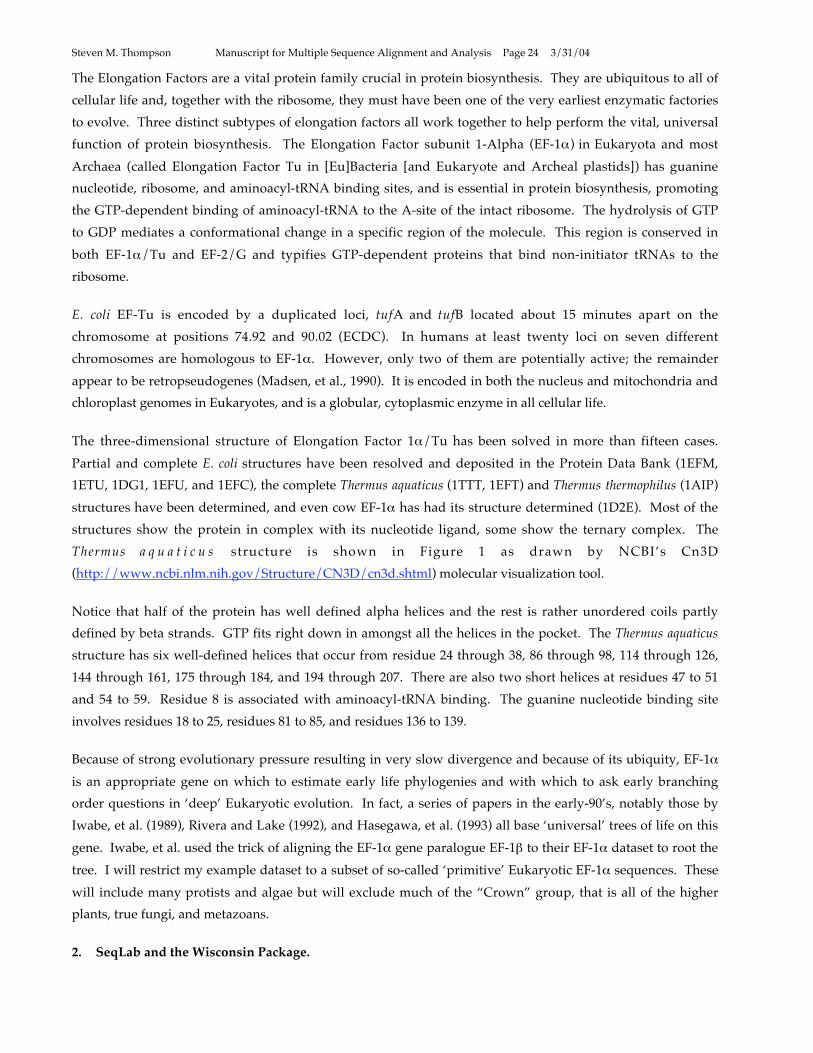
Thermus a q u a t i c u s structure is shown in Figure 1 as drawn by NCBI’s Cn3D
(http://www.ncbi.nlm.nih.gov/Structure/CN3D/cn3d.shtml) molecular visualization tool.
Notice that half of the protein has well defined alpha helices and the rest is rather unordered coils partly
defined by beta strands. GTP fits right down in amongst all the helices in the pocket. The Thermus aquaticus
structure has six well-defined helices that occur from residue 24 through 38, 86 through 98, 114 through 126,
144 through 161, 175 through 184, and 194 through 207. There are also two short helices at residues 47 to 51
and 54 to 59. Residue 8 is associated with aminoacyl-tRNA binding. The guanine nucleotide binding site
involves residues 18 to 25, residues 81 to 85, and residues 136 to 139.
Because of strong evolutionary pressure resulting in very slow divergence and because of its ubiquity, EF-1
is an appropriate gene on which to estimate early life phylogenies and with which to ask early branching
order questions in ‘deep’ Eukaryotic evolution. In fact, a series of papers in the early-90’s, notably those by
Iwabe, et al. (1989), Rivera and Lake (1992), and Hasegawa, et al. (1993) all base ‘universal’ trees of life on this
gene. Iwabe, et al. used the trick of aligning the EF-1 gene paralogue EF-1 to their EF-1 dataset to root the
tree. I will restrict my example dataset to a subset of so-called ‘primitive’ Eukaryotic EF-1 sequences. These
will include many protists and algae but will exclude much of the “Crown” group, that is all of the higher
plants, true fungi, and metazoans.
2. SeqLab and the Wisconsin Package.
Page 25
Steven M. Thompson Manuscript for Multiple Sequence Alignment and Analysis Page 25 3/31/04
The SeqLab graphical user interface (GUI) is included in the Accelrys Genetics Computer Group’s (GCG)
Wisconsin Package (http://www.accelrys.com/products/gcg_wisconsin_package/index.html). This
comprehensive package of sequence analysis programs began in 1982 in Oliver Smithies Genetics lab at the
University of Wisconsin, and is now used worldwide. The Wisconsin Package only runs on server computers
running one of its supported versions of the UNIX operating system, but it can be accessed from any
networked computer anywhere. It has arguably become the global ‘industry-standard’ in sequence analysis
software. The Wisconsin Package provides a comprehensive toolkit of almost 150 integrated DNA and
protein analysis programs — from database, pattern, and motif searching; fragment assembly; mapping; and
sequence comparison; to gene finding; protein and evolutionary analysis; primer selection; and DNA and
RNA secondary structure prediction. X-windows based SeqLab is a powerful ‘front-end’ to the package. It
provides an intuitive alternative to the UNIX command line by allowing menu-driven access to most of
GCG’s programs. SeqLab is based on Steve Smith’s (et al., 1994) GDE (the Genetic Data Environment) and
makes running the Wisconsin Package easier by providing a common editing interface from which most
programs can be launched and alignments can be manipulated.
Specialized “X-server” graphics communications software is required to use GCG’s SeqLab interface. X
server emulation software needs to be installed separately on personal style Microsoft Windows/Intel or pre
OS X Macintosh machines but genuine X-Windowing comes standard with most UNIX/Linux operating
systems. ‘Wintel’ machines are often set up with either XWin32 or eXceed to provide this function; pre OS X
Macintoshes are often loaded with either MacX or eXodus software. OS X Macs can have true X windowing
installed with the Apple’s own X11 package or with the public domain XDarwin package. The details of X
and of connecting to your local GCG server will not be covered in this chapter. Get assistance from your local
computer support personnel, if you need help. A couple of X-window tips should be mentioned though. X-
windows are only active when the mouse cursor is in that window, and always close windows when you are
through with them to conserve system memory. Furthermore, rather than holding mouse buttons down, to
activate items, just click on them. Also, buttons are turned on when they are pushed in and shaded. Finally,
do not close windows with the X-server software’s close icon in the upper right- or left-hand window corner,
rather, always use GCG’s “Close” or “Cancel” or “OK” button, usually at the bottom of the window.
3. Prepare Your Dataset.
You can use any of several different text string searching tools to find a particular biological molecular
sequence from a database. As described earlier, the collection of sequences used throughout this chapter
consists of representative EF-1 sequences from many ‘primitive’ Eukaryotes. This dataset was started using
GCG’s LookUp program, a Sequence Retrieval System (SRS) derivative (Etzold and Argos, 1993), because
LookUp creates an output file that can be used as an input list file to other GCG programs. However, it could
as well have been collected using Entrez at NCBI (http://www.ncbi.nlm.nih.gov/Entrez/), either through
the World Wide Web (WWW), or installed as their client/server NetEntrez application; or WWW SRS,
available at all EMBL and many other biocomputing sites around the world (see e.g. http://srs.ebi.ac.uk/).
After an entry has been identified, a natural next step is to use a sequence similarity searching program such
as FastA (Pearson and Lipman, 1988; and Pearson, 1998) and/or BLAST (Altschul et al. 1990 and 1997) to help
Page 26
Steven M. Thompson Manuscript for Multiple Sequence Alignment and Analysis Page 26 3/31/04
prepare a list of sequences to be aligned. Here we’ll use GCG’s version of FastA because of its flexible input
sequence specification requirements and its ability to output a valid GCG list file.
One of the more difficult aspects of multiple sequence alignment is knowing what sequences you should
attempt it with. Any list from any program will need to be restricted to only those sequences that actually
should be aligned. Make sure that the group of sequences that you align are in fact related, that they actually
all belong to the same gene family, that the alignment will be meaningful. Furthermore, in these days of huge
genome projects and massive databases, one important slant is a data mining question, that is, figuring out
just which sequences to align from a huge number available that are all homologous to your query. This
question is particularly appropriate here since there are an enormous number of Elongation Factors present in
the databases. So often it depends on the type of scientific question that you are asking in your research. Are
you interested in predicting the structure or the function of your particular research molecule; what about in
ascertaining the evolution of a paralogous gene family within a species as the result of gene duplications;
what about the evolution of several species based on an analysis of the orthologues present in several
different species? Clearly the dataset to be used is directly molded by the question that you ask.
3.1. GCG’s LookUp Program.
To follow along with my example log on to your local GCG server and launch SeqLab in an X environment. I
won’t be able to explain anything about achieving this for your individual situations — there are just way too
many variables — talk to your local biocomputing support personnel for assistance. In my example below all
commands, buttons, and menus that you are to use are printed in bold and exact phrases are quoted.
To identify entries of interest in GCG sequence databases you need to know their proper database names or
their accession codes. I’ll find relevant entries with LookUp to assemble a representative set of elongation
factor entries from the ‘primitive’ Eukaryotes. That is, those Eukaryotes that exclude the Fungi, Metazoans,
and true Plants. Launch “LookUp” through the “Functions” “Database Reference Searching” menu. In the
new “LookUp” window be sure that “Search the chosen sequence libraries” is checked and then select
“SwissProt” as well as “SPTREMBL” for the libraries to search. I recommend searching SwissProt and
SPTREMBL together in order to take advantage of the excellent annotation of the SwissProt database and yet
still find entries that have not yet been moved from their preliminary status in SPTREMBL. Under the main
query section of the window, type the words and symbols “elongation & factor & alpha” following the
category “Definition” and the words and symbols “eukaryota ! ( fungi | metazoa | viridiplantae )” in the
“Organism” category; next press the “Run” button. You need to use Boolean operator symbols to connect
the individual query strings because the databases are indexed using individual words for most fields. The
“Organism” field is an exception; it will accept ‘Genus species’ designations as well as any other single word
supported level of taxonomy, e.g. “fungi”. The Boolean operators supported by LookUp are the ampersand,
“&”, meaning “AND”, the pipe symbol, “|”, to denote the logical “OR”, and the exclamation point, “!”, to
specify “BUT NOT”. Other LookUp query construction rules are case insensitivity, parenthesis nesting, “*”
and “?” wildcard support, and automatic wildcard extension. This query should find most of the elongation
factor alpha’s from the ‘primitive’ Eukaryotes in the SwissProt and SPTREMBL databases and will provide a
Page 27
Steven M. Thompson Manuscript for Multiple Sequence Alignment and Analysis Page 27 3/31/04
reasonable and interesting starting dataset for the chapter. The “LookUp” window should look similar to
Figure 2. The results of the search will automatically display; Table 1 shows the beginning of mine.
Be careful that all of the sequences in the output from any text searching program are appropriate. In this
case the elongation factors found all look correct, but improper nomenclature and other database
inconsistencies can cause problems. If you find inappropriate sequences in the output, you can either edit the
output file to remove them, or comment out the undesired sequences by placing an exclamation point, “!” in
front of the unwanted lines, or “CUT” them from the SeqLab Editor display after loading the list.
Select the LookUp output file in the “SeqLab Output Manager”. This is a very important window and will
contain all of the output from your current SeqLab session. Files may be displayed, printed, saved in other
locations with other names, and deleted from this window. Press the “SeqLab Output Manager” “Add to
Main List” or “Add to Editor” button, depending on which mode you are in. Select the LookUp output, if in
List Mode, and then switch “Mode:” to “Editor”. This will load the file into the SeqLab Editor where you can
perform further analyses on those entries.
Notice that all of the sequences now appear in the Editor window with the amino acid residues color-coded.
The nine color groups are based on a UPGMA clustering of the BLOSUM62 amino acid scoring matrix, and
approximate physical property categories for the different amino acids. Expand the window to an
appropriate size by ‘grabbing’ the bottom-left corner of its ‘frame’ and ‘pulling’ it out as far as desired. The
display should look similar to Figure 3 after loading the dataset.
You can also directly load sequences into SeqLab with the “File” “Add sequences from” menu. Use the
online GCG databases with the “Databases. . .“ choice, if you know the sequences’ proper identifier names or
accession codes, or from the “Sequence Files. . .“ choice, if they are local GCG format sequences or list files.
Use SeqLab’s Editor “File” menu “Import” function to directly load GenBank format sequences or ABI style
binary trace files without the need to reformat. The “Add Sequences” window’s “Filter” box is very
important! By default files are filtered such that only those that end with the extension “.seq” are displayed.
This often won’t do you any good as the sequences that you may want to add may have other extensions.
Therefore, modify the text in the “Filter” box using the “*” wild card to identify your desired files. Press the
“Filter” button and select the file that you want from the “Files” box, and then check the “Add” and then
“Close” buttons at the bottom of the window to put the desired file into your current list, if you’re in List
Mode, or directly into the Editor, if you’re in “Editor Mode”.
While you have sequences loaded in the Editor explore the interface for a bit. Each protein sequence is listed
by its official SwissProt or SPTREMBL entry name (ID identifier). Use both scroll bars to move around within
the sequences. The scroll bar at the bottom allows you to move through the sequences linearly; the one at the
side allows you to scroll through all of your entries vertically. Quickly double click on various entries’ names
(or single click the “INFO” icon with the sequence entry name selected) to see the database reference
documentation on them. (This is the same information that you can get with the GCG command “typedata -
ref” at the command line.) “Close” the “Sequence Information” windows after reading them. You can also
change the sequences’ names and add any documentation that you want in this window. Change the
Page 28
Steven M. Thompson Manuscript for Multiple Sequence Alignment and Analysis Page 28 3/31/04
“Display:” box from “Residue Coloring” to “Feature Coloring” and then “Graphic Features”. Now the
display shows a schematic of the feature information from each entry with colors based on the information
from the database Feature Table for the entry. “Graphic Features” represents features using the same colors
but in a ‘cartoon’ fashion. Quickly double-click on one of the various colored regions of the sequences (or use
the “Features” choice under the “Windows” menu). This will produce a new window that describes the
features located at the cursor. Select the feature to show more details and to select that feature in its entirety.
All the features are fully editable through the “Edit” check box in this panel and new features can be added
with several desired shapes and colors through the “Add” check box.
Nearly all GCG programs are accessible through the “Functions” menu. Any portion of, or the entire
alignment loaded, is available for analysis by the programs. Select various entry’s names and then go to the
“Functions” menu to perform different analyses on them. You can select sequences in their entirety by
clicking on their names or you can select any position(s) within sequences by ‘capturing’ them with the
mouse. You can select a range of sequence names by <shift><clicking> the top-most and bottom-most name
desired, or <ctrl><click> sequence entry names to select noncontiguous entries. (However, there is a bug in
the Linux version of SeqLab. Use <ctrl><right-click> as a work-around.) The “pos:” and “col:” indicators
show you where the cursor is located on a sequence without including and with including gaps respectively.
The “1:1” scroll bar near the upper right-hand corner allows you to ‘zoom’ in or out on the sequences; move it
to 2:1 and beyond and notice the difference in the display.
It’s probably a good idea to save the sequences in the display at this point and multiple times down the road
as you work on a dataset. Do this occasionally the whole time you’re in SeqLab just in case there’s an
interruption of service for any reason. Go to the “File” menu and choose “Save As”. Accept the default
“.rsf” extension but give it any file name and directory specification you choose. RSF (Rich Sequence Format)
contains all the aligned sequence data as well as all the reference and feature annotation associated with each
entry. It is “Richer” than most other multiple sequence formats and is SeqLab’s default format.
3.2. Similarity Searching to Decrease (or Increase) Dataset Size.
A logical step in preparing a multiple sequence alignment might be to run a similarity based search to add
those most similar sequences from the database to your dataset. This would especially be true if your text-
based search did not find many entries. An advantage of running similarity searches within the context of
GCG is the results are immediately available for further analyses without the need for any sequence
downloading or reformatting because of the GCG list file format and the fact that all of the databases are
mounted locally. In your own research settings, and depending on the type of questions that you are asking,
you may want to create very large alignments by screening all available databases for sequences of significant
similarity to your query. Therefore, let’s talk about just how big you can go.
The Wisconsin Package’s restrictions, as of version 10.3, allow individual sequences to be a maximum of 350
Kb in length (longer entries are cut into overlaps in database creation steps), though SeqLab can display
longer sequences. You may want to load a longer sequence into SeqLab if you are working on genome scale
data, and want to extract subranges from that entry. The MSF file format can hold up to 500 sequences; RSF
can hold much more, limited by system memory. This allows programs such as HmmerAlign (described
Page 29
Steven M. Thompson Manuscript for Multiple Sequence Alignment and Analysis Page 29 3/31/04
later) to produce multiple sequence alignment output larger than 500 sequences. PileUp (described below)
can handle a sequence alignment up to 7,000 characters long, including gaps. PileUp input sequences are
restricted to a length of 5,000 characters by default. The 'overall surface-of-comparison' is restricted to
2,250,000 with the default program, a bit more than all the residues or bases plus all the gaps in the
alignment. Alternate executables are provided with the Package for allowing 10,000, 15,000, and 20,000
character input, though they are usually not scripted into SeqLab. Launch them from the command line with
“pileup_10000”, “pileup_15000”, and “pileup_20000” respectively. Take home message: really huge
alignments are possible; it depends on what you need for the biological questions that you are asking.
But what about the opposite situation, when you have too many homologues? I’ll use FastA to illustrate this
data mining function. FastA database similarity searching can be very helpful for sorting any collection of
GCG sequence specifications into order of alignment significance. Data mining this way allows you to easily
screen undesired sequences from the bottom of any list or combinations of lists. But, be warned, on some
systems with some versions of GCG, you can not run FastA on too small of a dataset without causing core
dumps! A trick is to add another small database such as NRL_3D, or the output list files from other database
searches, to your “Search List Set”. This provides the necessary background randomization to allow proper
normalization. Another data mining trick is to repeat a text-based search, not on a sequence database, but
rather, on the results of similarity searches. This can further restrict your dataset to only those taxonomic
groups of interest. Another point to remember is you can not use any of the BLAST programs to search
against any sequence set that has not been preformatted into a BLAST compatible database. Because of this,
BLAST is not an appropriate program to use for this type of list file sorting, data mining function. However,
the FastA family of programs support all GCG sequence specifications, so it works great for this purpose.
Here I’ll use FastA to search my existing LookUp output list file and a previous output list file from another
search. A really powerful thing to do is to run LookUp to create your own custom list file of all of the protein
sequences available from any desired level of taxonomy. You could then use that list file as input to FastA. I
will use the Giardia sequence as a query for my search because Giardia’s most ancient ancestor is often
considered to be rooted near the Eukaryote lineage’s base on the universal tree of life (see e.g. Sogin, et al.,
1996). Since my target dataset is all ‘primitive’ Eukaryotic, this should be an appropriate choice.
Select the “EF1A_GIALA” sequence and launch FastA off the “Functions” “Database Sequence Searching”
menu. If a “Which selection” window pops up asking if you want to use the “selected sequences” or
“selected region”, choose “selected sequences” to run the program on the full length of the selected protein.
At most sites the default protein database to search, “Search Set. . .“, will be “Using pir:*”, but, here I’m going
to change it to specify my LookUp output list file and a previous protein database search result list file.
Therefore, push the “Search Set. . .“ button, select “pir:*” in the “Build FastA’s Search Set” box that pops up,
and then “Remove from Search Set”. Next, press the “Add Main List Selection. . .” button and then select
your previous LookUp output from the “List Chooser” window that pops up; press “Add to Search Set”.
Repeat this process using the “Add Sequences Files. . .” button in the “Build FastA’s Search Set” window,
using the “Filter” function correctly to identify and load any other desired database search output files in
your working directory. “Close” the “List Chooser” and the “Build Search Set” windows. Decrease the
cutoff Expectation value in the main FastA window to something quite stringent like “0.01” to reduce the
Page 30
Steven M. Thompson Manuscript for Multiple Sequence Alignment and Analysis Page 30 3/31/04
output list size. Be sure that the “FastA” program window shows “How:” “Background Job”, and then press
the “Run” button. The output will quickly return since it’s a relatively small search set. The output is a
perfectly suitable GCG list file, complete with beginning and ending attributes, and reverse complement
strand warnings when necessary, if you had used a DNA query.
However you manage to get your dataset the size you want, you still need to load it into the SeqLab Editor.
Use the “Output Manager” window again, always available through the SeqLab “Windows” menu. You
need to use an extremely important “Output Manager” function at this point. Select your FastA output file in
the “Output Manager” and then press the “Add to Editor” button. Specify “Overwrite old with new” in the
“Reloading Same Sequence” window when prompted, to take the search output and merge it with sequences
already in the open Editor. Click “Interrupt Loading” in the “Loading sequences” window after as many
sequences have loaded as you care to work with. If loading a FastA file, they are loaded in order of similarity
to your query. In my example’s case I restricted my analysis to about the top 50 entries of my final FastA file.
The next prompt requires some thought, if you’re loading the results of a similarity search. You’ll be asked
whether to “Modify the sequences” or “Ignore all attributes” in a “List file attributes set” window. The
answer will depend on the type of alignment you are creating and the biological questions that you asking.
In many cases, especially if you are asking phylogenetic questions, then you will not want to modify the
sequences. Load their full length to maximize available signal. However, if dealing with extremely diverse
sequences and/or just domains of sequences, then trimming the sequences down to those most conserved
portions identified by FastA can be very helpful. In this case I will not trim them down, so I press the “Ignore
all attributes” button. “Close” the “Output Manager” after loading your FastA list file and return your
display to “1:1” and “Residue Coloring”. Take a look at the new sequences in the display. As before,
quickly double click on various entries’ names to see the database reference descriptions for them (or click on
the “INFO” button). Figure 4 shows the Editor display after loading the top part of my FastA file.
Now would be a good time to go back to the “File” menu and save the RSF file. “Overwrite” in the “File
exists” box if you’ve used the same name for this file earlier. I suggest you do this, as RSF files are quite large
and there’s no need to save all the various versions of the data.
4. MEME: Expectation Maximization.
Before actually performing multiple sequence alignment on your dataset a powerful de novo motif discovery
algorithm can be run. The algorithm is called Expectation Maximization; it uses Bayesian probabilities and
unsupervised learning to find conserved, unknown motifs among a group of unaligned, ungapped sequences
(Bailey and Elkan, 1994). The motifs do not have to be in congruent order among the different sequences; i.e.
it has the power to discover ‘unalignable’ motifs between sequences. This characteristic differentiates MEME
from most other profile building techniques. It is implemented in the Wisconsin Package as the MEME
program and it produces output containing multiple, ungapped profiles as well as a readable report file. Its
profile output serves as input to MotifSearch (Bailey and Gribskov, 1998). I strongly suggest reading the
MEME and MotifSearch chapters in the GCG Program Manual (“genmanual” at the command line or the
“Help” buttons in the program in SeqLab) — they explain the details of the algorithms quite well.
Page 31
Steven M. Thompson Manuscript for Multiple Sequence Alignment and Analysis Page 31 3/31/04
Select all of the sequences in the Editor window so that MEME runs on them all. Launch “MEME” off of the
“Functions” “Multiple Comparisons” menu. A "Which selection" window may pop up asking if you want
to use the "selected sequences" or "selected region;" choose "selected sequences" to run the program on the full
length of all the sequences. In most cases the default parameters will work fine but the algorithm can be sped
up at the cost of sensitivity by decreasing the number of motifs to be found, by restricting the number of
motifs found to exactly one in each sequence, and/or by decreasing the allowable motif window size. Again,
I suggest reading the relevant GCG Program Manual chapters.
MEME output consists of two files; a .meme readable text file and a .prf multiple profile text file. MotifSearch
will scan any dataset specified with the multiple profile file that MEME produced. A helpful thing to do is
scan the original ‘training’ dataset that you created the profiles with. This can annotate those regions that
MEME discovered in your SeqLab Editor RSF file. After alignment the MEME motifs that are alignable will
all line up. Go to the “Database Sequence Searching” menu and select “MotifSearch. . .”. Specify your
“query profile(s)”, the one you just made, and change the “Search set” to the RSF dataset that you now have
loaded in the Editor. Be sure to activate “Save motif features to the RSF file”. The output will return with
the .rsf file on top. Don’t bother trying to read it; just “Close” it. It contains the SeqLab format “Rich Text
Format” for all the feature data discovered by MEME in your dataset. The .ms file contains the readable
results of the search in list file format with Expectation value statistics and the number of motif hits for each
fit. After the list file portion a “Position diagram” schematically describes the hits in each sequence. Take a
moment to look it over by pressing the “Display” button in the Output Manager and then “Close” it.
Use the Output Manager to merge the motifsearch.rsf feature file with the existing data already in the open
SeqLab Editor. This will add the feature annotation created when you activated the MotifSearch -RSF option.
The location of each motif will be included in the Editor sequence display. To do this again use the extremely
important “Add to Editor” “Output Manager” function. As above, specify “Overwrite old with new” in the
next window when prompted. “Close” the “Output Manager” after loading your new RSF file. Change
“Display:” to “Graphic Features” and check out the additional annotation. Figure 5 illustrates my “Graphic
Features” display at a “4:1” zoom ratio.
5. Searching PROSITE: A ‘Quick and Dirty’ GCG Motifs Search.
The GCG Motifs program searches through the one-dimensional, regular expression, descriptions of the
PROSITE database (Bairoch, 1992), described in the previous chapter, for matches to your protein. The
program can tolerate mismatches with a -MisMatch option and it displays an abstract with selected references
for each motif signature found. This can be a tremendous timesaver, suggesting immediate routes of
experimentation. It should always be utilized — it’s just too fast and simple to ignore.
Start the Motifs program by selecting all of the protein entries’ names in SeqLab, as in the previous MEME
run, and then going to the “Functions” “Protein Analysis” menu and picking “Motifs. . .”. The "Motifs"
program window will be displayed. Check the “Save results as features in file motifs.rsf” button in the
“Motifs” program window. As with MotifSearch, this file contains annotation discovered by the program
and we’ll use it below. None of the other options are required for this run so press the “Run” button. After a
few moments you should get output. The file displayed, “motifs.rsf”, isn’t very interesting to read so
Page 32
Steven M. Thompson Manuscript for Multiple Sequence Alignment and Analysis Page 32 3/31/04
“Close” it and use the “Output Manager” to display the file with the “.motifs” extension. Carefully look over
the text file that is displayed. Notice the sites in Table 2 that have been characterized in these sequences and
the extensive bibliography associated with them.
Extensive abstract and reference lists follow the identified sequence locations for each site. This information
can save anybody a tremendous amount of work! The sites themselves are shown with their sequence
locations below each consensus pattern. Among the other motifs discovered, the characteristic P-Loop
described in the previous chapter is defined as (A,G)x4GK(S,T), i.e. either an alanine or a glycine, followed by
four of anything, followed by an invariant glycine-lysine pair, followed by either a serine or a threonine.
Exceptions are noted in the documentation. This particular site has been very well researched and many
three-dimensional structures are available for it. It always has a beta/alpha/beta secondary structure
conformation and is sometimes known as the “Rossman Fold”.
Post-translational modification sites commonly found in many proteins, such as glycosylation,
phosphorylation, amidation, and myristylation, will only be listed if you specify the -Frequent option.
However, realize that sites may be false positives, especially if you use the -Frequent option. This is always a
danger with simple one-dimensional, regular expression, consensus style searches. The GCG programs
ProfileScan and HmmerPfam use a much more sensitive profile matrix approach to search your sequence
with profiles including most of PROSITE and will be discussed further later. In this example Motifs
discovered the truly positive GTP-binding elongation factor signature and the ATP/GTP-binding P-loop site,
yet it also found two probable false positives, the Prokaryotic membrane lipoprotein lipid attachment site and
the FGGY family of carbohydrate kinases signature.
“Close” the “Motifs” output window when you’ve looked it over and then load the motifs.rsf file into
SeqLab. This will add the feature annotation created with the -RSF option. The location of the PROSITE
signatures will now be included in the Editor sequence display. Again use the “SeqLab Output Manager” to
do this, as discussed previously. Select the file “motifs.rsf”, then press the “Add to Editor” button and
specify “Overwrite old with new” to take the new motifs.rsf feature file and merge it with the old RSF file in
the open Editor. “Close” the “Output Manager” after loading your new RSF file. Look at your display using
“Features Coloring” or “Graphic Features” to display the new annotation and see if you can recognize the
differences. My dataset is illustrated in Figure 6 using “Features Coloring” now annotated with its original
database features as well as MEME discoveries and Motifs patterns.
6. Performing the Alignment: the PileUp Program.
Align all of the protein sequences in your dataset next. Select all of the entries in the Editor window and then
go to the “Functions” menu and select “Multiple comparison”. Click on “PileUp. . .“ to align the entries. A
new window will appear with the parameters for running PileUp. Often you’ll accept all of the program
defaults on a first run by pressing the “Run” button; however, here I am going to change the scoring matrix
for the alignment from the default BLOSUM62 to the alternate BLOSUM30 matrix.
Depending on the level of divergence in a data set, better multiple sequence alignments can often be
generated with alternate scoring matrices (the -Matrix option, specifying the desired matrix from the GCG
Page 33
Steven M. Thompson Manuscript for Multiple Sequence Alignment and Analysis Page 33 3/31/04
logical directory GenMoreData) and/or different gap penalties. Beginning with GCG version 9.0, the
BLOSUM62 (Henikoff and Henikoff, 1992) matrix file, “blosum62.cmp”, is used as the default scoring matrix
in most programs. Furthermore, appropriate gap creation and extension penalties are now coded directly
into the matrix, though they can still be adjusted within the program if desired. Formerly a normalized
Dayhoff PAM 250 table (Schwartz and Dayhoff, 1979) and program encoded penalty values were used. The
BLOSUM series are more robust at handling a wider range of sequence divergence than the PAM table ever
was — the BLOSUM30 table being most appropriate for the most divergent datasets.
Therefore, click on the “Options” button. To specify the BLOSUM30 matrix select the check button next to
and click on the “Scoring Matrix. . .“ box in the “Pileup Options” window. This will launch a “Chooser for
Scoring Matrix” window from which you can select the BLOSUM30 matrix file, “blosum30.cmp”. Double-
click the matrix’s name to see what it looks like; click “OK” to close both windows. Scroll through the rest of
“PileUp Options” window to see all those available. “Close” it when finished and then press “Run” in the
“PileUp” window to launch the program.
PileUp will first compare every sequence with every other one. This is the pairwise nature of the program;
then it will progressively merge them into an alignment in the order of determined similarity, from most to
least similar (Feng and Doolittle, 1987). The window will go away and then, after a few moments, depending
on the complexity of the alignment and the load on the server, new output windows will automatically
display. The top window will be the Multiple Sequence Format (MSF) output from your PileUp run. Notice
the BLOSUM30 matrix specification and the default gap introduction and extension penalties associated with
that matrix, 15 and 5 respectively. As mentioned above, in most cases the default gap penalties will work fine
with their respective matrixes, though they can be changed if desired. In fact, see below on improving
regions within alignments, where it is absolutely required.
Scroll through your alignment to check it out and then “Close” the window afterwards. My abridged output
file example is shown in Table 3. Notice the interleaved character of the sequences, yet they all have unique
identities, addressable through their MSF filename together with their own name in braces, {name}.
Return to the listing of sequence names near the top of the file. This listing contains an important number
called the checksum. All GCG sequence programs use this number as a unique sequence identifier. There is
a checksum line for the whole alignment as well as individual checksum lines for each member of the
alignment. If any two of the checksum numbers are the same, then those sequences are identical. If they are,
an editor can be used to place an exclamation point, “!” at the start of the checksum line in which the
duplicate sequence occurs. Exclamation points are interpreted by GCG as remark delineators, therefore, the
duplicate sequence will be ignored in subsequent programs. Or the sequence could be “CUT” from the
alignment with the SeqLab Editor. Another important number on the individual checksum lines is the
“Weight” designation. It determines how much importance each sequence contributes to a profile made from
the alignment (see below). It may be worth adjusting these values so that the contribution of a collection of
very similar sequences does not overwhelm the signal from a few more divergent sequences. In the SeqLab
interface the “Sequence Info . . .” window can be used to accomplish this, or you can use a text editor.
Page 34
Steven M. Thompson Manuscript for Multiple Sequence Alignment and Analysis Page 34 3/31/04
Scroll through the alignment and then “Close” its window. Again use the “Output Manager” to “Add to
Editor” and “Overwrite old with new”, to take your new MSF output and merge it with the old RSF file in
the open Editor. This will keep all of the database feature annotation intact, yet renumber all of its reference
locations based on the inclusion of gaps in the alignment. “Close” the “Output Manager” after loading your
new alignment. The next window will contain PileUp’s cluster dendrogram, in the EF-1 example, Figure 7.
PileUp automatically creates this dendrogram of the similarity clustering relationships between the
sequences. It can be very helpful for adjusting sequence Weight values to even out each sequences’
contribution to a profile. The lengths of the vertical lines are proportional to the differences in similarity
between the sequences. However, realize that this tree is not an evolutionary tree, and it should never be
presented as one. No phylogenetic inference algorithm, such as maximum likelihood, least-squares fit, or
parsimony, nor any molecular substitution, multiple-hit correction models, such as Jukes-Cantor, Kimura, or
any other subset of the GTR (General Time Reversible) model, nor any site rate heterogeneity models such as
a Gamma correction, are used in its construction. (It is roughly an uncorrected UPGMA tree, prone to all the
same errors seen with UPGMA.) PileUp’s dendrogram merely indicates the relative similarity of the
sequences based on the scoring matrix used, by default the BLOSUM62 but the BLOSUM30 in my example,
and, therefore, the clustering order used to create the alignment.
You can directly print from SeqLab graphics Figure windows to PostScript files by picking “Print . . .”
“[Encapsulated] PostScript File” (or some similar PostScript format specification) from the “Output Device:”
menu. If there is no PostScript choice there, run SetPlot to designate a graphics configuration for your
account. This can also be done with SeqLab’s “Options” “Graphics Devices. . .” button. Name the output file
to anything you want and click “Proceed” to create an EPSF output in your current directory. To actually
print this file you may need to transfer it to a local machine attached to a PostScript compatible printer unless
you have direct access to the GCG UNIX server system printer and it is PostScript compatible. (All
Macintosh compatible laser printers run PostScript by default. Carefully check any laser printer connected to
a ‘Wintel’ system to be sure that it is PostScript compatible.) “Close” the dendrogram window.
Notice that your residues now align by color. My Editor display looks like Figure 8 after loading the MSF file
using “Residue Coloring” and a “1:1” zoom ratio. The nice columns of color represent columns of aligned
residues. Change the “Display:” box from “Residue Coloring” to “Graphic Features”. Now the display
shows a schematic of the original database feature annotation from each entry, as well as all of the motifs
discovered by both programs Motifs and MotifSearch, and will look like Figure 9, at a “4:1” zoom.
Remember, quickly double clicking on any of the color coded feature regions in the Editor display will
produce a “Features” window where more information is available about that particular feature by selecting
the Feature entry in the new window. Clicking once in the colored region and then using the “Features”
option from the “Windows” menu will also produce the “Features” window. Now would also be another
good time to save your work as an updated RSF file!
6.1. Visualizing Conservation in Multiple Sequence Alignments.
Page 35
Steven M. Thompson Manuscript for Multiple Sequence Alignment and Analysis Page 35 3/31/04
The most conserved portions of an alignment are those most resistant to evolutionary change, often due to
some type of structural constraint. To easily visualize the positional conservation of a multiple sequence
alignment use the graphics program PlotSimilarity. The program draws a graph of the running average
similarity along a group of aligned sequences (or of a profile with the -Profile option). The PlotSimilarity
peaks of a protein alignment represent the most conserved areas of the alignment, but even more so, those
areas most resistant to evolutionary change due to the algorithm’s use of the BLOSUM matrix in its
calculations. PlotSimilarity is also a nice way to see those areas of an alignment that may need improving by
pointing out the most variable regions. Furthermore, PlotSimilarity can be helpful for ascertaining alignment
quality by noting changes in the overall average alignment similarity and in those regions of conservation
within the alignment, as it is adjusted and refined.
Select all of the sequences and then go to the “Functions” menu and under the “Multiple comparison”
section choose “PlotSimilarity . . .”. I recommend changing some of the program defaults so choose
“Options” in the program window. Check “Save SeqLab colormask to” and “Scale the plot between:” the
“minimum and maximum values calculated from the alignment”. The first option’s output file will be used
in the next step. The second specification launches the program’s command line -Expand option. This blows
up the plot, scaling it between the maximum and minimum similarity values observed, so that the entire
graph is used, rather than just the portion of the Y axis that your alignment happens to occupy. The Y-axis of
the resulting plot uses the similarity values from whichever scoring matrix you used to create your
alignment, unless you specify an alternative. The default matrix, BLOSUM62, begins its identity value at 4
and ranges up to 11; mismatches go as low as -4. “Close” the “Options” window; notice that the “Command
Line:” box reflects your updated options. Click “Run” to launch the program. The output will quickly
return. “Close” the plotsimilarity.cmask display and the “Output Manager” and then take a look at the
similarity plot. My example is shown in Figure 10.
My example shows a great deal of sequence similarity. Strong peaks are seen centered around positions 30,
100, and 375. The ordinate scale is dependent on the scoring matrix used by the program, here the
BLOSUM30 table, which ranges in score from -7 to +20. The dashed line across the middle shows the average
similarity value for the entire alignment, here about 4.4. Make a PostScript file of this plot too, if desired.
Regardless of whether you print this plot or not, take notes of where the similarity significantly falls off
within and at the beginning and end of the alignment. In my example this is the first 25 residues or so, a
region around 190 and 220, around 390, and about the last 25 residues. “Close” the “PlotSimilarity” window
after noting where these deepest valleys, the least similar regions of the alignment, lay.
Now go to the “File” menu and click on “Open Color Mask Files”. This will produce another window from
which you should select your new “plotsimilarity.cmask” file; click on “Add” and then “Close” the window.
This will produce a gray scale overlay on your sequences that describes their regional similarity where darker
gray corresponds to higher similarity values. My sample alignment, at a “4:1” zoom factor, is shown in
Figure 11. Notice the strong conservation peak centered just before residue 100 in the alignment, one of EF-
1 ’s GTP binding regions.
6.2. Improving Alignments in SeqLab.
Page 36
Steven M. Thompson Manuscript for Multiple Sequence Alignment and Analysis Page 36 3/31/04
The beauty of this representation is you can now easily select those regions of low similarity to try to improve
their alignment automatically. This is possible because of PileUp’s incredibly effective -InSitu option that can
realign regions within an alignment. Be sure that all of your sequences are selected and then zoom back in
your alignment to “1:1” so that you can see individual residues and then scroll to the carboxy end. It’s best to
start at the carboxy termini in this process so that the positions of the low similarity regions do not become
skewed as you proceed through the procedure. Next select a region of low similarity across the complete
sequence set. This can be done using the mouse if it’s all on the screen in front of you, which is not the case
here. Therefore, use the “Edit” “Select Range” function (determine the positions by placing your cursor at
the beginning and end of the range to be selected and noting the column number in the lower left-hand of the
Editor display). Once all of your sequences and the region that you wish to improve are selected, go to the
“Functions” menu and again select “Multiple comparison”. Click on “PileUp . . .“ to realign all of the
sequences within that region. (The “Windows” menu also contains a ‘shortcut’ listing of all of the programs
that you have used in the current session; you can launch any of them from there as well.) You will be asked
whether you want to use the “Selected sequences” or “Selected region;” it is very important to specify
“Selected region”. This will produce a new window with the parameters for running PileUp. Next, be sure
to click on “Options . . .” to change the way that PileUp will perform the alignment. In the “Options”
window check the gap creation and extension boxes and change their respective values to much less than the
default. Changing them to about a third the default value works pretty well for a start, so for the BLOSUM30
matrix change the values to “5” and “2” respectively. Most importantly, check “Realign a portion of an
existing alignment;” this calls up the command line -InSitu option. Otherwise only that portion of your
alignment selected will be retained in the output. Furthermore, we really don’t need another similarity
dendrogram, so uncheck the “Plot dendrogram” box. “Close” the window and notice the new options in the
PileUp “Command Line:” “Run” the program to improve your alignment. The window will go away and
your MSF result will return very quickly, since you are only realigning a portion of the alignment. Notice the
lowered gap introduction and extension penalties of 5 and 2. Scroll through your alignment to check it out
and then “Close” the window. The next window will be the “Output Manager”. Just like before, click on
“Add to Editor” and then specify “Overwrite old with new” in the “Reloading Same Sequences” window
to merge the new alignment with the old one and retain all feature annotation. This feature information may
help guide your alignment efforts in subsequent steps. “Close” the “Output Manager” window after loading
your new alignment.
Your alignment should now be better within the specified region. Repeat this process in all areas of low
similarity, again, working from the carboxy termini toward the amino end. Notice that all of the options that
you last specified are retained by the program so you don’t need to respecify them. You can also save run
parameters so that they will come up in subsequent sessions by clicking on the “Save Settings” box in any of
the program run windows. You may want to go to the “File” menu periodically to save your work using the
“Save as . . .” function in case of a computer or network problem. It’s also probably a good idea to reperform
the PlotSimilarity and color mask procedure after going through the entire alignment to see how things have
improved after you’ve finished the various InSitu PileUps. If you discover an area that you can not improve
through this automated procedure, then it is time to either manually ‘correct’ it or ‘throw it away’. Again,
note those ‘problem’ areas and then switch back to “Residue Coloring”. This will ease manual alignment by
allowing your eyes to work with columns of color.
Page 37
Steven M. Thompson Manuscript for Multiple Sequence Alignment and Analysis Page 37 3/31/04
Other things that can help manual alignment are “GROUP”ing and “Protections”. The “GROUP” function
allows you to manipulate ‘families’ of sequences as a whole — any change in one will be propagated
throughout them all. To “GROUP” sequences, select those that you want to behave collectively and then
click on the “GROUP” icon right above your alignment. You can have as many groups as you want. The
space bar will introduce a gap into the sequence and the delete key will take a gap away. However, you can
not delete a sequence residue without changing that sequence’s (or the entire alignment’s) “Protections”.
Click on the padlock icon to produce a “Protections” window. Notice that the default protection allows you
to modify “Gap Characters” and “Reversals” only. Check “All other characters” to allow you to “CUT”
regions out of your alignment and/or delete individual residues and then click “OK” to close the window. A
very powerful manual alignment function can be thought of as the ‘abacus’ function. To take advantage of
this function select the region that you want to slide and then press the shift key as you move the region with
the right or left arrow key. You can slide residues greater distances by prefacing the command keystrokes
with the number of spaces that you want them to slide.
Make subjective decisions regarding your alignment. Is it good enough; do things line up the way that they
should? If, after all else, you decide that you just can’t align some region, or even an entire sequence, then
perhaps get rid of it with the “CUT” function. Another alternative is the mask function that I will describe
below. Cutting out an entire sequence may leave some columns of gaps in your alignment. If this is the case,
then reselect all of your sequences and go to the “Edit” menu and select “Remove Gaps . . .“ “Columns of
gaps”. Notice the extreme amino and carboxy ends of the alignment. Amino and carboxy termini seldom
align properly and are often jagged and uncertain. This is fairly common in multiple sequence alignments
and subsequent analyses should probably not include these regions. If loading sequences from a similarity
search, allowing SeqLab to trim the ends automatically based on beginning and ending constraints
considerably improves this situation. Overall, things to look for include columns of strongly conserved
residues such as tryptophans, cysteines, and histidines, important structural amino acids such as prolines,
tyrosines and phenylanines, and conserved isoleucine, leucine, valine substitutions; make sure they all align.
After you have finished tweaking, evaluating, and readjusting your alignment to make it as ‘satisfying’ as
possible, change back to “Feature Coloring” “Display”. Those features that are annotated should now align
perfectly. This is another way to assure that your alignment is as biologically ‘correct’ as possible.
Everything you do from this point on, and especially later if you use alignments to ascertain molecular
phylogenies, is absolutely dependent on the quality of the alignment! You need a very clean, unambiguous
alignment that you can have a very high confidence in — truly a biologically meaningful alignment. Each
column of symbols must actually contain homologous characters.
Other alignment editors are available for refining multiple sequence alignments. However, I think that you
will find SeqLab very satisfying, and only using a GCG compatible editor assures that the format will not be
corrupted. If you do make any changes to a GCG sequence data file with a non-GCG compatible editor, you
must reformat the alignment afterwards. However, reformatting GCG MSF or RSF files requires a couple of
tricks. If you do need to do this, you must use the appropriate Reformat option (either -MSF or -RSF
respectively) and you must specify all the sequences within the file using the brace specifier, “{*}”, e.g:
> reformat -msf your_favorite.msf{*}
Page 38
Steven M. Thompson Manuscript for Multiple Sequence Alignment and Analysis Page 38 3/31/04
You should never need to do this, unless for some perverse reason you decide to edit an alignment with a
non-GCG compliant editor; however, it may prove necessary in some situations. After reformatting, the new
MSF or RSF file will follow GCG convention, with updated format, numbering, and checksums.
7. SeqLab Editor On-Screen Annotation.
You may want to add text annotation to your alignment after you’ve gotten it all cleaned up. Changing the
entries’ names for presentation purpose might also be helpful. Both are easy to do in the SeqLab Editor.
Double-click on an entry’s name to get its “Sequence Information” window and directly edit the name there.
Selecting the entry name and then pressing the “INFO” icon does the same thing. To put text lines directly
into your display go to the SeqLab “File” menu “New sequence . . .“ entry and select the “Text” button to the
“What type of sequence?” question. This will put a “NewText” line at the bottom of the Editor display that
you can directly type annotation into. You can also add customized “Graphic Features” and “Features
Coloring” annotation with the “Windows” “Features” window. Select a desired region across an alignment
and launch the “Features” window. Press “Add” to get a “Feature Editor” window where you can designate
the feature’s “Shape:” “Color:” and “Fill:” as well as give the region a “Keyword:” and “Comments:”.
Warning: You can add feature annotation to a region across an entire alignment, but you can not delete or
edit the annotation from the whole region collectively afterwards. You can only edit or delete feature
annotation from an RSF file with the SeqLab Editor one sequence feature at a time!
Subsequent screen shots of my example dataset will reflect changed entry names and onscreen annotation, as
described above. I’ll also pare down my dataset to 38 sequences by excluding the farthest outliers, least
similar to Giardia EF-1 , and by removing redundancies where two sequences were nearly identical.
8. Profile Analysis.
As described in the previous chapter, the original form of profile analysis, what I am calling ‘traditional’
profile analysis, was developed by Michael Gribskov and relies on a considerable amount of subjectivity and
heuristics. John Devereux authored an excellent overview essay about traditional profile analysis in the GCG
Program Manual. It’s well worth the read. The Gribskov et al. (1987 and 1989) method is implemented in the
Wisconsin Package with a series of five programs:
ProfileMake creates the profile from a multiple sequence alignment.
ProfileSearch searches other sequences (a database) with a profile.
ProfileSegments aligns the output list of a ProfileSearch.
ProfileGap aligns individual sequences to a profile.
ProfileScan searches sequences against a validated profile library based on PROSITE.
Traditional profiles are created with ProfileMake. All members of a profile should be appropriately
weighted. Each sequence, by default, contributes an equal importance, i.e. “Weight”, to the profile. This may
or may not be appropriate for your situation. Consider a multiple sequence alignment with several very
similar sequences and a few more divergent ones. In this case the contribution of the more divergent
sequences would be ‘lost’ among the overpowering signal of all the similar ones. It is appropriate to increase
the weight of the more divergent sequences to even out each sequences’ contribution. This is often done in an
Page 39
Steven M. Thompson Manuscript for Multiple Sequence Alignment and Analysis Page 39 3/31/04
ad hoc manner, though a similarity dendrogram, such as that produced by PileUp, can aid. Those clusters
with less than their ‘fair share’ of contribution, have their weights increased. To estimate appropriate
weighting factors, choose the largest cluster, assign each member a weight of one and then propagate that up
throughout the clusters. The process of weighting your sequences appropriately and repeatedly searching
the database with your profile and then adjusting the weights and including or excluding subsequent
members of the profile is known as “validating” your profile. Following the validation procedures outlined
in the GCG Program Manual ProfileScan description is prudent, if using traditional profile analysis in your
own research. A ‘motif’ style profile library prepared by Gribskov, based on over 600 PROSITE patterns, is
available within the Wisconsin Package. The program ProfileScan searches your query against this library.
To run ProfileMake in SeqLab be sure that all of your alignment sequences are selected and then, based on
your previous observations and your experimental objectives, select the longest, most conserved, overall
sequence length available. Restrict the length of your profile so that jagged ends in the alignment are
excluded. Do this through the “Edit” “Select Range. . .“ menu. “Select” and then “Close” the box. Another
effective strategy is to develop multiple shorter profiles centered about the similarity peaks of your
alignment. These most likely will correspond to functional or structural domains in your protein. Do not
select any mask or annotation sequences (masks will be explained later); profiles need to include all of the
information content of the desired region of the alignment. After your range is selected use the “Functions”
“Multiple Comparison” “ProfileMake” menu and reply “Selected region”, rather than “Selected
sequences”, if restricting your profile’s length, in the “Which selection” dialog box. You can also use the
“Options. . .“ menu from the “ProfileMake” dialog box to specify the -SeqOut command option by checking
“Write the consensus into a sequence file” and giving it an appropriate name. This will generate a normal
sequence file of the consensus in addition to the profile file. Adjust any of the other appropriate options, such
as the scoring matrix, and then “Close” the “Options” box and “Run” ProfileMake. After running
ProfileMake, the top window returned will display your profile consensus sequence. The header contains
information relating to the sequence’s creation through ProfileMake. “Close” the consensus window. The
“Output Manager” will also list a “.prf” file. This is the profile itself.
The greatest amount of conservation of the P-Loop region is centered about absolute residue position twenty
or so. What happens if I prepare a profile of just this region; what does it look like? This small profile is
shown in Table 4. The matrix doesn’t make a whole lot of sense at first. On closer inspection, it begins to
make some sense. Across the top are all possible residues. The first column is that residue that received the
highest score in the program — the consensus. But notice the interior of the matrix. Numbers bounce all over
the place, from 150 to -87. What’s that all about? Well, without going into the mathematics, based on the
alignment we fed it, and on the initial scoring matrix used (by default the BLOSUM62 matrix but you can
specify others), the program has scaled those positions that are most important up and those positions least
important down. The initial scoring matrix provides a ‘pseudocount’ background frequency, the alignment
provides the scaling factor. For instance the threonine at position 27 in our alignment is the only residue
absolutely conserved throughout — it gets the highest score! The aspartate at position 22 substituted with a
tryptophan would never happen, hence the -87 score. Tryptophan is the most conserved residue in the
BLOSUM matrix and aspartate is conserved at all positions in our alignment that have residues at that
position — the negative matrix score of any substitution to tryptophan times the high conservation at that
Page 40
Steven M. Thompson Manuscript for Multiple Sequence Alignment and Analysis Page 40 3/31/04
position for aspartate equals the most negative score in the profile. How about those positions where the
conservation is not as striking? Position 16 is a good one to pick on. Valine is the assigned consensus residue
because it has the highest score, 37, but glycine also occurs several times, a score of 20. However, other
residues are ranked in the substitution matrices as being quite similar to valine; therefore isoleucine and
leucine also get similar scores, 24 and 14, and alanine occurs some of the time in the alignment so it gets a
comparable score, 15. But realize that all of these numbers are way less than the highest numbers in the
matrix — because the position is not well conserved all the values are fairly mediocre at that position.
OK, but what about the last two columns in the matrix, and the last row? The last row is the composition of
the whole profile. Our alignment has twenty alanines overall and no cysteines — big deal. However, the last
two columns are very important! They relate to gap penalties in any subsequent analysis with this particular
profile. Gaps are more easily introduced into variable regions than conserved regions in profile analysis.
Well, this is where that comes from. The first column is the gap opening penalty and the second is the gap
extension penalty for that particular spot in any subsequent analysis (both as a percentage). Unlike other
implementations of dynamic programming, the penalties are not constant throughout the length of the
profile. Those regions where conservation is highest, receive 100% of the assigned gap penalty. Those
regions with less conservation, receive less gap penalty. Here, everywhere else only gets 12% of the assigned
gap penalty!
“Save As . . .“ the profile in your “Output Manager” giving it an appropriate name that you can recognize;
retain the “.prf” extension. “Close” the “Output Manager”.
ProfileSearch is launched through SeqLab with the “Functions” menu; select “Database Sequence
Searching” “ProfileSearch”. Specify the “Query profile. . .” in the “File Chooser” and click “OK”. Search
whichever protein database you prefer, though to reduce cpu load I suggest you just use “NRL_3D” for now.
I like to run ProfileSegments separately after my ProfileSearch is done. Therefore, uncheck
“ProfileSegments. . .” to prevent ProfileSearch’s output from automatically being passed to ProfileSegments.
This way I can edit the ProfleSearch output file so that ProfileSegments only makes pairwise or multiple
alignments of the sequences that I am interested in to my profile. Also, under “Options. . .” I like to use the -
MinList option by changing “Lowest Z score to report in output list” from 2.5 to 3.5 or higher. -MinList sets
a list Z score cut-off value — a handy way to limit your output list size. “Close” the “Options” window and
and then click “Run”.
As in BLAST and FastA searches, ProfileSearch estimates a realistic significance parameter. In this case it is a
Z score based on the normalized distance, in the number of standard deviations, from the rest of the
‘insignificant’ database matches. Here, rather than randomizing sequences to evaluate a Z score, as is done in
Monte Carlo approaches (see previous discussion on significance), it is calculated based on all of the
nonsimilar sequences from the database search, similar to the way that FastA calculates its Expectation
values. As with Monte Carlo approaches, Z scores below 3 are probably not worth considering, from around
4 to 7 may be interesting, and above 7 are most probably significant and should be checked further.
8.1. Interpreting Profile Analysis: Why Even Bother; What Can it Show Us?
Page 41
Steven M. Thompson Manuscript for Multiple Sequence Alignment and Analysis Page 41 3/31/04
Even though ProfileSearches require some work to setup and run — a meaningful multiple sequence
alignment must be assembled and refined, ProfileMake needs to be run, and the search job itself takes quite a
long time to run — it is well worth the bother. ProfileSearches are also incredibly CPU intensive, together
with HmmerSearch some of the most so in the Wisconsin package, so be sure to submit them as early as
possible. (Use “Background Job” in SeqLab or, if launched from the command line, use the -Batch option.)
Carefully evaluate ProfileSearch’s output. There is a good chance that other search algorithms will have
missed some of the sequences listed as significant matches.
A greatly abridged screen trace of the output from a PIR/NBRF ProfileSearch with a profile built from most
of the length of my Elongation Factor 1 alignment is shown in Table 5. I’ve excluded many of the entries
that I would expect and left some of the surprises. ProfileSearch finds all of the Elongation Factors in the PIR
database plus many other nucleotide binding proteins. The nucleotide binding motifs in the EF-1 profile are
among the most highly conserved portions of the alignment; therefore, more importance is placed on them by
the search resulting in other proteins with similar domains also being found. Notice the very clean
demarcation in Z scores between the EF-1 orthologues, with Z scores above around 100, and all the GTP-
binding proteins below that with Z scores from below 100 to almost 5, and what is most likely just noise, with
Z scores of around 5 and less.
The program ProfileSegments makes BestFit style alignments from the results of a ProfileSearch. A
ProfileSegments option, -MSF, allows you to prepare a multiple sequence alignment of the ProfileSearch
segments. This can be helpful for merging ever-increasingly distant sequences into an alignment. The full
information content of the profile including the importance of the conserved portions of your alignment is
used in this alignment procedure. Something I’ll often do to ProfileSearch output is edit it to exclude (or
comment out by placing an exclamation point at the beginning of the entry’s line) the sequences that I
expected to be found by the search; i.e. in my example most of the EF-1 ’s. If you ever do this, be sure not
mess with the header portion of the file, it specifies your profile’s directory location! Alignments are made
from the modified ProfileSearch output file with ProfileSegments. When running ProfileSegments be sure to
set your list size big enough to include all of the sequences you wish to align. Another handy option is -
Global versus the -Local default; this will force full-length alignments, which might be what you would want,
especially if you are trying to build up a multiple sequence alignment.
A screen snapshot centered about the t-RNA binding region of a ProfileSegments -MSF -Global alignment
made from entries from the ProfileSearch example aligned against my example EF-1 profile is shown in
Figure 12. Profile alignments are often ‘gappier’ than other alignments, more so than just that caused by the
extreme divergence of this example. The conserved portions of the profile do not allow the corresponding
portion of alignment to gap. Yet gaps are easily put in the non-conserved regions of the alignment; residue
‘clustering’ becomes more critical. This is because of profile analysis’ variable gap penalties.
8.2. HMMER: Hidden Markov Modeling and Profiles.
In collaboration with the author, Sean Eddy (1996 and 1998), GCG has incorporated the HMMER
(pronounced “hammer”) package into the Wisconsin Package. As mentioned in the previous chapter
HMMER uses the full statistical rigor of Hidden Markov modeling to create HMMER profiles, databases,
Page 42
Steven M. Thompson Manuscript for Multiple Sequence Alignment and Analysis Page 42 3/31/04
multiple sequence alignments, and searches. Again, GCG has taken the time to write an excellent essay in the
Program Manual on HMMER, on what Hidden Markov Models are, and on how the algorithms work. I urge
you to read it, as well as each individual HMMER program description.
As with Gribskov profiles, HMMER profiles are built from a set of prealigned sequences. It’s just not as
important that the alignment be as comprehensive and perfect. To build a HMMER profile of an alignment in
SeqLab, select all of the relevant dataset, minus any mask or annotation sequences. Unlike ProfileMake, if
you want to build a HMMER profile of a subset of your alignment, either in number of sequences or in
overall length, it is best to prepare a copy of your original RSF file missing the excluded portions and then
build the HMMER profile off of the smaller RSF file. This will prevent HmmerAlign from becoming confused
and failing in a subsequent step below. Go to the “Functions” “HMMER” menu and pick “HmmerBuild”.
Accept the default “create a new HMM” and specify some “Internal name for profile HMM”. Also specify
the “Type of HMM to be Built” — “multiple global” is the default. This is a big difference between
HmmerBuild and other profile building programs; when the profile is built you need to specify the type of
eventual alignment it will be used with, rather than when that subsequent alignment is built. The HMMER
profile will either be used for global or local alignment, and it will occur multiply or singly on a given
sequence. Weighting is also handled differently in HMMER than it is with Gribskov profiles. To use a
custom weighting scheme, e.g. if you’ve modified your RSF file weight values for ProfileBuild, you need to
tell HmmerBuild not to use one of its built-in weighting schemes with the -Weighting=N option. Otherwise
HmmerBuild’s internal weighing algorithm will calculate the best weights for you automatically based on the
sequences’ similarities using a cluster analysis approach. It again becomes important to understand the types
of biological questions that you are asking to rationally set many of the program parameters.
Notice HmmerCalibrate is checked by default. The completion of HmmerBuild automatically launches a
calibration procedure that increases the speed and accuracy of subsequent analyses with the resultant profile.
The other HmmerBuild options can be explored, but read the Program Manual first. For now accept the
default HmmerBuild parameters and press “Run”. The output is an ASCII text profile representation of a
statistical model, a Hidden Markov Model, of the consensus of a sequence family, deduced from a multiple
sequence alignment. A utility program, HmmerConvert, can change HMMER style profiles into Gribskov
profiles, however information is lost in the process. Normally you would directly use your new HMMER
profile for database searching or as a template upon which to build ever-larger multiple sequence alignments.
To use a HMMER profile as a search probe go to the “Funct ions” menu and pick “HMMER”
“HmmerSearch”. Specify the new HMMER profile by clicking “Profile HMM to use as query. . .” and using
the “File Chooser” window to select the correct HMMER profile. Either accept the default “Sequence search
set. . .” “PIR:*” specification or choose other sequences to search. HmmerSearch has similar cutoff parameters
as other GCG database searches, that is, you can restrict the size of the output based on significance scores
and you can limit the number of pairwise alignments displayed. HmmerSearch is quite slow because it uses
no heuristics, just dynamic programming, searching a HMMER profile matrix against a whole database. Run
it in the background if using SeqLab or, if at a terminal session, use the -Batch command line option. If your
server has multiple processors, HmmerSearch supports the multithreading -Processors=x option to speed
things up. “Run” the program when you’ve got the options set the way you want them. The output is huge
Page 43
Steven M. Thompson Manuscript for Multiple Sequence Alignment and Analysis Page 43 3/31/04
but very informative. Everything is based on significance Expectation value scores. The top portion is a list
of best hits based on all domains, the second section is the GCG list file portion of the best domain hits, next
pairwise alignments are given, and finally a score distribution is plotted. Since it is a GCG list file, it can be
read by other GCG programs, in particular HmmerAlign.
HmmerAlign can help people working with very large multiple alignments by adding newly found
sequences to an existing alignment regardless of size. Somewhat similar in concept to the -MSF option of
ProfileSegments, it takes a specified profile, in this case a HMMER profile, and aligns a specified set of
sequences to it, to produce a multiple sequence alignment based on that profile. Unlike ProfileSegments,
HmmerAlign takes any GCG sequence specification as input, not just the output from its own database
searching program. It is much faster and more accurate to create very large multiple alignments this way,
versus using PileUp, on a large dataset. The rationale being — take the time to make a good small seed
alignment and HMMER profile, then use that to build up the original larger and larger. The alignment
procedure used by HmmerAlign is a full-blown, recursive, dynamic programming implementation, the
HMMER profile’s matrix against every sequence individually, until an entire alignment is built.
HmmerAlign can also use its profile to align one multiple alignment to another and produce a merged result
of the two. Using the original alignment, the one that you made the profile with, against another sequence set
is very fast; it’s the -MapAlignment=some.rsf{*} command line option and provides an exact, non-heuristic
alignment. A heuristic (optimality is not guaranteed) solution is provided if you use “another alignment”
(the command line -Heuristic=some.msf{*} option). Launch HmmerAlign off the “Functions” “HMMER”
menu by picking “HammerAlign. . .“. Specify the correct HMMER profile with the “profile HMM to use . .
.“ button and pick the sequences that you want to align to the profile with the “Sequences to align . . .”
button. Press the “Options” button next and choose “Combine output alignment and . . .” “Original HMM
alignment” and then press the “select alignment. . .” button. Use the next window to “Add Main List
Selection. . .” specifying the RSF file you prepared the HMMER profile with. Close the “Build
HmmerAlign’s Search Set” window and the options window and press “Run” in the main program window.
1EFT is one of the most similar Elongation Factor 1 homologues to my example ‘primitive’ Eukaryote EF-1
profile that has a solved structure. Therefore, an alignment of its primary sequence with structural
annotation against my sample dataset should allow a decent inference of secondary structure across the entire
alignment. This is the basis of homology modeling. Here I’ve loaded the results of a HmmerAlign run on
NRL_3D:1EFT, the EF-Tu structure from Thermus aquaticus, against my example EF-1 HMMER profile and
its associated alignment. My inferred secondary structure is illustrated in Figure 13; “Features Coloring”
highlights alpha helices in red.
8.3. HmmerPfam: Searching a Hidden Markov Model Library.
As with Motifs and MotifSearch, HmmerPfam can help build up the annotation of an RSF file. This program
scans sequences against a library of HMMER profiles, by default the Pfam library (A database of protein
domain family alignments and HMMs 1996-2000 The Pfam Consortium). Select all of your protein
sequences (do not select annotation or mask lines) and launch the program through the “Functions”
Page 44
Steven M. Thompson Manuscript for Multiple Sequence Alignment and Analysis Page 44 3/31/04
“HMMER” “HmmerPfam. . .” menu. “Save the best scoring profile HMMs as an RSF file” and give an
appropriate name. You can check out the options if desired; you may want to reduce the Expectation cutoff
values. “Run” the program. When its finished (it can take quite a while to run — don’t wait for it to finish)
add it’s RSF output file to the Editor display as before with the “Output Manager”’s “Add to Editor” and
“Overwrite old with new” functions. The output .hmmerpfam file lists Pfam domain matches ranked by
Expectation values and with the -RSF option writes the domain identification and Expectation value as a
feature in an RSF file. The screen snapshot in Figure 14 shows my sample alignment over the same span as
before but now including additional HmmerPfam annotation using “Graphic Features” “Display:” mode.
Inferred alpha helices are now seen as transparent red coils.
9. Consensus and Masking Issue: GCG’s Mask operation.
Consensus methods are another powerful way to visualize similarity within an alignment besides the
PlotSimilarity program. The SeqLab “Edit” menu allows you to easily create several types of consensus
representations. To create a standard protein sequence consensus select all your sequences and use the
“Edit” “Consensus . . .” menu and specify “Consensus type:” “Protein Sequence”. When making a normal
sequence consensus of a protein alignment you can generate figures with black highly similar residues, gray
intermediate similarities, and white non-similar amino acids. This is a nice way to prepare alignment figures
for publication. The default mode is to create an identity consensus at the 2/3’rds plurality level (“Percent
required for majority”) with a threshold of 5 (“Minimum score that represents a match”). Try different lower
plurality and threshold values as well as different scoring matrices to see the difference that it can make in the
appearance of your alignment. Be sure that “Shade based on similarity to consensus” is checked to generate
a color mask overlay on the display to help in the visualization process. Figure 15 illustrates a region near the
carboxy termini of my example using the BLOSUM30 matrix, a “Percent required for majority” (plurality) of
33%, and a “Minimum score that represents a match” (threshold) cutoff value of 4.
A print option is always available in SeqLab’s main window. After you’ve found a plurality, threshold, and
scoring matrix combination that you like, go to the “File” “Print. . .” command and change the “Output
Format:” to “PostScript” to prepare a PostScript file of your alignment. The PostScript file will capture the
color scheme being displayed by the Editor at the time. Play around with the parameters — notice that as
you change the font size the number of pages to be printed varies. In the “Print Alignment” menu specify
“Destination. . . File” and give it an appropriate filename and then click “OK”. This will produce a
PostScript language graphics file in the directory that you launched SeqLab from and is a great way to
prepare presentations of your research. This PostScript file can be imported into PostScript savvy graphics
programs, or directly sent to a color PostScript printer or to a black and white PostScript printer, that will
simulate the colors with gray tones. Unfortunately, if it’s longer than one page, ‘raw’ PostScript format is so
different from standard single page Encapsulated PostScript format that you may have to use a different
UNIX print queue. Discuss these matters with your system administrator. It may require some variation of
the following type of command:
> lpr -PPostScript_que seqlab_alignment.ps
Page 45
Steven M. Thompson Manuscript for Multiple Sequence Alignment and Analysis Page 45 3/31/04
In addition to standard consensus sequences using various similarity schemes, SeqLab also allows you to
create consensus “Masks” that screen specified areas of your alignment from further analyses by specifying 0
or 1 weights for each column. A SeqLab mask allows the user to differentially weight different parts of their
alignment to reflect their confidence in it. It can be a handy trick with some data sets, especially those with
both highly conserved and highly variable regions. Masks can be modified by hand and/or they can be
created manually through the “New Sequences” menu. They can have position values all the way up to 9,
though I doubt anyone would want any column of an alignment to be nine times as important as some other
column. Masking is especially helpful for phylogenetic analysis by excluding those less reliable columns in
your alignment where you are not confident in the positional homology without actually getting rid of the
data.
Once a mask has been created in SeqLab, most of the programs available through the “Functions” menu will
use that mask, if the mask is selected along with the desired sequences, to weight the columns of the
alignment data matrix appropriately. This only occurs through the “Functions” menu. However, as
previously mentioned, do not try to use a mask while creating any type of sequence alignment profile.
To create a mask style sequence consensus select all your sequences and then use the “Edit” “Consensus . . .”
menu and specify “Consensus type:” “Mask Sequence”. As above, the default mode uses an identity
consensus at the 2/3’rds plurality level with a threshold of 5. However, these are very high values for
phylogenetic analysis and would likely not leave much phylogenetically informative data. Therefore,
experiment with different lower pluralities, threshold values, and scoring comparison matrices. Be sure that
“Shade based on similarity to consensus” is still checked. Figure 16 illustrates the carboxy terminal end of
my example using a weight mask generated from the BLOSUM30 matrix, a plurality of 15%, and a threshold
of 4. Few areas are excluded by the mask in this alignment because of the high similarity of this group of
sequences. This is as it should be, for excluding many more columns in this particular alignment would
likely leave nearly identical sequences and it would be impossible to ascertain how they are related.
10. Complications: Conversion to Two Phylogenetics Formats.
One of the biggest problems in computational biology is that of molecular sequence data format. Each suite
of programs to come along requires its own different sequence format. The major databases all have their
own; Clustal has its own; even the database similarity searching program FastA has a sequence format
associated with it. GCG Wisconsin Package sequence format exists as single sequence, Multiple Sequence
Format (MSF), and SeqLab’s Rich Sequence Format (RSF) that contains both sequence data and reference and
feature annotation. PAUP* has a required format called the NEXUS file and PHYLIP has its own unique
format requirements. The PAUP* interfaces in the Wisconsin Package, PAUPSearch and PAUPDisplay,
automatically generate NEXUS format directly from the GCG formatted files. Most systems are not nearly so
helpful. Several different programs are available to convert formats back and forth between the required
standards, but it all can get quite confusing. One public domain program, ReadSeq by Don Gilbert at Indiana
University (1993 and 1999), allows for the back and forth conversion between several different formats. It
comes as a ‘tried-and-trued’ C version or a GUI JAVA version. I would heartily recommend installing it on
all of your computers.
Page 46
Steven M. Thompson Manuscript for Multiple Sequence Alignment and Analysis Page 46 3/31/04
Alignment gaps are another problem. Different programs may use different symbols to represent them.
Most use hyphens, “-”; the Wisconsin Package uses periods, “.”. Furthermore, not all gaps in sequences
should be interpreted as deletions. Interior gaps are probably okay to represent this way, as regardless of
whether a deletion, insertion or a duplication event created the gap, logically they will be treated the same by
the algorithms. These are indels. However, gaps at the ends and beginnings of sequences probably should
not be represented as indels, because a lack of information beyond the length of a given sequence may not be
due to a deletion or insertion event. It may have nothing to do with the particular stretch being analyzed at
all. It may just not have been sequenced! These gaps are just place holders for the sequence. Therefore, it is
safest to manually edit an alignment to change leading and trailing gap symbols to “x”’s which mean
“unknown amino acid”, or “n”’s which mean “unknown base”, or “?”’s which is supported by many
programs, but not all, and means “unknown residue or indel”. This will assure that the programs do not
make incorrect assumptions about your sequences.
10.1. GCG’s Interface to PAUP* and the NEXUS Format.
GCG implements David Swofford’s PAUP* (pronounced ‘pop star’) phylogenetic analysis package (Swofford,
1989–2003) with the paired programs PAUPSearch and PAUPDisplay. These interface programs provide an
easy to use access to a subset of PAUP* within GCG. However, PAUP*’s use for evolutionary inference is
way beyond the scope of this chapter. Furthermore, the version of PAUP*, included in the Wisconsin
Package version 9.1 through 10.3 (but not included at all with the Linux version!), either run in native mode
or through the PAUPSearch and PAUPDisplay programs, is an old 4.0.0d55 version. For serious phylogenetic
analysis you may want to consider running PAUP* exterior to GCG by getting the latest version directly from
Sinauer Associates, the publishing company that distributes the software (http://www.sinauer.com/), and
installing it on your personal computer or having it installed on your local biocomputing server. If you do
not have access to the latest and greatest version of PAUP*, which contains many bugs fixes and
enhancements since 4.0.0d55, then using it within GCG is a legal alternative. Use the following command in a
terminal window to read the license agreement with GCG, if you’re curious:
> typedata paup-license.txt
The PAUP package was originally written to only perform parsimony analysis with either DNA sequences or
morphological character data using a Macintosh. It latest incarnation, version 4.0+, changed the package’s
name by adding the asterisk which means “and other methods” referring to the incorporation of the
minimum evolution distance method and the maximum likelihood method to the package. It was also
expanded into a “portable” package capable of being run on many different platforms using a command line
interface in addition to its original Macintosh GUI. PAUP* doesn’t deal with protein sequences very well as it
has no protein models of evolution other than a crude identity model. However, more sophisticated protein
models can be used by embedding the necessary commands and matrices in the NEXUS file used as input to
the package. Though, as discussed previously, many people prefer to perform evolutionary inference with
DNA sequences anyway. Furthermore, PAUP*’s DNA models are perhaps the most sophisticated available
in any molecular phylogenetic inference software, and I, therefore, heartily recommend using it for DNA
datasets.
Page 47
Steven M. Thompson Manuscript for Multiple Sequence Alignment and Analysis Page 47 3/31/04
NEXUS format files are most easily and reliably built from GCG alignments with GCG’s PAUPSearch
interface to PAUP*. The resultant NEXUS format files can then be fed directly to any version of PAUP*.
Begin the NEXUS conversion process by selecting all relevant sequences, and any desired weight masks, in
the “Main Window” display. Select “PAUPSearch. . .” from the “Functions” “Evolution” menu to launch the
dialogue box. To only generate a NEXUS file, run PAUPSearch in its fastest mode without actually
performing a search. Accept the default “Tree Optimality Criterion” “maximum parsimony” and the
“heuristic tree search (fast)” “Method for Obtaining Best Tree(s)”. Be sure that the “perform bootstrap
replications. . .” button is not pressed and then launch the “Options” menu by pressing the appropriate
button. In the “PAUPSearch Options” window check in the top box to save the PAUPscript file. This is not
required for running the program, but since we are just generating NEXUS format, it is essential. You can
change or leave the file name as you wish. The PAUPscript output file results from the automatic conversion
of the alignment to NEXUS format and contains all the PAUP commands as well as the alignment. (If
needed, the PAUPlog file keeps track of all that happened during the program run and is a good place to look
for any error messages. It is, therefore, a handy file to save to avoid otherwise frustrating troubleshooting.)
Uncheck the next box, “Perform the analysis”. This makes the program do the conversion to generate the
NEXUS script but prevents it from performing the heuristic search for the best tree (equivalent to the
command line option -NoRun). “Close” the options menu. Normally PAUPSearch and PAUPDisplay are
linked to each other when you run them from the SeqLab interface. Therefore, uncheck the “PAUPDisplay. .
.“ button in PAUPSearch’s main window to turn PAUPDisplay off and then press “Run”. Table 6 shows my
abridged Elongation Factor protein PAUPscript file.
The PAUPscript file contains the NEXUS format file that was generated by GCG to run PAUP*. Notice that
columns of your alignment with zeroes in their mask are excluded from the NEXUS alignment. This file can
be used to run the latest version of PAUP*, if available, in its native mode by transferring it to an appropriate
machine. Using a Macintosh may be desirable in order to take advantage of PAUP*’s Macintosh GUI. Since
GCG automatically creates this file for you, correctly encoding all of the required format data, when you run
PAUPSearch, there is no need to hassle with a later conversion of your alignment to NEXUS. File format
conversion can be a huge headache and here GCG has done all of that work for you. When using this file as
input to native PAUP* you will want to comment or edit out any inappropriate commands within the
command block near the end of the file. Likewise, this file can be greatly expanded by encoding any desired
commands and rate matrices within its command block. I would recommend running the latest version of
PAUP* available, but whatever version you run, learn how to run the most robust searches possible, before
accepting any output as valid phylogenetic inference.
10.2. PHYLIP Format.
Joseph Felsenstein’s PHYLIP (PHYLogenetic Inference Package [1980-2003]) suite from the University of
Washington (http://evolution.genetics.washington.edu/phylip.html) uses its own distinct file format.
PHYLIP is a comprehensive freeware suite of thirty different programs for inferring phylogenies that can
handle molecular sequence, restriction digest, gene frequency, and morphological character data. Complete
documentation comes with the package. Methods available in the package include parsimony, distance
matrix, and likelihood, as well as bootstrapping and consensus techniques. A menu controls the programs
Page 48
Steven M. Thompson Manuscript for Multiple Sequence Alignment and Analysis Page 48 3/31/04
and asks for options to set and starts the computation. Data is automatically read into the program from a
text file in PHYLIP format called "infile”. If it is not found, the user types in the proper data file name.
Output is written into special files with names like "outfile" and "outtree”. Trees written in the Newick
format, an informal standard agreed upon in 1986 by authors of a number of major phylogeny packages.
PHYLIP has been in distribution since 1980, and has over 6,000 registered users. It is the most widely
distributed phylogeny package worldwide, and competes with PAUP/PAUP* as that responsible for the
largest number of published trees.
To reliably generate PHYLIP format from GCG alignments in SeqLab we’ll use a combination approach —
GCG’s ToFastA and Don Gilbert’s ReadSeq (1993 and 1999). But first go to the “SeqLab Main Window”
“File” “Export” menu; click “Format” and notice that “MSF”, “GenBank”, and “GDE2.2” are all available for
saving a copy of an RSF file in some alternative formats. Do not export any of these formats and “Cancel” the
window. Realize that using this export route does not use the mask data to include or exclude columns from
your alignment. To take advantage of the mask data for subsequent phylogenetic analyses, export your
alignment using the “Functions” menu. Therefore, after being sure that all of the relevant sequences, as well
as any mask sequence that you wish to use, are selected. Next, go to the “Functions” menu, where all choices
will be affected by the mask that you’ve chosen, and choose “Importing/Exporting” “ToFastA. . .”. No
options are required here; just press “Run” to convert the portion of the alignment that is not masked out into
FastA format. FastA is a good intermediate format on the way to PHYLIP's required format. The new file
will be displayed by SeqLab. The first part of my protein dataset FastA format output file is shown in Table
7.
Notice that it excludes those positions that were masked with zero and that it now follows all FastA format
conventions including the automatic conversion of all GCG style gap periods and tildes to the more universal
gap hyphen representation. This step, therefore, circumvents the common ‘dot to dash’ problem often
encountered in sequence format conversion. “Close” the ToFastA output window. You may want to use the
“Output Manager” to save the file under a name that makes more sense to you through the “Save As . . .“
menu. Next, ReadSeq is used to convert this FastA format file to PHYLIP compatible format.
To do this either exit SeqLab with the “File” menu “Exit” choice, or temporarily switch to your background
terminal window. If you exit, you will probably be asked if you want to save your RSF file and any changes
in your list. Accept the suggested changes giving appropriate names, if you’re interested in saving your data,
and SeqLab will close. This will return you to your terminal window, formerly behind the SeqLab display,
where we can run ReadSeq. This program can be used to change your FastA format file into something
acceptable for PHYLIP use. A limitation of ReadSeq is it does not allow you to only choose a portion of an
alignment, nor does it automatically convert dots and tildes to hyphens. However, since we’ve taken care of
these points while in SeqLab, it’ll work just fine for us here. I’ll illustrate the command line version of
ReadSeq; it runs a bit backward from what most people are used to.
Begin the program by typing “readseq” at your command prompt in the terminal window. ReadSeq first
prompts you for an appropriate output file name, not an input file. Do not make a mistake in this step by
giving the name of your input file first. If you do, you will overwrite the input file while running the
Page 49
Steven M. Thompson Manuscript for Multiple Sequence Alignment and Analysis Page 49 3/31/04
program, and then when it tries to read it, there will be nothing left to read! Next choose “12” off of the
ReadSeq menu for the current PHYLIP format and then designate the input sequence. (Do not use the GCG
{*} designator; this is not a GCG program.) Finally, after the program has read all of the input sequences,
specify “All” the sequences by typing the word “all”. When the program again asks for an input sequence,
press return to inform it that you are done, and let it do its thing. A sample terminal session screen trace is
shown in Table 8; user responses are in bold.
Never mind if you get a “. . . padded to fit” error message — the program is just doing what it is supposed to
do. Realize, though, that had I not used ReadSeq on the output from ToFastA to convert to PHYLIP, and had
rather used a GCG MSF file as input, then an essential change would have to be made before it would be
correct for PHYLIP. As mentioned before, periods and tildes will not work to represent indels (gaps); they
must all be changed to hyphens (dashes). The following, UNIX command works well for this step from the
command line, but you should not need to use it, if you’ve followed my suggested procedure:
> tr \~\. \- < infile.phy > outfile.phy
The first part of my example PHYLIP output file is displayed in Table 9. Notice that the file begins with two
numbers; the first shows the number of sequences in the matrix and the second lists the length of the matrix
including any gaps and ambiguities. The next section lists the names of the sequences truncated to ten
characters, if necessary, along with all the sequences printed in an ‘interleaved’ fashion. Only the first
sequence block lists the names, all others just give the sequence data itself.
Regardless of how you go from GCG format to acceptable PHYLIP format, one more technicality requires
discussion. As mentioned above, you should evaluate the terminal ends of your data matrix. If any of the
implied indels are uncertain (especially true if sequence lengths were different), then question marks, “?”’s,
are usually more appropriate than hyphens. Leaving them hyphens could be misleading. This is also an
excellent point to verify that the sequence names are exactly as you wish them to appear in final PHYLIP
plots. PHYLIP sequence names can contain very limited punctuation and mixed capitalization, and can be up
to ten characters in length. Be very careful with these edits so that the alignment doesn’t shift out of phase.
11. Coding DNA Issues and some Conclusions.
Aligning DNA sequences along with their corresponding proteins can be very informative. SeqLab’s
“GROUP” function enables this to be accomplished. Phylogenetic analyses can then performed on the DNA,
rather than on the proteins. This is especially important when dealing with datasets that are quite similar
since the proteins may not reflect many differences hidden in the DNA. Furthermore, many people prefer to
run phylogenetic analyses on DNA rather than protein regardless of how similar they are — the evolutionary
multiple substitution models are much more robust for DNA. In fact, many phylogenetic inference
algorithms do not even take advantage of amino acid similarity when dealing with protein sequences; they
only count identities! However, the more diverged a dataset becomes, the more random third and eventually
first codon positions become, which introduces noise (error) into the analysis. Therefore, often third positions
and sometimes first positions are masked out of datasets. Just like in most of computational molecular
Page 50
Steven M. Thompson Manuscript for Multiple Sequence Alignment and Analysis Page 50 3/31/04
biology, one is always balancing signal against noise. Too much noise or too little signal both degrade the
analysis to the point of nonsense.
The logic to this paired protein and DNA alignment approach is as follows:
• The easy case where you can align the DNA directly. If the DNA sequences are directly alignable
because they are quite similar, then merely create your DNA alignment. Next use the “Edit” menu
“Translate” function and the “align translations” option to create aligned corresponding protein
sequences. Select the region to translate based on the CDS reference in each DNA sequence’s annotation.
Be careful of CDS entries that do not begin at position 1 — the GenBank CDS feature annotation
“/codon_start=” identifies which position the translation begins within the first codon listed. You may
also have to trim sequences down to just the relevant gene, especially if they’re genomic. You’ll have to
change their protections with the padlock icon, if this is the case. Group each protein to its corresponding
DNA sequence so that subsequent manipulations will keep them together.
• The way more difficult case where you need to use the protein sequences to create the alignment because
the DNA is not directly alignable. In this case you need to load the protein sequences first, create their
alignment, and then load their corresponding DNA sequences. You can find the DNA sequence
accession codes in the annotation of the protein sequence entries. Next translate the unaligned DNA
sequences into new protein sequences with the Edit-Translate function using the “align translations”
option and Group these to their corresponding DNA sequences, just as above. However, this time the
DNA along with their translated sequences are not aligned as a set, just the other protein set is aligned.
Also, Group all of the aligned protein dataset together, separately from the DNA/aligned translation set.
Now comes the manual part; rearrange your display to place the DNA, its aligned translation, and the
original aligned protein sequence side-by-side and then manually slide one set to match the other. Use
the “CUT” and “PASTE” buttons to move the sequences around. When pasting realize that the
“Sequence clipboard” contains complete sequence entries, whereas the “Text clipboard” only contains
sequence data, amino acid residues or DNA bases, as the case may be. The translated sequence entries
can be “CUT” away after they’re aligned to the rest of the set. Merge the newly aligned sequences into
the existing alignment Group as you go and then start on the next one. It sounds difficult, but since
you’re matching up two identical protein sequences, the DNA translation and the original aligned
protein, it’s really not too bad. The Group function keeps everything together the way it should be so
that you don’t lose your original alignment as you space residues apart to match them up to their
respective codons. Some codons may become spaced apart in this process and will have to be adjusted
afterwards. As usual, save your work often.
My final, completely aligned, sample data RSF file with the Thermus aquaticus sequences aligned to the
‘primitive’ Eukaryotic protein and DNA sequences, and all annotation, is available in my WWW pages in the
Data_Files directory, in case you wish to play with it (http://bio.fsu.edu/~stevet/Data_Files/EF1a-
primitive.rsf). A screen dump of my sample dataset part way through the DNA-protein alignment process is
shown in Figure 17.
Page 51
Steven M. Thompson Manuscript for Multiple Sequence Alignment and Analysis Page 51 3/31/04
This chapter largely used GCG’s suggested defaults. This will usually work just fine, but it is a good idea to
think about what these default values imply and adjust them accordingly, especially if the results seem
inappropriate after a first pass with the default parameters. Furthermore, keep in mind that the chapter was
illustrated with a very similar, quite easily aligned dataset. This was done so that individuals working
through the text would be able to proceed in ‘real-time’ on-line. However, many datasets that you will
encounter, especially the ‘very-interesting’ ones, will not have so many obvious homologues, or you’ll be
trying to align distantly related domains, or you’ll be working on a paralogous system; the list goes on and
on. These are the situations that will present vexing alignment problems and difficult editing decisions. A
comprehensive multiple sequence editor such as the Wisconsin Package SeqLab graphical user interface can
help, but these are the times that you’ll really have to think.
References.
Altschul, S.F., Gish, W., Miller, W., Myers, E. W., and Lipman, D. J. 1990. Basic local alignment search tool. J.
Mol. Biol. 215: 403–410.
Altschul, S.F., Madden, T.L., Schaffer, A.A., Zhang, J., Zhang, Z., Miller, W., and Lipman, D.J. 1997. Gapped
BLAST and PSI-BLAST: a new generation of protein database search programs. N.A.R. 25: 3389–3402.
Bailey, T.L. and Elkan, C. 1994. Fitting a mixture model by expectation maximization to discover motifs in
biopolymers. In: Proceedings of the Second International Conference on Intelligent Systems for Molecular
Biology, AAAI Press, Menlo Park, California. p. 28–36.
Bailey, T.L. and Gribskov, M. 1998. Combining evidence using p-values: application to sequence homology
searches. Bioinformatics. 14: 48–54.
Bairoch A. 1992. PROSITE: A Dictionary of Sites and Patterns in Proteins. N.A.R. 20: 2013–2018.
ECDC. The E. col i Database Collection: The K12 Chromosome h t t p : / / w w w . u n i -
giessen.de/~gx1052/ECDC/ecdc.htm Justus-Liebig-Universitaet, Giessen.
Eddy, S.R. 1996. Hidden Markov models. Curr. Op. Struct. Biol. 6: 361–365.
Eddy, S.R. 1998. Profile hidden Markov models. Bioinformatics. 14: 755–763.
Etzold, T. and Argos, P. 1993. SRS — an indexing and retrieval tool for flat file data libraries. CABios. 9:
49–57.
Page 52
Steven M. Thompson Manuscript for Multiple Sequence Alignment and Analysis Page 52 3/31/04
Felsenstein, J. 1980–2003. PHYLIP (Phylogeny Inference Package), version 3.5+. public domain software
distributed by the author. http://evolution.genetics.washington.edu/phylip.html Department of
Genetics, University of Washington, Seattle, Washington.
Feng, D.F. and Doolittle, R. F. 1987. Progressive sequence alignment as a prerequisite to correct phylogenetic
trees. J. Mol. Evol. 25: 351–360.
Genetics Computer Group (GCG ). 1982–2004. Program Manual for the Wisconsin Package , version 10.3.
http://www.accelrys.com/products/gcg_wisconsin_package/index.html Accelrys, a wholly owned
subsidiary of Pharmacopeia Inc., San Diego, California.
Gilbert, D.G. 1993 (C release) and 1999 (Java release). ReadSeq, public domain software distributed by the
author. http://iubio.bio.indiana.edu/soft/molbio/readseq/ Bioinformatics Group, Biology Department,
Indiana University, Bloomington, Indiana.
Gribskov M., McLachlan M., Eisenberg, D. 1987. Profile analysis: detection of distantly related proteins. Proc.
Natl. Acad. Sci. USA. 84: 4355–4358.
Gribskov, M., Luethy, R., and Eisenberg, D. 1989. Profile analysis. In: Methods in Enzymology 183. R.F.
Doolittle, ed. Academic Press, San Diego, California. p. 146–159.
Hasegawa, M., Hashimoto, T., Adachi, J., Iwabe, N., and Miyata, T. 1993. Early branchings in the evolution of
Eukaryotes: ancient divergence of Entamoeba that lacks mitochondria revealed by protein sequence data.
J. Mol. Evol. 36: 380–388.
Henikoff, S. and Henikoff, J.G. 1992. Amino acid substitution matrices from protein blocks. Proc. Natl. Acad.
Sci. USA. 89: 10915–10919.
Iwabe, N., Kuma, E.-I., Hasegawa, M., Osawa, S., and Miyata, T. 1989. Evolutionary relationship of
Archaebacteria, Eubacteria, and Eukaryotes inferred from phylogenetic trees of duplicated genes. Proc.
Natl. Acad. Sci. USA. 86: 9355–9359.
Madsen, H.O. Poulsen, K., Dahl, O., Clark, B.F., and Hjorth, J.P. 1990. Retropseudogenes constitute the major
part of the human elongation factor 1 alpha gene family. N.A.R. 18: 1513–1516.
Page 53
Steven M. Thompson Manuscript for Multiple Sequence Alignment and Analysis Page 53 3/31/04
National Center for Biotechnology Information (NCBI) Entrez, public domain software distributed by the
authors. http://www.ncbi.nlm.nih.gov/Entrez/ National Library of Medicine, National Institutes of
Health, Bethesda, Maryland.
Pearson, W.B. 1998. Empirical statistical estimates for sequence similarity searches. J. Mol. Biol. 276: 71–84.
Pearson, W.R. and Lipman, D.J. 1988. Improved tools for biological sequence analysis. Proc. Natl. Acad. Sci.
USA. 85: 2444–2448.
Rivera, M.C. and Lake, J.A. 1992. Evidence that Eukaryotes and Eocyte Prokaryotes Are immediate relatives.
Science 257: 74–76.
Schwartz, R.M. and Dayhoff, M.O. 1979. Matrices for detecting distant relationships. In: Atlas of Protein
Sequences and Structure 5. M.O. Dayhoff, ed. National Biomedical Research Foundation, Washington,
D.C. p. 353–358.
Smith, S.W., Overbeek, R., Woese, C.R., Gilbert, W., and Gillevet, P.M. 1994. The Genetic Data Environment
an expandable GUI for multiple sequence analysis. CABios 10: 671–675.
Sogin, M.L., Morrison, H.G., Hinkle, G., and Silberman, J.D. 1996. Ancestral relationships of the major
Eukaryotic lineages. Microbiolgia Sem 12: 17–28.
Swofford, D.L. 1989–2004. PAUP* (Phylogenetic Analysis Using Parsimony and other methods), version 4.0+.
Florida State University, Tallahassee, Florida. http://paup.csit.fsu.edu/. distributed through Sinaeur
Associates, Inc. http://www.sinauer.com/ Sunderland, Massachusetts.