Multiplex Assays for Ubiquitous Signal Transduction Cascades: MAPK, Apoptosis and Akt/mTOR Protein phosphorylation is the major cellular mechanism used to regulate protein function including enzyme activity, protein-protein interactions, subcellular distribution, stability and degradation. These functions in turn control cell growth, death, differentiation, inflammation and apoptosis among other responses. The ability to assay phosphorylation status throughout signaling cascades is paramount to drug discovery and life science research. Ideally, multiplex assays allow simultaneous interrogation of multiple members of a signaling cascade. We have developed MULTI-ARRAY TM assays that simultaneously interrogate the phosphorylation state of key components of MAPK (ERK1/2, p38 and JNK), Apoptosis (Caspase-3, PARP, Bcl-2, BAD and p53) and Akt/mTOR (Akt, p70S6K, GSK-3‘, GSK-3a and MEK1/2) signaling pathways. Furthermore, the percentage of protein that is phosphorylated at a given site can be estimated by simultaneously measuring the phosphorylated and total pools. The results closely mimic those seen in western blots. MULTI-ARRAY assays offer the advantages of multiplexing while being very sensitive and rapid. The combination of multiplexing and rapid protocols results in tremendous gains in productivity compared to conventional western blot and ELISA analyses. ® Meso Scale Discovery Applications Nisar Pampori, Jennifer Lewis, Bruk G. Leta, Laura K. Schaefer, Robert M. Umek, Paula Denney Eason and Jacob N. Wohlstadter
Transcript
Multiplex Assays for Ubiquitous Signal TransductionCascades: MAPK, Apoptosis and Akt/mTOR
Protein phosphorylation is the major cellular mechanism used to regulate protein function including
enzyme activity, protein-protein interactions, subcellular distribution, stability and degradation. These
functions in turn control cell growth, death, differentiation, inflammation and apoptosis among other
responses. The ability to assay phosphorylation status throughout signaling cascades is paramount to
drug discovery and life science research. Ideally, multiplex assays allow simultaneous interrogation of
multiple members of a signaling cascade. We have developed MULTI-ARRAYTM assays that simultaneously
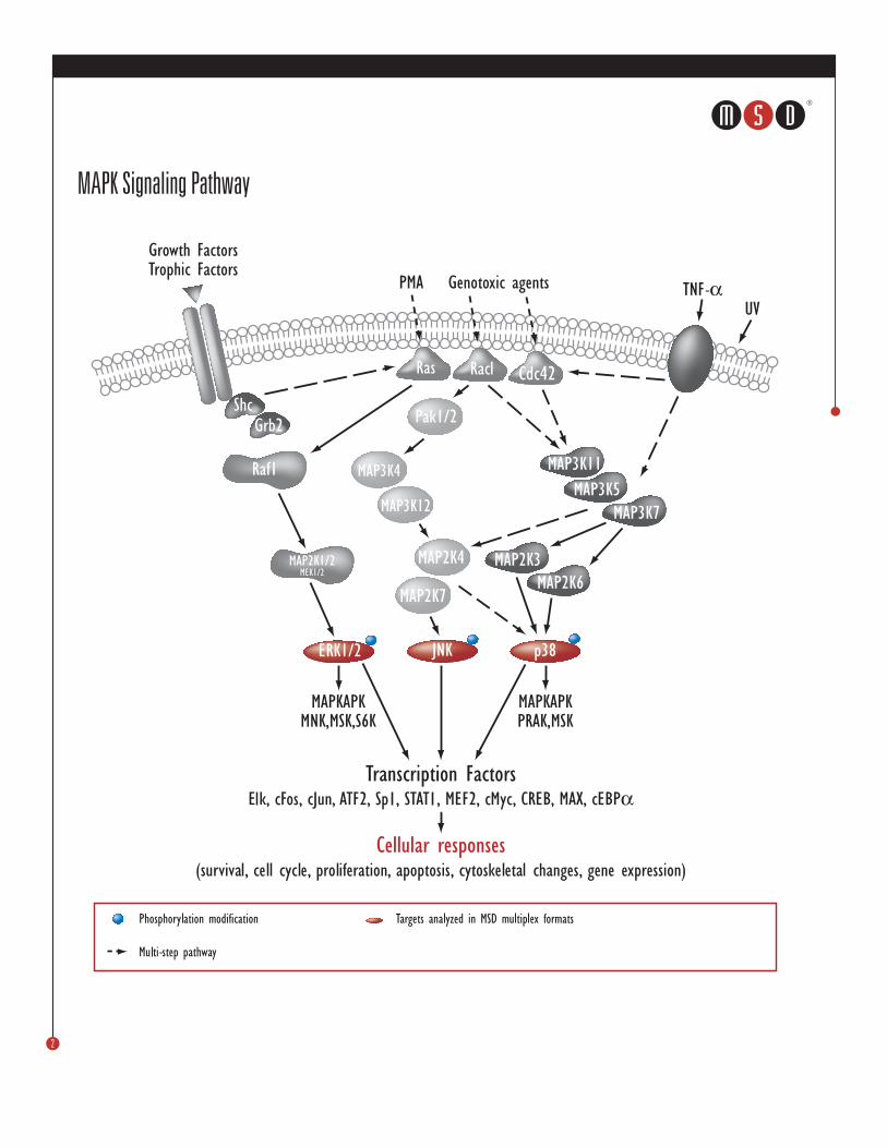
interrogate the phosphorylation state of key components of MAPK (ERK1/2, p38 and JNK), Apoptosis
(Caspase-3, PARP, Bcl-2, BAD and p53) and Akt/mTOR (Akt, p70S6K, GSK-3 , GSK-3 and MEK1/2)
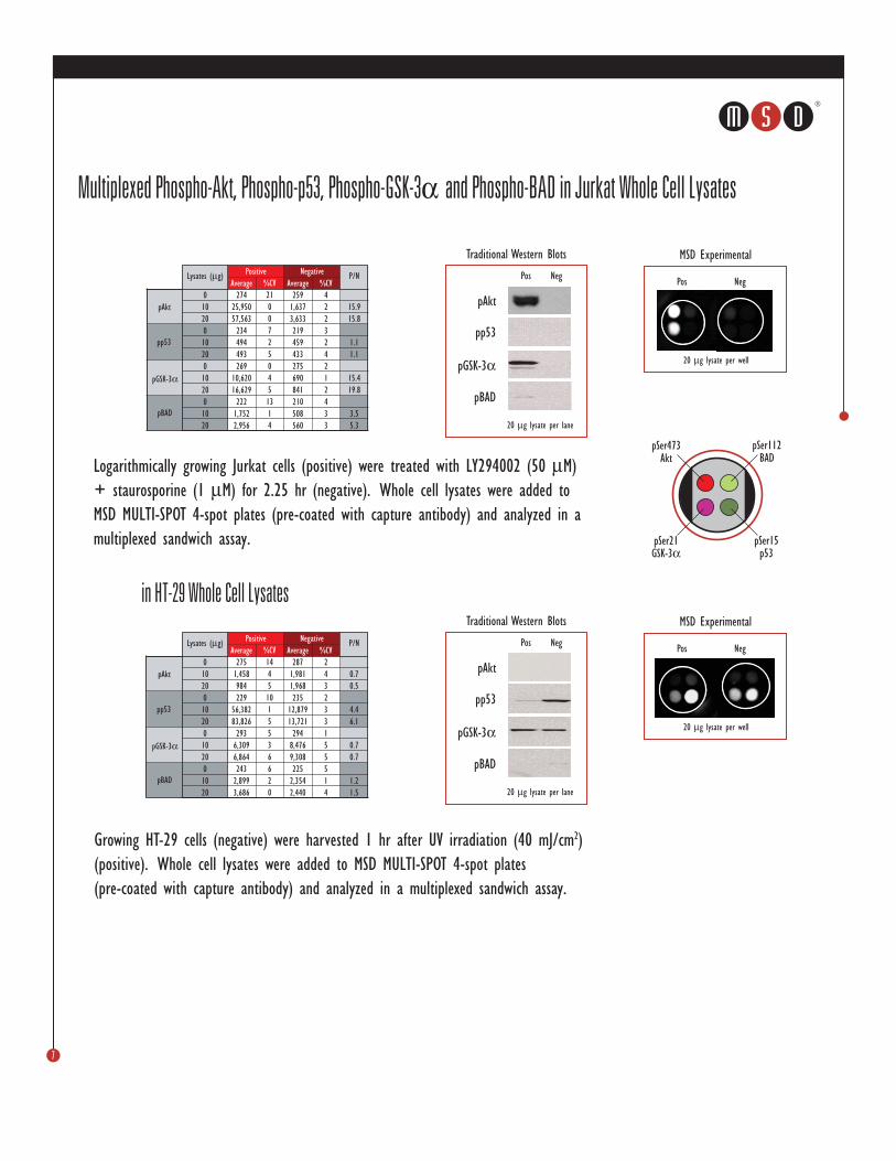
signaling pathways. Furthermore, the percentage of protein that is phosphorylated at a given site can
be estimated by simultaneously measuring the phosphorylated and total pools. The results closely
mimic those seen in western blots. MULTI-ARRAY assays offer the advantages of multiplexing while
being very sensitive and rapid. The combination of multiplexing and rapid protocols results in
tremendous gains in productivity compared to conventional western blot and ELISA analyses.
®
Mes
o Sc
ale
Disc
over
y Ap
plic
atio
ns
Nisar Pampori, Jennifer Lewis, Bruk G. Leta, Laura K. Schaefer, Robert M. Umek, Paula Denney Eason and Jacob N. Wohlstadter
®
1
MSD MULTI-ARRAY Technology and MULTI-SPOT® Plates
1. MULTI-SPOT 4 Spot 96-Well Plates precoated with capture antibodies are blocked with
100 L of MSD Blocker-A for 1 hr and washed with TBST.
2. 25 L of cell lysates are added to the wells and incubated for 1-3 hr with shaking,
followed by washing with TBST.
3. 25 L MSD SULFO-TAGTM labeled antibodies (in antibody dilution buffer) are added to
the wells and incubated for 1 hr with shaking, followed by washing with TBST.
4. 150 L MSD Read Buffer T (with surfactant) are added to the wells and analyzed on


![[VII]. Regulation of Gene Expression Via Signal Transduction Reading List VII: Signal transduction Signal transduction in biological systems.](https://static.documents.pub/doc/80x56/56649e385503460f94b28319/vii-regulation-of-gene-expression-via-signal-transduction-reading-list-vii.jpg)


