Toyoake and Takasaki, Japan; Gaithersburg, Maryland; and Irvine, California
Objectives To evaluate the feasibility of noninvasive assessment of the characteristics of disrupted atheroscleroticplaques, the authors interrogated the culprit lesions in acute coronary syndromes (ACS) by multislice com-puted tomography (CT).
Background Disrupted atherosclerotic plaques responsible for ACS histopathologically demonstrate large lipid cores and posi-tive vascular remodeling. It is expected that plaques vulnerable to rupture should bear similar imaging signa-tures by CT.
Methods Either 0.5-mm � 16-slice or 64-slice CT was performed in 38 patients with ACS and compared with 33 patientswith stable angina pectoris (SAP) before percutaneous coronary intervention. The coronary plaques in ACS andSAP were evaluated for the CT plaque characteristics, including vessel remodeling, consistency of noncalcifiedplaque (NCP �30 HU or 30 HU �NCP �150 HU), and spotty or large calcification.
Results In the CT profile of culprit ACS and SAP lesions, the frequency of 30 HU �NCP �150 HU (100% vs. 100%, p �
NS) was not different, and large calcification (22% vs. 55%, p � 0.004) was significantly more frequent in thestable lesions. Positive remodeling (87% vs. 12%, p � 0.0001), NCP �30 HU (79% vs. 9%, p � 0.0001), andspotty calcification (63% vs. 21%, p � 0.0005) were significantly more frequent in the ACS lesions. Presence ofall 3 (i.e., positive remodeling, NCP �30 HU, and spotty calcification) showed a high positive predictive value,and absence of all 3 showed a high negative predictive value for the culprit plaques associated with ACS.
ublished by Elsevier Inc. doi:10.1016/j.jacc.2007.03.044
crspdtclMupto
isruption of an atherosclerotic plaque is responsible for ateast two-thirds of acute coronary events (1,2). Rupturedlaques are histopathologically characterized by large plaqueolumes and large necrotic cores that are covered by atten-ated fibrous cap often inflamed with monocyte-acrophage infiltration (3). The vessels are positively re-odeled at the site of plaque disruption (4,5). In addition,
mall calcific concretions in fibrous caps have been shown to
rom the *Department of Cardiology, Fujita Health University, Toyoake, Japan;Department of Cardiology, Takase Clinic, Takasaki, Japan; ‡Department ofadiology, Fujita Health University, Toyoake, Japan; §International Registry ofathology, Gaithersburg, Maryland; and the ¶Division of Cardiology, Universityf California Irvine, Irvine, California. Dr. James E. Muller acted as the Guest Editoror this article.
tManuscript received July 25, 2006; revised manuscript received March 13, 2007,
ccepted March 15, 2007.
ontribute to plaque instability (6,7). Plaques vulnerable toupture are termed thin cap fibroatheroma (TCFA) andhow histopathological characteristics similar to disruptedlaques except that the fibrous caps are still intact. Variousiagnostic techniques have identified the imaging charac-eristics of disrupted plaques and proposed that theseharacteristics may allow recognition of coronary lesionsikely to result in acute coronary syndromes (ACS) (3,8,9).
ultiple catheter-based techniques, such as intravascularltrasound (IVUS) (5,10–12), optical coherence tomogra-hy (13,14), intravascular magnetic resonance (15,16), andhermography (17,18), have been used for the identificationf unstable plaques.Recent improvements in computed tomography (CT)
echnology and the advent of the multislice computed tomog-
tpPpcmps
Cc(peeoCt
New
rftpsbwSot
SniSsdwgA(wPptAo
320 Motoyama et al. JACC Vol. 50, No. 4, 2007MSCT in Acute Coronary Syndromes July 24, 2007:319–26
raphy (MSCT) have spurred in-terest in noninvasive detectionof morphologic characteristics ofvulnerable plaques (19–23). Toevaluate the feasibility of CT as-sessment of plaque characteris-tics, we interrogated the culpritlesions in ACS by MSCT andcompared them with atheroscle-rotic plaques in patients present-ing with stable angina pectoris(SAP). Based on the availableCT literature and histopatholog-ical data, remodeling of the ves-sel, plaque consistency, and theextent of calcification were eval-uated by 0.5-mm � 16-slice or0.5-mm � 64-slice CT.
Methods
We started the study in 2004with 0.5-mm � 16-slice CT.After 56 consecutive studies(male, 40; female, 16; age 65 �12 years), the same study proto-col was initiated with 0.5-mm �64-slice CT and 15 more pa-tients were added (male, 14; fe-male, 1; age 61 � 10 years). Thestudy was approved by the insti-tutional review board and theethics committee, and all pa-
ients voluntarily consented to participate in the studyrotocol.atients. Preinterventional CT images were obtained inatients presenting with ACS (ST-segment elevation myo-ardial infarction [STEMI], non–ST-segment elevationyocardial infarction [NSTEMI], and unstable angina
ectoris [UAP]) or SAP. Overall, 441 patients werecreened for recruitment of 71 patients (16%) (Fig. 1).
The SAP patients were classified based on the Canadianardiovascular Society Classification System. Patients with
lass 1 or 2 were included as candidates in the SAP group24). Coronary angiography (CAG) was performed, and theatients with single-vessel disease who were advised to havelective percutaneous coronary intervention (PCI) werenrolled in this study. Culprit lesion was determined basedn significant stenosis in CAG. These patients underwentT examination before PCI. After screening of 149 pa-
ients, 33 patients were included.In the ACS group, patients diagnosed with STEMI,STEMI, and UAP were included as follows. A 12-lead
lectrocardiogram (ECG) and cardiac-specific troponin
ere performed in all patients presenting to the emergency S
oom with chest discomfort. Patients with ischemic discom-ort, ST-segment elevation on the ECG, and elevation ofroponin level were defined as STEMI. In symptomaticatients with 12-lead ECG ST-segment elevation, reperfu-ion intervention was initiated as soon as possible withouteing contingent on a biomarker assay (25); such patientsere excluded from the present study. On the other hand,TEMI patients brought to the hospital �24 h after thenset of chest pain and free of symptoms were enrolled inhe study.
Patients with ischemic discomfort presenting withoutT-segment elevation on the ECG but elevation of tropo-in level were defined as having NSTEMI. Patients with
schemic discomfort that was Canadian Cardiovascularociety class 3 or 4 without electrocardiographic ST-egment elevation and elevation of troponin level wereefined as having UAP. Patients with NSTEMI and UAPere classified into high-, intermediate-, and low-riskroups based on the 2002 American College of Cardiology/merican Heart Association UAP/NSTEMI guidelines
26). High-risk patients admitted within 24 h after onsetere excluded because these patients needed early CAG andCI. High-risk patients presenting 24 h after onset of chestain or intermediate- and low-risk patients were included inhe present study. A CAG was performed in all enrolledCS patients. The culprit lesion was identified on the basisf CAG, location of asynergy in ECG, and location of
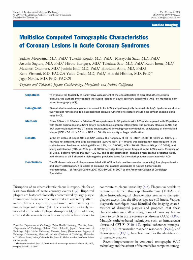
Figure 1 The Patient Population
Overall, 441 patients were screened and 71 were enrolled in the study. Ofthese 71 patients, 10 had STEMI, 9 had NSTEMI, 19 had UAP, and 33 hadSAP. CAG � coronary artery grafting; MVD � multivessel disease; NSTEMI �
T-segment elevation in STEMI. The patients with only
sw
e9plc1atrpmpM(wttempt0ccaapc(Trrwido
dcd6opdtgp�i(c
D
VwstearrpwdcPcodsHwaccct
(1neptwcbs1�N�ItwiaaaouTvcc
321JACC Vol. 50, No. 4, 2007 Motoyama et al.July 24, 2007:319–26 MSCT in Acute Coronary Syndromes
ingle-vessel disease were enrolled in this study and under-ent CT examination before PCI.Overall 441 patients were screened (Fig. 1), and 71 were
nrolled in the study. Of these 71 patients, 10 had STEMI,had NSTEMI, 19 had UAP, and 33 had SAP. The CT
laque characteristics from the culprit ACS and stableesions were tabulated. Because we have recently reported aomparison of IVUS with plaque composition observed by6-slice CT (27), we did not feel comfortable performingdditional IVUS imaging during coronary catheterization inhe first batch of 56 patients. However, we have noteported a comparison of IVUS data with 64-slice CTlaque characteristics earlier. Therefore, we obtained per-ission for additional IVUS studies in the group of 15
atients studied by 64-slice CT.SCT protocol. In the first 56 patients, 16-slice CT
Aquilion 16, Toshiba Medical Systems, Otawara, Japan)as used with collimation 16 � 0.5 mm, detector pitch 3.2
o 3.6, pixel size 0.39 � 0.39 mm, rotation time 400 ms,ube current 360 mA, and voltage 135 kV. For the contrast-nhanced scan, 60 ml of contrast media was injected at 3.0l/s followed by 40 ml at 1.5 ml/s. In the remaining 15
atients, 64-slice CT (Aquilion 64) was used with collima-ion 64 � 0.5 mm; detector pitch 9.8 to 11.2; pixel size.39 � 0.39 mm; rotation time 350, 375, or 400 ms; tubeurrent 400 or 450 mA; and voltage 135 kV. For theontrast-enhanced scan, 60 ml contrast media was injectedt 4.0 ml/s followed by 20 ml at 2.0 ml/s. This strategyllowed the application of dedicated spiral algorithms thatrovided up to 75 ms of temporal resolution. The start ofontrast-enhanced scan was adapted to sure-start images28). All scans were performed during a single breath hold.he raw data of the scans were reconstructed using algo-
ithms optimized for retrograde ECG-gated multislice spi-al reconstruction. The reconstructed image data of the CTere transferred to a computer workstation for postprocess-
ng (ZIO M900, Amin/ZIO, Tokyo, Japan). For plaqueetection, both cross-sectional and curved multiplanar ref-rmation images were analyzed.Because the rate of infusion of contrast medium was
ifferent in the 2 protocols and may affect the diffusion ofontrast material into the plaque, we measured the lumenensity in both the ACS and the SAP group by 16- and4-slice CT. Although the Kruskal-Wallis test comparisonf the luminal contrast in 4 groups followed by the Fisherrotected least significant difference test showed significantifference in the luminal densities in 16- and 64-slice scans,here was no significant difference in the ACS and SAProups by either 16-slice (262 � 55 HU vs. 259 � 29 HU,� 0.74) or 64-slice (318 � 43 HU vs. 304 � 51 HU, p0.45) CT. Therefore, the diffusion of contrast material
nto the plaques may be different in 16- and 64-slice CTsee plaque consistency), but the comparison of the plaque
haracteristics between ACS and SAP is valid. S
efinition of CT Plaque Characteristics
ascular remodeling. The coronary arterial remodelingas defined as a change in the vessel diameter at the plaque
ite in comparison with the reference segment set proximalo the lesion in a normal-appearing vessel segment (refer-nce segment). Manual inspection, in both cross-sectionnd longitudinal reconstruction, was used for defining theemodeling index (lesion diameter/reference diameter). Theemodeling index on MSCT was calculated and reported asositive remodeling when the diameter at the plaque siteas at least 10% larger than the reference segment. Theelineation of the outer vessel boundary was difficult in 6ases (8%) because of severe calcification.laque consistency. The plaques were reported as eitheralcified or noncalcified plaques (NCP). Further, based onur comparative 16-slice CT and IVUS data (27), weivided the NCP into 2 categories; NCP �30 HU (corre-ponding to IVUS lipid cores) and 30 HU �NCP �150U (corresponding to IVUS fibrous plaques). Calcificationas recognized as the plaque with the density of �220 HU,
nd was classified as spotty or large calcification. Spottyalcification was defined when �3 mm in size (11) onurved multiplanar reformation images and 1-sided onross-sectional images. Large calcification was defined ashe calcification larger than spotty calcification.
Based on our comparative 16-slice CT and IVUS data27), the HU density of IVUS-identified soft plaques was1 � 12 and that of fibrous plaque was 78 � 21; there waso overlap in standard deviation values. Accordingly, westablished 30 HU as the cutoff point, and IVUS was noterformed in patients with 16-slice CT examination. Onhe other hand, all 15 patients included in the present studyith 64-slice CT underwent IVUS examination during
oronary angiography; 10 of 15 patients had NCP �30 HUy 64-slice CT. Compared with IVUS, sensitivity andpecificity of CT for detecting soft plaque was 91% and00%. Therefore, we have maintained the value of NCP30 HU as the cutoff point, and stratified the presence ofCP into 2 groups of NCP �30 HU and 30 HU �NCP150 HU.
nterpretation of CT scans, data presentation, and sta-istical analysis. The observers interpreting the CT studiesere blinded to the clinical data. Two observers were
nvolved in the interpretation of data, and the interobservergreement has been calculated by kappa statistic. Thegreement (�) for the interpretation of vascular remodelingnd plaque consistency was 0.884 and 0.913 for the 2bservers, respectively. The plaque characteristics were tab-lated in ACS and SAP patients, and reported in percent.he sensitivity, specificity, positive and negative predictive
alues (PPV and NPV), and diagnostic accuracy of eachharacteristic alone or in combination were calculated. Theomparison of the plaque characteristics in the ACS and
AP groups was made using the chi-square test for inde-
ps
R
TTap
lC
i((psrwcwlt
(dlmO0cfsiPPT76tsaw
CC
Tc
aSu
322 Motoyama et al. JACC Vol. 50, No. 4, 2007MSCT in Acute Coronary Syndromes July 24, 2007:319–26
endence, and a p value of �0.05 was consideredignificant.
esults
he clinical characteristics of all 71 patients are shown inable 1. There was no statistically significant difference in
ge, gender, presence of risk factors, or culprit vessel amongatients with ACS or SAP.The characteristics of culprit lesions in ACS and target
esions in SAP are presented in Figures 2 through 4. TheT examination of angiographically defined culprit lesions
linical and Angiographicharacteristics of the Patients
Table 1 Clinical and AngiographicCharacteristics of the Patients
ACS (n � 38)(STEMI 10, NSTEMI 9, UAP 19) SAP (n � 33) p Value
Age, yrs 62 � 11 67 � 9 0.053
Male/female 31/7 23/10 0.242
Diabetes 8 (21%) 7 (22%) 0.987
Hypertension 16 (42%) 19 (58%) 0.295
Hyperlipidemia 23 (61%) 22 (67%) 0.592
Smoking 19 (51%) 18 (55%) 0.702
Obesity 11 (29%) 11 (33%) 0.690
Lesion location 0.650
LAD 19 (50%) 14 (42%)
LCX 8 (22%) 6 (18%)
RCA 11 (29%) 12 (36%)
here was no statistically significant difference in age, gender, presence of risk factors, andoronary vessel involvement between ACS and SAP patient groups.ACS � acute coronary syndrome; LAD � left anterior descending artery; LCX � left circumflex
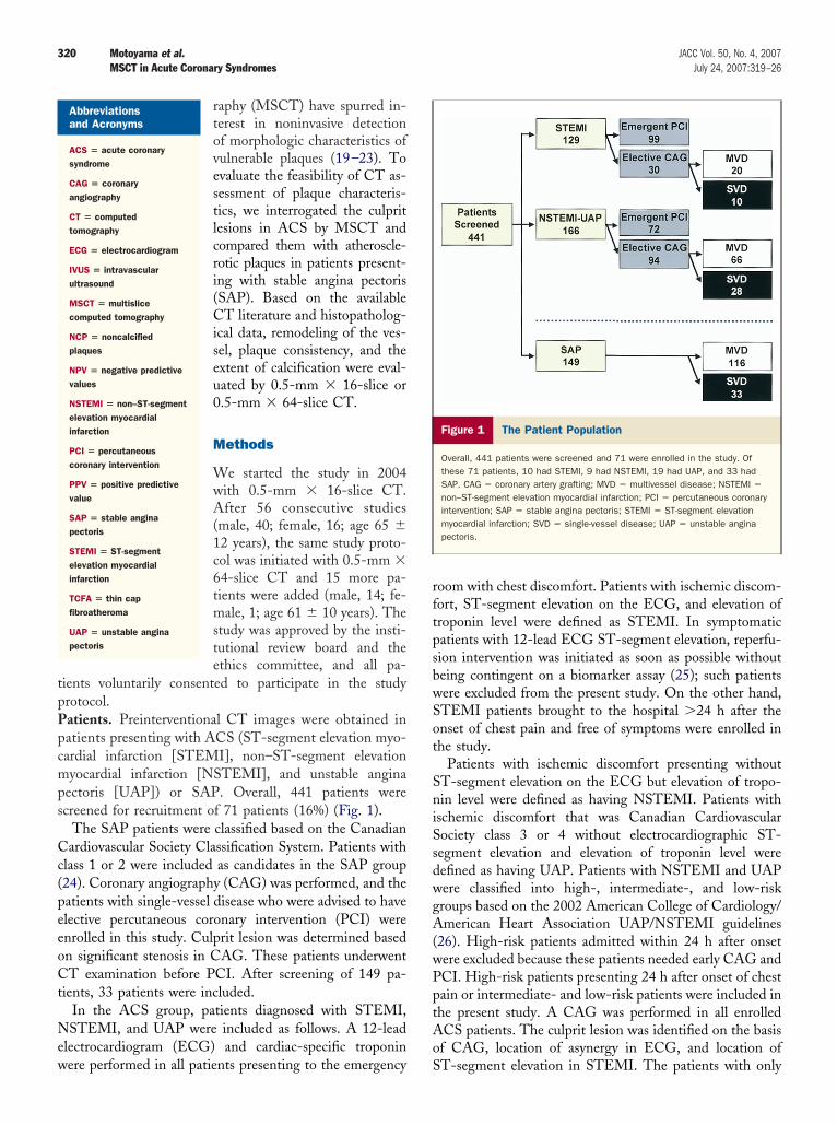
The CT characteristics of a culprit lesion in a 40-year-old male patient presenting wview of the region of interest from (C). (D) Coronary angiogram. The white arrowssolid yellow arrows at 2 sites in the culprit lesion in (C), the lesion is positively re(denoted by interrupted arrows). Remodeling index in this patient was 1.43. An Nthe course of low attenuation), and 30 HU �NCP �150 HU denotes a fibrous plaqartery; MPR � multiplanar reformation; NCP � noncalcified plaque.
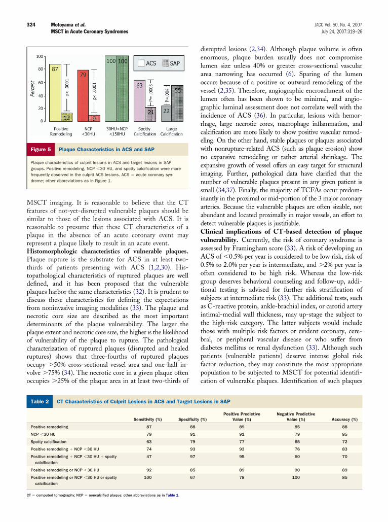
n ACS revealed positive expansive remodeling of the vessel87%), NCP �30 HU (79%), and spotty calcification (63%)Fig. 5). In addition, 30 HU �NCP �150 HU wereresent in all culprit lesions, and larger calcifications wereeen in 22%. On the other hand, the vessels were positivelyemodeled only in 12% of stable lesions, and all plaquesere characterized as 30 HU �NCP �150 HU. Spotty
alcification was observed in 21%, but larger calcific plaquesere found in 55%. The mean remodeling index in the ACS
esions was 1.20 � 0.17, which was significantly greaterhan the SAP plaques (0.96 � 0.12, p � 0.0001).
Although the frequency of 30 HU �NCP �150 HU100% vs. 100%, p � NS) on the MSCT was not significantlyifferent between the lesions associated with ACS and SAP,
arge calcification (22% vs. 55%, p � 0.0044) was significantlyore frequent in the lesions associated with SAP than ACS.n the other hand, positive remodeling (87% vs. 12%, p �
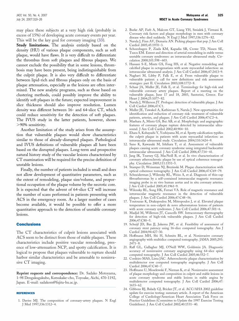
.0001), NCP �30 HU (79% vs. 9%, p � 0.0001), and spottyalcification (63% vs. 21%, p � 0.0005) were significantly morerequent in the culprit ACS lesions than SAP (Fig. 5). Theensitivity, specificity, PPV, and NPV of various characteristicsndependently or in combination are provided in Table 2.ositive remodeling showed the best sensitivity, specificity,PV, and NPV of 87%, 88%, 89%, and 85%, respectively.hese values for the NCP �30 HU were 79%, 91%, 91%, and9%, and for spotty calcification were 63%, 79%, 77%, and5%, respectively. However, when the plaque was described byhe absence of all (i.e., positive remodeling, NCP �30 HU, orpotty calcification), NPV was 100%; therefore, plaque associ-ted with an acute event was not missed. On the other hand,hen the plaque showed all 3 features (i.e., positive remodel-
ute coronary syndrome. (A) Volume rendering. (B) Curved MPR. (C) Magnifiedand (D) show the site of luminal obstruction or culprit lesion. As shown by the
led as compared with the normal coronary segment proximal to the lesion0 HU represents the probability of a soft plaque (red circles are placed alongreen squares). CT � computed tomography; LAD � left anterior descending
ith acin (A)mode
CP �3ue (g
ip
D
TsNcpvlerNbcl
bsipMdci(aaIrltt1
323JACC Vol. 50, No. 4, 2007 Motoyama et al.July 24, 2007:319–26 MSCT in Acute Coronary Syndromes
ng, NCP �30 HU, and spotty calcification), the PPV of thelaque associated with an acute event increased to 95%.
iscussion
he present study. The culprit atherosclerotic lesions as-ociated with ACS showed positive vascular remodeling,CP �30 HU, and spotty calcification on MSCT. These
haracteristics were uncommonly associated with lesions inatients presenting with SAP. Positive remodeling of theessel was the most important accompaniment of the culpritesions and confirms the data described earlier by the IVUSxamination (4,5) and the classic pathological description ofuptured plaques on autopsy (2,29,30). The presence ofCP �30 HU offered higher specificity and PPV of 90%,
ut sensitivity and NPV were �80%. Based on our recentorrelation of CT plaque characteristics and IVUS, it isikely that NCP �30 HU represent a necrotic core and may
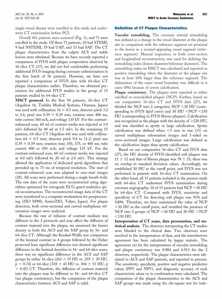
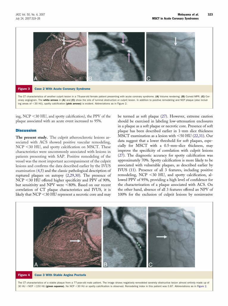
Figure 3 Case 2 With Acute Coronary Syndrome
The CT characteristics of another culprit lesion in a 79-year-old female patient preonary angiogram. The white arrows in (A) and (C) show the site of luminal obstruing areas of �30 HU), spotty calcification (pink arrows) is evident. Abbreviations
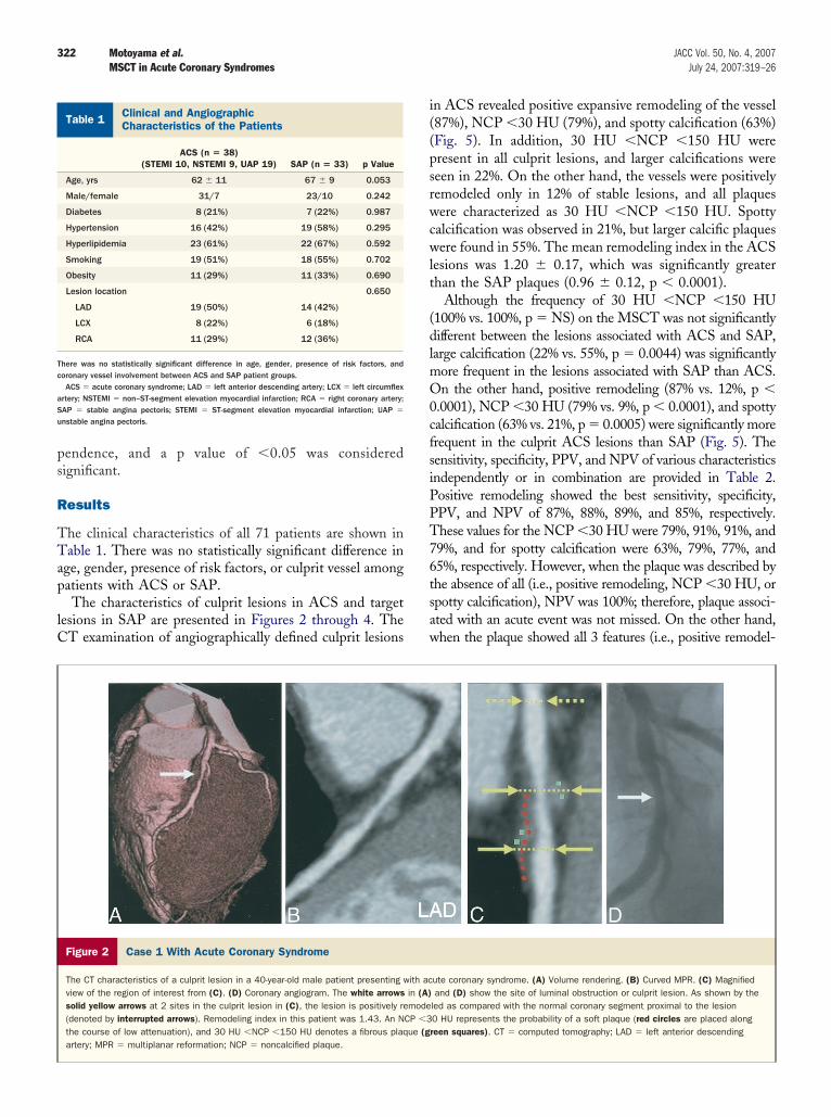
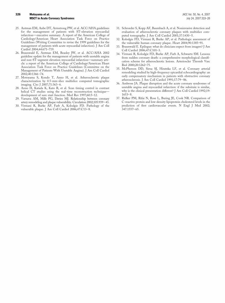
Figure 4 Case 3 With Stable Angina Pectoris
The CT characteristics of a stable plaque from a 77-year-old male patient. The ima30 HU �NCP �150 HU (green squares). No NCP �30 HU or spotty calcification i
e termed as soft plaque (27). However, extreme cautionhould be exercised in labeling low-attenuation enclosuresn a plaque as a soft plaque or necrotic core. Presence of softlaque has been described earlier in 1-mm slice thicknessSCT examination as a lesion with �50 HU (22,31). Our
ata suggest that a lower threshold for soft plaques, espe-ially for MSCT with a 0.5-mm-slice thickness, maymprove the specificity of correlation with culprit lesions27). The diagnostic accuracy for spotty calcification waspproximately 70%. Spotty calcification is more likely to bessociated with vulnerable plaques, as described earlier byVUS (11). Presence of all 3 features, including positiveemodeling, NCP �30 HU, and spotty calcification, al-owed PPV of 95%, providing a high level of confidence forhe characterization of a plaque associated with ACS. Onhe other hand, absence of all 3 features offered an NPV of00% for the exclusion of culprit lesions by noninvasive
with acute coronary syndrome. (A) Volume rendering. (B) Curved MPR. (C) Cor-r culprit lesion. In addition to positive remodeling and NCP plaque (also includ-igure 2.
ows negatively remodeled severely obstructive lesion almost entirely made up ofrved. Remodeling index in this patient was 0.87. Abbreviations as in Figure 2.
sentingction oas in F
ge shs obse
MfsrprHPttdpdfndpocrovo
delaovlgircewneinsiaadCvaA0ogtsaittbdpfpc
C
C
324 Motoyama et al. JACC Vol. 50, No. 4, 2007MSCT in Acute Coronary Syndromes July 24, 2007:319–26
SCT imaging. It is reasonable to believe that the CTeatures of not-yet-disrupted vulnerable plaques should beimilar to those of the lesions associated with ACS. It iseasonable to presume that these CT characteristics of alaque in the absence of an acute coronary event mayepresent a plaque likely to result in an acute event.
istomorphologic characteristics of vulnerable plaques.laque rupture is the substrate for ACS in at least two-
hirds of patients presenting with ACS (1,2,30). His-opathological characteristics of ruptured plaques are wellefined, and it has been proposed that the vulnerablelaques harbor the same characteristics (32). It is prudent toiscuss these characteristics for defining the expectationsrom noninvasive imaging modalities (33). The plaque andecrotic core size are described as the most importanteterminants of the plaque vulnerability. The larger thelaque extent and necrotic core size, the higher is the likelihoodf vulnerability of the plaque to rupture. The pathologicalharacterization of ruptured plaques (disrupted and healeduptures) shows that three-fourths of ruptured plaquesccupy �50% cross-sectional vessel area and one-half in-olve �75% (34). The necrotic core in a given plaque oftenccupies �25% of the plaque area in at least two-thirds of
Figure 5 Plaque Characteristics in ACS and SAP
Plaque characteristics of culprit lesions in ACS and target lesions in SAPgroups. Positive remodeling, NCP �30 HU, and spotty calcification were morefrequently observed in the culprit ACS lesions. ACS � acute coronary syn-drome; other abbreviations as in Figure 1.
T Characteristics of Culprit Lesions in ACS and Target Lesions in
Table 2 CT Characteristics of Culprit Lesions in ACS and Targe
Sensitivity (%) Speci
Positive remodeling 87
NCP �30 HU 79
Spotty calcification 63
Positive remodeling � NCP �30 HU 74
Positive remodeling � NCP �30 HU � spottycalcification
47
Positive remodeling or NCP �30 HU 92
Positive remodeling or NCP �30 HU or spottycalcification
100
T � computed tomography; NCP � noncalcified plaque; other abbreviations as in Table 1.
isrupted lesions (2,34). Although plaque volume is oftennormous, plaque burden usually does not compromiseumen size unless 40% or greater cross-sectional vascularrea narrowing has occurred (6). Sparing of the lumenccurs because of a positive or outward remodeling of theessel (2,35). Therefore, angiographic encroachment of theumen often has been shown to be minimal, and angio-raphic luminal assessment does not correlate well with thencidence of ACS (36). In particular, lesions with hemor-hage, large necrotic cores, macrophage inflammation, andalcification are more likely to show positive vascular remod-ling. On the other hand, stable plaques or plaques associatedith nonrupture-related ACS (such as plaque erosion) showo expansive remodeling or rather arterial shrinkage. Thexpansive growth of vessel offers an easy target for structuralmaging. Further, pathological data have clarified that theumber of vulnerable plaques present in any given patient ismall (34,37). Finally, the majority of TCFAs occur predom-nantly in the proximal or mid-portion of the 3 major coronaryrteries. Because the vulnerable plaques are often sizable, notbundant and located proximally in major vessels, an effort toetect vulnerable plaques is justifiable.linical implications of CT-based detection of plaque
ulnerability. Currently, the risk of coronary syndrome isssessed by Framingham score (33). A risk of developing anCS of �0.5% per year is considered to be low risk, risk of.5% to 2.0% per year is intermediate, and �2% per year isften considered to be high risk. Whereas the low-riskroup deserves behavioral counseling and follow-up, addi-ional testing is advised for further risk stratification ofubjects at intermediate risk (33). The additional tests, suchs C-reactive protein, ankle-brachial index, or carotid arteryntimal-medial wall thickness, may up-stage the subject tohe high-risk category. The latter subjects would includehose with multiple risk factors or evident coronary, cere-ral, or peripheral vascular disease or who suffer fromiabetes mellitus or renal dysfunction (33). Although suchatients (vulnerable patients) deserve intense global riskactor reduction, they may constitute the most appropriateopulation to be subjected to MSCT for potential identifi-ation of vulnerable plaques. Identification of such plaques
ions in SAP
%)Positive Predictive
Value (%)Negative Predictive
Value (%) Accuracy (%)
89 85 88
91 79 85
77 65 72
93 76 83
95 60 70
89 90 89
78 100 85
SAP
t Les
ficity (
88
91
79
93
97
85
67
meTSdptcbtbpmcisdcT�
tsabnCu
nttItAbql
C
TAcelhs
R1J
R
1
1
1
1
1
1
1
1
1
1
2
2
2
2
2
325JACC Vol. 50, No. 4, 2007 Motoyama et al.July 24, 2007:319–26 MSCT in Acute Coronary Syndromes
ay place these subjects at a very high risk (probably inxcess of 15%) of developing acute coronary events per year.his will be the key goal for coronary imaging (33).tudy limitations. The analysis entirely based on theensity (HU) of various plaque components, such as softlaque, would have flaws. It is very difficult to differentiatehe thrombus from soft plaques and fibrous plaques. Weannot exclude the possibility that in some lesions, throm-osis may have been present, being indistinguishable fromhe culprit plaque. It is also very difficult to differentiateetween lipid-rich and fibrous plaques only on the basis oflaque attenuation, especially as the lesions are often inter-ixed. The new analytic programs, such as those based on
lustering methods, could possibly improve the ability todentify soft plaques in the future; expected improvement inlice thickness should also improve resolution. Lumenensity was different between 16- and 64-slice CT, whichould reduce sensitivity for the detection of soft plaques.he IVUS study in the latter patients, however, shows90% sensitivity.Another limitation of the study arises from the assump-
ion that vulnerable plaques would show characteristicsimilar to those of already-disrupted plaques. Pathologicalnd IVUS definitions of vulnerable plaques all have beenased on the disrupted plaques. Long-term and prospectiveatural history study of the vascular lesions characterized byT examination will be required for the precise definition ofnstable lesions.Finally, the number of patients included is small and does
ot allow development of quantitative parameters, such ashe extent of remodeling, plaque size/volume, and propor-ional occupation of the plaque volume by the necrotic core.t is expected that the advent of 64-slice CT will increasehe number of scans performed in patients presenting withCS in the emergency room. As a larger number of casesecome available, it would be possible to offer a moreuantitative approach to the detection of unstable coronaryesions.
onclusions
he CT characteristics of culprit lesions associated withCS seem to be distinct from those of stable plaques. These
haracteristics include positive vascular remodeling, pres-nce of low-attenuation NCP, and spotty calcification. It isogical to propose that plaques vulnerable to rupture shouldarbor similar characteristics and be amenable to noninva-ive CT imaging.
eprint requests and correspondence: Dr. Sadako Motoyama,-98 Dengakugakubo, Kutsukake-cho, Toyoake, Aichi, 470-1192,apan. E-mail: [email protected].
EFERENCES
1. Davies MJ. The composition of coronary-artery plaques. N EnglJ Med 1997;336:1312–4.
2. Burke AP, Farb A, Malcom GT, Liang YH, Smialek J, Virmani R.Coronary risk factors and plaque morphology in men with coronarydisease who died suddenly. N Engl J Med 1997;336:1276–82.
3. Narula J, Finn AV, Demaria AN. Picking plaques that pop. J Am CollCardiol 2005;45:1970–3.
4. Schoenhagen P, Ziada KM, Kapadia SR, Crowe TD, Nissen SE,Tuzcu EM. Extent and direction of arterial remodeling in stable versusunstable coronary syndromes: an intravascular ultrasound study. Cir-culation 2000;101:598–603.
5. Hassani S-E, Mintz GS, Fong HS, et al. Negative remodeling andcalcified plaque in octogenarians with acute myocardial infarction: anintravascular ultrasound analysis. J Am Coll Cardiol 2006;47:2413–9.
6. Naghavi M, Libby P, Falk E, et al. From vulnerable plaque tovulnerable patient: a call for new definitions and risk assessmentstrategies: part II. Circulation 2003;108:1772–8.
7. Schaar JA, Muller JE, Falk E, et al. Terminology for high-risk andvulnerable coronary artery plaques. Report of a meeting on thevulnerable plaque, June 17 and 18, 2003, Santorini, Greece. EurHeart J 2004;25:1077–82.
8. Narula J, Willerson JT. Prologue: detection of vulnerable plaque. J AmColl Cardiol 2006;47:C1.
9. Muller JE, Tawakol A, Kathiresan S, Narula J. New opportunities foridentification and reduction of coronary risk: treatment of vulnerablepatients, arteries, and plaques. J Am Coll Cardiol 2006;47:C2–6.
0. Maehara A, Mintz GS, Bui AB, et al. Morphologic and angiographicfeatures of coronary plaque rupture detected by intravascular ultra-sound. J Am Coll Cardiol 2002;40:904–10.
1. Ehara S, Kobayashi Y, Yoshiyama M, et al. Spotty calcification typifiesthe culprit plaque in patients with acute myocardial infarction: anintravascular ultrasound study. Circulation 2004;110:3424–9.
2. Sano K, Kawasaki M, Ishihara Y, et al. Assessment of vulnerableplaques causing acute coronary syndrome using integrated backscatterintravascular ultrasound. J Am Coll Cardiol 2006;47:734–41.
3. Jang IK, Tearney GJ, MacNeill B, et al. In vivo characterization ofcoronary atherosclerotic plaque by use of optical coherence tomogra-phy. Circulation 2005;111:1551–5.
4. Stamper D, Weissman NJ, Brezinski M. Plaque characterization withoptical coherence tomography. J Am Coll Cardiol 2006;47:C69–79.
5. Schneiderman J, Wilensky RL, Weiss A, et al. Diagnosis of thin-capfibroatheromas by a self-contained intravascular magnetic resonanceimaging probe in ex vivo human aortas and in situ coronary arteries.J Am Coll Cardiol 2005;45:1961–9.
6. Wilensky RL, Song HK, Ferrari VA. Role of magnetic resonance andintravascular magnetic resonance in the detection of vulnerableplaques. J Am Coll Cardiol 2006;47:C48–56.
7. Toutouzas K, Drakopoulou M, Mitropoulos J, et al. Elevated plaquetemperature in non-culprit de novo atheromatous lesions of patientswith acute coronary syndromes. J Am Coll Cardiol 2006;47:301–6.
8. Madjid M, Willerson JT, Casscells SW. Intracoronary thermographyfor detection of high-risk vulnerable plaques. J Am Coll Cardiol2006;47:C80–5.
9. Schuijf JD, Bax JJ, Jukema JW, et al. Feasibility of assessment ofcoronary stent patency using 16-slice computed tomography. Am JCardiol 2004;94:427–30.
0. Hoffmann MH, Shi H, Schmitz BL, et al. Noninvasive coronaryangiography with multislice computed tomography. JAMA 2005;293:2471–8.
1. Raff GL, Gallagher MJ, O’Neill WW, Goldstein JA. Diagnosticaccuracy of noninvasive coronary angiography using 64-slice spiralcomputed tomography. J Am Coll Cardiol 2005;46:552–7.
2. Cordeiro MAS, Lima JAC. Atherosclerotic plaque characterization bymultidetector row computed tomography angiography. J Am CollCardiol 2006;47:C40–7.
3. Hoffmann U, Moselewski F, Nieman K, et al. Noninvasive assessmentof plaque morphology and composition in culprit and stable lesions inacute coronary syndrome and stable lesions in stable angina bymultidetector computed tomography. J Am Coll Cardiol 2006;47:1655–62.
4. Gibbons RJ, Balady GJ, Bricker JT, et al. ACC/AHA 2002 guidelineupdate for exercise testing: summary article. A report of the AmericanCollege of Cardiology/American Heart Association Task Force onPractice Guidelines (Committee to Update the 1997 Exercise Testing
Guidelines). J Am Coll Cardiol 2002;40:1531–40.
2
2
2
2
2
3
3
3
3
3
3
3
3
326 Motoyama et al. JACC Vol. 50, No. 4, 2007MSCT in Acute Coronary Syndromes July 24, 2007:319–26
5. Antman EM, Anbe DT, Armstrong PW, et al. ACC/AHA guidelinesfor the management of patients with ST-elevation myocardialinfarction—executive summary. A report of the American College ofCardiology/American Heart Association Task Force on PracticeGuidelines (Writing Committee to revise the 1999 guidelines for themanagement of patients with acute myocardial infarction). J Am CollCardiol 2004;44:671–719.
6. Braunwald E, Antman EM, Beasley JW, et al. ACC/AHA 2002guideline update for the management of patients with unstable anginaand non-ST-segment elevation myocardial infarction—summary arti-cle: a report of the American College of Cardiology/American HeartAssociation Task Force on Practice Guidelines (Committee on theManagement of Patients With Unstable Angina). J Am Coll Cardiol2002;40:1366–74.
7. Motoyama S, Kondo T, Anno H, et al. Atherosclerotic plaquecharacterization by 0.5-mm-slice multislice computed tomographicimaging. Circ J 2007;71:363–6.
8. Anno H, Katada K, Kato R, et al. Scan timing control in contrasthelical CT studies using the real-time reconstruction technique—development of sure start function. Med Rev 1997;60:5–12.
9. Varnava AM, Mills PG, Davies MJ. Relationship between coronaryartery remodeling and plaque vulnerability. Circulation 2002;105:939–43.
0. Virmani R, Burke AP, Farb A, Kolodgie FD. Pathology of the
vulnerable plaque. J Am Coll Cardiol 2006;47:C13–8.
1. Schroeder S, Kopp AF, Baumbach A, et al. Noninvasive detection andevaluation of atherosclerotic coronary plaques with multislice com-puted tomography. J Am Coll Cardiol 2001;37:1430–5.
2. Kolodgie FD, Virmani R, Burke AP, et al. Pathologic assessment ofthe vulnerable human coronary plaque. Heart 2004;90:1385–91.
3. Braunwald E. Epilogue: what do clinicians expect from imagers? J AmColl Cardiol 2006;47:C101–3.
4. Virmani R, Kolodgie FD, Burke AP, Farb A, Schwartz SM. Lessonsfrom sudden coronary death: a comprehensive morphological classifi-cation scheme for atherosclerotic lesions. Arterioscler Thromb VascBiol 2000;20:1262–75.
5. McPherson DD, Sirna SJ, Hiratzka LF, et al. Coronary arterialremodeling studied by high-frequency epicardial echocardiography: anearly compensatory mechanism in patients with obstructive coronaryatherosclerosis. J Am Coll Cardiol 1991;17:79–86.
6. Ambrose JA. Plaque disruption and the acute coronary syndromes ofunstable angina and myocardial infarction: if the substrate is similar,why is the clinical presentation different? J Am Coll Cardiol 1992;19:1653–8.
7. Ridker PM, Rifai N, Rose L, Buring JE, Cook NR. Comparison ofC-reactive protein and low-density lipoprotein cholesterol levels in theprediction of first cardiovascular events. N Engl J Med 2002;